

Polyarteritis Nodosa-Associated with IgA Nephropathy – Case ...
Polyarteritis Nodosa-Associated with IgA Nephropathy – Case ...
Polyarteritis Nodosa-Associated with IgA Nephropathy – Case ...

You also want an ePaper? Increase the reach of your titles
YUMPU automatically turns print PDFs into web optimized ePapers that Google loves.
Acta Nephrologica 26(1): 32-36, 2012<br />
DOI: 10.6221/2012.26(1)032<br />
<strong>Case</strong> Report<br />
<strong>Polyarteritis</strong> <strong>Nodosa</strong>-<strong>Associated</strong> <strong>with</strong> <strong>IgA</strong><br />
<strong>Nephropathy</strong> <strong>–</strong> <strong>Case</strong> Report and<br />
Literature Review<br />
Wei Lin 1 , Hsin-Fan Chen 1 , I-Wen Wu 1 , Yeon-Jian Jan Wu 2 , and Mai-Szu Wu 1<br />
1<br />
School of Medicine, Chang Gung University; Division of Nephrology<br />
2<br />
Division of Rheumatology<br />
Chang Gung Memorial Hospital, Keelung, Taiwan, Republic of China<br />
Abstract<br />
<strong>Polyarteritis</strong> nodosa (PAN)-associated glomerulonephritis presenting <strong>with</strong> nephrotic syndrome is<br />
not common. We describe a 69-year-old hypertensive man in whom PAN coexisted <strong>with</strong> <strong>IgA</strong> nephropathy<br />
presenting <strong>with</strong> severe nephrotic syndrome, livedo reticularis, testicular pain, and microaneurysm of<br />
renal vessels. Extensive literature review of PAN-associated glomerulonephritis was summarized. PANassociated<br />
<strong>IgA</strong> nephropathy should be considered in a patient having PAN <strong>with</strong> nephrotic syndrome.<br />
Steroid therapy is an effective therapy in cases where these 2 diseases coexisted.<br />
KEY WORDS: glomerulonephritis, <strong>IgA</strong> nephropathy, nephrotic syndrome, polyarteritis nodosa<br />
Introduction<br />
<strong>Polyarteritis</strong> nodosa (PAN), first described by<br />
Kussmaul and Maier in 1866, is an uncommon systemic<br />
necrotizing vasculitis and is characterized by<br />
inflammatory reactions of medium-sized muscular arteries,<br />
which lead to aneurysm formation <strong>with</strong> the involvement<br />
of multiple organs, including skin, kidneys,<br />
peripheral nerves, muscles, and gastrointestinal tract<br />
(1). The clinical features are tissue infarction, hemorrhage,<br />
and organ dysfunction (2). Renal involvement<br />
of PAN is frequent and constitutes 93.4% of all visceral<br />
manifestations (3). Common renal manifestations include<br />
loin pain and hematuria due to renal infarction,<br />
renal dysfunction, and renin-dependent hypertension<br />
secondary to ischemia, microaneurysm of renal vasculature,<br />
perirenal hematoma because of microaneurysm<br />
rupture (2), and bilateral reversible hydronephrosis<br />
(4, 5).<br />
PAN-associated glomerulonephritis presenting<br />
<strong>with</strong> nephrotic syndrome is not common. Some investigators<br />
have described the association of PAN <strong>with</strong><br />
cryoglobulinemic glomerulonephritis (6) hepatitis B<br />
Corresponding author: Mai-Szu Wu, M.D., Division of Nephrology, Chang Gung Memorial Hospital, No. 222, Mai-Chin Rd., Keelung,<br />
Taiwan, R.O.C. Tel: +886-2-24313131 ext. 3169, Fax: +886-2-24335342, E-mail: maxwu1@adm.cgmh.org.tw<br />
Received: March 21, 2011; Revised: May 16, 2011; Accepted: September 28, 2011. .<br />
32<br />
virus-related membranous nephropathy (7) antiglomerular<br />
basement membrane disease (7). Berger<br />
disease (8) and in particular, the Henoch-Schonlein<br />
purpura (HSP) (9, 10). All these cases manifested <strong>with</strong><br />
hematuria and/or non-nephrotic-range proteinuria.<br />
Herein, we describe a patient <strong>with</strong> PAN presenting<br />
<strong>with</strong> nephrotic syndrome; renal biopsy for this patient<br />
revealed <strong>IgA</strong> nephropathy.<br />
<strong>Case</strong> Report<br />
A 69-year-old hypertensive man was admitted<br />
because of diffuse skin lesions and bilateral leg edema<br />
for 1 week. There was no fever, itching, swelling, local<br />
heat, or tenderness. The patient complained of<br />
testicular pain and livedo reticularis was observed.<br />
On admission, the blood pressure was 144/93 mmHg;<br />
respiratory rate, 12; heart beat, 99/min; and temperature,<br />
36.5°C. Physical examination revealed diffuse<br />
purpuric plaque, <strong>with</strong> necrotic change over the lower<br />
limbs, which extended into the trunk and upper limbs.<br />
Both legs also had pitting edema over the pretibial<br />
and pedal areas. The auscultation of the lungs and
heart was normal. No lymphoadenopathy, hepato-, or<br />
splenomegaly was found. The patient had a medical<br />
history of ruptured anterior communicating artery aneurysm<br />
and right carotid artery stenosis. He had also<br />
undergone a ventriculo-peritoneal shunt for normal<br />
pressure hydrocephalus. The patient received aspirin<br />
for secondary prevention of old ischemic stroke since<br />
September, 2000. The patient did not receive any<br />
ACEI or ARB. He denied having received any new<br />
medication or herbal medicine.<br />
Laboratory findings showed that the hemoglobin<br />
level was 12.3 g/dL; hematocrit value, 37.8%; white<br />
cell count, 6,200/mm 3 (differential count: 74.8% neutrophils,<br />
20.3% lymphocytes, 4.2% monocytes); and<br />
platelet count, 260,000/mm 3 . The erythrocyte sedimentation<br />
rate (ESR) was 31 mm/h. Serum electrolyte<br />
(sodium, potassium, calcium, and phosphate), amylase,<br />
lipase, and blood glucose levels were normal;<br />
the creatinine level was 0.9 mg/dL and urea nitrogen<br />
was 8 mg/dL. The albumin level was 2.5 g/dL; globulin,<br />
1.9 g/dL; total cholesterol, 206 mg/dL; and triglyceride,<br />
102 mg/dL.<br />
Anti-nuclear antibody (ANA) titer was 1:40<br />
(weakly positive). The C3 level was 140 mg/dL (normal<br />
73-134 mg/dL) and the C4 level was 20.3 mg/dL<br />
(normal, 18.2-45.5 mg/dL). The serum concentrations<br />
of the antibodies were as follows: IgG, 885.0 mg/dL;<br />
<strong>IgA</strong>, 121.0 mg/dL; IgM, 88.4 mg/dL; and IgE, < 1.50<br />
IU/mL. Urinalysis showed proteinuria (500 mg/dL),<br />
hematuria (blood: 3+), and 61-70 erythrocytes in each<br />
high-power field. In a 24-h specimen of urine, the<br />
amount of protein was 5.6 g. Nephrotic syndrome is<br />
impressed. Antibodies to double-strand DNA and extractable<br />
nuclear antigen (ENA), including Smith antigen<br />
and cryoglobulins, were absent. The hepatitis B<br />
surface antigen (HBsAg) and antibodies against hepitits<br />
C virus (HCV) were absent. The sample was<br />
weakly positive for perinuclear anti-neutrophilic cytoplasmic<br />
antibodies (P-ANCA) and negative for cytoplasmic<br />
anti-neutrophilic cytoplasmic antibodies<br />
(C-ANCA) and anti-cardiolipin antibodies.<br />
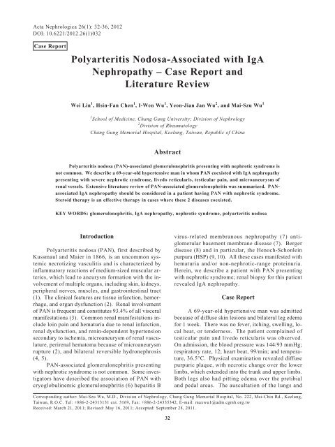
Skin biopsy revealed abundant neutrophil infiltrates<br />
around medium-sized dermal vessels <strong>with</strong> fibrinoid<br />
necrosis, hemorrhage, and nuclear dusts, which<br />
was consistent <strong>with</strong> features of necrotizing vasculitis<br />
(Fig. 1A). Study of skin biopsy by direct immunofluorescence<br />
showed negative findings. Percutaneous<br />
renal biopsy was performed and light microscopy<br />
showed mild mesangial hyperplasia <strong>with</strong> interstitial<br />
fibrosis. The tubules contained protein casts. Arterioles<br />
and capillaries had a normal appearance (Fig.<br />
1B). An immunofluorescence study revealed marked<br />
granular deposits of <strong>IgA</strong> and C3 in the mesangium<br />
(Fig. 1C), indicating <strong>IgA</strong> nephropathy. Computedtomographic<br />
angiography disclosed irregular caliber<br />
and multiple narrowing in the segmental and interlobar<br />
<strong>Polyarteritis</strong> <strong>Nodosa</strong>-<strong>Associated</strong> <strong>IgA</strong> <strong>Nephropathy</strong> 33<br />
(A)<br />
(B)<br />
(C)<br />
Fig. 1. (A) Skin biopsy shows abundant neutrophil infiltrates<br />
around medium-sized dermal vessels <strong>with</strong> fibrinoid necrosis<br />
(black arrows) (40×). (B) Renal specimen shows<br />
mild mesangial hyperplasia (200×, light microscopy);<br />
and (C) intense <strong>IgA</strong> staining (3+) in the mesangium<br />
(immunofluorescence microscopy).<br />
arteries of both kidneys (Fig. 2).<br />
The patient received pulse hydrocortisone therapy<br />
(400 mg daily, for 5 days), followed by oral pred-
34 Lin, Chen, Wu, Wu and Wu<br />
Fig. 2. Computed-tomographic angiography shows irregular<br />
caliber and multiple narrowing in the segmental and<br />
interlobar arteries of both the kidneys (white arrows).<br />
nisolone 80 mg daily, which was then tapered to 50<br />
mg daily. The skin lesions, edema, and proteinuria<br />
regressed rapidly after steroid therapy. The followup<br />
at outpatient clinic revealed that the patient was<br />
negative for proteinuria.<br />
Discussion and Review of Literature<br />
The clinical constellation of the patient included<br />
nephrotic syndrome, livedo reticularis, testicular pain,<br />
diastolic BP of above 90 mmHg, necrotizing vasculitis<br />
of medium-sized dermal vessels, and microaneurysm<br />
formation of segmental and interlobar arteries of both<br />
kidneys. PAN is conclusively diagnosed if 5 out of<br />
the 10 American College of Rheumatology (ACR)<br />
criteria are fulfilled (11).<br />
Nephrotic syndrome is rarely associated <strong>with</strong><br />
PAN. The renal histology of classic PAN comprises<br />
fibrinoid necrosis of arcuate or interlobular arteries<br />
<strong>with</strong> marked inflammatory response <strong>with</strong>in and surrounding<br />
a vessel (12). The renal manifestation of<br />
PAN often presents as hematuria and hypertension.<br />
However, the renal specimen of our patient revealed<br />
normal arterioles and capillaries and marked <strong>IgA</strong><br />
deposition in the mesangium. This microscopic picture<br />
suggested the coexistence of <strong>IgA</strong> nephropathy in this<br />
PAN patient. The skin biopsy revealed medium-sized<br />
vessel <strong>with</strong> fibrinoid necrosis; however, it was negative<br />
for <strong>IgA</strong> staining. Furthermore, the dermal vasculitis<br />
may come from PAN rather than <strong>IgA</strong> nephropathy.<br />
Mesangial deposit of <strong>IgA</strong> in PAN patients has<br />
been reported in the literature, and all the cases in the<br />
literature had hematuria or non-nephrotic-range proteinuria<br />
at presentation. The presence of nephrotic<br />
syndrome in our patient is peculiar. Praderio et al. (8)<br />
first described the causal relationship between <strong>IgA</strong><br />
nephropathy and PAN. Subsequently, other authors<br />
described fatal cases of PAN associated <strong>with</strong> HSP (9,<br />
10, 13, 14) and termed this condition as polyangitis<br />
overlap syndrome (15). Literature review revealed a<br />
few cases of PAN associated-glomerulonephritis. The<br />
proteinuria in all cases was mild, and the outcome of<br />
these cases was dismal (Table 1). Coexistence of<br />
PAN and <strong>IgA</strong> nephropathy has rarely been reported in<br />
the literature. The exact mechanism for this association<br />
is unclear. <strong>IgA</strong> nephropathy is associated <strong>with</strong><br />
prominent, globular deposits of <strong>IgA</strong> (often accompanied<br />
by C3 and IgG) in the mesangium but also, to<br />
a lesser degree, along the glomerular capillary wall.<br />
A possible explanation for the heavy proteinuria of<br />
our patient is the possible glomerular capillary or<br />
podocytes involvement. Electron microscopy in <strong>IgA</strong><br />
nephropathy typically reveals electron-dense deposits<br />
that are primarily limited to the mesangium but may<br />
also occur in the subendothelial and subepithelial<br />
spaces. Electron microscopy may add additional information<br />
but is lacking in our patient.<br />
The association of <strong>IgA</strong> nephropathy <strong>with</strong> HSP is<br />
particularly strong, and the two diseases may actually<br />
share the same mechanism of pathogenesis. In view<br />
of the absence of gastrointestinal and musculoskeletal<br />
manifestation and negative immunoglobulin deposition<br />
on the skin biopsy of our patient, we thought that<br />
HSP was an unlikely diagnosis. Praderio et al. (8) described<br />
a patient <strong>with</strong> a past infection <strong>with</strong> hepatitis B<br />
virus (HBV) who presented <strong>with</strong> gross hematuria and<br />
a mild proteinuria of 0.6 g/day. The patient responded<br />
successfully to prednisolone therapy. It was thus<br />
concluded that the circulating immune complex might<br />
have a pathogenic role in the development of systemic<br />
vasculitis. Whether the presence of the HBV marker<br />
could trigger immune complex formation in such<br />
patients remains unknown. The serology of our patient<br />
was negative for hepatitis B infection. The pathogenic<br />
mechanism of <strong>IgA</strong> nephropathy involved mesangial<br />
precipitation of the circulating immune complex <strong>with</strong><br />
altered O-glycosylation of serum <strong>IgA</strong>1 (16). In contrast,<br />
confocal microscopy analysis revealed a direct<br />
deposition of undergalactosylated <strong>IgA</strong>1 in the<br />
mesangium independent of the immune complex formation<br />
(17). It is possible that the immune complex<br />
formation in PAN may contain aberrant <strong>IgA</strong> that precipitates<br />
in the mesangium. However, the exact mechanism<br />
eliciting abnormal <strong>IgA</strong> glycosylation in PAN<br />
patients remains unclear.<br />
The outcome of PAN-associated glomerulonephritis<br />
is poor (9, 10, 13, 14). Glucocorticoid ther-
Table 1. Glomerulonephritis associated <strong>with</strong> PAN: a literature review<br />
<strong>Polyarteritis</strong> <strong>Nodosa</strong>-<strong>Associated</strong> <strong>IgA</strong> <strong>Nephropathy</strong> 35<br />
<strong>Case</strong> Author Year Glomerulonephritis Age Gender Initial presentation Renal presentation HBVsAg ANCA Treatment Outcome<br />
1 Praderio (8) 1989 <strong>IgA</strong>N 17 M Intermittent Gross hematuria N NA Steroid Alive<br />
claudication<br />
2 Kirkland (7) 1996 Anti-GBM disease 72 F Diarrhea, abdominal Proteinuria, NA N Cyclophosphamide Alive<br />
pain, fever hematuria + steroid<br />
3 Mouthon (20) 1995 MGN 70 M Weight loss, Nephrotic P N Plasmaphresis Alive<br />
arthralgias, syndrome + IFN α<br />
myalgias<br />
4 Yokose (14) 1993 HSP 77 M Leg skin eruptions Proteinuria, N NA Pulse steroid Dead<br />
hematuria<br />
5 Birchmore (13) 1996 HSP 50 M Purpuric rash, Proteinuria, NA N Cyclophosphamide Dead<br />
abdominal pain, hematuria + steroid<br />
arthralgia, myalgia<br />
6 Cnada (6) 2006 Cryoglobulinemic 53 M Hypertensive Proteinuria, N N Cyclophosphamide Alive<br />
GN encephalopathy, hematuria + steroid<br />
purpuric lesions<br />
7 Leavitt (15) 1986 Churg-Strauss 21 M NA Microaneurysm N NA Cyclophosphamide Alive<br />
+ steroid<br />
8 Leavitt (15) 1986 Vasculitis 54 F NA Microaneurysm N NA Cyclophosphamide Alive<br />
+ steroid<br />
9 Leavitt (15) 1986 HSP 33 M NA Vasculitis N NA Cyclophosphamide Alive<br />
+ steroid<br />
10 Watanabe (9) 2003 HSP 56 M Arthralgia, purpura, Proteinuria, N N Hemodialysis Dead<br />
nasal bleeding, tarry hematuria<br />
stool<br />
11 Lin 2010 <strong>IgA</strong>N 69 M Purpuric lesions Nephrotic syndrome N p-ANCA Steroid Alive<br />
Abbreviations: P-ANCA, perinuclear anti-neutrophilic cytoplasmic antibodies; Anti-GBM, anti-glomerular basement membrane; GN, glomerulonephritis; y, years-old;<br />
M, male; F, female; P, positive; N, negative; NA, not available; IFNα, interferon alpha; HSP, Henoch-Schonlein Purpura; <strong>IgA</strong>N, <strong>IgA</strong> nephropathy; MGN, membranous<br />
nephropathy.
36 Lin, Chen, Wu, Wu and Wu<br />
apy is beneficial in most patients <strong>with</strong> PAN (18, 19).<br />
However, a cytotoxic agent should be used in most<br />
cases when PAN coexists <strong>with</strong> vasculitis. In contrast,<br />
the results of Praderio (8) and our patient demonstrated<br />
that predisolone therapy alone was effective for PANassociated<br />
<strong>IgA</strong> nephropathy.<br />
In conclusion, we found a patient <strong>with</strong> concurrent<br />
<strong>IgA</strong> nephropathy and PAN. Steroid therapy is an effective<br />
therapy in cases where these two diseases coexisted.<br />
References<br />
1. Bae YD, Choi HJ, Lee JC, Park JJ, Lee YJ, Lee EB, et al. Clinical<br />
features of polyarteritis nodosa in Korea. J Korean Med Sci 21:<br />
591-595, 2006.<br />
2. Balow JE. Renal vasculitis. Kidney Int 27: 954-964, 1985.<br />
3. Klimenko OV, Semenkova EN, Krivosheev OG. The peculiarities<br />
of renal lesion in nodular polyarteritis. Klin Med (Mosk) 84: 44-50,<br />
2006.<br />
4. Casserly LF, Reddy SM, Rennke HG, Carpinito GA, Levine JS.<br />
Reversible bilateral hydronephrosis <strong>with</strong>out obstruction in hepatitis<br />
β-associated polyarteritis nodosa. Am J Kidney Dis 34: e11, 1999.<br />
5. Melin JP, Lemaire P, Birembaut P, Aubert L, Cohen J, Lardennois<br />
B, et al. <strong>Polyarteritis</strong> nodosa <strong>with</strong> bilateral ureteric involvement.<br />
Nephron 32: 87-89, 1982.<br />
6. Canada R, Chaudry S, Gaber L, Waters B, Martinez A, Wall B.<br />
<strong>Polyarteritis</strong> nodosa and cryoglobulinemic glomerulonephritis related<br />
to chronic hepatitis C. Am J Med Sci 331: 329-333, 2006.<br />
7. Kirkland GS, Savige J, Sinclair RA, Hennessy O. <strong>Polyarteritis</strong><br />
nodosa and antiglomerular basement membrane disease <strong>with</strong>out<br />
antineutrophil cytoplasm antibodies. Am J Nephrol 16: 442-445,<br />
1996.<br />
8. Praderio L, Corti C, Sposato E, Taccagni GL, Volpi A, Sabbadini<br />
MG. Berger’s disease <strong>with</strong> polyarteritis nodosa. Br J Rheumatol<br />
28: 161-163, 1989.<br />
9. Watanabe K, Abe H, Mishima T, Ogura G, Suzuki T. Polyangitis<br />
overlap syndrome: a fatal case combined <strong>with</strong> adult Henoch-<br />
Schonlein purpura and polyarteritis nodosa. Pathol Int 53: 569-573,<br />
2003.<br />
10. Madridano Cobo O, Sendino Revuelta A, Martinez Hernandez PL,<br />
Arnalich Fernandez F, Vazquez Rodriguez JJ. The polyangiitis<br />
overlap-Schonlein-Henoch purpura-PAN syndrome: a case of more<br />
than a rare association. An Med Interna 12: 195-196, 1995.<br />
11. Lightfoot RW, Jr., Michel BA, Bloch DA, Hunder GG, Zvaifler NJ,<br />
McShane DJ, et al. The American College of Rheumatology 1990<br />
criteria for the classification of polyarteritis nodosa. Arthritis<br />
Rheum 33: 1088-1093, 1990.<br />
12. Segelmark M, Selga D. The challenge of managing patients <strong>with</strong><br />
polyarteritis nodosa. Curr Opin Rheumatol 19: 33-38, 2007.<br />
13. Birchmore D, Sweeney C, Choudhury D, Konwinski MF, Carnevale<br />
K, D’Agati V. <strong>IgA</strong> multiple myeloma presenting as Henoch-<br />
Schonlein purpura/polyarteritis nodosa overlap syndrome. Arthritis<br />
Rheum 39: 698-703, 1996.<br />
14. Yokose T, Aida J, Ito Y, Ogura M, Nakagawa S, Nagai T. A case<br />
of pulmonary hemorrhage in Henoch-Schonlein purpura accompanied<br />
by polyarteritis nodosa in an elderly man. Respiration 60: 307-<br />
310, 1993.<br />
15. Leavitt RY, Fauci AS. Polyangitis overlap syndrome: classification<br />
and prospective clinical experience. Am J Med 81: 79-85, 1986.<br />
16. Allen AC, Feehally J. <strong>IgA</strong>1 glycosylation and the pathogenesis of<br />
<strong>IgA</strong> nephropathy. Am J Kidney Dis 35: 551-556, 2000.<br />
17. Giannakakis K, Feriozzi S, Perez M, Faraggiana T, Muda AO.<br />
Aberrantly glycosylated <strong>IgA</strong>1 in glomerular immune deposits of<br />
<strong>IgA</strong> nephropathy. J Am Soc Nephrol 18: 3139-3146, 2007.<br />
18. Langford CA. Treatment of polyarteritis nodosa, microscopic<br />
polyangiitis, and Churg-Strauss syndrome: where do we stand?<br />
Arthritis Rheum 44: 508-512, 2001.<br />
19. Leib ES, Restivo C, Paulus HE. Immunosuppressive and corticosteroid<br />
therapy of polyarteritis nodosa. Am J Med 67: 941-947, 1979.<br />
20. Mouthon L, Deblois P, Sauvaget F, Meyrier A, Callard P, Guillevin<br />
L. Hepatitis B virus-related polyarteritis nodosa and membranous<br />
nephropathy. Am J Nephrol 15: 266-269, 1995.
