Neck anatomy - Anterior triangle - MRCS
Neck anatomy - Anterior triangle - MRCS
Neck anatomy - Anterior triangle - MRCS

Create successful ePaper yourself
Turn your PDF publications into a flip-book with our unique Google optimized e-Paper software.
<strong>MRCS</strong> <strong>Neck</strong> <strong>anatomy</strong> - <strong>Anterior</strong> <strong>triangle</strong><br />
Introduction<br />
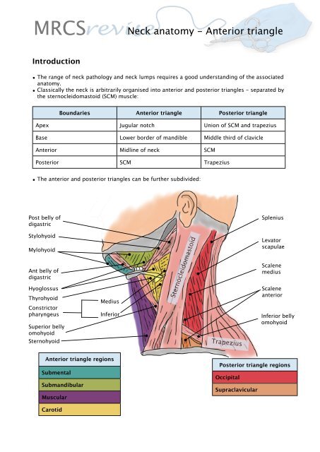
• The range of neck pathology and neck lumps requires a good understanding of the associated<br />
<strong>anatomy</strong>.<br />
• Classically the neck is arbitrarily organised into anterior and posterior <strong>triangle</strong>s - separated by<br />
the sternocleidomastoid (SCM) muscle:<br />
Boundaries <strong>Anterior</strong> <strong>triangle</strong> Posterior <strong>triangle</strong><br />
Apex Jugular notch Union of SCM and trapezius<br />
Base Lower border of mandible Middle third of clavicle<br />
<strong>Anterior</strong> Midline of neck SCM<br />
Posterior SCM Trapezius<br />
• The anterior and posterior <strong>triangle</strong>s can be further subdivided:<br />
Post belly of<br />
digastric<br />
Stylohyoid<br />
Mylohyoid<br />
Ant belly of<br />
digastric<br />
Hyoglossus<br />
Thyrohyoid<br />
Constrictor<br />
pharyngeus<br />
Superior belly<br />
omohyoid<br />
Sternohyoid<br />
Medius<br />
Inferior<br />
<strong>Anterior</strong> <strong>triangle</strong> regions<br />
Submental<br />
Submandibular<br />
Muscular<br />
Carotid<br />
Sternocleidomastoid<br />
Trapezius<br />
Splenius<br />
Levator<br />
scapulae<br />
Scalene<br />
medius<br />
Scalene<br />
anterior<br />
Inferior belly<br />
omohyoid<br />
Posterior <strong>triangle</strong> regions<br />
Occipital<br />
Supraclavicular
<strong>MRCS</strong> <strong>Neck</strong> <strong>anatomy</strong> - <strong>Anterior</strong> <strong>triangle</strong><br />
Submandibular region<br />
• The region immediately below the mandible.<br />
Boundaries <strong>Anterior</strong> <strong>triangle</strong><br />
Superior Body of mandible<br />
<strong>Anterior</strong> <strong>Anterior</strong> belly of digastric<br />
Posterior Posterior belly of digastric<br />
Roof Skin, superficial fascia, platysma, deep fascia<br />
Floor Mylohyoid muscle<br />
Superficial submandibular structures:<br />
Platysma - broad sheet of superficial muscle arising from<br />
the fascia that overlaps sternocleidomastoid.<br />
Facial vein<br />
Cervical branch of facial nerve - supplies platysma<br />
Submandibular gland:<br />
• Accounts for ≈ 70% of salivary production<br />
• Wharton’s duct (submandibular duct) arises from the<br />
gland and passes between mylohyoid, hyoglossus and<br />
genioglossus - eventually opening either the side of the<br />
lingula frenulum.<br />
• Parasympathetic supply - from a branch of the facial<br />
nerve the chorda tympani (CNVII) which runs in the<br />
sheath of the lingual nerve (branch of mandibular nerve<br />
CNV3) to synapse at the submandibular ganglion -<br />
which supplies the submandibular gland and lies on<br />
hyoglossus.<br />
Deep submandibular structures:<br />
Facial artery<br />
Lingual nerve -branch of mandibular nerve (CNV3)<br />
Submandibular ganglion<br />
Hypoglossal nerve (CNXII) - supplies all muscles of<br />
tongue except palatoglossus.<br />
Lingual artery
<strong>MRCS</strong> <strong>Neck</strong> <strong>anatomy</strong> - <strong>Anterior</strong> <strong>triangle</strong><br />
Submental region<br />
• The region between the two anterior bellies of digastric.<br />
Boundaries <strong>Anterior</strong> <strong>triangle</strong><br />
Superior Body of mandible<br />
<strong>Anterior</strong> Midline<br />
Posterior <strong>Anterior</strong> belly of digastric<br />
Roof Skin, superficial fascia, platysma, deep fascia<br />
Floor Mylohyoid muscle<br />
• Contents:<br />
• Submental lymph nodes.<br />
• Superficial veins which form tributaries of the anterior jugular veins.<br />
Carotid <strong>triangle</strong><br />
• Contains the carotid sheath.<br />
<strong>Anterior</strong> belly of digastric<br />
Submental lymph nodes<br />
Stylohyoid<br />
Posterior belly of digastric<br />
<strong>Anterior</strong> jugular veins<br />
Hyoid<br />
Boundaries <strong>Anterior</strong> <strong>triangle</strong><br />
Superior Stylohyoid and posterior belly of digastric<br />
Inferior Superior belly of omohyoid<br />
Posterior Sternocleidomastoid<br />
Roof Skin, superficial fascia, platysma, deep fascia<br />
Floor Thyrohyoid, hyoglossus, constrictor<br />
pharyngeus medius/inferior.
<strong>MRCS</strong> <strong>Neck</strong> <strong>anatomy</strong> - <strong>Anterior</strong> <strong>triangle</strong><br />
Carotid <strong>triangle</strong><br />
Hypoglossal nerve continues to supply all muscles<br />
of the tongue except palatoglossus.<br />
Superficial structures<br />
The superficial veins are the first structures encountered<br />
on removing the roof of the carotid <strong>triangle</strong>:<br />
Common facial vein<br />
Retromandibular vein<br />
Posterior auricular vein<br />
External jugular vein - commences at level of the angle of<br />
the mandible runs obliquely over sternocleidomastoid. At<br />
the midpoint of the clavicle - in the subclavian <strong>triangle</strong><br />
penetrates the deep fascia to drain into the subclavian<br />
vein.<br />
Internal jugular vein - deeper contained within the carotid<br />
sheath (see below).<br />
<strong>Anterior</strong> jugular vein (note not within carotid <strong>triangle</strong>)<br />
Post removal/reflection of sternocleidomastoid -<br />
overlying nerves and internal jugular vein<br />
Hypoglossal nerve and branch of first cervical - a branch<br />
of the first cervical nerve courses in the same sheath as<br />
the hypoglossal nerve then leaves as two branches in the<br />
carotid <strong>triangle</strong>:<br />
1. Superior root of ansa cervicalis - supplies infrahyoid<br />
muscles (see muscular <strong>triangle</strong>).<br />
2. C1 nerve directly to thyrohyoid.<br />
Ansa cervicalis - a loop of nerves derived from the<br />
cervical plexus - receives supply from C1-3 nerve roots.<br />
Innervates the infrahyoid muscles: sternohyoid,<br />
sternothyroid and omohyoid.<br />
Internal jugular vein - lies lateral to the internal carotid<br />
and common carotid. At the root of the neck joins the<br />
subclavian vein to form the brachiocephalic vein.<br />
Combined with the carotid and vagus is contained within<br />
the carotid sheath (see below).
<strong>MRCS</strong> <strong>Neck</strong> <strong>anatomy</strong> - <strong>Anterior</strong> <strong>triangle</strong><br />
Carotid <strong>triangle</strong><br />
Post removal of internal jugular vein<br />
External carotid artery<br />
Occipital artery<br />
Facial artery<br />
Lingual artery<br />
Ascending pharyngeal artery<br />
Superior thyroid artery<br />
Internal carotid artery<br />
Post removal of carotid artery<br />
Vagus nerve - contained within carotid sheath (with<br />
carotid and internal jugular vein)<br />
Internal branch of superior laryngeal nerve - supplies<br />
sensation to the epiglottis - irritation causes coughing.<br />
External branch of superior laryngeal nerve- supplies<br />
cricothyroid muscle - Clinical note: Susceptible to<br />
damage during thyroidectomy as lies inferior and close to<br />
superior thyroid artery (which is ligated during<br />
thyroidectomy). Palsy results in loss of high pitch voice.
<strong>MRCS</strong> <strong>Neck</strong> <strong>anatomy</strong> - <strong>Anterior</strong> <strong>triangle</strong><br />
Muscular <strong>triangle</strong><br />
Boundaries <strong>Anterior</strong> <strong>triangle</strong><br />
Superior Superior belly of omohyoid<br />
<strong>Anterior</strong> Midline of neck<br />
Inferior Sternocleidomastoid<br />
Roof Skin, superficial fascia, platysma, deep fascia<br />
• The muscular <strong>triangle</strong> contains the infrahyoid muscles (also known as the strap muscles) whose<br />
primary function is to depress the hyoid and larynx during swallowing.<br />
• All are supplied by the ansa cervicalis (see above) except thyrohyoid which is directly supplied<br />
by a branch of C1 nerve.<br />
Superficial layer<br />
Superior belly omohyoid - omohyoid consists of 2 bellies<br />
separated by an intermediate tendon. It arises from the<br />
scapula. The inferior belly then courses along the clavicle<br />
bound by a fibrous expansion. Behind the<br />
sternocleidomastoid it becomes tendinous then courses<br />
vertically upwards as the superior belly to insert onto the<br />
hyoid.<br />
Sternohyoid- sternum to hyoid<br />
Deep layer<br />
Thyrohyoid - thyroid cartilage to hyoid<br />
Sternothyroid - sternum to thyroid cartilage<br />
Thyroid - see separate revision sheet<br />
Inferior belly omohyoid