Anterior triangle of the neck
Anterior triangle of the neck
Anterior triangle of the neck

Create successful ePaper yourself
Turn your PDF publications into a flip-book with our unique Google optimized e-Paper software.
<strong>Anterior</strong> <strong>triangle</strong> <strong>of</strong> <strong>the</strong> <strong>neck</strong><br />
Written by : Hisham Saleem<br />
Dr.Zuhair Fadhl<br />
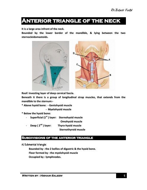
It is a large area infront <strong>of</strong> <strong>the</strong> <strong>neck</strong>.<br />
Bounded by <strong>the</strong> lower border <strong>of</strong> <strong>the</strong> mandible, & lying between <strong>the</strong> two<br />
sternocleidomastoids.<br />
Ro<strong>of</strong>: Investing layer <strong>of</strong> deep cervical fascia.<br />
Beneath it <strong>the</strong>re is a group <strong>of</strong> longitudinal strap muscles, that extends from <strong>the</strong><br />
mandible to <strong>the</strong> sternum:-<br />
* Above hyoid bone: - Geniohyoid muscle<br />
- Myelohyoid muscle<br />
* Below <strong>the</strong> hyoid bone:<br />
- Superficial (1 st ) layer: Sternohyoid muscle<br />
Omohyoid muscle<br />
- Deep ( 2 nd ) layer: Thyro-hyoid muscle<br />
Sternothyroid muscle<br />
Subdivisions <strong>of</strong> <strong>the</strong> anterior <strong>triangle</strong><br />
A) Submental <strong>triangle</strong>:<br />
Bounded by : <strong>the</strong> 2 bellies <strong>of</strong> digastric & <strong>the</strong> hyoid bone.<br />
Floor formed by : <strong>the</strong> myelohyoid muscle<br />
Occupied by : lymphnodes.<br />
1
B) Digastric (Submandibular) <strong>triangle</strong>:<br />
Written by : Hisham Saleem<br />
Dr.Zuhair Fadhl<br />
Bounded by : <strong>the</strong> ant. belly <strong>of</strong> <strong>the</strong> digastric muscle, stylohyoid muscle & <strong>the</strong> lower<br />
border <strong>of</strong> <strong>the</strong> mandible.<br />
Floor formed by: myelohyoid & hyoglossus muscles.<br />
Occupied by: Submandibular lymphnodes & Submandibular salivary glands.<br />
C) Carotid <strong>triangle</strong>:<br />
Bounded by: <strong>the</strong> sternocleidomastoid muscle, posterior belly <strong>of</strong> digastric & <strong>the</strong><br />
superior belly <strong>of</strong> omohyoid muscles.<br />
Floor formed by: <strong>Anterior</strong>ly; formed by <strong>the</strong> hyoglossus & thyrohyoid muscles<br />
Posteriorly; by middle & inferior constrictor muscles.<br />
Occupied by: <strong>the</strong> vertical nervovascular bundle <strong>of</strong> <strong>the</strong> <strong>neck</strong>, covered by <strong>the</strong><br />
sternocleidomastoid muscle.<br />
D) Muscular <strong>triangle</strong>:<br />
Bounded by: <strong>the</strong> sternocleidomastoid, superior belly <strong>of</strong> omohyoid & median plane<br />
(mid line).<br />
Occupied by: <strong>the</strong> infra hyoid muscles.<br />
Supra Hyoid muscles ( muscles form <strong>the</strong> floor <strong>of</strong><br />
mouth)<br />
1) Myelohyoid<br />
2) Geniohyoid muscle<br />
2
Infra Hyoid muscles<br />
Sternohyoid muscle:<br />
Written by : Hisham Saleem<br />
Dr.Zuhair Fadhl<br />
Flat strap muscle situated close to each o<strong>the</strong>r above but diverge from each o<strong>the</strong>r<br />
below.<br />
Origin: from lower border <strong>of</strong> hyoid bone<br />
Insertion: to <strong>the</strong> back <strong>of</strong> sterno clavicular joint & adjoining parts <strong>of</strong> manubrium &<br />
clavicle.<br />
Nerve supply: Branch from ansa cervicalis C1,2,3 enter through its lower end &<br />
supply it segmentally.<br />
Tendinous insections may be present which indicate segmental origin <strong>of</strong> <strong>the</strong> muscle.<br />
3
Omohyoid muscle:<br />
Long muscle formed <strong>of</strong> 2 bellies and intermediate tendon.<br />
Written by : Hisham Saleem<br />
Dr.Zuhair Fadhl<br />
Superior belly:<br />
Origin: from lateral part <strong>of</strong> <strong>the</strong> inferior border <strong>of</strong> hyoid bone, descends obliquely<br />
downward passing beneath <strong>the</strong> sternomastiod muscle & over <strong>the</strong> carotid<br />
sheath.<br />
Insertion : muscle fibers are replaced by flat intermediate tendon.<br />
Inferior belly:<br />
Origin: attached to <strong>the</strong> transverse scapular ligament & upperborder <strong>of</strong> <strong>the</strong><br />
scapula. Situated horizontal to <strong>the</strong> upper border <strong>of</strong> <strong>the</strong> clavicle.<br />
Insertion: Intermediate tendon, which is bound down to <strong>the</strong> clavicle by facial sling<br />
(from deep cervical fascia).<br />
Nerve supply: segmentally by C1,2,3 by <strong>the</strong> ansa cervicalis.<br />
Action: when <strong>the</strong> head is rotated to one side, <strong>the</strong> opposite omohyoid is straightened &<br />
can exert a more downward pull on <strong>the</strong> hyoid bone.<br />
Thyro hyoid muscle:<br />
Flat broad strap-like muscle, lies beneath sternohyoid & omohyoid muscles.<br />
Origin: greater horn <strong>of</strong> hyoid bone.<br />
Insertion: oblique line <strong>of</strong> thyroid cartilage, end to end with sterno thyroid muscle.<br />
Nerve supply: branch <strong>of</strong> hypoglossal nerve carries C1 fibers.<br />
Sterno thyroid muscle:<br />
Broad flat muscle.<br />
Origin: oblique line <strong>of</strong> thyroid cartilage.<br />
Insertion: posterior surface <strong>of</strong> manubrium below Sternohyoid & from manubrium<br />
to first costal cartilage.<br />
Nerve supply: ansa cervicalis C2, 3.(C1 to thyrohyoid)<br />
4
Action <strong>of</strong> infrahyoid muscles.<br />
Written by : Hisham Saleem<br />
Dr.Zuhair Fadhl<br />
Depression <strong>of</strong> <strong>the</strong> larynx: sternothyroid acts directly on thyroid cartilage, <strong>the</strong><br />
o<strong>the</strong>rs indirectly via hyoid bone.<br />
Opponent <strong>of</strong> <strong>the</strong> elevators <strong>of</strong> <strong>the</strong> larynx (myelohyoid, palatopharyngeus,<br />
stylopharyngeus & salpingopharyngeus).<br />
Prevent ascent <strong>of</strong> <strong>the</strong> hyoid bone when <strong>the</strong> digastric muscle is in action.<br />
Digastric muscle:<br />
Composed <strong>of</strong> 2 bellies & an intermediate tendon.<br />
It is formed by fusion <strong>of</strong> component <strong>of</strong> 1st & 2nd pharyngeal arches.<br />
<strong>Anterior</strong> belly: a fleshy belly<br />
Origin: from digastric notch on <strong>the</strong> medial surface <strong>of</strong> <strong>the</strong> mandible.<br />
Insertion: it tapers down to <strong>the</strong> intermediate tendon.<br />
Posterior belly:<br />
Origin: medial surface <strong>of</strong> mastoid process.<br />
Insertion: Intermediate tendon.<br />
Intermediate tendon:<br />
Held by fibrous sling to <strong>the</strong> lesser cornu <strong>of</strong> <strong>the</strong> hyoid bone, which Is lubricated by<br />
synovial sheath.<br />
Nerve supply: <strong>Anterior</strong> belly: myelohyoid nerve (V).<br />
Posterior belly: facial nerve (VII).<br />
Action: depresses & retracts <strong>the</strong> chin.<br />
5