Pulpal and Periapical Pathoses & Osteomyelitis
Pulpal and Periapical Pathoses & Osteomyelitis
Pulpal and Periapical Pathoses & Osteomyelitis

Create successful ePaper yourself
Turn your PDF publications into a flip-book with our unique Google optimized e-Paper software.
<strong>Pulpal</strong> <strong>and</strong> <strong>Periapical</strong> <strong>Pathoses</strong><br />
& <strong>Osteomyelitis</strong><br />
All pictures are intellectual property of the Division of Oral <strong>and</strong><br />
Maxillofacial Pathology or its Faculty. Duplication or any unauthorized<br />
use is prohibited.<br />
• Acute or chronic<br />
• Subtotal or generalized<br />
• Infected or sterile<br />
Pulpitis<br />
Reversible Pulpitis<br />
Temperature extremes, sweet or sour food<br />
–Mild to moderate pain<br />
–Sudden<br />
–Short duration<br />
<strong>Pulpal</strong> Pathology<br />
• Pulpitis<br />
– Similar characteristics with other inflammatory lesions<br />
– Difference: Confined area<br />
– Dilatation, edema, strangulation of capillary flow,<br />
vessel damage, inflammation <strong>and</strong> necrosis<br />
– Mechanical, Thermal, Chemical, Bacterial<br />
• Reversible<br />
• Irreversible<br />
• Chronic hyperplastic<br />
Pulpitis<br />
Reversible Pulpitis<br />
• Pain DOES NOT occur without stimulation<br />
• Subsides seconds after removal of stimulus<br />
• EPT: lower levels than tooth control<br />
• No mobility, no sensitivity to percussion<br />
• If stimulus continuous irreversible<br />
1
Irreversible Pulpitis<br />
•Early<br />
– Sharp, severe pain upon thermal stimulation<br />
– Pain continues after removal of stimulus<br />
– COLD uncomfortable (also warm <strong>and</strong> sweet)<br />
– Spontaneous or continuous<br />
– EPT: lower levels<br />
– Pain can be localized<br />
– Patient may be able to point to the offending tooth<br />
• With increasing discomfort, patient may be unable<br />
Irreversible Pulpitis<br />
• NO BLACK OR WHITE<br />
• Patients may have no symptoms<br />
• Severe pulpitis <strong>and</strong> abscess formation may be<br />
asymptomatic<br />
• Mild pulpitis may cause excruciating pain<br />
Irreversible Pulpitis<br />
•Late<br />
– Pain increases in intensity<br />
– Throbbing pressure (night owl)<br />
– Heat increases pain<br />
– Cold MAY PROVIDE RELIEF<br />
– EPT: HIGHER OR NO RESPONSE<br />
– Usually no mobility or sensitivity to percussion<br />
– If the inflammation spreads beyond the apical area you may<br />
get sensitivity to percussion<br />
Chronic hyperplastic pulpitis<br />
• Pulp polyp<br />
• Large exposure<br />
• Children or youth<br />
• Deciduous teeth<br />
• Hyperplastic granulation tissue that can become<br />
epithelialized from shedding epithelial cells<br />
• Open apex decreases the chances of pulpal necrosis<br />
2
Chronic localized osteitis<br />
• Apical inflammatory lesion<br />
• Defensive reaction<br />
• Bacteria in the pulp <strong>and</strong> spread of toxins<br />
• Defense in the beginning<br />
• With time the reaction less effective<br />
• Can arise after quiescence of periapical abscess,<br />
or may develop as initial periapical pathosis<br />
• Static or development of periapical cyst<br />
Chronic localized osteitis<br />
• RCT<br />
• Lesion may fail to heal because of<br />
– Cyst formation<br />
– Inadequate RCT<br />
– Root fracture<br />
– <strong>Periapical</strong> foreign material<br />
– Periodontal disease<br />
– Maxillary sinus penetration<br />
– Fibrous scar (no bone fill)<br />
Chronic apical periodontitis<br />
Chronic localized osteitis<br />
So-called dental granuloma<br />
True dental granuloma<br />
Chronic localized osteitis<br />
• Asymptomatic, pain or sensitivity if acute<br />
exacerbation occurs<br />
• No mobility or significant sensitivity to percussion<br />
• Soft tissue overlying lesion may be tender<br />
• No response on EPT or thermal tests<br />
• X-ray: Radiolucency, circumscribed or ill-defined,<br />
usually small; root resorption may be present<br />
Chronic localized osteitis<br />
• Repeat RCT<br />
• <strong>Periapical</strong> surgery <strong>and</strong> retrofill<br />
• Histopathologic examination because<br />
– You must have a record<br />
– The patient may not have periapical inflammatory lesion<br />
after all<br />
3
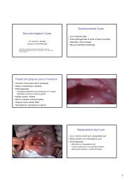
<strong>Periapical</strong> Cyst<br />
• Well-defined radiolucency with sometimes<br />
sclerotic border<br />
• Size or shape of radiolucency cannot differentiate<br />
between osteitis or granuloma <strong>and</strong> cyst<br />
<strong>Periapical</strong> Cyst<br />
• Rests of Malassez<br />
• Crevicular epithelium<br />
• Sinus epithelium<br />
• Lateral location (perio or pulpal disease)<br />
• Residual cyst<br />
• No symptoms generally<br />
• Mobility may be present<br />
• NO RESPONSE<br />
4
<strong>Periapical</strong> Abscess<br />
• People talk about acute <strong>and</strong> chronic abscesses<br />
• THEY ARE MISINFORMING YOU<br />
• IN ABSCESSES YOU HAVE ACUTE<br />
INFLAMMATION. (PERIOD)<br />
<strong>Periapical</strong> Abscess<br />
• Thickening of apical periodontal ligament<br />
• Ill-defined radiolucency<br />
• No alterations detected sometimes<br />
<strong>Periapical</strong> Abscess<br />
• Can be the initial pathosis<br />
• Usually carious teeth but also trauma<br />
• Acute apical periodontitis (acute localized osteitis)<br />
may or may not proceed abscess formation<br />
– Usually non-vital tooth<br />
– Tooth may be vital in cases of trauma<br />
• Occlusal contacts, or wedging a foreign object<br />
<strong>Periapical</strong> Abscess<br />
• Symptomatic or asymptomatic<br />
• Phoenix abscess (acute exacerbation of chronic inflammatory<br />
process)<br />
• Initially tenderness that can be relieved by pressure<br />
• With progression more intense pain, extreme sensitivity to<br />
percussion, extrusion of tooth <strong>and</strong> swelling of tissues<br />
• No Response to cold or EPT<br />
• General symptoms<br />
6
<strong>Periapical</strong> Abscess<br />
• If sinus tract develops you may have presence of<br />
little mass on the alveolus or palate or soft tissues<br />
or skin with an opening.<br />
• Buccal surface<br />
• Maxillary laterals, palatal roots of molars <strong>and</strong><br />
m<strong>and</strong>ibular 2 nd <strong>and</strong> 3 rd molars may drain lingual<br />
•PUS<br />
• Less symptoms because of drainage<br />
<strong>Periapical</strong> Abscess<br />
MAY LEAD TO:<br />
–OSTEOMYELITIS<br />
–CELLULITIS<br />
<strong>Periapical</strong> Abscess<br />
• Histopathology<br />
– Well delineated accumulation of PMNs, exudate,<br />
cellular debris, necrotic material, bacteria<br />
7
<strong>Osteomyelitis</strong><br />
• Acute or chronic<br />
• Different form osteoradionecrosis<br />
• Variations<br />
– Focal or diffuse sclerosing<br />
– Proliferative periostitis<br />
– Alveolar osteitis (dry socket)<br />
<strong>Osteomyelitis</strong><br />
• Predisposing factors<br />
– Chronic systemic diseases<br />
– Immunocompromised status<br />
– Tobacco use, alcohol abuse, drug abuse<br />
– Diabetes mellitus<br />
– Infections<br />
– Tumors or tumor-like processes<br />
• Fever<br />
• Leukocytosis<br />
• Lymphadenopathy<br />
• Swelling<br />
• Sensitivity<br />
Acute osteomyelitis<br />
<strong>Osteomyelitis</strong><br />
• True osteomyelitis is uncommon<br />
– Odontogenic infection or fracture<br />
– Associated with ANUG Noma<br />
•Acute<br />
– Symptoms of acute inflammation<br />
– Fever, leukocytosis, lymphadenopathy, significant<br />
sensitivity, swelling; sequestrum, involucrum<br />
• Chronic<br />
– May arise without acute phase<br />
Acute osteomyelitis<br />
• Insufficient time for reaction by the body<br />
• Spreads in the medullary spaces<br />
• X-ray: Spectrum (No lesion ill-defined radiolucency)<br />
• Sequestrum<br />
• Involucrum<br />
Acute osteomyelitis<br />
• Antibiotics <strong>and</strong> drainage<br />
• Penicillin, clindamycin, cephalexin, gentamycin<br />
• Sequestra should be removed<br />
8
Chronic osteomyelitis<br />
• Features similar to acute<br />
• Patchy, ill-defined radiolucencies<br />
• Radiopaque sequestra (pts. can loose significant bone<br />
proper)<br />
• Periosteal bone reaction<br />
Chronic osteomyelitis<br />
• May arise without acute phase<br />
• Granulation tissue<br />
• Scar formation<br />
• Reservoir of bacteria<br />
• Antibiotics do not reach easily the area<br />
• Aggressive management<br />
• Intravenous antibiotics<br />
• Removal of necrotic bone<br />
• Immobilization of jaws<br />
• Hyperbaric oxygen<br />
Chronic osteomyelitis<br />
9
Condensing Osteitis<br />
• Teeth have pathosis or restoration<br />
• Sclerotic bone<br />
• No clinical expansion<br />
• Density without lucent border<br />
• Vs. osteosclerosis: Not separated from apex<br />
Osteosclerosis<br />
10
Dry socket (alveolar osteitis)<br />
• Destruction of blood clot in the socket of an<br />
extracted tooth<br />
• Fibrinolysis <strong>and</strong> formation of kinins pain<br />
•Causes<br />
– Inexperience<br />
– Trauma<br />
– Oral contraceptives<br />
– Smoking<br />
– Estrogens<br />
Proliferative Periostitis<br />
Proliferative periostitis<br />
• Garrè osteomyelitis (wrong term)<br />
• Periosteal reaction<br />
• Children<br />
• Caries, dental inflammatory disease<br />
• Occlusal or lateral oblique radiographs show<br />
opaque laminations like onion skin<br />
Onion-skin<br />
Cellulitis<br />
• Spread of abscess in fascial planes of soft tissues<br />
• Ludwig’s angina<br />
– Subm<strong>and</strong>ibular region<br />
–Lower molars<br />
– Trauma, lacerations, peritonsillar infections<br />
– Extension to pharyngeal <strong>and</strong> mediastinal spaces<br />
• Cavernous sinus thrombosis<br />
– Maxillary molars <strong>and</strong> premolars<br />
– Maxillary sinus, infratemporal fossa, orbit <br />
cavernous sinus at the cranial vault<br />
11
Cellulitis<br />
• Spread of abscess in fascial planes of soft tissues<br />
• Ludwig’s angina<br />
– Swelling: floor of mouth, tongue, subm<strong>and</strong>ibular region<br />
– Woody tongue <strong>and</strong> bull neck<br />
• Cavernous sinus thrombosis<br />
– Edematous periorbital enlargement<br />
– Protrusion <strong>and</strong> fixation of eyelid <strong>and</strong> pupil dilatation<br />
• Blindness<br />
– CNS involvement, sometimes brain abscess<br />
– Deepening stupor, delirium<br />
Cellulitis<br />
• Ludwig’s angina<br />
– Maintenance of airway<br />
– Antibiotic treatment<br />
– Surgical drainage<br />
– Tracheostomy<br />
• Cavernous sinus thrombosis<br />
– Antibiotics<br />
– Extraction of tooth<br />
– Corticosteroids to avoid vascular collapse from pituitary<br />
dysfunction<br />
12