Identify and Define All Diagnostic Terms for Pulpal - American ...
Identify and Define All Diagnostic Terms for Pulpal - American ...
Identify and Define All Diagnostic Terms for Pulpal - American ...

Create successful ePaper yourself
Turn your PDF publications into a flip-book with our unique Google optimized e-Paper software.
Review Article<br />
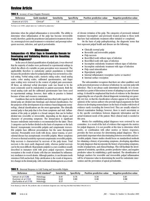
TABLE 8. Accuracy of Laser Doppler Flowmetry<br />
Reference Gold st<strong>and</strong>ard Sensitivity Specificity Positive predictive value Negative predictive value<br />
Evans et al (121) Clinical a<br />
1.0 1.0 — —<br />
a In Evans et al (1999), pulpal status was ‘‘confirmed by pulpectomy.’’<br />
determine when the pulpal inflammation is irreversible. The ability to<br />
determine when inflammation of the pulp has become irreversible<br />
would, there<strong>for</strong>e, guide the practitioner <strong>and</strong> patient in treatment choices<br />
(ie, nonsurgical root canal treatment vs extraction) <strong>and</strong> preempt subsequent<br />
necrosis, infection, <strong>and</strong> apical periodontitis.<br />
Discussion<br />
Subquestion #5: What Gaps in Knowledge Remain <strong>for</strong><br />
Developing <strong>and</strong> Validating Metrics <strong>and</strong> the Resulting<br />
<strong>Pulpal</strong> Diagnoses?<br />
In the area of clinical quantification of pulpal pain, it was observed<br />
that the majority of studies were per<strong>for</strong>med in experimental settings in<br />
which the effects of a variable on pain perception were measured. The<br />
applicability there<strong>for</strong>e to endodontic patient populations is limited<br />
because the predictive value <strong>for</strong> pulpal pathology was not tested in a clinical<br />
setting. Verbal rating scales, numeric rating scales, visual analog<br />
scales, color analog scales, calibrated questionnaires, <strong>and</strong> finger<br />
span scaling were reviewed in the context of pulpal pain assessment.<br />
Of these, an in<strong>for</strong>mal verbal descriptor scale was found to be the<br />
most commonly used by endodontists in patient assessment. Both the<br />
visual analog scale <strong>and</strong> the calibrated questionnaire have been used<br />
in experimental settings; however, their utility in practice is limited<br />
because of time <strong>and</strong> resource constraints.<br />
Conditions that can be identified <strong>and</strong> described with regard to the<br />
dental pulp are divided into histologic <strong>and</strong> clinical classifications. For<br />
the purposes of the development of an evidence-based diagnostic terminology,<br />
clinical classifications are the most appropriate. The clinically<br />
normal pulp is that pulp that is free from symptoms <strong>and</strong> vital. Inflammation<br />
of the pulp or pulpitis is a broad category that can be further<br />
divided into reversible or irreversible, depending on the degree <strong>and</strong><br />
character of presenting symptoms. The demarcation is significant<br />
because endodontic intervention is recommended <strong>for</strong> the latter. These<br />
2 categories can be further divided on the basis of symptoms or the lack<br />
thereof. Asymptomatic irreversible pulpitis <strong>and</strong> symptomatic irreversible<br />
pulpitis have different presentations but the same therapeutic<br />
outcome. Presumably every tooth with decay, minor trauma, or periodontal<br />
disease has asymptomatic reversible pulpitis. Minor symptoms<br />
of sweet or thermal sensitivity represent symptomatic reversible pulpitis.<br />
Pulp necrosis is characterized by necrosis of the pulp tissue. Total<br />
necrosis is the most easily diagnosed entity, whereas partial necrosis<br />
can be the most difficult. Hyperplastic pulpitis is a rare condition usually<br />
described in immature teeth with gross pulpal exposures. Internal<br />
resorption is the result of clastic cells that are stimulated by inflammatory<br />
mediators to resorb dentin. Although painless, it can threaten tooth<br />
retention if left unchecked. Pulp calcification is the result of degenerative<br />
changes in the dental pulp, with exuberant dentinogenesis as a result<br />
of chronic irritation of the pulp. The categories of previously initiated<br />
treatment (incomplete) <strong>and</strong> previously treated pertain to those teeth<br />
that have had endodontic treatment either initiated or completed.<br />
On the basis of pulp pathophysiology, the diagnostic terms that<br />
best represent pulpal health <strong>and</strong> disease are the following:<br />
Clinically normal pulp<br />
Reversible <strong>and</strong> irreversible pulpitis<br />
Pulp necrosis<br />
Root-filled tooth without signs of infection<br />
Root-filled tooth with signs of infection<br />
Incomplete endodontic treatment without signs of infection<br />
Incomplete endodontic treatment with signs of infection<br />
Pulp canal mineralization<br />
Hyperplastic pulpitis<br />
Internal inflammatory resorption (active or inactive)<br />
Internal surface resorption<br />
The subcommittee recognizes that there are other qualifiers such<br />
as the perceived presence or absence of infection (ie, necrotic pulp with<br />
infection). This is not always easily determined clinically. It is recommended<br />
as a point of discussion in terms of adopting it as part of terminology.<br />
It should be emphasized that levels of evidence in the literature<br />
supporting the use of specific clinical diagnostic terminology are generally<br />
very low, in that the classification schemes appear to be mainly the<br />
opinions of the various authors who provide logical arguments <strong>for</strong> their<br />
choices in developing nomenclature on the basis of studies with levels of<br />
evidence rarely exceeding the lowest level. They are usually related to<br />
clinical examination findings; however, there is much uncertainty as<br />
to the specific correlations between diagnostic in<strong>for</strong>mation <strong>and</strong> the<br />
actual treatment needs of the patient. More clinical study is needed in<br />
this area.<br />
Metrics <strong>for</strong> establishing pulpal diagnoses were reviewed by our<br />
committee. As a result of the lack of evidence that supports the metrics<br />
<strong>for</strong> pulpal diagnosis, it is not possible at this time to determine which<br />
metric, or combination with other metrics or history responses,<br />
provides the best accuracy <strong>for</strong> determining pulpal diagnoses. This is<br />
particularly important when discriminating between reversible <strong>and</strong> irreversible<br />
pulpitis. Future studies should focus on st<strong>and</strong>ardized methods<br />
<strong>for</strong> obtaining a history of presenting symptoms, developing algorithms<br />
<strong>for</strong> pulp diagnoses that incorporate the history of presenting symptoms,<br />
results of pulpal tests, <strong>and</strong> clinical findings. This will facilitate the development<br />
of sensitivity, specificity, positive predictive values, <strong>and</strong> negative<br />
predictive values by establishing a gold st<strong>and</strong>ard. The identification of<br />
biologic markers <strong>for</strong> reversible <strong>and</strong> irreversible pulpal inflammation<br />
will be of immense value in determining the need <strong>for</strong> endodontic intervention<br />
<strong>and</strong> the prevention of apical periodontitis.<br />
TABLE 9. Abnormal Response to Cold Testing <strong>and</strong> Irreversible Pulpitis<br />
Reference Gold st<strong>and</strong>ard’’ Sensitivity Specificity Positive predictive value Negative predictive value<br />
Seltzer et al (128) Histology 0.41 0.76 0.34 0.81<br />
Dummer et al (132) Histology 0.63 0.80 0.48 0.88<br />
Garfunkle et al (119) Histology 0.57 — — —<br />
From Hyman JJ, Cohen M. The predictive value of endodontic diagnostic tests. Oral Surg Oral Med Oral Pathol 1984;58:343–6.<br />
1654 Levin et al. JOE — Volume 35, Number 12, December 2009