Identify and Define All Diagnostic Terms for Pulpal - American ...
Identify and Define All Diagnostic Terms for Pulpal - American ...
Identify and Define All Diagnostic Terms for Pulpal - American ...

Create successful ePaper yourself
Turn your PDF publications into a flip-book with our unique Google optimized e-Paper software.
Review Article<br />
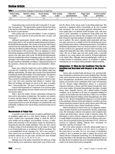
TABLE 1. Use of Dolorimetry Technique in Observations of <strong>Pulpal</strong> Pain<br />
Verbal rating<br />
scales<br />
Numeric rating<br />
scales<br />
Visual analog<br />
scales<br />
Visual analog scales consist of a line with 2 end points of ‘‘no pain’’<br />
<strong>and</strong> ‘‘worst pain ever.’’ The patient marks a point on the line that relates<br />
to the intensity of their pain. The distance of that point from ‘‘no pain’’ is<br />
the measure of pain intensity.<br />
Color analog scales are used with children. A series of graded in<br />
intensity colors are anchored at each end by the terms ‘‘no pain’’ <strong>and</strong><br />
‘‘worst pain.’’<br />
Calibrated questionnaires should really be calibrated questionnaire<br />
because there is only one that has gained widespread acceptance,<br />
the McGill Pain Questionnaire. This consists of 20 groups of descriptors<br />
selected from the medical literature that describe the sensory qualities<br />
of the pain, the affective qualities of the pain, or are evaluative describing<br />
the overall intensity of the experience. These are displayed on a <strong>for</strong>m<br />
that includes diagrams used <strong>for</strong> localization. A pain rating index is determined<br />
on the rank values of the words. The McGill Pain Questionnaire<br />
has been translated into at least 16 languages <strong>and</strong> is very widely used. Its<br />
advantage is that it allows measurement of the different components of<br />
the pain experience individually, providing a 3-dimensional measure of<br />
the experience, whereas the other scales are predominantly 1-dimensional.<br />
Finger span scaling has largely been used in children because it<br />
overcomes the complexities of other scales that children might have<br />
difficulty underst<strong>and</strong>ing. The finger span concept is first demonstrated<br />
by holding the thumb <strong>and</strong> <strong>for</strong>efinger of one h<strong>and</strong> together. The patient is<br />
told that the fingers in this position represent ‘‘no hurt’’ (or ‘‘no pain’’).<br />
Then a spread of a small distance between the fingers is shown to represent<br />
a ‘‘tiny’’ hurt, <strong>and</strong> a somewhat wider distance is ‘‘medium’’ hurt.<br />
When the <strong>for</strong>efinger <strong>and</strong> thumb are moved as far apart as possible,<br />
this is ‘‘most possible hurt.’’ The span in each instance is measured.<br />
Cortical evoked potentials are components of an electroencephalogram<br />
taken while applying a noxious stimulus <strong>and</strong> can be used with an<br />
unconscious subject.<br />
Table 1 shows the number of times a dolorimetry technique has<br />
been used in observations of pulpal pain. The numbers are numbers<br />
of reports <strong>and</strong> thus biased by investigators who have used the same<br />
approach in multiple studies. In some studies more than 1 approach<br />
to pain measurement was used. <strong>All</strong> techniques are included in the count<br />
individually, resulting in some reports being counted more than once in<br />
the table.<br />
Measurement of <strong>Pulpal</strong> Pain<br />
A systematic review of the literature revealed no published reports<br />
of quantifying pulpal pain in a truly clinical situation. <strong>All</strong> available<br />
reports resulted from experimental settings in which the effect of<br />
some variable such an analgesic, local anesthetic, exercises, or orthodontic<br />
tooth movement on the perception of pain was determined by<br />
measuring the pain after pulpal stimulation. There are many reports<br />
of efficacy testing of local anesthetics that use the failure to respond<br />
to an electrical pulp tester as an indicator of effective anesthesia. This<br />
is not quantification but the reporting of an ‘‘all or none response.’’<br />
These reports were not included in this survey. Some studies of local<br />
anesthetic solutions do use pain scales, <strong>and</strong> they have been included<br />
(8–16, 18, 20, 22–29, 33, 36, 38, 39, 44, 45).<br />
Although some of the studies reviewed <strong>for</strong> this article are of high<br />
level in that they were r<strong>and</strong>omized, clinical trials, none of them exam-<br />
Color analog<br />
scales<br />
Calibrated<br />
questionnaires<br />
Finger Span<br />
Scale<br />
Cortical evoked<br />
potentials<br />
16 3 16 2 2 3 5<br />
ined the efficacy of the various scales in describing pulpal pain. This<br />
represents a significant deficit of knowledge in the area of pulpal<br />
pain assessment. The most prevalent approach endodontists use to<br />
assess pulpal pain is an in<strong>for</strong>mal verbal descriptor scale, with terms<br />
such as severe, intermittent, or spontaneous being widely used. The<br />
visual analog scale has achieved wide acceptance in the experimental<br />
field, having the important attributes of simplicity <strong>and</strong> a facile conversion<br />
to numbers. The scale is clinically useful, particularly with longterm<br />
pain, <strong>and</strong> serves as a valuable tool <strong>for</strong> the monitoring <strong>and</strong> assessment<br />
of clinical interventions. Calibrated questionnaires (essentially the<br />
McGill Pain Questionnaire) have very broad acceptance in many areas,<br />
but they would be less appropriate <strong>and</strong> more time-consuming in the<br />
setting of the dental office than either verbal descriptor or visual analog<br />
scales. The use of finger span <strong>and</strong> color analog scales is generally<br />
confined to very young subjects <strong>and</strong> would be of limited application<br />
in the dental office. Although electroencephalography would be an<br />
exciting extension to endodontic practice, its acceptance is unlikely,<br />
rendering the use of cortical evoked potentials a distant possibility.<br />
Subquestion #2: What Are the Conditions That Can Be<br />
Identified <strong>and</strong> Described with Respect to the Dental<br />
Pulp?<br />
Various states of pulpal health <strong>and</strong> disease exist, <strong>and</strong> historically,<br />
many classification systems have been used to designate them. The diagnostic<br />
systems that have been advocated can be combined into 2 main<br />
types, histopathologic classification systems <strong>and</strong> clinical classification<br />
systems, yet most have used a combination of the 2 types of terminology<br />
(46–53). Because pulpal inflammatory disease is a progressive<br />
temporal continuum, a disease state that changes through time, there<br />
exist a large number of potential histopathologic descriptors of pulpal<br />
disease states. Clinically, however, only a limited number of pulpal<br />
conditions can be described on the basis of examination findings <strong>for</strong><br />
a patient. Several studies have shown that there is little or no correlation<br />
between clinical diagnostic findings <strong>and</strong> the histopathologic state of the<br />
pulp (54–63). Because histopathologic diagnosis is not truly available<br />
to the endodontic clinician <strong>and</strong> because diagnosis is needed to per<strong>for</strong>m<br />
clinical endodontic treatment, then the various disease states of the pulp<br />
must be described by using a clinical classification scheme.<br />
Clinical classification is based on the use of a diagnostic methodology<br />
to produce data that can be interpreted to develop a pulpal diagnosis.<br />
The in<strong>for</strong>mation collected is the patient’s chief complaint, their<br />
medical <strong>and</strong> dental history, <strong>and</strong> the results of objective testing. The<br />
in<strong>for</strong>mation is used to develop a diagnosis <strong>and</strong> a plan of treatment. It<br />
is usually helpful to <strong>for</strong>mat the process to increase efficiency <strong>and</strong> consistency.<br />
One such systematic <strong>for</strong>mat is given the name S.O.A.P., which is an<br />
acronym <strong>for</strong> Subjective findings, Objective tests, Assessment (or<br />
Appraisal), <strong>and</strong> Plan of treatment (49).<br />
One of the earlier attempts to describe clinical pulpal states of<br />
health <strong>and</strong> disease was by Morse et al (51), <strong>and</strong> it is a variation of<br />
this system that we use today (50, 64). New systems of classification<br />
continue to arise as attempts are made to enhance the accuracy <strong>and</strong><br />
clinical relevance of diagnostic terminology (65). By eliminating terminology<br />
that relates to the clinically inaccessible histopathologic state of<br />
the pulp, the list of conditions that can be identified <strong>and</strong> described with<br />
respect to the dental pulp becomes manageable.<br />
1646 Levin et al. JOE — Volume 35, Number 12, December 2009