... page 10 - University of Missouri College of Veterinary Medicine
... page 10 - University of Missouri College of Veterinary Medicine
... page 10 - University of Missouri College of Veterinary Medicine

You also want an ePaper? Increase the reach of your titles
YUMPU automatically turns print PDFs into web optimized ePapers that Google loves.
M E D I C A L R E V I E W<br />
■ Dr. Kornegay Named Dean<br />
■ Developing Quadramet<br />
■ Helping an Osprey Catch<br />
an Important Flight<br />
■ Taking a Bite Out <strong>of</strong> Pet<br />
Health Costs<br />
■ Diagnostic Detectives<br />
■ Old Books, Old Wisdom<br />
■ Dog’s Best Friend<br />
New<br />
Flashback!<br />
See Some Surprises!<br />
... <strong>page</strong> <strong>10</strong>
VETERINARY MEDICAL REVIEW<br />
is published twice a year by the<br />
<strong>College</strong> <strong>of</strong> <strong>Veterinary</strong> <strong>Medicine</strong>,<br />
<strong>University</strong> <strong>of</strong> <strong>Missouri</strong>-Columbia<br />
Editorial Office<br />
W-203 <strong>Veterinary</strong> <strong>Medicine</strong> Building<br />
<strong>University</strong> <strong>of</strong> <strong>Missouri</strong>-Columbia<br />
Columbia, MO 65211<br />
<strong>College</strong> <strong>of</strong> <strong>Veterinary</strong> <strong>Medicine</strong> Dean<br />
Dr. Joe Kornegay<br />
Associate Dean for Academic Affairs<br />
Dr. C.B. Chastain<br />
Quadramet<br />
...8<br />
Editor<br />
Randy Mertens<br />
Photography<br />
Howard Wilson<br />
Randy Mertens<br />
Graphic Design<br />
Sandy Whitter<br />
MU Printing Services<br />
Graphic Support<br />
Don Conner<br />
Telephone<br />
(573) 884-2215<br />
Web Page Address<br />
www.cvm.missouri.edu<br />
www.vmth.missouri.edu<br />
M E D I C A L R E V I E W<br />
Spring/Summer 1999 Volume 16, Number 2<br />
Inside this Issue<br />
Message from the Dean 3<br />
Around the <strong>College</strong> 4<br />
Developing Quadramet 8<br />
Revealing the Mysteries <strong>of</strong> the Ancient Egyptian Animal Mummies <strong>10</strong><br />
Taking a Bite Out <strong>of</strong> Pet Health Costs 14<br />
Helping an Osprey Catch an Important Flight 16<br />
<strong>Veterinary</strong> Medical Diagnostic Laboratory, Diagnostic Detectives 18<br />
Old Books, Old Wisdom 20<br />
Dogs’ Best Friend 24<br />
Catching Up With Dr. Kenneth Niemeyer 28<br />
Flashback! 30<br />
Pet Health Insurance<br />
...14<br />
Helping an Osprey<br />
...16<br />
Old Books<br />
...20<br />
MU does not discriminate on the basis <strong>of</strong> race, color, religion, national origin, ancestry, sex, age, disability, or status as a disabled veteran <strong>of</strong> the Vietnam era.<br />
For more information, call Human Resource Services at (573) 882-4256 or the U.S. Department <strong>of</strong> Education, Office <strong>of</strong> Civil Rights.<br />
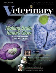
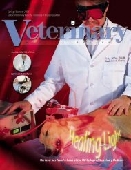
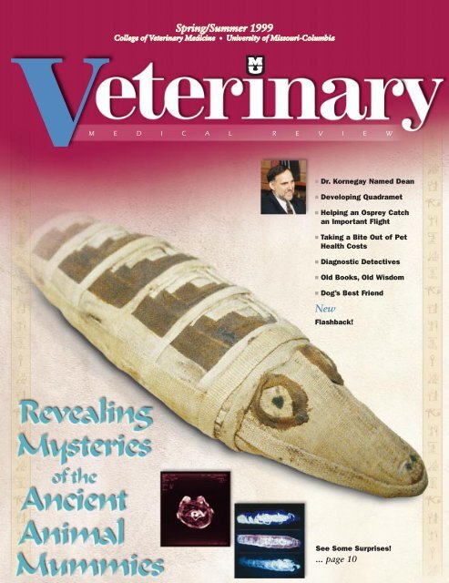
ON THE COVER<br />
B EST PRESERVATION<br />
Of the three mummies investigated,<br />
the baby crocodile was in<br />
the best condition on the outside.<br />
Its crosshatched linen<br />
wrappings are still nearly perfect<br />
in spite <strong>of</strong> 2,000<br />
years <strong>of</strong> age.<br />
Flashback !<br />
...30
“I look<br />
forward to<br />
working with<br />
our alumni<br />
and friends.”<br />
D R . J O E N . K O R N E G A Y , D E A N O F T H E C O L L E G E<br />
Moving Forward Together<br />
By now, many <strong>of</strong><br />
you will have heard <strong>of</strong> my<br />
appointment as Dean <strong>of</strong> the MU<br />
<strong>College</strong> <strong>of</strong> <strong>Veterinary</strong> <strong>Medicine</strong>.<br />
This is a tremendous honor for<br />
me. I look forward to working<br />
with our alumni and friends to<br />
advance the <strong>College</strong>’s programs.<br />
First, thanks so much for your support<br />
and advice over my tenure as interim<br />
dean. At the time I accepted the interim<br />
position in May, 1998, a number <strong>of</strong> key<br />
issues faced the <strong>College</strong>. These included<br />
a review <strong>of</strong> the DVM curriculum, our<br />
anniversary endowment campaign,<br />
recruitment <strong>of</strong> several endowed pr<strong>of</strong>es-<br />
sorships, mission enhancement for Uni-<br />
versity programs through the state<br />
legislature, and the American <strong>Veterinary</strong><br />
Medical Association’s (AVMA) accredita-<br />
tion visit. Our students, staff, and fac-<br />
ulty have really "stepped up to the<br />
plate" and worked tirelessly to address<br />
these areas. The good news is that<br />
progress has been made on all fronts!<br />
I’ll keep you updated on what should<br />
be some exciting developments in the<br />
months ahead.<br />
Preparation for the AVMA accredita-<br />
tion visit required a substantial self-study<br />
document. We decided to take advantage<br />
<strong>of</strong> the resulting discussions and incorpo-<br />
rated strategic planning at the unit level<br />
in this process. I’ve reviewed these mate-<br />
rials and want to now develop a more<br />
comprehensive <strong>College</strong> plan. Your input<br />
will be needed. With this in mind, over<br />
the coming months, I’ll be getting out in<br />
the state to meet with many <strong>of</strong> you and<br />
other key constituency groups. It’s really<br />
pretty simple. You’ve been there for us<br />
time and time again when we’ve needed<br />
you. It’s important that we be there for<br />
you when you need us.<br />
As I’ve stressed in the past, a natural<br />
synergism exists among our teaching,<br />
service, and research missions. I’m con-<br />
fident that, working together, we can<br />
capitalize on this synergism and really<br />
make a difference!<br />
Joe N. Kornegay, Dean<br />
3
4 A R O U N D T H E C O L L E G E<br />
Dr. Joe N. Kornegay,<br />
Interim Dean <strong>of</strong> the<br />
<strong>University</strong> <strong>of</strong> <strong>Missouri</strong>-Columbia<strong>College</strong><br />
<strong>of</strong> <strong>Veterinary</strong><br />
<strong>Medicine</strong>, was named Dean<br />
<strong>of</strong> the <strong>College</strong>. Dr. Kornegay took over<br />
as Interim Dean in May 1998, succeeding<br />
Dr. H. Richard Adams, who resigned<br />
after 6 years as Dean to become dean <strong>of</strong><br />
veterinary medicine at his alma mater,<br />
Texas A&M <strong>University</strong>.<br />
“Dean Kornegay will be an important<br />
addition to the leadership team<br />
<strong>of</strong> this campus at a very<br />
important time in the history<br />
<strong>of</strong> the <strong>College</strong> <strong>of</strong> <strong>Veterinary</strong><br />
<strong>Medicine</strong> and the<br />
educational mission <strong>of</strong> this<br />
university,” Provost Brady<br />
Deaton said.<br />
“His<br />
Dr. Joe Kornegay Named Dean<br />
distinguished record as an exemplary<br />
teacher, national prominence as an academic<br />
clinician and scholar, and his commitment<br />
to the statewide role <strong>of</strong> the<br />
<strong>College</strong> make him an ideal choice for this<br />
position. Not only will Dean Kornegay<br />
continue to develop research and scholarship<br />
on the campus, but he will continue<br />
to evaluate and build programs that<br />
strengthen the statewide role <strong>of</strong> the <strong>College</strong>.<br />
His leadership will enrich the strong<br />
programs <strong>of</strong> the <strong>College</strong> and provide students<br />
with the training and conceptual<br />
understanding to be leaders in the practice<br />
<strong>of</strong> veterinary medicine. I am<br />
excited by this appointment and am<br />
looking forward to working closely<br />
with Dr. Kornegay.”<br />
“I appreciate the confidence<br />
shown in me by Provost Deaton<br />
and look forward to working with<br />
him to achieve our common goals for<br />
the <strong>College</strong> <strong>of</strong> <strong>Veterinary</strong> <strong>Medicine</strong>,”<br />
Dr. Kornegay said. “Through the<br />
collective efforts <strong>of</strong> our students,<br />
staff and faculty, as<br />
well as alumni and other<br />
supporters in the state,<br />
the <strong>College</strong> has made<br />
tremendous progress in<br />
recent years. It is important<br />
to me that all groups<br />
served through our programs<br />
benefit from this<br />
momentum. With this in<br />
mind, I will work particularly<br />
hard to capitalize on<br />
the natural synergism that<br />
exists among the <strong>College</strong>’s<br />
teaching, service, and research<br />
missions.”<br />
“I am pleased by this appointment,<br />
which places a very strong<br />
academician and administrator in<br />
such an important position for the<br />
<strong>College</strong>, the <strong>University</strong> and our<br />
state,” Chancellor Richard Wallace<br />
said. “Importantly, Dr. Kornegay has<br />
excellent credentials as an educator<br />
S P R I N G / S U M M E R 1 9 9 9 V E T E R I N A R Y M E D I C A L R E V I E W<br />
and clinician in addition to his record in<br />
scholarship. The breadth <strong>of</strong> his background<br />
will help ensure that <strong>Missouri</strong>’s<br />
only <strong>College</strong> <strong>of</strong> <strong>Veterinary</strong> <strong>Medicine</strong><br />
continues its prominent role <strong>of</strong> service<br />
to the people at an exciting time when<br />
we are strengthening the life sciences on<br />
our campus and planning for the opportunities<br />
<strong>of</strong>fered by the state’s mission<br />
enhancement program. Dr. Kornegay is<br />
the right person at the right time to provide<br />
leadership for this effort.”<br />
Dr. Kornegay joined MU in 1994 as<br />
pr<strong>of</strong>essor and chair <strong>of</strong> veterinary medicine<br />
and surgery, director <strong>of</strong> the <strong>Veterinary</strong><br />
Medical Teaching Hospital and<br />
investigator for the Dalton Cardiovascular<br />
Research Center. Previously, he was a<br />
pr<strong>of</strong>essor <strong>of</strong> companion animal and special<br />
species medicine at North Carolina<br />
State <strong>University</strong> in Raleigh, where he<br />
spent 11 years, received numerous teaching<br />
awards and remains an adjunct pr<strong>of</strong>essor.<br />
There, Dr. Kornegay gained a<br />
national reputation for his research on a<br />
canine model <strong>of</strong> Duchenne muscular dystrophy,<br />
which he continues at MU. He<br />
also spent several years working as a veterinarian<br />
in private practice. Dr. Kornegay<br />
earned a bachelor’s degree in veterinary<br />
sciences and a doctorate in veterinary<br />
medicine from Texas A&M <strong>University</strong>,<br />
and a master’s degree in veterinary<br />
anatomy and doctorate in veterinary<br />
pathology from the <strong>University</strong> <strong>of</strong> Georgia<br />
in Athens. He is board certified in the<br />
neurology specialty <strong>of</strong> the American <strong>College</strong><br />
<strong>of</strong> <strong>Veterinary</strong> Internal <strong>Medicine</strong>.<br />
“The <strong>College</strong> <strong>of</strong> <strong>Veterinary</strong> <strong>Medicine</strong><br />
plays an extremely important role at MU<br />
and throughout the state,” Dr. Kornegay<br />
said. “We are the only <strong>Missouri</strong> university<br />
that awards the DVM degree and<br />
also have critical responsibilities in clinical<br />
service and outreach. In addition,<br />
<strong>College</strong> faculty are committed to work<br />
with colleagues from other divisions to<br />
increase MU’s research productivity.”<br />
VMR
Bald Eagle Returns to Wild<br />
with Help from Vet Students<br />
On a warm October day, a mature bald<br />
eagle named Itsan was released back into<br />
the wild south <strong>of</strong> Columbia, Mo. after six<br />
months <strong>of</strong> intensive rehabilitation.<br />
Itsan arrived at MU’s <strong>Veterinary</strong> Medical<br />
Teaching Hospital in April. He was<br />
underweight, sick, and had a dislocated<br />
shoulder. Students in the <strong>College</strong>’s Raptor<br />
Rehabilitation Project first worked to<br />
cure an infection. Later, the group sought<br />
to increase the eagle’s body weight.<br />
After a month <strong>of</strong> cage rest, the eagle<br />
began a rehabilitation process that<br />
included limited flying. A large crowd <strong>of</strong><br />
students, faculty, staff, and television<br />
reporters were on hand to observe the<br />
release near the <strong>Missouri</strong> River.<br />
The MU Raptor Rehabilitation Project<br />
is an educational and service activity <strong>of</strong><br />
the <strong>College</strong> <strong>of</strong> <strong>Veterinary</strong> <strong>Medicine</strong> with<br />
support from the <strong>Missouri</strong> Department<br />
<strong>of</strong> Conservation. The project is managed<br />
by veterinary medical students and community<br />
volunteers under the supervision<br />
<strong>of</strong> veterinary medical hospital faculty.<br />
The group seeks to rehabilitate and<br />
release birds <strong>of</strong> prey back into the wild,<br />
learn more about <strong>Missouri</strong>’s large birds,<br />
and educate the public about raptors and<br />
their importance to the environment.<br />
Marla Gray, a fourth year veterinary<br />
medical student and president <strong>of</strong> the raptor<br />
project who supervised much <strong>of</strong><br />
Itsan’s rehabilitation, warned that<br />
untrained people should not approach an<br />
injured raptor. These birds, she said, are<br />
wild animals with powerful beaks and<br />
claws. Additionally, raptors are protected<br />
by state and federal law. People who find<br />
an injured bird should call the MU <strong>Veterinary</strong><br />
Teaching Hospital at (573) 882-<br />
7821 for assistance.<br />
The group is available to give presentations<br />
on birds-<strong>of</strong>-prey featuring some <strong>of</strong><br />
its resident raptors—injured birds who<br />
cannot be returned to the wild because <strong>of</strong><br />
their injuries. The Raptor Rehabilitation<br />
Project also coordinates a raptor adoption<br />
program in which individuals or<br />
companies can sponsor an injured bird’s<br />
care. Funds also are used to improve the<br />
group’s bird holding and training facilities.<br />
Each sponsor receives a certificate <strong>of</strong><br />
adoption and a photo <strong>of</strong> the adopted<br />
bird. Fees range from $25 to adopt a<br />
screech owl, to $<strong>10</strong>0 to adopt a golden<br />
eagle, to $150 to adopt a bald eagle.<br />
New CVM Clinical Trials Offer<br />
Reduced Fees for Dogs<br />
VMR<br />
Dogs with certain types <strong>of</strong> cancer may<br />
be eligible for reduced-fee medical<br />
treatment by participating in one <strong>of</strong> a<br />
series <strong>of</strong> clinical studies at<br />
MU’s <strong>Veterinary</strong> Medical<br />
Teaching Hospital.<br />
One study will clinically<br />
test a new chemotherapy<br />
protocol designed to<br />
induce remission <strong>of</strong> canine transitional<br />
cell carcinoma <strong>of</strong> the bladder. A second<br />
trial will investigate a new chemotherapy<br />
treatment for prostatic cancers. A third<br />
study is designed to help develop a medical<br />
strategy to prevent the recurrence <strong>of</strong><br />
certain s<strong>of</strong>t tissue tumors after cancer<br />
surgery.<br />
In the first study, the MU <strong>Veterinary</strong><br />
Medical Teaching Hospital is the lead<br />
institution in a <strong>Veterinary</strong> Cooperative<br />
Oncology Group (VCOG) trial for the<br />
treatment <strong>of</strong> bladder cancer in dogs.<br />
S P R I N G / S U M M E R 1 9 9 9 V E T E R I N A R Y M E D I C A L R E V I E W<br />
A R O U N D T H E C O L L E G E<br />
Marla Gray, a fourth year veterinary medical student and president <strong>of</strong> the <strong>College</strong> Raptor Rehabilitation<br />
Project, struggles to get a bald eagle from its cage to release back into the wild. She is assisted by<br />
Nicole Griffin also a fourth year student and vice president <strong>of</strong> the group. Two local television stations<br />
witnessed the release.<br />
Dogs with a diagnosis <strong>of</strong> transitional cell<br />
carcinoma (TCC), a common bladder<br />
cancer, and whose disease is visible with<br />
ultrasound, are eligible for the study.<br />
To be eligible, the dogs must not have<br />
received previous treatment with piroxicam<br />
and must not have any other complicating<br />
disease, Dr. Carolyn Henry,<br />
assistant pr<strong>of</strong>essor <strong>of</strong> oncology said.<br />
In the bladder and prostatic tumor<br />
clinical trials, dogs receive four doses <strong>of</strong><br />
mitoxantrone chemotherapy, given on an<br />
outpatient basis every three weeks at the<br />
MU <strong>Veterinary</strong> Medical Teaching Hospital.<br />
In the bladder study, owners also<br />
administer an oral medication, piroxicam,<br />
at home. The tumors are measured<br />
before the first and third treatments,<br />
using ultrasound. Mitoxantrone is provided<br />
free <strong>of</strong> charge. “This is nearly $900<br />
in savings for a 60-pound dog,” Dr.<br />
Henry said.<br />
Clients are responsible for other costs<br />
associated with diagnosis and treatment.<br />
“We have enrolled 21 dogs in the bladder<br />
cancer study thus far and our target<br />
enrollment is 50 cases by May 1999,”<br />
Dr. Henry said.<br />
The third trial is designed to find a<br />
treatment that will prevent post surgical<br />
recurrence <strong>of</strong> a type <strong>of</strong> s<strong>of</strong>t tissue tumor<br />
5
6 A R O U N D T H E C O L L E G E<br />
(hemangiopericytoma) that occurs most<br />
<strong>of</strong>ten on the limbs. This trial is designed<br />
for use in cases where radiation therapy<br />
is not feasible. “Here, we are investigating<br />
the use <strong>of</strong> mitoxantrone chemotherapy<br />
post operatively,” Dr. Henry said.<br />
Dogs participating in the study will<br />
receive treatment on an outpatient basis<br />
every three weeks for four doses. Regular<br />
follow-up care at the teaching hospital<br />
is required after completion <strong>of</strong><br />
treatment. The drug is provided free <strong>of</strong><br />
charge, as are thoracic radiographs. To<br />
date, two dogs have been enrolled in the<br />
study that has a target enrollment <strong>of</strong> 40<br />
cases in a one-year period.<br />
A fee reduction also is available for the<br />
treatment <strong>of</strong> dogs with oral malignant<br />
melanoma. This is the most common oral<br />
cancer in dogs and also serves as a model<br />
for treatment <strong>of</strong> melanoma in people.<br />
Dr. Henry can be reached at the <strong>Veterinary</strong><br />
Medical Teaching Hospital,<br />
573/882-7821.<br />
VMR<br />
MU Researcher Named<br />
Chief USDA Scientist<br />
Dr. Michael Roberts, adjunct pr<strong>of</strong>essor and<br />
the previous chair <strong>of</strong> pathobiology in MU’s<br />
<strong>College</strong> <strong>of</strong> <strong>Veterinary</strong> <strong>Medicine</strong>, has been<br />
named chief scientist at the U.S. Department<br />
<strong>of</strong> Agriculture’s National Research<br />
Institute (NRI) in Washington, D.C.<br />
Dr. Roberts divides his time between<br />
Columbia and Washington D.C., where<br />
he works as a policy leader for the<br />
National Research Initiative Competitive<br />
Program. The program funds research on<br />
key problems <strong>of</strong> national and regional<br />
importance in biological, environmental,<br />
physical, and social sciences relevant to<br />
agriculture, food, and the environment.<br />
The grant proposals are judged on a<br />
peer-reviewed, competitive, basis.<br />
Dr. Roberts has received several<br />
awards and honors. He was elected to<br />
membership in the prestigious National<br />
Academy <strong>of</strong> Sciences in 1996. He also<br />
was the 1994 Amoroso Lecturer at the<br />
Society <strong>of</strong> the Study <strong>of</strong> Fertility, the 1992<br />
U.S. Department <strong>of</strong> Agriculture Distinguished<br />
Scientist, and the 1990 Sydney<br />
A. Asdell Lecturer at Cornell <strong>University</strong><br />
for the Study <strong>of</strong> Reproduction’s Research<br />
Award. VMR<br />
Scholarship Honors Dean Adams<br />
A scholarship will be established honoring<br />
former MU <strong>College</strong> <strong>of</strong> <strong>Veterinary</strong><br />
<strong>Medicine</strong> Dean H. Richard Adams.<br />
Donations are now being sought.<br />
The scholarship will be awarded to a<br />
graduating veterinary medical student<br />
who demonstrates exceptional interest<br />
and understanding <strong>of</strong> how the humananimal<br />
bond will influence the future <strong>of</strong><br />
veterinary medicine.<br />
Donations to this fund should be sent<br />
to David Horner, director <strong>of</strong> development,<br />
<strong>College</strong> <strong>of</strong> <strong>Veterinary</strong> <strong>Medicine</strong>,<br />
W203 <strong>Veterinary</strong> Medical Building, <strong>University</strong><br />
<strong>of</strong> <strong>Missouri</strong>, Columbia, Mo.<br />
65211 (phone: (573) 884-5972). Checks<br />
should be payable to the <strong>University</strong> <strong>of</strong><br />
<strong>Missouri</strong> and designated for the H.<br />
Richard Adams CVM Tribute Fund.<br />
New Editions <strong>of</strong> Books by<br />
Faculty and Alumni<br />
VMR<br />
New versions <strong>of</strong> two books edited by<br />
<strong>College</strong> faculty and alumni were recently<br />
introduced.<br />
Dr. John Bonagura, the <strong>College</strong>’s<br />
Gilbreath-McLorn Endowed Pr<strong>of</strong>essor<br />
<strong>of</strong> Cardiology, recently completed editing<br />
Kirk’s Current <strong>Veterinary</strong> Therapy,<br />
Volume 13, Small Animal Practice.<br />
Dr. Robert Paddleford, DVM ’70,<br />
recently saw the second edition <strong>of</strong> his<br />
book Manual <strong>of</strong> Small Animal Anesthesia<br />
published.<br />
VMR<br />
Time is Best <strong>Medicine</strong> for Some Eye<br />
Injuries, MU Report Indicates<br />
It’s best to let nature take its course with<br />
some eye diseases, according to a recent<br />
MU <strong>College</strong> <strong>of</strong> <strong>Veterinary</strong> <strong>Medicine</strong> study.<br />
The study found that healing <strong>of</strong><br />
bruised and swollen corneas was actually<br />
delayed when treated with corticosteroid<br />
ointments or drops. In fact, in<br />
some cases, the treatment can lead to<br />
permanent damage to the eye.<br />
The study was conducted by Dr. Cecil<br />
Moore, acting chair <strong>of</strong> <strong>Veterinary</strong> <strong>Medicine</strong><br />
and Surgery and the MU <strong>Veterinary</strong><br />
Medical Teaching Hospital.<br />
Corneal bruises, usually caused by a<br />
blow to the eyelid or brow, impart a<br />
S P R I N G / S U M M E R 1 9 9 9 V E T E R I N A R Y M E D I C A L R E V I E W<br />
Dr. Cecil Moore, left, examines the eyes <strong>of</strong> a small<br />
dog at the <strong>College</strong>’s <strong>Veterinary</strong> Medical Teaching<br />
Hospital. Dr. Moore is one <strong>of</strong> the nation’s leading<br />
veterinary ophthalmologists.<br />
cloudy appearance to the intact surface<br />
<strong>of</strong> the eye. Corticosteroid preparations<br />
have already been linked to destructive<br />
fungal growth in cases where the surface<br />
<strong>of</strong> the eye has been disrupted, such as a<br />
corneal scratch. According to Dr.<br />
Moore, a veterinary medical ophthalmologist,<br />
use <strong>of</strong> these preparations on<br />
an intact cornea can also be detrimental.<br />
Dr. Moore’s study involved nine cases<br />
<strong>of</strong> horses with bruised corneas. He found<br />
that the six that initially received cortisone<br />
took, on average, two weeks longer<br />
to heal than the three that did not.<br />
One way the medication inhibited the<br />
healing process was by preventing small<br />
blood vessels from migrating into the<br />
bruised cornea. The migration helps clean<br />
out the damaged tissue, Dr. Moore said.<br />
Animal owners understandably want<br />
the eye to look normal as soon as possible,<br />
Dr. Moore said. Corticosteroids<br />
seem to initially achieve that goal. The<br />
eye, however, will flare up again as the<br />
tissues try to heal. The longer the healing<br />
process, the more potential for permanent<br />
damage to the eye and loss <strong>of</strong><br />
sight, he continued.<br />
For bruises <strong>of</strong> the cornea, Dr. Moore<br />
recommends using a systemic or topical<br />
nonsteroidal anti-inflammatory drugs,<br />
which help to control the irritation while<br />
allowing the surface <strong>of</strong> the eye to heal on<br />
its own. VMR
Honoring a Friend<br />
Classmates <strong>of</strong> Dr. Steven Bumgarner, class <strong>of</strong> 1987, gather around a lecturn dedicated in Dr.<br />
Bumgarner’s honor. The lecturn is located in the <strong>College</strong> <strong>of</strong> <strong>Veterinary</strong> <strong>Medicine</strong>’s H. Richard Adams<br />
Conference Center. Dedication <strong>of</strong> the lecturn occurred in May ’98, one year after Dr. Bumgarner died <strong>of</strong><br />
cancer. Attending the dedication ceremony were Dr. Craig Franklin, MU veterinary pathobiology<br />
assistant pr<strong>of</strong>essor; Dr. Kitty Gepford-Barnett, Fulton, Mo.; Dr. Stacy Messner, Blue Springs, Mo.;<br />
and Dr. Lonnie Blum, Holden, Mo.<br />
Accolades<br />
Dr. Allen Hahn, pr<strong>of</strong>essor<br />
<strong>of</strong> veterinary medicine and<br />
surgery, received the Best <strong>of</strong><br />
Symposium Award for<br />
Investigative Informatics by<br />
the Association for <strong>Veterinary</strong><br />
Informatics and Idexx Informatics.<br />
The award was made at the groups’<br />
meeting last Summer. Dr. Hahn’s presentation<br />
was judged as contributing most<br />
to the improvement <strong>of</strong> veterinary medical<br />
patient care through the application<br />
<strong>of</strong> informatics by the practitioner.<br />
Dr. Robert Livingston, a MU post doctoral<br />
fellow, was certified as a Diplomate<br />
<strong>of</strong> the American <strong>College</strong> <strong>of</strong> Laboratory<br />
Animal <strong>Medicine</strong>. The specialty board,<br />
recognized by the American <strong>Veterinary</strong><br />
Medical Association, consists <strong>of</strong> more<br />
than 600 active specialists in the field <strong>of</strong><br />
laboratory animal medicine.<br />
Dr. Stan Casteel, associate<br />
pr<strong>of</strong>essor <strong>of</strong> veterinary<br />
pathology, was invited to<br />
participate as a panelist in<br />
Protecting Children from<br />
Lead Exposure: Assessing<br />
the Relationship Between Soil Lead to<br />
Blood Lead. The discussion was held in<br />
October in Washington, D.C., and was<br />
hosted by the National Environmental<br />
Policy Institute.<br />
Dr. Lynelle Johnson, veterinary biomedical<br />
sciences graduate student, received<br />
the 1998 American Physiological Society’s<br />
Outstanding Presentation by a<br />
Graduate Student Award for her presentation<br />
“Exercise Training Produces<br />
Enhanced Acetylcholine-Induced Vasorelaxation<br />
in Porcine Pulmonary Arteries.”<br />
She made the presentation at the society’s<br />
recent annual conference.<br />
Dr. James Thorne, associate pr<strong>of</strong>essor <strong>of</strong><br />
pathobiology, recently<br />
chaired the<br />
annual Epidemiology and<br />
Economics <strong>of</strong> Animal<br />
Health Management meeting<br />
in Lansing, Mich.<br />
The MU <strong>College</strong> <strong>of</strong> <strong>Veterinary</strong> <strong>Medicine</strong><br />
Alumni Organization recently<br />
received Honor Chapter status from the<br />
MU Alumni Association. Only 20 <strong>of</strong><br />
MU’s 70 alumni chapters received honor<br />
status this year. The recognition is based<br />
on the chapter’s activities, projects, membership,<br />
and service. An Honor Chapter<br />
receives additional scholarship and operating<br />
funding. VMR<br />
S P R I N G / S U M M E R 1 9 9 9 V E T E R I N A R Y M E D I C A L R E V I E W<br />
’99<br />
Calendar <strong>of</strong> Events<br />
M E S SA AR GO E U NFD R OT M H E T HC E O L DL E AG NE<br />
7<br />
March 15-19<br />
Spring Vacation Week for VM 1 & 2<br />
March 20-28<br />
MU Spring Break<br />
April 1<br />
Research Day<br />
Sponsored by Phi Zeta & the <strong>College</strong><br />
April <strong>10</strong><br />
<strong>College</strong> <strong>of</strong> <strong>Veterinary</strong> <strong>Medicine</strong><br />
Parents Day<br />
H. Richard Adams<br />
Multipurpose Conference Center<br />
8 am – 2:30 pm<br />
April <strong>10</strong><br />
Gentle Doctor Benefit<br />
MU Hearnes Center Field House<br />
4–<strong>10</strong> pm<br />
April 16-17<br />
MU <strong>College</strong> <strong>of</strong> <strong>Veterinary</strong><br />
<strong>Medicine</strong> Open House<br />
CVM campus<br />
9am-3pm<br />
May 2-8<br />
National Pet Week<br />
May 7<br />
<strong>College</strong> <strong>of</strong> <strong>Veterinary</strong> <strong>Medicine</strong><br />
Commencement<br />
Jesse Hall, UMC campus<br />
July <strong>10</strong>-14<br />
American <strong>Veterinary</strong> Medical Assn.<br />
New Orleans, La.<br />
September 25<br />
<strong>College</strong> Alumni Day<br />
NOTE<br />
Times and places here<br />
may change.<br />
Check with the sponsoring<br />
organization<br />
for the latest information.
8<br />
R E S E A R C H P R O G R A M S<br />
Dr. Jimmy Lattimer, associate pr<strong>of</strong>essor at<br />
the MU <strong>College</strong> <strong>of</strong> <strong>Veterinary</strong> <strong>Medicine</strong>,<br />
investigated the ability <strong>of</strong> a new drug to<br />
relieve the pain <strong>of</strong> cancer in dogs. Dr.<br />
Lattimer was a member <strong>of</strong> a university-wide<br />
team <strong>of</strong> investigators evaluating the<br />
radioactive drug, Quadramet, in dogs and<br />
humans.<br />
The Quadramet story is a<br />
remarkable testament to the<br />
power <strong>of</strong> collaboration and an<br />
excellent example <strong>of</strong> the ‘one<br />
medicine concept.’ Working<br />
together, faculty from the various<br />
divisions contributing to the life<br />
sciences will continue to make<br />
discoveries that impact both<br />
human and animal health.<br />
—Dr. Joe Kornegay<br />
Dean, <strong>College</strong> <strong>of</strong> <strong>Veterinary</strong> <strong>Medicine</strong><br />
<strong>University</strong><br />
Team Effort Helps<br />
Cut Pain to the Bone<br />
The pain <strong>of</strong> metastatic prostate<br />
and breast cancer in humans<br />
that has spread to the bone can<br />
be debilitating. Thirteen years ago, a<br />
group <strong>of</strong> MU researchers began a study<br />
to develop a new drug to more effectively<br />
treat that pain. Today, the fruits <strong>of</strong><br />
their labor are helping cancer patients in<br />
the form <strong>of</strong> Quadramet, a radiotherapeutic<br />
approved last spring by the U.S.<br />
Food and Drug Administration.<br />
The effort not only yielded the new<br />
drug, but also helped create a <strong>University</strong><br />
wide team approach to develop and test<br />
new drugs. These team players included<br />
faculty in the <strong>University</strong>’s <strong>College</strong> <strong>of</strong> Arts<br />
and Science, <strong>College</strong> <strong>of</strong> Engineering, Ellis<br />
Fischel Cancer Center, Research Reactor<br />
Center, School <strong>of</strong> <strong>Medicine</strong>, <strong>University</strong><br />
Hospital and Clinics, and <strong>College</strong> <strong>of</strong> <strong>Veterinary</strong><br />
<strong>Medicine</strong>. Staff in the Truman<br />
Veterans Hospital also aided in the effort.<br />
The effort showed that the “one medicine<br />
concept,” a joint effort where<br />
diverse health-related organizations lend<br />
their particular expertise to a project,<br />
could yield tangible results.<br />
“Without the teamwork this may<br />
never have gotten <strong>of</strong>f the ground,” said<br />
Dr. Wynn Volkert, lead investigator <strong>of</strong><br />
the project and pr<strong>of</strong>essor <strong>of</strong> radiology at<br />
the MU School <strong>of</strong> <strong>Medicine</strong>.<br />
Metastatic cancer afflicts more than<br />
200,000 people a year, according to the<br />
American Cancer Society and National<br />
Cancer Institute. The chronic, unremitting<br />
pain <strong>of</strong> the cancer is caused by rapidly<br />
growing skeletal tumors.<br />
“The quality <strong>of</strong> life deteriorates rather<br />
quickly because the pain <strong>of</strong> this cancer<br />
makes life difficult,” said Dr. Allen<br />
Ketring, associate director for research<br />
S P R I N G / S U M M E R 1 9 9 9 V E T E R I N A R Y M E D I C A L R E V I E W<br />
and development at the <strong>University</strong> <strong>of</strong><br />
<strong>Missouri</strong> Research Rector Center.<br />
The Story <strong>of</strong> Quadramet<br />
The story <strong>of</strong> Quadramet began in<br />
1982 with a cooperative development<br />
agreement between the <strong>University</strong>’s<br />
Health Sciences Center and Dow Chemical<br />
Co. to find applications for Dow’s<br />
existing stock <strong>of</strong> chelating agents—drugs<br />
that attach themselves to specific parts <strong>of</strong><br />
the body. Work was begun to see if these<br />
agents had any value in treating cancer.<br />
The best candidate had the unwieldy<br />
name <strong>of</strong> ethylenediaminetetramethylenephosphoric<br />
acid, EDTMP for short.<br />
EDTMP was strongly attracted to the<br />
rapidly growing bone cells that comprise<br />
bone cancer tumors. This made EDTMP a<br />
likely carrier to deliver a radioactive element<br />
that would help kill the cancerous<br />
cells. To be effective, EDTMP would have<br />
to concentrate its effects without lingering<br />
too long in the body causing unwanted<br />
side effects.<br />
After reviewing several candidates for the<br />
cancer-treating component,<br />
the radioactive<br />
isotope <strong>of</strong> samarium-<br />
152, a rare metal in the<br />
earth’s crust, was chosen.<br />
Samarium-152 is<br />
not naturally radioactive,<br />
bringing the <strong>University</strong>’s<br />
research<br />
reactor into the picture.<br />
After a week-long<br />
bath in the core <strong>of</strong> a<br />
nuclear reactor, samarium-152<br />
is converted<br />
in radioactive samarium-153.
Samarium-153 looked promising as a<br />
cancer-fighting drug. With a half-life <strong>of</strong> 46<br />
hours, it decays naturally in a couple <strong>of</strong><br />
days leaving no lingering radiation. It also<br />
emits enough gamma radiation to be picked<br />
up by a gamma camera, allowing clinicians<br />
to make sure the drug localized in the targeted<br />
tumor and not healthy tissue.<br />
Strontium-89, a current cancer-fighting<br />
isotope, has a half-life <strong>of</strong> 50 days, produces<br />
a number <strong>of</strong> side effects including<br />
nausea and hair loss as radiation<br />
destroys adjoining tissue. It also allows<br />
few opportunities for monitoring.<br />
Samarium-153 also blended well with<br />
EDTMP. But would they work well in a<br />
living organism and help destroy tumor<br />
cells?<br />
1983<br />
In 1983, samarium-153/EDTMP passed<br />
its first tests on healthy laboratory rabbits<br />
and rats. What would it do against<br />
bone cancer? Dr. Jimmy Lattimer, associate<br />
pr<strong>of</strong>essor at the MU <strong>College</strong> <strong>of</strong> <strong>Veterinary</strong><br />
<strong>Medicine</strong> had a plan: beagles.<br />
“As it happens, dogs are the only<br />
other species that commonly contract<br />
bone cancer,” Dr. Lattimer said. “They<br />
get it at about <strong>10</strong> times the rate <strong>of</strong><br />
humans.”<br />
Dr. Lattimer structured two tests. The<br />
first used 24 laboratory-bred beagles.<br />
The second test used 40 canine cancer<br />
patients at the <strong>Veterinary</strong> Medical Teaching<br />
Hospital. Under Dr. Lattimer’s direction,<br />
samarium-153/EDTMP was used<br />
against cancer for the first time in living<br />
subjects.<br />
From 1984 to 1988, Dr. Lattimer and<br />
Dr. Louis Corwin Jr, pr<strong>of</strong>essor <strong>of</strong> veterinary<br />
pathobiology, worked on the study.<br />
Pet owners from as far away as New<br />
York City and Philadelphia flew their<br />
sick dogs to MU to undergo the experimental<br />
treatment.<br />
Initial results were promising. Samarium-153/EDTMP<br />
ignored healthy tissue<br />
and settled in large quantities in tumor<br />
sites. Excess medication passed harmlessly<br />
through urine within six to 12<br />
hours.<br />
The drug produced no typical side<br />
effects such as hair loss, although it did<br />
temporarily suppress the dog’s production<br />
<strong>of</strong> bone marrow and white blood<br />
cell count, causing a reduction in platelet<br />
production. Dogs that entered the hospital<br />
unwilling to stand, walk, or eat<br />
because <strong>of</strong> pain reverted to a more normal<br />
attitude and lifestyle.<br />
“We were able to exhibit a reduction<br />
in bone pain in more than 90 percent <strong>of</strong><br />
the dogs we treated,” Dr. Lattimer said.<br />
In 1987, based on the successful dog<br />
studies, the first phase <strong>of</strong> human testing<br />
began. The first human patient, who<br />
received the medication on an outpatient<br />
basis, responded as positively as Dr. Lattimer’s<br />
patients did. Next, 22 patients at<br />
MU’s Ellis Fischel Cancer Center, suffering<br />
from the pain <strong>of</strong> metastatic bone<br />
cancer, volunteered for the treatment.<br />
The results again mirrored Dr. Lattimer’s<br />
studies, with 17 <strong>of</strong> the 22 reporting a<br />
significant reduction in pain.<br />
1990<br />
S P R I N G / S U M M E R 1 9 9 9 V E T E R I N A R Y M E D I C A L R E V I E W<br />
R E S E A R C H P R O G R A M S<br />
In 1990, samarium-153/EDTMP testing<br />
shifted from MU to Dow’s hands. In<br />
phase two testing, clinics in Europe and<br />
North America confirmed MU’s phase<br />
one results. In phase three trials, the final<br />
step before FDA approval, 50 human<br />
medical clinics performed testing. Last<br />
March, samarium-153/EDTMP, marketed<br />
under the name Quadramet, was<br />
approved by the FDA for human use.<br />
The effort to produce Quadramet has<br />
come full circle on the MU campus. The<br />
<strong>University</strong>’s hospitals use it in humans as<br />
a palliative therapy, the research reactor<br />
irradiates samarium-152 into samarium-<br />
153, and, at the <strong>College</strong> <strong>of</strong> <strong>Veterinary</strong><br />
<strong>Medicine</strong>, Dr. Lattimer and his colleagues<br />
have another tool to fight the<br />
pain <strong>of</strong> cancer in dogs. VMR<br />
Christmas Cheer<br />
For the needy <strong>of</strong> mid-<br />
<strong>Missouri</strong>, Santa didn’t<br />
come by reindeer but by<br />
pickup trucks this past<br />
Christmas when the<br />
<strong>College</strong> Class <strong>of</strong> 2001<br />
donated 670 pounds <strong>of</strong><br />
food to the Central<br />
<strong>Missouri</strong> Food Bank.<br />
The Class <strong>of</strong> 2001 won a<br />
friendly competition<br />
with the Class <strong>of</strong> 2002<br />
who also collected and<br />
donated food to the<br />
charity.<br />
Mark Your Calendar for <strong>College</strong> Events in April<br />
The MU <strong>College</strong> <strong>of</strong> <strong>Veterinary</strong> <strong>Medicine</strong> will be center<br />
stage for three events in April 1999.<br />
On April <strong>10</strong>, the annual CVM Parents Day will occur.<br />
The event will begin in the <strong>College</strong>’s H. Richard Adams<br />
Conference Center and extend from 9-11:30 am.<br />
The <strong>College</strong>’s annual Gentle Doctor Benefit is scheduled<br />
for 4-<strong>10</strong> pm on April <strong>10</strong> and will take place in the<br />
MU Hearnes Center.<br />
The <strong>College</strong>’s Open House is scheduled for Friday, April<br />
16 (9 am-3 pm), and Saturday, April 17 (<strong>10</strong> am-3 pm).<br />
Admission is free. There will be demonstrations <strong>of</strong> police<br />
dogs and German protection dogs, as well as mule rides, a<br />
petting zoo, tours, and a teddy bear clinic. VMR<br />
9
Sometime between 300 B.C. and<br />
200 A.D, an Egyptian felt compelled<br />
to make a prayer <strong>of</strong>fering to<br />
the crocodile god Sobek. With a prayer<br />
for strength or endurance, he reverently<br />
left a brown-and-tan baby crocodile<br />
mummy in the temple <strong>of</strong> this god.<br />
Twenty centuries later, that same 18inch-long<br />
mummified crocodile, on loan<br />
to the <strong>University</strong> <strong>of</strong> <strong>Missouri</strong>-Columbia’s<br />
Museum <strong>of</strong> Art and Archaeology, came<br />
under the laser positioning crosshairs <strong>of</strong><br />
the CT Scanner at MU’s <strong>Veterinary</strong> Medical<br />
Teaching Hospital. After thousands<br />
<strong>of</strong> years <strong>of</strong> mystery and conjecture, the<br />
animal was to yield its secrets to one <strong>of</strong><br />
the most sophisticated medical imaging<br />
devices devoted to veterinary medicine.<br />
Without disturbing its fragile linen wrappings,<br />
the scanner would peer into the<br />
animal and look for the remains <strong>of</strong> a<br />
skeleton, internal organs, or any clues<br />
about the Egyptian rituals that caused<br />
these animals to be worshiped<br />
and mummified. It was also<br />
hoped the scans would<br />
show any mummification<br />
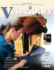
A CT cross-section slice<br />
<strong>of</strong> the crocodile from its<br />
eyes through its jaw shows<br />
that the protruding eyes were<br />
made from coiled linen. The<br />
internal organs, including the brain,<br />
had been removed by Egyptian<br />
specialists and replaced with fabric.<br />
The animal’s spine, however, was left intact.<br />
by Randy Mertens<br />
incisions, disease pathology, and, if the<br />
stomach was present, the animal’s last<br />
meal.<br />
Joining the baby crocodile in the investigation<br />
was a 3,000-year-old hawk and<br />
2,000-year-old kitten. Egyptologists had<br />
studied the outside <strong>of</strong> the animals for<br />
decades, even X-raying them in 1931.<br />
The CT scanner, a computer-enhanced<br />
X- ray machine that can yield threedimensional<br />
s<strong>of</strong>t tissue images that conventional<br />
radiography cannot, promised<br />
to show more.<br />
<strong>College</strong> Is Asked to Assist<br />
Dr. Jane Biers, curator at the MU<br />
Museum <strong>of</strong> Art and Archaeology, and<br />
Dr. Patricia Podzorski, assistant director<br />
and an Egyptologist, requested the assistance<br />
<strong>of</strong> the MU <strong>College</strong> <strong>of</strong> <strong>Veterinary</strong><br />
<strong>Medicine</strong> to learn more about the mummies<br />
on loan from the Field Museum in<br />
Chicago.<br />
A 1931 X-ray yielded some fuzzy<br />
skeletal information on two <strong>of</strong> the animals,<br />
showing little beyond that animals<br />
did exist under the wrappings. In addition<br />
to looking for evidence <strong>of</strong> the<br />
mummification technique<br />
used, Drs. Biers and<br />
Podzorski<br />
hoped to<br />
see if<br />
Of the three mummies investigated, the baby<br />
crocodile was in the best condition on the outside. Its<br />
crosshatched linen wrappings are still nearly perfect in<br />
spite <strong>of</strong> 2,000 years <strong>of</strong> age.<br />
any amulets (Egyptian good luck<br />
charms) were inside the wrappings.<br />
Knowing the gender, species, and age<br />
might help Egyptologists determine if<br />
these played roles in religious rituals.<br />
Another question: Did the mummies<br />
die natural deaths, or were they killed to<br />
satisfy the market for animal <strong>of</strong>ferings?<br />
The 1931 X-ray did not reveal a cat<br />
under its linen wrapping. Egyptologists<br />
speculated that an unscrupulous ancient<br />
vendor, knowing the buyer wouldn’t<br />
unwrap the mummy, sold a fake cat to<br />
an unsuspecting customer. Because <strong>of</strong><br />
this, the cat mummy commanded only<br />
moderate historical interest.<br />
Dr. Biers said Egyptologists could only<br />
guess the mummies’ ages as they were<br />
unearthed by grave robbers and sold as<br />
tourist trinkets.<br />
Because the practice <strong>of</strong> mummifying<br />
animals took place during a 1,000-year<br />
time span, with several millions <strong>of</strong> animals<br />
sacrificed, there are thousands <strong>of</strong><br />
catacombs filled with these relics, Dr.<br />
Podzorski said.<br />
CT Scanner<br />
The MU <strong>Veterinary</strong> Medical Teaching<br />
Hospital’s CT Scanner serves the companion-animal<br />
clinic, equine hospital,<br />
and food animal efforts as the state’s<br />
only CT scanner dedicated to animal<br />
use. The scanner is particularly helpful in<br />
the hospital’s cancer treatment program.<br />
The program is one <strong>of</strong> the few veterinary<br />
cancer treatment operations in the country.<br />
Here, many <strong>of</strong> the same cancerfighting<br />
techniques and<br />
technology are used as in<br />
human hospitals. Cancer<br />
diagnosis is aided by use<br />
<strong>of</strong> the CT scanner and<br />
treatment is aided<br />
by one <strong>of</strong> the<br />
few linear<br />
accelerators<br />
in<br />
the
world dedicated to animal use.<br />
CT stands for Computed Tomography,<br />
previously known as Computed Axial<br />
Tomography (CAT). “Tomo” means<br />
slice in Greek.<br />
In a conventional X-ray exam, X-rays<br />
pass through the body to a film that<br />
records anatomy mainly as shadows. Only<br />
the outside shape <strong>of</strong> organs is visible.<br />
CT scanning uses X-ray information<br />
taken from around an object to construct<br />
a series <strong>of</strong> initial black and white<br />
cross-sectional images. These images are<br />
like slices <strong>of</strong> bread, revealing a detailed<br />
image inside the targeted bones and s<strong>of</strong>t<br />
tissue. In a live animal, injectable, contrast-enhancing<br />
dyes may be given in<br />
conjunction with the scan.<br />
The data is sent to a computer for processing<br />
in a step called reconstruction.<br />
This converts the images into a series <strong>of</strong><br />
pixels that can be manipulated into a<br />
computer-generated three-dimensional<br />
image.<br />
Jeffrey Wilcox, registrar <strong>of</strong> the MU Museum <strong>of</strong> Art and Archeology, positions the<br />
three animal mummies into the MU <strong>Veterinary</strong> Medical Teaching Hospital’s CT<br />
Scanner.<br />
CT scans are generally used to determine<br />
the condition <strong>of</strong> s<strong>of</strong>t organs such as<br />
the brain, liver, lung tissue, and kidneys.<br />
It also can be used on complex bony<br />
structures like the spine, pelvis, and hip.<br />
The CT is a valuable tool in searching<br />
for large space-occupying lesions such as<br />
tumors.<br />
CT scanning is particularly useful clinically<br />
because it can be performed rapidly.<br />
Only a few minutes are required<br />
after scanning for the initial data to<br />
show up on computer monitors in the<br />
control room. CT scanning is becoming<br />
the method <strong>of</strong> choice for imaging trauma<br />
patients as the fast and simple exam that<br />
allows a quick overview <strong>of</strong> possibly lifethreatening<br />
pathology and rapidly<br />
enables a surgical intervention.<br />
During a CT exam, the patient lies in a<br />
large device that has a doughnut-like<br />
opening. Inside the doughnut is a sophisticated<br />
X-ray tube that spins around the<br />
patient to take pictures <strong>of</strong> the patient<br />
from every angle. The beam <strong>of</strong> X-rays<br />
passes through the patient and is<br />
detected according to the density <strong>of</strong> the<br />
tissue encountered.<br />
Each slice is then reconstructed into<br />
the initial two-dimensional image that<br />
appears on the control room monitor.<br />
The good s<strong>of</strong>t tissue and spatial resolution<br />
possible with CT imaging allows<br />
its use in orthopedics. It can be used in<br />
imaging <strong>of</strong> complex joints like the shoulder<br />
or hip as a functional unit. Fractures,<br />
especially those affecting the spine, are<br />
also common CT scanning targets.<br />
The computerized post processing<br />
three-dimensional images can be rotated<br />
to view from several angles, further<br />
enhancing the value <strong>of</strong> CT imaging for<br />
surgeons. CT images also are used as the<br />
basis <strong>of</strong> radiotherapy planning purposes<br />
and for interventional work like CT<br />
guided biopsies.<br />
Dr. Jimmy Lattimer, associate pr<strong>of</strong>essor<br />
<strong>of</strong> veterinary medicine and surgery<br />
The baby crocodile was the most difficult <strong>of</strong> the three animals to render in the<br />
CT Scanner’s computer. The crocodile was quite young when it died. Its<br />
skeleton had not yet transformed into bone from cartilage, something that<br />
does not show well in either x-rays or CT scans. Also, unlike the other animals,<br />
the crocodile had been mummified in the same way as humans. The internal<br />
organs <strong>of</strong> the animal had been removed and replaced with coiled linen.
While the cat mummy was relatively small on the outside, the CT scan<br />
indicated an even smaller kitten was inside. The top <strong>of</strong> the kitten’s head was<br />
bent forward, putting the top <strong>of</strong> the kitten’s head just<br />
behind the painted face on the outside <strong>of</strong><br />
the mummy. This rendering shows<br />
that the internal organs <strong>of</strong> the cat<br />
are still present.<br />
The 2,000-year-old kitten’s<br />
wrappings are somewhat tattered.<br />
A cat’s face was painted on the linen.<br />
The cat’s ears are really linen fashioned<br />
to look like ears.<br />
One <strong>of</strong> the questions<br />
Egyptologists wanted answered was<br />
whether the cat’s neck had been broken.<br />
This was sometimes done by “cat farms” to ensure<br />
plenty <strong>of</strong> animal <strong>of</strong>ferings. The CT scan’s computerized<br />
rendering <strong>of</strong> the cat inside the linen indicates that the cat’s neck was indeed broken.<br />
A cat looks back from 2,000 years. One slice <strong>of</strong> the<br />
initial scan <strong>of</strong> the kitten mummy was directed to<br />
the front <strong>of</strong> the cat’s skull to verify that a cat was<br />
indeed in the linen. The face <strong>of</strong> the long-dead cat<br />
emerged on the computer monitor in the CT<br />
scanning room, confirming that the remains <strong>of</strong><br />
the animal were inside the wrappings.<br />
(far left) Dr. Cristi Reeves Cook, <strong>College</strong> <strong>of</strong><br />
<strong>Veterinary</strong> <strong>Medicine</strong> radiologist, and Heather King,<br />
senior veterinary technician, position the cat<br />
mummy on the CT Scanner. The scan revealed the<br />
skeleton and internal organs <strong>of</strong> a kitten, disproving<br />
the idea that the mummy was a fake.<br />
“Oh, it’s real.<br />
Cool.”<br />
and director <strong>of</strong> radiology, coordinated<br />
the mummy scans. His first job was to<br />
choose scanning options and gather the<br />
raw imagery data for placement into a<br />
computer database. Part two would be<br />
to selectively reconstruct the data into a<br />
computerized, three-dimensional, virtual<br />
computer mummy for analysis.<br />
It’s Real<br />
As the high-resolution thin-slice scans<br />
<strong>of</strong> the cat mummy began, something<br />
unexpected emerged on the television<br />
screens <strong>of</strong> the darkened CT scanning<br />
control room. The skeleton <strong>of</strong> a kitten,<br />
not the sawdust expected, was under the<br />
cat mummy’s wrapping.<br />
“Oh, it’s real. Cool,” Dr. Podzorski<br />
said. The 1931 X-ray and subsequent<br />
Egyptology catalogs were wrong.<br />
The initial black and white scans<br />
showed more. A skeleton, a brain, and<br />
other internal organs appeared. Even the<br />
cat’s fur could be detected. That these<br />
items were still in place provides worthwhile<br />
clues about life in ancient Egypt.<br />
As the CT scan moved along the front<br />
<strong>of</strong> the cat’s skull, an ancient face looked<br />
back at the dozen people in the CT scanning<br />
room over a gulf <strong>of</strong> 2,000 years.<br />
When the scanner sliced through the cat’s<br />
neck, broken vertebra appeared on the<br />
monitor. The cat was probably killed to<br />
satisfy the need for religious <strong>of</strong>ferings.<br />
From the damage and the position <strong>of</strong> the<br />
broken bones, the guess was that the kitten<br />
was grabbed by the head, face down,<br />
and snapped like a whip.<br />
Because <strong>of</strong> the severity <strong>of</strong> the spinal<br />
injuries, the position <strong>of</strong> the real face <strong>of</strong><br />
the cat does not correspond with the<br />
face painted on the outside <strong>of</strong> the mum-
mification linen. The real cat’s head is<br />
bowed forward, putting the top <strong>of</strong> its<br />
head just behind the painted face on the<br />
outside <strong>of</strong> the cat’s linen covering.<br />
The kitten was probably only twomonths-old<br />
when it died, as its skeleton<br />
is mostly cartilage, not bone, Dr. Lattimer<br />
observed, which is probably why<br />
little was seen by the conventional X-ray.<br />
As the scan, consisting <strong>of</strong> one-millimeter<br />
slices <strong>of</strong> the kitten, continued, more<br />
internal organs began to appear. Dr. Lattimer<br />
and the Egyptologists conferred,<br />
agreeing that the kitten was not mummified,<br />
but probably chemically dried. Dr.<br />
Podzorski, watching the computer image,<br />
said Egyptologists believe small animals<br />
began their mummification process in<br />
large terra-cotta jars full <strong>of</strong> drying salts<br />
and other chemicals. The kitten probably<br />
went through this process.<br />
The initial scans revealed no evidence<br />
<strong>of</strong> sickness. Up until 40 years ago and<br />
before vaccinations became common,<br />
more than half <strong>of</strong> all kittens died <strong>of</strong> disease<br />
before they were three months old.<br />
In fact, the evidence from the scan indicated<br />
the kitten was healthy, creating<br />
speculation that the cat was raised in one<br />
<strong>of</strong> several Egyptian “cat farms.” Here,<br />
cats were bred and pampered as revered<br />
symbols <strong>of</strong> the gods. Based on the evidence<br />
<strong>of</strong> the broken neck, however, this<br />
good life was short-lived as the commercial<br />
enterprises had to satisfy thousands<br />
<strong>of</strong> customers seeking religious <strong>of</strong>ferings.<br />
The Hawk<br />
The scan on the hawk indicated that it<br />
had not been mummified, either. Intact<br />
feathers, wings, feet, skeleton, heart,<br />
liver, lungs, brain, and other organs<br />
showed up on the initial scan. Most<br />
human mummies went through an elaborate<br />
process <strong>of</strong> drying with the body<br />
organs removed and stored in jars. Since<br />
the hawk was preserved with organs<br />
intact, some sort <strong>of</strong> chemical process<br />
must have been used.<br />
The hawk appeared to be a fully<br />
grown bird—probably a peregrine falcon.<br />
Part <strong>of</strong> its last meal appeared on the<br />
monitors as the scan passed through the<br />
bird’s digestive tract.<br />
As the scan moved from the hawk’s<br />
feet to head, the feathers appeared to be<br />
arranged in a pinwheel form—the technique<br />
<strong>of</strong> the person who preserved the<br />
animal or something <strong>of</strong> religious consequence,<br />
perhaps?<br />
The scan <strong>of</strong> the crocodile revealed one<br />
quick surprise. While the<br />
wrappings are 18 inches long,<br />
the animal inside is only nine<br />
inches long. Was the animal<br />
wrapped with extra linen to<br />
make it more salable?<br />
Looking at the scan, Dr.<br />
Lattimer noted that little <strong>of</strong><br />
the crocodile’s skeleton had<br />
transformed from cartilage to<br />
bone, indicating a very young<br />
animal. This was bad news in<br />
terms <strong>of</strong> a detailed look at the<br />
interior <strong>of</strong> the animal as cartilage<br />
is one <strong>of</strong> the few<br />
things that yield little<br />
information to the<br />
CT Scanner.<br />
The CT scan did<br />
reveal that the nineinch-long<br />
baby crocodile<br />
was mummified in<br />
much the same way as<br />
Egyptians mummified people.<br />
No internal organs showed on the CT<br />
scan—the spaces were stuffed with linen<br />
to help the outside <strong>of</strong> the animal keep its<br />
external shape. Some <strong>of</strong> this linen was<br />
tightly and precisely wrapped while other<br />
linen was haphazardly crumpled. Also,<br />
the crocodile’s back legs were missing.<br />
The animal apparently underwent an<br />
extensive and expensive mummification<br />
process. The remarkable appearance <strong>of</strong><br />
the linen after thousands <strong>of</strong> years indicated<br />
a special craftsmanship. Were the<br />
legs removed for a religious reason or to<br />
enhance the smooth look <strong>of</strong> the finished<br />
product, thus, boosting its salability?<br />
Another clue indicating special treatment:<br />
The skin <strong>of</strong> the crocodile was surprisingly<br />
well preserved.<br />
As the scanning continued there was<br />
yet another surprise: Small hard spheres<br />
appeared in the body. While these could<br />
be rocks, dirt, or chemical pellets left<br />
over from the mummification<br />
process, the spheres<br />
are too consistent in<br />
size and shape to be<br />
dismissed lightly.<br />
Another CT scan on<br />
decorative beads sometimes<br />
inserted into human<br />
mummies would have to be<br />
conducted to see if the scanner<br />
recorded similar readings. If<br />
these are amulets, it might<br />
indicate the crocodile was<br />
even more than an aboveaverage<br />
religious <strong>of</strong>fering.<br />
Did the hawk mummy still have its feathers? An initial CT scan<br />
indicated that the feathers were still present under the linen<br />
wrappings.<br />
Of the three animal mummies, the hawk’s<br />
linen was the most tattered. The talons and beak<br />
<strong>of</strong> the 3,000-year-old hawk peek from its linen.<br />
Computer Imagery Unlocks Secrets<br />
After the initial scanning, Dr. Lattimer<br />
took the raw data to his reconstruction<br />
computer for a more complete, threedimensional<br />
look into the animals. It<br />
took 24 hours <strong>of</strong> computer time for the<br />
first phase <strong>of</strong> this part <strong>of</strong> the investigation.<br />
Computer “wire” renderings,<br />
where the outlines <strong>of</strong> internal shapes<br />
were reduced to lines, showed the location<br />
<strong>of</strong> heart, lungs, and other organs.<br />
“Solid” form computer imagery showed<br />
what the animals look like under their<br />
linen wrappings.<br />
These are to be the maps for a more<br />
detailed examination. Here, as questions<br />
are asked, detailed examination <strong>of</strong> specific<br />
parts <strong>of</strong> the animals can be undertaken.<br />
With more information <strong>of</strong> what’s<br />
under the animals’ wrappings, it is<br />
hoped even more secrets will be revealed<br />
from the ancient animal mummies.<br />
A computer “wire” rendering shows the condition<br />
<strong>of</strong> the hawk’s skeleton and digestive track.
14 V E T E R I N A R Y B U S I N E S S<br />
Article Courtesy <strong>of</strong> The Columbia (Mo.) Daily Tribune, Photos courtesy <strong>of</strong> <strong>Veterinary</strong> Pet Insurance<br />
Taking a Bite Out <strong>of</strong><br />
Pet Heath Care Costs<br />
Asimple question led Dr. Jack<br />
Stephens to start a revolution<br />
in pet health care. “Pet owners<br />
were asking, “Why don’t<br />
we have insurance for our pets like we<br />
do for our children?” said Dr. Stephens,<br />
DVM Class <strong>of</strong> ’72 and California veterinarian<br />
who saw too many pets being put<br />
to sleep because <strong>of</strong> economics.<br />
President <strong>of</strong> the Southern California<br />
<strong>Veterinary</strong> Medical Association at the<br />
time, Dr. Stephens first tried to persuade<br />
traditional insurance companies to cover<br />
pet health. That didn’t pan out, so he<br />
started his own company, <strong>Veterinary</strong> Pet<br />
Insurance.<br />
When VPI started selling policies in<br />
1982 on a shoestring budget, it was the<br />
first pet-insurance company in the<br />
nation. Today, the Anaheim, Calif.based<br />
company is still the largest, with<br />
1<strong>10</strong>,000 policies in force in 48 states.<br />
Revenues are doubling every year,<br />
from premium volume <strong>of</strong> $8.7 million in<br />
1997 to $18 million this year. The company<br />
expects revenue <strong>of</strong> between $36<br />
million and $40 million in 1999.<br />
“Jack has been the only person I know<br />
<strong>of</strong> who’s been successful in actually<br />
implementing a comprehensive petinsurance<br />
program,” said David Horner<br />
Jr., director <strong>of</strong> development for MU’s<br />
<strong>College</strong> <strong>of</strong> <strong>Veterinary</strong> <strong>Medicine</strong>. “It’s<br />
going to impact the overall pr<strong>of</strong>ession<br />
someday.”<br />
Pet health insurance is common in<br />
some European countries, but fewer than<br />
one percent <strong>of</strong> pet owners in the United<br />
States own policies. Meantime, vet costs<br />
are climbing faster than human health<br />
care—30 percent a year by one estimate.<br />
A recent survey in the veterinary trade<br />
journal DVM Magazine suggests that<br />
American dog and cat owners begin to<br />
consider euthanasia when faced with vet<br />
By Andrew Walters<br />
Dr. Stephens, pictured with his miniature<br />
pinscher, Skeeter, has built <strong>Veterinary</strong> Pet<br />
Insurance into a nationwide company. He<br />
plans to return next year to speak at MU.<br />
Dr. Jack Stephens<br />
Age: 52<br />
Born: Hobart, Oklahoma<br />
Hometown: Anaheim, California<br />
Education: <strong>University</strong> <strong>of</strong> <strong>Missouri</strong><br />
doctor <strong>of</strong> veterinary medicine, ’72<br />
Family: wife, Vicki, four children,<br />
seven dogs and a cat<br />
Hobbies: Hiking, backpacking and bird<br />
hunting<br />
Sidekick: Skeeter, a miniature pinscher<br />
that Dr. Stephens takes to work with<br />
him every day<br />
bills in excess <strong>of</strong> $576. Dr. Stephens<br />
believes a third-party payer can help<br />
keep that to a minimum.<br />
Pet insurance “is really becoming<br />
attractive for pet owners who care about<br />
their pets,” Dr. Stephens said. “Veterinarians<br />
can do anything they can do in<br />
human health care. And even though it’s<br />
only one-fifteenth <strong>of</strong> the cost, it’s still<br />
expensive.”<br />
VPI’s basic policy costs $89 a year and<br />
S P R I N G / S U M M E R 1 9 9 9 V E T E R I N A R Y M E D I C A L R E V I E W<br />
covers up to $2,000 per incident, with a<br />
maximum <strong>of</strong> $7,500 per policy. The<br />
deluxe policy costs $159 and covers up<br />
to $4,000 per incident, with a maximum<br />
<strong>of</strong> $12,000 per policy. Both plans carry a<br />
$40 deductible.<br />
A California policyholder submitted a<br />
$<strong>10</strong>,000 claim for cancer treatment for a<br />
Great Dane that included chemotherapy,<br />
radiation and surgery at Colorado State<br />
Medical School. VPI paid $8,000.<br />
VPI also paid to install a pacemaker in<br />
a 15-week-old poodle in Santa Cruz that<br />
lived another 12 years.<br />
“Pets give you so much love. They’re<br />
great companions,” Dr. Stephens said.<br />
“Why wouldn’t you give back to them?”<br />
Dr. Stephens’ success in the dog-eatdog<br />
world <strong>of</strong> insurance comes as somewhat<br />
<strong>of</strong> a surprise to former classmates<br />
at MU, who remember him as someone<br />
who took his work seriously but also<br />
wanted to make sure everyone else had a<br />
good time.<br />
“No one expected him to be the big<br />
businessman <strong>of</strong> the crowd,” said classmate<br />
Dr. John Williams, who graduated from<br />
the <strong>College</strong> <strong>of</strong> <strong>Veterinary</strong> <strong>Medicine</strong> with<br />
Dr. Stephens in 1972 and now is co-owner<br />
<strong>of</strong> Horton Animal Hospitals in Columbia,<br />
Mo. “I don’t think anybody thought he<br />
had the disposition to do that.”<br />
It wasn’t easy. In 1991, VPI fell short<br />
<strong>of</strong> California’s capital requirements for<br />
insurance companies and was taken over<br />
by state regulators for three months.<br />
In the midst <strong>of</strong> the company’s financial<br />
turmoil, Dr. Stephens was going through<br />
a personal ordeal <strong>of</strong> his own. In 1989 he<br />
was diagnosed with throat cancer. Doctors<br />
recommended surgery and gave him<br />
a year to live.<br />
Ironically, it was a veterinarian who<br />
suggested an alternative treatment that<br />
ended up saving his life. Dr. Stephens
MU graduate Dr. Jack Stephens has built a fledgling California company into the nation’s largest pet health<br />
insurance company. <strong>Veterinary</strong> Pet Insurance has 1<strong>10</strong>,000 policies in force in 48 states. “Pets give you so<br />
much love. They’re great companions,” Dr. Stephens says. “Why wouldn’t you give back to them?”<br />
was treated with radiation implants<br />
inserted into his throat and tongue,<br />
along with chemotherapy.<br />
Eight years later, the cancer is gone,<br />
and he doesn’t even go in for checkups.<br />
The procedure dried out his throat permanently—he<br />
has to drink plenty <strong>of</strong><br />
water with food—but, as he points out,<br />
it’s better than the alternative.<br />
“They wanted to do radical surgery;<br />
remove my tongue and jawbone. I<br />
wouldn’t have been able to eat or talk,”<br />
he said. “Now I’m talking and leading a<br />
normal life. It was all from a doggie doctor.<br />
It just shows you how sophisticated<br />
a veterinary doctor can be.”<br />
The episode could have taken Dr.<br />
Stephens out <strong>of</strong> commission for months<br />
and ruined his fledgling company. Instead,<br />
he missed only two weeks <strong>of</strong> work.<br />
“The company was still struggling at<br />
the time,” he said. “They wanted me to<br />
take six months <strong>of</strong>f, but I couldn’t do it.<br />
The company couldn’t survive if I did.”<br />
Dr. Stephens said he felt a personal<br />
responsibility to some 950 investors —<br />
700 <strong>of</strong> them veterinarians — who had<br />
put up an average <strong>of</strong> $2,000. “I didn’t<br />
want them saying I lost their money.<br />
They’d never forgive me,” he said. “I<br />
used to be the goat, and now I’m the<br />
hero. It’s all due just to perseverance, I<br />
guess.”<br />
Dr. Stephens said he has no plans to<br />
take his company public. Money won’t be<br />
an issue for a number <strong>of</strong> years, he said,<br />
thanks to investments by two major shareholders—Scottsdale<br />
Insurance Co. and<br />
<strong>Veterinary</strong> Centers <strong>of</strong> America Inc., each<br />
<strong>of</strong> which has a 23 percent stake in VPI.<br />
Santa Monica, Calif.-based VCA,<br />
which operates the nation’s largest chain<br />
<strong>of</strong> animal hospitals and is the largest<br />
provider <strong>of</strong> diagnostic laboratory services<br />
in the country, invested $4.5 million<br />
in VPI last year.<br />
“We’re really at the beginning <strong>of</strong> the<br />
technology boom inside <strong>of</strong> veterinary<br />
care,” said Bob Antin, president and<br />
CEO <strong>of</strong> VCA. “As the diagnostic world<br />
expands and pet owners can treat their<br />
pets using techniques once only available<br />
for humans, the cost <strong>of</strong> it will somewhat<br />
justify having insurance.”<br />
Antin said he’s known Dr. Stephens for<br />
11 years and has been impressed by the<br />
“labor <strong>of</strong> love” approach he takes to pet<br />
insurance.<br />
“Nobody has done it like Jack has —<br />
with heart and compassion and the personal<br />
inner drive for success,” he said.<br />
“It’s truly entrepreneurial.”<br />
A drawback Antin and others see in<br />
VPI’s approach, however, is the company’s<br />
strategy <strong>of</strong> marketing its product<br />
mostly through veterinarians using point<strong>of</strong>-purchase<br />
displays at clinics across the<br />
country.<br />
S P R I N G / S U M M E R 1 9 9 9 V E T E R I N A R Y M E D I C A L R E V I E W<br />
V E T E R I N A R Y B U S I N E S S<br />
“Veterinarians are not insurance salesmen,”<br />
said Dr. Williams. “We’re too busy<br />
to sell it. When I’m done with a client, I’ve<br />
got another one waiting to come in. I can’t<br />
spend another five minutes saying, ‘Wait a<br />
minute. Here’s something you need.’ ”<br />
Dr. Stephens said the company is beefing<br />
up its budget for advertising and promotions<br />
from about $1.8 million this<br />
year to more than $4 million in 1999.<br />
It’s still chicken feed, Dr. Stephens<br />
acknowledges. But then again, the company<br />
doesn’t have much competition.<br />
The only other national pet-health<br />
insurer, Canton, Ohio-based Petshealth<br />
Insurance Agency Inc., has about 4,000<br />
policies in force and 1998 annualized<br />
premium volume <strong>of</strong> about $500,000,<br />
said co-founder Russell Smith.<br />
Petshealth policies are underwritten by<br />
American Home Assurance Co., a member<br />
<strong>of</strong> giant American International<br />
Group. Premiums begin at $120 a year<br />
for dogs and $83 a year for cats. Policies<br />
cover 80 percent <strong>of</strong> vet costs after a<br />
$<strong>10</strong>0 annual deductible.<br />
Petshealth expects to grow rapidly<br />
next year, with a goal <strong>of</strong> 40,000 policies<br />
in force and premium volume <strong>of</strong> $<strong>10</strong><br />
million by the end <strong>of</strong> its first fiscal year<br />
in June 1999. The publicly traded company<br />
is planning a $5 million stock<br />
<strong>of</strong>fering early next year.<br />
Dr. Stephens said VPI has been successful<br />
in part because <strong>of</strong> his clinical<br />
background. “We understand pets and<br />
pet diseases,” he says. And VPI tries to<br />
keep its policyholders happy by paying<br />
claims within five days.<br />
“A lot <strong>of</strong> companies have gone into the<br />
market and gone out <strong>of</strong> the market. Jack<br />
is unique. He had a mission and stuck<br />
with the mission,” said Horner <strong>of</strong> MU’s<br />
vet college. “He’s devoted years <strong>of</strong> his life<br />
and all <strong>of</strong> his financial resources to this.”<br />
Dr. Stephens has kept his ties to MU as<br />
an advisor and financial contributor to<br />
the <strong>Veterinary</strong> <strong>College</strong>. In 1993, he was<br />
named the school’s alumnus <strong>of</strong> the year,<br />
and he plans to accept an invitation to<br />
speak to the graduating class next year.<br />
“I feel an affiliation with the school. I<br />
worked for the school in pre-vet and all<br />
through vet college,” he said. “Some <strong>of</strong><br />
my pr<strong>of</strong>essors are still there. If it wasn’t<br />
for them, I wouldn’t have had the opportunity<br />
in life I’ve enjoyed.” VMR<br />
15
16<br />
S E R V I N G T H E C O M M U N I T Y<br />
Dr. James Cook, clinical instructor in small animal<br />
surgery and orthopedics specialist, examines<br />
Ishmael’s broken wing. Assisting Dr. Cook is Nicole<br />
Griffin, a fourth-year veterinary medical student and<br />
vice president <strong>of</strong> the MU Raptor Rehabilitation<br />
Project.<br />
Students and Surgeons<br />
at the <strong>College</strong> <strong>of</strong><br />
<strong>Veterinary</strong> <strong>Medicine</strong><br />
Help a Broken-Winged<br />
Osprey to Migrate<br />
with His Fellows<br />
(above right) Dr. James Cook uses the hospital’s C-arm<br />
intraoperative fluoroscope to view Ishmael’s broken<br />
wing. The C-arm allows surgeons to see a series <strong>of</strong><br />
x-ray images on a TV-like screen.<br />
Ishmael has an important flight to catch—<br />
probably the most important <strong>of</strong> his life and one that<br />
can’t be delayed. It was not the best time to suffer a<br />
broken wing.<br />
Ishmael is a fledgling osprey brought to <strong>Missouri</strong> as<br />
part <strong>of</strong> a five-year project to repopulate the birds to<br />
the state. The plan was to bring the just-hatched bird to<br />
<strong>Missouri</strong> in July, have it “imprint” on the state in August<br />
through September, migrate south in October, and return<br />
to <strong>Missouri</strong> the next spring.<br />
Ishmael arrived in <strong>Missouri</strong> with a broken wing. If he can’t migrate with his<br />
fellows, he will, in essence, be forever lost.<br />
That’s why the <strong>Missouri</strong> Department <strong>of</strong> Conservation brought Ishmael to<br />
MU’s <strong>Veterinary</strong> Medical Teaching Hospital. Here, Ishmael had four things<br />
going for him: an orthopedic surgeon with almost <strong>10</strong>0 avian surgeries to his<br />
credit, a sophisticated X-ray machine, anesthesiologists familiar with avian<br />
respiratory systems, and veterinary medical students with specialized knowledge<br />
in rehabilitating birds <strong>of</strong> prey.<br />
With this help, Ishmael, named by veterinary medical students helping his<br />
recovery, has a good chance <strong>of</strong> making his flight.<br />
Ospreys in <strong>Missouri</strong><br />
Text and photos by Randy Mertens<br />
Ishmael is part <strong>of</strong> the fourth installment <strong>of</strong> a Department <strong>of</strong> Conservation<br />
program to restore these fish-eating hawks to <strong>Missouri</strong>. Ospreys used to breed<br />
in <strong>Missouri</strong> but disappeared in the late 1800s.<br />
“Their habitat was destroyed by pioneers digging stream channels, polluting<br />
waterways, draining wetlands, and sawing down nesting areas,” said John<br />
Meyer, <strong>Missouri</strong> Department <strong>of</strong> Conservation (MDC) wildlife biologist<br />
and osprey reintroduction project leader. “They were also<br />
subjected to the unlimited killing that took place in the<br />
early 1900s before the rise <strong>of</strong> the conservation movement.<br />
The insecticide, DDT, finished <strong>of</strong>f the rest <strong>of</strong><br />
the osprey population.”<br />
As a result, their Midwest breeding range<br />
shrank to Minnesota and Wisconsin. Before<br />
the reintroduction work, ospreys only flew<br />
through <strong>Missouri</strong> on their spring<br />
and fall migrations to the tropics<br />
between North and South<br />
America.<br />
When mature, ospreys are<br />
21 to 24 inches long, have<br />
five- to six-foot wingspans,<br />
and weigh about three<br />
pounds. They are dark<br />
S P R I N G / S U M M E R 1 9 9 9 V E T E R I N A R Y M E D I C A L R E V I E W
own with white undersides and heads.<br />
Prominent dark stripes set <strong>of</strong>f their yellow<br />
eyes. In flight, their light-colored wings<br />
show a pronounced crook with a dark<br />
spot at the wrist. Ospreys eat shad, catfish,<br />
yellow perch, carp, and suckers, which<br />
they catch by plunging into the water feet<br />
first to grab prey swimming near the surface.<br />
The program’s goal is to have ospreys<br />
live in <strong>Missouri</strong> in summer as they once<br />
did. Ospreys become attached to the<br />
location where they hatch and try to<br />
come back each spring. By raising chicks<br />
at state lakes, MDC hopes to “imprint”<br />
the young transplants on <strong>Missouri</strong> sites,<br />
so they will return and raise their young.<br />
To help chicks become attached to<br />
these sites, they are raised in four-foot<br />
boxes on towers eight to 12 feet tall.<br />
These towers overlook the lakes and<br />
mimic conditions <strong>of</strong> natural nests. Boxes<br />
are covered with wire mesh, allowing the<br />
birds to see their surroundings. For<br />
osprey chicks to imprint on their nesting<br />
site, they need 360-degree visibility,<br />
Meyer said. They remember the area and<br />
come back to nest.<br />
Until the ospreys can feed themselves,<br />
conservationists catch enough fish to feed<br />
each bird about one pound per day—usually<br />
three <strong>10</strong>-inch live grass carp.<br />
When the birds are strong enough to<br />
fly, their boxes are opened and the birds<br />
begin life on their own. Spray-painted<br />
patches on the birds’ wings and numbered<br />
leg bands help biologists follow<br />
the ospreys’ progress.<br />
Ishmael’s part <strong>of</strong> the plan went bad<br />
early. As a four-week-old fledgling, he<br />
suffered a broken wing on the flight<br />
from his Wisconsin birthplace to<br />
<strong>Missouri</strong>. For Ishmael to survive,<br />
his wing would have to<br />
be healed in time for him<br />
to imprint on a lake<br />
and for the October departure with this<br />
fellow osprey.<br />
Dr. James Cook, clinical instructor in<br />
veterinary surgery and orthopedics specialist,<br />
performed Ishmael’s operation.<br />
Ishmael had two wing bone fractures—<br />
one through the growth plate that separates<br />
the long wing bone from the wing’s<br />
“wrist” joint. This cartilage is the growing<br />
part <strong>of</strong> the bone. It was the first<br />
avian growth plate fracture that Dr.<br />
Cook had seen.<br />
Treating growth plate fractures is<br />
tricky. The bone is small, and imprecise<br />
alignment can cause it to heal crooked.<br />
Dr. Cook inserted a temporary pin the<br />
length <strong>of</strong> the long bone, growth plate, and<br />
fragment to ensure correct alignment.<br />
Avian surgery is also difficult because<br />
birds carry virtually no extra blood—<br />
any blood loss during surgery is critical.<br />
Also, bird respiratory systems are more<br />
complex and faster than humans, making<br />
anesthesia difficult to administer.<br />
Prolonged surgery can be fatal to most<br />
birds. Luckily, anesthesiologists experienced<br />
in avian metabolism and physiology<br />
were on hand for Ishmael.<br />
Dr. Cook’s operation on Ishmael took<br />
only 20 minutes with help <strong>of</strong> a C-arm<br />
machine. Named for its shape, the C-arm<br />
is an intraoperative fluoroscope, an Xray<br />
machine that provides surgeons with<br />
movie-like images during surgery. By having<br />
exact and immediate visual information,<br />
the surgeon can make smaller and<br />
more precise incisions. There is less blood<br />
loss and the operation is faster.<br />
The C-arm is usually used in human<br />
hospitals. MU’s <strong>Veterinary</strong> Medical<br />
Teaching Hospital is one <strong>of</strong> the few veterinary<br />
medical colleges to use this technology<br />
for animal surgery.<br />
The X-ray movies allowed Dr. Cook<br />
to move Ishmael’s wing through its full<br />
motion and watch for obstructions or<br />
One day after surgery, the osprey, named Ishmael by veterinary medical<br />
students, is alert and recuperating in the raptor rehabilitation ward <strong>of</strong> the<br />
MU <strong>College</strong> <strong>of</strong> <strong>Veterinary</strong> <strong>Medicine</strong>’s Teaching Hospital.<br />
S E R V I N G T H E C O M M U N I T Y<br />
problems on the TV screen. The imagery<br />
was videotaped for teaching purposes.<br />
“The C-arm in this surgery made all the<br />
difference in the world,” Dr. Cook said.<br />
Overseeing Ishmael’s post-operative<br />
healing and rehabilitation is the <strong>College</strong>’s<br />
Raptor Rehabilitation Project. This<br />
group <strong>of</strong> about 25 third- and fourth-year<br />
veterinary medical students, clinicians,<br />
and volunteers administers the special<br />
care needed to rehabilitate injured birds<br />
<strong>of</strong> prey so that they can be returned to<br />
the wild.<br />
The Raptor Rehabilitation Project<br />
maintains a ward for birds in the hospital<br />
requiring critical care and the nearby<br />
Raptor Compound where the birds<br />
undergo rehabilitation and restoration to<br />
flight status. The compound also is host<br />
to about <strong>10</strong> eagles, owls, hawks, and<br />
kestrels who are unable to be returned to<br />
the wild, said Nichole Griffin, vice president<br />
<strong>of</strong> the raptor project and overseeing<br />
Ishmael’s recovery.<br />
Normally, post surgical birds are<br />
moved to the compound where they are<br />
kept in one <strong>of</strong> ten mews—open-air buildings<br />
that allow birds to be outside but<br />
not escape. As the birds recover their<br />
strength, they visit the flight cage—a<br />
barn large enough for the birds to fly<br />
from perch to perch. This helps the birds<br />
regain muscle strength and flying skills.<br />
Later, when the birds are strong enough,<br />
they are connected to a creance, several<br />
hundred yards <strong>of</strong> strong string that<br />
allows the birds to fly without escaping.<br />
When the birds are ready, the Raptor<br />
Project releases them, Ms. Griffin said.<br />
The Future<br />
The MDC’s osprey reintroduction program<br />
will continue until the year 2000<br />
with a total <strong>of</strong> 32 to 48 birds being<br />
released from <strong>10</strong> different sites.<br />
Birds released in previous years have<br />
made their migrations and have returned<br />
to <strong>Missouri</strong>. They have been observed<br />
living near their original nesting sites,<br />
flying and catching fish.<br />
“We have already positively identified<br />
osprey released in 1995 at Mark Twain,<br />
and we think we have identified all four<br />
birds from Thomas Hill,” Meyer said. “If<br />
the next few years are also successful, <strong>Missouri</strong><br />
will soon have a growing population<br />
<strong>of</strong> ospreys fishing in the state’s waters.”<br />
With luck, Ishmael will be back, too. VMR<br />
17
18<br />
S E R V I N G T H E C O M M U N I T Y<br />
Worried large animal veterinarians<br />
and cattle producers from across<br />
<strong>Missouri</strong> noticed sick cattle with<br />
symptoms ranging from stiffness and<br />
lack <strong>of</strong> appetite to fever, swollen<br />
tongues, and oral and nasal ulcers.<br />
The problem seemed to follow no<br />
known pattern. Blood tests and other<br />
observations taken from the field<br />
were sent to the <strong>Veterinary</strong> Medical<br />
Diagnostic Laboratory for tests.<br />
The<br />
<strong>Veterinary</strong><br />
Medical<br />
Diagnostic<br />
Laboratory<br />
MU’s Animal Disease Detectives<br />
It was during the warm days <strong>of</strong> late<br />
September that the phones began to ring<br />
at the MU <strong>College</strong> <strong>of</strong> <strong>Veterinary</strong> <strong>Medicine</strong><br />
<strong>Veterinary</strong> Medical Diagnostic Laboratory.<br />
Worried cattle producers and largeanimal<br />
veterinarians from across <strong>Missouri</strong><br />
had sick cattle with symptoms<br />
ranging from stiffness and lack <strong>of</strong><br />
appetite to fever, swollen tongues, and<br />
oral and nasal ulcers. The problem<br />
seemed to follow no known pattern.<br />
The pathologists and staff at the Diagnostic<br />
Laboratory handled the concerns<br />
the way they always do, with a watchful,<br />
well-trained eye, and a full range <strong>of</strong> diagnostic<br />
services. As its name implies, the<br />
Diagnostic Laboratory conducts scientific<br />
analyses to determine the causes <strong>of</strong><br />
the most vexing animal and agricultural<br />
disease problems. It features the laboratory<br />
equipment and techniques to conduct<br />
simple chemical tests to<br />
state-<strong>of</strong>-the-art DNA analysis. The laboratory<br />
seeks out the toxins, viruses, contaminates,<br />
infections, and biological<br />
hazards that threaten the state’s agriculture<br />
industry.<br />
S P R I N G / S U M M E R 1 9 9 9 V E T E R I N A R Y M E D I C A L R E V I E W<br />
The Lab Comes to State Aid<br />
The laboratory helps <strong>Missouri</strong> in<br />
numerous and varied ways. An outbreak<br />
<strong>of</strong> aggressiveness in rabbits was traced to<br />
lead in their blood caused by eating<br />
paint chips. Diagnostic Laboratory staff<br />
acted like detectives to determine a sudden<br />
increase in deaths in feedlot cattle.<br />
Cause: Discarded automobile mufflers<br />
were leaking chemicals into the drinking<br />
stream. A dog food company suspected<br />
contamination in hundreds <strong>of</strong> bags <strong>of</strong><br />
feed. The Diagnostic Laboratory determined<br />
there was no contamination, and<br />
the company was saved from a massive<br />
recall.<br />
Still other cases involved young goats<br />
that died suddenly. Post-mortem tests<br />
revealed clues that led to the discovery <strong>of</strong><br />
a poisonous vine that the animals had<br />
eaten. In addition to identifying various<br />
bacteria and viruses that silently afflict<br />
animals, the laboratory also is called in<br />
to help in cases such as satanic sacrifice<br />
<strong>of</strong> cattle and dogs.<br />
The laboratory also helped identify the<br />
causes <strong>of</strong> a wheat fungus and helped<br />
farmers deal with the problem.
“We are the only full-service laboratory<br />
in the state <strong>of</strong> <strong>Missouri</strong>,” said Dr.<br />
Harvey Gosser, director <strong>of</strong> the Diagnostic<br />
Laboratory. “We can examine whole<br />
animals, tissues, blood and specimens,<br />
and perform a wide range <strong>of</strong> diagnostic<br />
tests. It is helpful to have all <strong>of</strong> this evidence<br />
in one facility in situations like<br />
this so that we can act quickly determine<br />
the causes <strong>of</strong> problems.”<br />
The laboratory has more than 50 faculty<br />
and staff who handle more than<br />
75,000 specimens and perform more<br />
than 300,000 diagnostic tests annually.<br />
The number <strong>of</strong> cases has been growing.<br />
Last year the number <strong>of</strong> tests increased<br />
15 percent. Early in the laboratory’s history,<br />
it mainly dealt with food and<br />
equine problems. In the past decade,<br />
there has been increasing requests for<br />
diagnostic services relating to companion-animal<br />
health.<br />
In addition to <strong>of</strong>fering diagnostic assistance<br />
and consultation with veterinary<br />
medical practitioners and animal owners,<br />
the laboratory also has a place in<br />
educating the next generation <strong>of</strong> veterinarians.<br />
Each MU DVM student and<br />
pathology resident learn about laboratory<br />
procedures and animal disease<br />
investigation in the Diagnostic<br />
Laboratory.<br />
An Anniversary Virus<br />
The Diagnostic Laboratory had just<br />
celebrated its 30th anniversary when the<br />
flood <strong>of</strong> September phone calls from cattle<br />
producers began coming in.<br />
The symptoms led laboratory personnel<br />
to suspect a virus that historically affected<br />
mainly deer populations, epizootic hemorrhagic<br />
disease (EHD), among other possible<br />
causes. The possibility <strong>of</strong> EHD<br />
alarmed the veterinarians because cattle<br />
with the disease, which is caused by an<br />
insect bite, normally do not show any<br />
symptoms. On the other hand, infected<br />
deer experience serious symptoms and<br />
<strong>of</strong>ten die as a result <strong>of</strong> the virus.<br />
Using laboratory analysis <strong>of</strong> cattle<br />
specimens, Diagnostic Laboratory personnel<br />
confirmed the presence <strong>of</strong> EHD in<br />
29 <strong>of</strong> 44 submitted cases. Twenty-two <strong>of</strong><br />
those samples were <strong>Missouri</strong> cattle; the<br />
others were deer, elk, and sheep.<br />
“What we saw was a cluster <strong>of</strong> cases<br />
with similar clinical signs that are testing<br />
positive for EHD,” said Dr. Eileen<br />
Ostlund, assistant pr<strong>of</strong>essor and veterinary<br />
virologist. “Cattle in <strong>Missouri</strong> have<br />
had EHD in past years. What’s unusual<br />
here is that the presence <strong>of</strong> the disease<br />
has been accompanied by relatively<br />
severe manifestations.”<br />
After Dr. Ostlund and her colleagues<br />
received several requests to test for the<br />
unusual disease, they began to keep a<br />
chart to compare the cases, their history,<br />
location, symptoms, and test results.<br />
After initial blood tests showed antibodies<br />
to the EHD virus, they began to seek<br />
more conclusive evidence that EHD<br />
could be causing the cattle’s sickness.<br />
Because isolation <strong>of</strong> the virus that causes<br />
EHD is extremely difficult and time-consuming,<br />
they implemented an alternate<br />
type <strong>of</strong> sophisticated testing to identify<br />
genetic material <strong>of</strong> the virus present in<br />
the samples, called polymerase chain<br />
reaction (PCR) testing, for concerned<br />
owners and veterinarians. PCR detects<br />
An historical review<br />
indicated the last<br />
significant outbreak<br />
<strong>of</strong> hemorrhagic<br />
disease in <strong>Missouri</strong> deer<br />
occurred in 1988<br />
DNA patterns in infectious organisms<br />
leading to a rapid diagnosis from small<br />
test samples.<br />
As information from laboratory, clinical,<br />
historic, and on-site testing began to<br />
come together, the details <strong>of</strong> the problem<br />
began to emerge.<br />
A historical review indicated the last<br />
significant outbreak <strong>of</strong> hemorrhagic disease<br />
in <strong>Missouri</strong> deer occurred in 1988,<br />
and mortality estimates in the wild deer<br />
population ranged from six percent to<br />
16 percent. No cases in cattle were<br />
described at that time. EHD isn’t typically<br />
fatal in cattle unless the animal suffers<br />
from concurrent disease. Humans<br />
are unaffected by the disease, and are<br />
not harmed by eating meat with the<br />
virus present.<br />
S P R I N G / S U M M E R 1 9 9 9 V E T E R I N A R Y M E D I C A L R E V I E W<br />
S E R V I N G T H E C O M M U N I T Y<br />
As the data were examined, the cause<br />
was determined. The disease was caused<br />
and transmitted by biting midges, Culicoides<br />
variipenis, also known as gnats,<br />
punkies, and no-see-ums. The disease is<br />
seen most commonly in <strong>Missouri</strong> in the<br />
summer and fall when the midges are<br />
active.<br />
Diagnosis: EHD is self-limiting; it will<br />
run its course like the common cold<br />
virus in humans.<br />
“Take a little extra care, avoid stress,<br />
and make sure water is easily accessible,”<br />
said Dr. John Kreeger, associate<br />
pr<strong>of</strong>essor and veterinary pathologist.<br />
“While the virus can’t be ‘treated,’ it is<br />
probably a good idea to give antibiotics<br />
to affected animals to reduce the chance<br />
<strong>of</strong> secondary bacterial infections, but it<br />
does nothing to inhibit the course <strong>of</strong> the<br />
viral infection. Analgesics can be used to<br />
relieve discomfort.”<br />
With the diagnosis, the immediate job<br />
was done. As always, the Diagnostic<br />
Laboratory continues to monitor the<br />
cases as a whole to learn more about the<br />
virus. And, as it has for its 30-year history,<br />
they’re ready for a new challenge<br />
when the phones ring again. VMR<br />
19
20 V E T E R I N A R Y E D U C A T I O N<br />
The 1683 book, “Anatomy <strong>of</strong> the Horse”, by<br />
Andrew Snape, is beautifully illustrated.<br />
This illustration was considered so<br />
desirable that thieves would tear out the<br />
<strong>page</strong> and frame it like a painting.<br />
CURE FOR BLOATED CATTLE<br />
After cutting <strong>of</strong>f the end <strong>of</strong> the<br />
tail, boil a strong dose <strong>of</strong><br />
thoroughwort and tanzey.<br />
Pour it down the throat as<br />
soon as it is cool enough, then<br />
turn the animal into the yard and<br />
drive it for a few minutes. This will soon start<br />
wind. Repeat treatment in the nose, if necessary.<br />
CURE FOR SCOURES IN CALVES<br />
Break two eggs into a basin. Beat them up. Pour<br />
them, by means <strong>of</strong> a tin funnel, down the calf’s<br />
throat.<br />
Repeat<br />
twice<br />
or trice. An almost infallible cure.<br />
Old Books,<br />
T H E C O L L E G E ’ S C O L L E C T I O N O F O L D V E T E R I N A R Y<br />
—Yankee Farmer’s Manual and Practical Farrier<br />
Boston, 1846<br />
In a room behind the circulation desk<br />
<strong>of</strong> the MU <strong>College</strong> <strong>of</strong> <strong>Veterinary</strong> <strong>Medicine</strong>’s<br />
Library are about 700 books that<br />
help tell the history <strong>of</strong> veterinary medicine.<br />
This is one <strong>of</strong> only a handful <strong>of</strong> collections<br />
<strong>of</strong> rare veterinary medical books.<br />
Before the age <strong>of</strong> computers, genetic engineering,<br />
and cellular biology, these books<br />
represented the sum <strong>of</strong> humankind’s wisdom<br />
in treating animals.<br />
The books in the collection range<br />
from the text <strong>of</strong> the first known veterinary<br />
medical book, circa 500 A.D., to<br />
books printed in the early 1900s. The<br />
books detail everything from homemade<br />
cures using grease to kill lice to surgical<br />
techniques that that would be recognized<br />
by today’s veterinarian.<br />
C. Trenton Boyd<br />
The person compiling the collection is<br />
C. Trenton Boyd, <strong>College</strong> librarian since<br />
1970.<br />
He became interested in rare books<br />
through his interest in history. While he<br />
saw a need to capture historical information<br />
about the veterinary medical pr<strong>of</strong>ession,<br />
he had no budget to make that<br />
happen.<br />
In the late 1980s, long-time <strong>College</strong><br />
faculty member Dr. Arthur Case died,<br />
and the family suggested donations to<br />
the <strong>College</strong> library as a memorial. With<br />
help from the Dean’s <strong>of</strong>fice, this memorial<br />
evolved into an endowment fund to<br />
subscribe to current scientific journals<br />
and buy the latest veterinary medical<br />
books. It also allowed for a modest<br />
sum to be used for the acquisition<br />
<strong>of</strong> rare veterinary medical books.<br />
The acquisition <strong>of</strong> rare books<br />
began in earnest when Boyd<br />
met Norman Comben, considered<br />
the foremost<br />
authority on rare veterinary<br />
medical books. The<br />
74-year-old Comben, who has an <strong>of</strong>fice<br />
in his Berkhamsted house north <strong>of</strong> London,<br />
<strong>of</strong>fered advice for the first purchase<br />
for the <strong>College</strong> library, a 1683 book<br />
titled Anatomy <strong>of</strong> the Horse.<br />
It was through Comben that Boyd discovered<br />
and purchased the first known<br />
book on veterinary medicine, Mulomedicina.<br />
This book, written in Latin, was<br />
penned by the Roman author Vegetius<br />
Renatus who lived from 450 to 500 A.D.<br />
The college’s version <strong>of</strong> this book is a<br />
1574 reprint <strong>of</strong> the original text. The<br />
book was reprinted 1,000 years after its<br />
author’s death both as a historical reference<br />
and a current textbook. The concepts<br />
and techniques detailed in 500 A.D.<br />
had changed little by the 16th century.<br />
“This book dovetails perfectly in two<br />
areas that we are collecting comprehensively—mule<br />
and significant veterinary<br />
medical books,” Boyd said. “This book<br />
deals with horses and mules because they<br />
were so important economically. No one<br />
bothered with dogs and cats. The first<br />
book about dogs didn’t appear until the<br />
late 1800s.”<br />
Though more than four centuries old,<br />
the book’s 200 paper <strong>page</strong>s are in excellent<br />
condition. The raised black ink has<br />
almost a paint quality to it, indicating it<br />
was printed on a Gutenberg-style letterpress.<br />
Sometime in this century, the original<br />
bookbinding was replaced.
Historical Plagarism<br />
Anatomy <strong>of</strong> the Horse, by Andrew<br />
Snape, “ferrier to the king,” is another<br />
book in the collection. This 1683 book<br />
features an elaborate woodcut “plate” <strong>of</strong><br />
Snape.<br />
Like the woodcut <strong>of</strong> Snape, the 49<br />
woodcuts <strong>of</strong> the horse’s anatomy are<br />
incredibly elaborate, and detailed. But,<br />
there’s a problem. The illustrations were<br />
stolen from another source. The book is<br />
one <strong>of</strong> the earliest examples <strong>of</strong> plagiarism,<br />
Boyd said.<br />
“Of the 49 plates in the book, 22 were<br />
taken in their entirety from Anatomia del<br />
Cavallo (Anatomy <strong>of</strong> the Horse) written<br />
by Ruini, an earlier Italian author,” Boyd<br />
said. “Four are taken partly from Ruini<br />
or suggested by his figures.”<br />
When Snape copied the illustrations,<br />
possibly by tracing the originals, he accidentally<br />
reversed the drawings in his<br />
book, putting organs like the heart on the<br />
wrong side <strong>of</strong> the horse. Anatomia del<br />
Cavallo was published in Venice in 1598.<br />
While Anatomy <strong>of</strong> the Horse is complete,<br />
one <strong>of</strong> the book’s <strong>page</strong>s is a recent<br />
reproduction. The drawing in the front <strong>of</strong><br />
the book <strong>of</strong> a horse<br />
in a field was so<br />
beautiful that a<br />
thief, <strong>10</strong>0 or so<br />
years ago, removed<br />
the original <strong>page</strong> to<br />
frame it.<br />
The first “encyclopaedic”<br />
work<br />
where the word<br />
“veterinary”<br />
appeared in a<br />
title was the Dictionary<br />
<strong>of</strong> <strong>Veterinary</strong><br />
Art,<br />
written in 1805. This work, at least two<br />
inches thick with no <strong>page</strong> numbers, was<br />
published in London. It proudly proclaimed<br />
that it was illustrated with copper<br />
plates.<br />
Somewhat arranged like the encyclopedia<br />
<strong>of</strong> today, its text detailed the author’s<br />
conversations with farmers, blacksmiths,<br />
and veterinarians about issues and practices<br />
<strong>of</strong> the day. Cooling <strong>of</strong>f an exhausted<br />
horse, for example, was best done by<br />
running the horse into a cold pond, one<br />
commentator proudly told the author.<br />
M EV SE ST AE GR E I N FA R OY M E DT HU E C A DT EI AO N<br />
Old Wisdom<br />
B O O K S T E L L S M U C H A B O U T T H E P R O F E S S I O N ’ S B E G I N N I N G S<br />
“Le Cheval” by Edouard Cuyer has loose <strong>page</strong>s that<br />
show the different gaits <strong>of</strong> horses portrayed in the<br />
same way a dance class portrays footsteps on paper.<br />
The articulated legs <strong>of</strong> a cardboard horse can be<br />
positioned on these steps to show what a horse looks<br />
like at a walk, trot, or gallop.<br />
The 1861 “Atlas der Anatomie des Pferdes” by August Leisering features incredibly detailed hand-drawn<br />
black and white line illustrations <strong>of</strong> the anatomy <strong>of</strong> the horse overprinted with red, blue, and yellow ink.<br />
An even more practical<br />
book is almost no<br />
book at all, but a<br />
combination book<br />
and wall hanging.<br />
Chevaux &<br />
Mulets (Horses and<br />
Mules) was published<br />
in France<br />
around 1900. The<br />
book features <strong>10</strong><br />
tablets designed to<br />
fold out and hang<br />
on a wall on eyelets.<br />
Each tablet<br />
details, by plastic-like reproduction,<br />
horse’s teeth at various stages <strong>of</strong> wear.<br />
The purpose was to create a standardized<br />
method <strong>of</strong> judging an animal’s age.<br />
The first tablet shows a reproduction <strong>of</strong><br />
“milk teeth,” the teeth <strong>of</strong> a neonatal<br />
horse. The last tablet shows the teeth <strong>of</strong><br />
an aged horse whose teeth are almost<br />
gone from wear.<br />
The sub-title <strong>of</strong> the book reads: “Caracteres<br />
de L “age” and shows a picture<br />
<strong>of</strong> a horse. Translation: Characteristics<br />
<strong>of</strong> Age <strong>of</strong> the Horses.<br />
Another French book on the horse, Le<br />
Cheval (the Horse) by Edouard Cuyer,<br />
also has a fold-out section showing teeth<br />
wear and anatomy. The 1886 book is<br />
unique, however, in that a pocket on the<br />
inside back cover contains loose <strong>page</strong>s<br />
that show the different gaits <strong>of</strong> horses<br />
portrayed in the same way an Arthur<br />
Murray dance class portrays footsteps<br />
on paper. The articulated legs <strong>of</strong> a cardboard<br />
horse can be positioned on these<br />
steps to show what a horse looks like at<br />
a walk, trot, or gallop.<br />
This book is one <strong>of</strong> the first printed<br />
with color overlays—a technological<br />
challenge in the late 1880s that meant<br />
several perfectly-aligned passes through<br />
a hand-fed press. The <strong>College</strong>’s version<br />
<strong>of</strong> this book may be the only complete<br />
example <strong>of</strong> its kind.<br />
Probably the most striking use <strong>of</strong> illustrations<br />
in the <strong>College</strong> collection is the<br />
21
22<br />
V E T E R I N A R Y E D U C A T I O N<br />
C. Trenton Boyd, <strong>College</strong> <strong>of</strong> <strong>Veterinary</strong><br />
<strong>Medicine</strong> librarian.<br />
“Although we’re ecstatic to add any<br />
rare book to the MU <strong>Veterinary</strong><br />
Medical Library collection, we want<br />
our collection to be unique from others<br />
in the world. We’ve decided to<br />
specialize in pamphlets and booklets<br />
published in the late 19th and early<br />
20th centuries by veterinary patent<br />
medicine companies with an emphasis<br />
on those from <strong>Missouri</strong>. No other<br />
library in the world has attempted to<br />
collect these materials. They document<br />
an important period <strong>of</strong> American<br />
veterinary history. They need to be<br />
collected and preserved while they are<br />
still obtainable. Most <strong>of</strong> these materials<br />
were printed on highly acidic paper<br />
and are literally being destroyed by the<br />
acid within the paper. It is critical to<br />
amass as complete a collection as soon<br />
as possible so we can treat the paper<br />
and stop the deterioration. If this<br />
project isn’t started now, in another 15<br />
to 20 years an important period <strong>of</strong><br />
veterinary history will be lost forever.<br />
The library is always interested in<br />
hearing from anyone who would like<br />
to donate items <strong>of</strong> this nature.”<br />
—C. Trenton Boyd<br />
Atlas der Anatomie des Pferdes by August<br />
Leisering. This 1861 German work features<br />
incredibly detailed hand-drawn<br />
black-and-white line illustrations <strong>of</strong> the<br />
anatomy <strong>of</strong> the horse overprinted with<br />
red, blue, and yellow ink. The overprinted<br />
ink (muscles, nerves, and blood vessels)<br />
is translucent and the black<br />
ink underneath shows<br />
through somewhat, creating<br />
an almost threedimensional<br />
effect.<br />
A Book <strong>of</strong> Value<br />
The most expensive<br />
book in the collection,<br />
valued at about<br />
$6,000, is also the<br />
largest. Weighing about<br />
25 pounds, it is almost two<br />
by one feet in size. Written<br />
by Phillippe-Etienne Lafosse, the<br />
book is four inches thick.<br />
Money has always been a significant<br />
consideration with this book. As there<br />
were no publishing houses to sponsor<br />
and market books, Lafosse paid a printer<br />
70,000 French livres (predecessor to the<br />
franc) in 1770 to produce the work.<br />
The book, Cours D’hippiatrique, ou<br />
Traite Complet De La Medecine des<br />
Chevaux (Treatise on the <strong>Veterinary</strong> Art<br />
...Mr. John Lawrence...<br />
alone gives rheumatism a place;<br />
and from which we extract the<br />
following particulars. He defines<br />
rheumatism as “a chronic local cold.”<br />
“It’s feat,” says he, “is among the<br />
integuments <strong>of</strong> the muscles and...<br />
consists <strong>of</strong> inspiffated mucus<br />
left upon the fascia, occasioning<br />
pain on every move....”<br />
From the<br />
Dictionary <strong>of</strong> the <strong>Veterinary</strong> Art<br />
London, 1805<br />
with Complete Studies on the <strong>Medicine</strong><br />
<strong>of</strong> Horses) features 65 incredibly detailed<br />
line drawings <strong>of</strong> the anatomy <strong>of</strong> horses.<br />
“It is truly amazing to see the detail <strong>of</strong><br />
the drawings <strong>of</strong> the horse’s anatomy,”<br />
Boyd said. “Imagine how many had to<br />
be destroyed to gather this level <strong>of</strong> detail<br />
on muscles, arteries, and nerves. There<br />
was no means <strong>of</strong> preservation at that<br />
time. Can you imagine how it would<br />
have stank as they dissected and studied<br />
all those animals?”<br />
The <strong>College</strong>’s copy is considered by<br />
collectors to be one <strong>of</strong> the best examples<br />
<strong>of</strong> this book in the world.<br />
The <strong>College</strong>’s Cours D’hippiatrique, ou<br />
Traite Complet De La Medecine des<br />
Chevaux is a rare find as it is a complete<br />
work. The last chapter deals with horse<br />
shoeing. Blacksmiths would tear this section<br />
out <strong>of</strong> the book to use as a reference<br />
over their forges. The book features two<br />
full <strong>page</strong>s on different horseshoes.<br />
Another <strong>page</strong> details blacksmithing tools.<br />
“The significance <strong>of</strong> these books, in my<br />
mind, is that they trace the development<br />
<strong>of</strong> veterinary medicine as a pr<strong>of</strong>ession,”<br />
Boyd said. “Sometimes<br />
it’s interesting to see what<br />
they knew, or didn’t<br />
know, <strong>10</strong>0 to 200 years<br />
ago. We think we know<br />
a lot today until you<br />
see some <strong>of</strong> the amazingly<br />
detailed illustrations<br />
they did back<br />
then. Also, we don’t<br />
know much more about<br />
conditions such as moon<br />
blindness, for example,<br />
than they did.”<br />
Boyd hopes to create a rare<br />
book room and display the more interesting<br />
books in a display case. He hopes<br />
to expand the collection into books and<br />
sales pamphlets on <strong>Missouri</strong> veterinary<br />
patent medicines as a way <strong>of</strong> capturing<br />
the development <strong>of</strong> pharmaceuticals.<br />
The book collection is available for<br />
viewing, by request, at the <strong>College</strong><br />
library. VMR<br />
Want to help the MU <strong>College</strong> <strong>of</strong> <strong>Veterinary</strong> <strong>Medicine</strong> Medical Library? A cash donation will assist the<br />
library in helping educate the next generation <strong>of</strong> veterinarians by making sure the latest journals and<br />
books are available. To make a gift, contact the Office <strong>of</strong> the Dean, W-203, <strong>College</strong> <strong>of</strong> <strong>Veterinary</strong><br />
<strong>Medicine</strong>, <strong>University</strong> <strong>of</strong> <strong>Missouri</strong>-Columbia, Columbia, Mo. 65211. Phone: (573) 884-6774.<br />
S P R I N G / S U M M E R 1 9 9 9 V E T E R I N A R Y M E D I C A L R E V I E W
■ Besides helping educate veterinarians, the <strong>University</strong> <strong>of</strong> <strong>Missouri</strong>’s<br />
<strong>Veterinary</strong> Medical Teaching Hospital provides outstanding primary<br />
and referral care for horses and food animals from all over the<br />
Midwest.<br />
■ This state-<strong>of</strong>-the-art facility <strong>of</strong>fers specialty service in areas including<br />
surgery, medicine, reproductive, and herd consultative services.<br />
■ Most importantly, the Equine and Food Animal Clinics are staffed by<br />
caring and competent people with a true conviction <strong>of</strong> helping you and<br />
your livestock.<br />
<strong>Veterinary</strong> Medical Teaching Hospital<br />
EQUINE CLINIC 573/882-3513 • FOOD ANIMAL CLINIC 573/882-6857 • EMERGENCIES 573/882-4589<br />
23
24 R E S E A R C H S T U D I E S<br />
Mission: Create a Genetic Canine Map<br />
Dr. Gary Johnson is one <strong>of</strong> a number <strong>of</strong><br />
scientists around the world pooling their<br />
research to create a genetic map <strong>of</strong> the dog.<br />
The effort is designed to help advance<br />
cures for more than 300 inherited canine<br />
diseases, including cancer, the biggest killer<br />
<strong>of</strong> older dogs.<br />
The plan was agreed to earlier this year<br />
when more than 400 scientists and dog<br />
breeders convened at the Molecular<br />
Genetics and Canine Health Conference in<br />
St. Louis. The events were sponsored by<br />
the American Kennel Club’s Canine Health<br />
Foundation.<br />
The map will allow researchers to<br />
pinpoint the DNA location for genetic<br />
characteristics, including genetic diseases.<br />
A DNA test directed to the known location<br />
<strong>of</strong> a disease will determine whether the dog<br />
carries that trait.<br />
The genetic map will help dog breeders<br />
identify dogs that are disease carriers so<br />
they can avoid breeding them, Dr. Johnson<br />
said. One DNA test that should lead to the<br />
eradication <strong>of</strong> von Willebrand’s disease in a<br />
few generations already is available.<br />
Canine genetics research is important in<br />
combating human diseases, as well.<br />
Because human and canine genomes<br />
are 85 percent similar, there are significant<br />
advantages to conducting genetic research<br />
with dogs rather than humans. The canine<br />
genetic map will be an invaluable resource<br />
for scientists researching inherited human<br />
diseases because the map will represent<br />
more than 140 genetically unique breeds<br />
with extensive pedigrees that may cover <strong>10</strong><br />
generations.<br />
Almost every working day in his<br />
third floor lab <strong>of</strong> the newly renovated<br />
Connaway Hall, Dr. Gary<br />
Johnson, associate pr<strong>of</strong>essor <strong>of</strong> veterinary<br />
pathobiology, studies hazy lines<br />
etched on sheets <strong>of</strong> photographic film.<br />
The lines don’t look like much, but to<br />
Dr. Johnson’s eyes, they are the clues to<br />
the causes <strong>of</strong> genetic canine diseases.<br />
Dr. Johnson’s hazy lines are DNA<br />
markers. DNA markers show some <strong>of</strong><br />
the most basic information about life—<br />
the genetic structure <strong>of</strong> the DNA molecule.<br />
These molecules are the cell’s<br />
“s<strong>of</strong>tware” that controls hair color, phys-<br />
ical characteristics, and, most important<br />
for Dr. Johnson, a predilection to certain<br />
diseases.<br />
When the results <strong>of</strong> Dr. Johnson’s<br />
research studies are completed, the scientific<br />
community will have a greater<br />
understanding <strong>of</strong> almost a dozen genetic<br />
dog diseases from epilepsy and kidney<br />
disease to cataracts.<br />
With this information, individual dogs<br />
can be tested for genetic diseases before<br />
the disease manifests itself. Breeders can<br />
use these tests to better select which dogs<br />
to breed, which will help eliminate certain<br />
genetic diseases in only a few generations.<br />
In the future, this genetic<br />
information may lead to better treatments,<br />
or even cures, for these diseases.<br />
To his colleagues, Dr. Johnson is a<br />
genetic detective and a leading DNA<br />
researcher. To a variety <strong>of</strong> canines, he is<br />
a dog’s best friend.<br />
DNA<br />
Mention DNA and most people think<br />
<strong>of</strong> evidence in murder trials or cloning<br />
dinosaurs from mosquito stingers as in<br />
the movie, Jurassic Park.<br />
DNA (deoxyribonucleic acid) carries<br />
the individual genetic blueprint for living<br />
things. DNA is comprised <strong>of</strong> only<br />
four basic units (called nucleotides), but<br />
the arrangement <strong>of</strong> these nucleotides<br />
S P R I N G / S U M M E R 1 9 9 9 V E T E R I N A R Y M E D I C A L R E V I E W<br />
produces an incredible amount—virtually<br />
infinite—<strong>of</strong> coded information on<br />
the gene. The information-carrying<br />
DNA molecules, double helices with two<br />
strands that look like a twisted ladder,<br />
are identical in each cell <strong>of</strong> a living<br />
being. Researchers can analyze a part <strong>of</strong><br />
a gene or chromosome from a cell from<br />
any part <strong>of</strong> the body.<br />
On this twisted ladder, individual<br />
genes sit like beads <strong>of</strong> a necklace. The<br />
genes and chromosomes come in pairs—<br />
one gene coming from the individual’s<br />
father, and the other gene from the<br />
mother. Most genes carry inherited DNA<br />
Dog’s Best Friend<br />
Cutting-Edge DNA Testing Aids Research Into Diseases Afflicting Dogs<br />
instructions on how to assemble particular<br />
proteins. Combinations <strong>of</strong> proteins<br />
are responsible for an individual function<br />
or trait such as hair color or muscle<br />
strength.<br />
Sometimes, a gene malfunctions and<br />
the “instructions” are lost or scrambled.<br />
When Dr. Johnson studies the hazy<br />
lines, he is looking for evidence <strong>of</strong> a<br />
malfunctioning gene.<br />
Two things make identification <strong>of</strong> specific<br />
disease-causing genes difficult. First,<br />
there are hundreds <strong>of</strong> millions <strong>of</strong> places<br />
to look on the DNA strand. Second, not<br />
all dogs that have a malfunctioning gene<br />
exhibit disease symptoms. It is a mystery<br />
worthy <strong>of</strong> Sherlock Holmes.<br />
Many genetic canine diseases are recessive<br />
diseases. Here, overt clinical symptoms<br />
occur when both members <strong>of</strong> the<br />
gene pair, the father’s and mother’s, are<br />
defective. More difficult are the carriers<br />
who have one normal and one defective<br />
member <strong>of</strong> the gene pair. Carriers appear<br />
healthy, but produce <strong>of</strong>fspring that, if<br />
bred to another carrier or affected dog,<br />
will exhibit the genetic disease.<br />
Sometimes, one defective member <strong>of</strong><br />
the gene pair is sufficient to produce the<br />
symptoms <strong>of</strong> a genetic disease. In these<br />
instances, the disease is considered the<br />
dominant trait. These are the plot twists<br />
<strong>of</strong> a DNA researcher.
The genetic map will help dog breeders<br />
identify dogs that are disease carriers so<br />
they can avoid breeding them.<br />
It doesn’t help the investigation that<br />
only a fraction <strong>of</strong> the genes that control<br />
diseases have been identified. If the<br />
investigator doesn’t see anything, it may<br />
be because it is not there, or the investigator<br />
hasn’t looked in the right place.<br />
There is hope, however. Dr. Johnson is<br />
part <strong>of</strong> a worldwide DNA research<br />
effort to create a “map” <strong>of</strong> the entire<br />
canine gene structure, called a genome.<br />
When this project is complete, it will be<br />
possible to consult a “map” to readily<br />
determine if a dog carries a disease-causing<br />
gene for poorly understood diseases<br />
such as epilepsy and ataxia.<br />
Parallel to the canine studies, other<br />
researchers are using the human<br />
genome map to identify malfunctioning<br />
genes implicated in such conditions as<br />
cystic fibrosis, polycystic kidney disease,<br />
inherited nonpolyposis colon cancer,<br />
retinitis pigmentosa, as well as hundreds<br />
<strong>of</strong> others. Many human discoveries<br />
have implications to the animal<br />
world and vice-versa. Dr. Johnson’s<br />
work with epilepsy in dogs may provide<br />
clues that will help people suffering<br />
from this malady.<br />
Dr. Johnson predicts that in the next<br />
20 years, researchers will pinpoint not<br />
only the genes responsible for such diseases<br />
as hip dysplasia and epilepsy, but<br />
also the genes responsible for physical<br />
and behavioral traits. Nonetheless, identifying<br />
the genetic determinants <strong>of</strong> traits<br />
and diseases is not easy. The canine<br />
genome contains approximately seventy<br />
thousand different genes and about<br />
three billion possible sites for mutation.<br />
Genetic Markers<br />
Until the canine genome map is complete,<br />
Dr. Johnson uses an alternative<br />
strategy to find DNA markers for<br />
genetic canine diseases, the candidate<br />
gene approach.<br />
This is the real detective work <strong>of</strong> gene<br />
research. Here, Dr. Johnson uses clues<br />
from other diseases or research studies<br />
to look for a possible disease-causing<br />
gene. With this approach, suspect genes<br />
are selected and tested for mutation.<br />
Sometimes the results are promising,<br />
sometimes the result is another deadend.<br />
Once the authentic disease gene has<br />
been identified, a DNA marker assay for<br />
carriers can be developed.<br />
DNA analysis generally starts with a<br />
blood sample. While DNA information<br />
can be extracted from the cells <strong>of</strong> any<br />
living tissue, current technology works<br />
best with blood.<br />
Dr. Johnson’s DNA maker assay test is<br />
designed to cheaply and efficiently<br />
reveal the presence <strong>of</strong> either a normal or<br />
mutant gene (or both) in a DNA sample<br />
from a dog. In addition to the scientific<br />
data collected for other researchers<br />
looking for disease cures, the information<br />
is invaluable to dog breeders who<br />
can selectively breed a disease out <strong>of</strong> a<br />
line <strong>of</strong> dogs.<br />
Dr. Johnson’s appointment is in the<br />
<strong>College</strong>’s Department <strong>of</strong> <strong>Veterinary</strong><br />
Pathobiology. He came to MU in 1980.<br />
Before that he earned a DVM at the <strong>University</strong><br />
<strong>of</strong> Minnesota and a Ph.D at<br />
Kansas State <strong>University</strong>. He performed<br />
post doctoral research at Johns Hopkins<br />
<strong>University</strong> and the New York State<br />
Department <strong>of</strong> Health. In the 1980s, Dr.<br />
Johnson’s research was focused on the<br />
analysis <strong>of</strong> proteins associated with diseases<br />
that cause abnormal bleeding.<br />
About ten years ago, the emphasis <strong>of</strong><br />
bleeding disease research switched from<br />
protein biochemistry to DNA analysis.<br />
“DNA studies were so much fun, I<br />
decided to expand my horizons and<br />
apply them to as many different inherited<br />
diseases as possible,” he said.<br />
The Bottom Line<br />
S P R I N G / S U M M E R 1 9 9 9 V E T E R I N A R Y M E D I C A L R E V I E W<br />
R E S E A R C H S T U D I E S<br />
Almost a dozen inherited diseases are<br />
now under study in Dr. Johnson’s laboratory.<br />
Most are funded by the American<br />
Kennel Club’s Canine Health Foundation.<br />
So far, Dr. Johnson’s markers have led<br />
to the eradication <strong>of</strong> hemophilia in Portuguese<br />
water dogs and cerebellar ataxia<br />
(inherited clumsiness) in Pointers. The<br />
ataxia study was done in collaboration<br />
with Dr. Dennis O’Brien, a veterinary<br />
neurologist in the MU Department <strong>of</strong><br />
<strong>Veterinary</strong> <strong>Medicine</strong> and Surgery.<br />
Drs. Johnson and O’Brien are also<br />
studying ataxia in several other breeds<br />
including Chinese crested dogs, Jack<br />
Russell terriers, Kerry blue terriers, and<br />
Old English sheepdogs. In addition, Drs.<br />
Johnson and O’Brien are collaborating<br />
in a study <strong>of</strong> canine epilepsy. Although<br />
they are interested in epilepsy in all<br />
breeds, they are now focusing on<br />
epilepsy in standard schnauzers and<br />
English Springer Spaniels. Dr. Johnson is<br />
excited about recent findings that suggest<br />
they are close to a breakthrough in<br />
the schnauzer epilepsy study.<br />
Dr. Johnson also is collaborating with<br />
MU veterinary ophthalmologist Dr.<br />
Cecil Moore to study inherited diseases<br />
<strong>of</strong> the canine ocular lens.<br />
Among the other genetic diseases<br />
under investigation are Shar Pei fever<br />
(inflammation and renal amyloidosis) in<br />
Chinese Shar Peis, Fanconi syndrome<br />
(renal resorption deficiency) in basenjis,<br />
and ceroid lip<strong>of</strong>uscinosis (a neurodegeneration)<br />
in Tibetan terriers. In addition,<br />
Dr. Johnson’s laboratory is involved in<br />
studies <strong>of</strong> inherited diseases <strong>of</strong> farm animals.<br />
Dr. Johnson is most proud <strong>of</strong> work he<br />
and his colleague, Dr. Hisashi Shibuya,<br />
did to find a DNA marker for protoporphyria<br />
(a metabolic disease causing photosensitivity)<br />
in Limousin beef cattle.<br />
Through selective breeding, this disease<br />
was eradicated.<br />
Like many <strong>of</strong> his projects, the ataxia<br />
case has human implications—the disease<br />
resembles a degenerative human<br />
nerve disease that causes a slow cell<br />
death similar to Alzheimer’s disease. Dr.<br />
Johnson’s research may yield clues to<br />
help prevent or cure this human condition,<br />
possibly making Dr. Johnson more<br />
than just a dog’s best friend. VMR<br />
25
26 A L U M N I A T L A R G E<br />
Dr. Caroline Truss, Class <strong>of</strong> ’84, owns the Barrett Station <strong>Veterinary</strong> Medical clinic near St. Louis.<br />
Alumnae Presents the Greatest Christmas Gift<br />
Caroline Truss, DVM, Class <strong>of</strong> ’84, gave<br />
the most important Christmas gift that<br />
one <strong>of</strong> her clients ever received, a lifesaving<br />
kidney.<br />
In a December 30 operation, Dr. Truss<br />
donated one <strong>of</strong> her kidneys to a client<br />
and friend dying <strong>of</strong> lupus-induced kidney<br />
failure. The operation “went like<br />
clockwork” according to the surgeons<br />
involved, and both donor and recipient<br />
are doing well.<br />
Dr. Truss owns the Barrett Station<br />
<strong>Veterinary</strong> Clinic in St. Louis. In the<br />
weeks after the operation, both her<br />
<strong>of</strong>fice and home were flooded with flowers<br />
and get well cards.<br />
Dr. Truss and the recipient’s paths had<br />
crossed only three times before the operation.<br />
They met in second grade when<br />
both were in the same Girl Scout troop.<br />
Later they became reacquainted as<br />
candy striper hospital volunteers, ironically<br />
in the same St. Louis hospital<br />
where the kidney transplant occurred. It<br />
was at that time that the recipient was<br />
diagnosed with lupus.<br />
About four years ago, Dr. Truss and<br />
the recipient crossed paths again. This<br />
time the recipient was looking for a veterinarian<br />
for her pet when she came<br />
across Dr. Truss’s practice.<br />
At that time, the recipient had been on<br />
dialysis for more than five years. The<br />
prognosis was grim as more than 300<br />
kidney donors were tested and found<br />
medically unsuitable. Dr. Truss agreed to<br />
be tested. The test revealed that Dr.<br />
Truss “was a miracle match.” The doctors<br />
said the match was better than<br />
between most siblings.<br />
With this, the decision to donate the<br />
kidney to her client was easy, Dr. Truss<br />
said. “This was being directed by a<br />
higher authority,” she said.<br />
Both donor and recipient experienced<br />
expected complications immediately after<br />
the operation. The recipient suffered<br />
unexpected problems with the anti-rejection<br />
drugs and Dr. Truss experienced<br />
more than usual post-operative pain.<br />
Within a week <strong>of</strong> the operation, both<br />
problems subsided. Both are expected to<br />
need about three to six weeks to recuperate.<br />
Dr. Truss said she was able to spend so<br />
much time away from her small practice<br />
because <strong>of</strong> the help from her two colleagues,<br />
Dr. Laura Blazek and Dr. Kimberly<br />
Sanford, DVM, Class <strong>of</strong> ’93, her<br />
<strong>of</strong>fice manager Diane Mansfield, and her<br />
staff. VMR<br />
S P R I N G / S U M M E R 1 9 9 9 V E T E R I N A R Y M E D I C A L R E V I E W<br />
Hahn Sets the Pace at the<br />
<strong>Missouri</strong> Seniors Game<br />
As Allen Hahn, DVM, Class <strong>of</strong> ’58,<br />
stepped up to the swimming pool platform,<br />
waiting for the start <strong>of</strong> the <strong>10</strong>0meter<br />
freestyle swim, he only hoped to<br />
place well in the race. Given his history<br />
<strong>of</strong> dominating the swimming events at<br />
last summer’s <strong>Missouri</strong> State Seniors<br />
Games, the crowd at Columbia’s Hickman<br />
High School had higher expectations.<br />
Dr. Hahn did not disappoint those<br />
who cheered him on. Far and away the<br />
standout <strong>of</strong> the group, this year he took<br />
gold medals in the <strong>10</strong>0 breaststroke, 50<br />
fly, 50 breast, <strong>10</strong>0 freestyle, and 50<br />
freestyle. He won each race by more<br />
than three seconds over second place.<br />
In fact, members <strong>of</strong> the Columbia<br />
Swim Club, an organization founded by<br />
Dr. Hahn in the 1970s dominated the<br />
games. The club boasts about 50 members<br />
and has been coached by MU swim<br />
coach Brian Offer and former Tigers<br />
coach Joe Goldfarb.<br />
“I’ve been doing competitive swimming<br />
for 25 years, but I am still surprised<br />
at how well I do here,” Dr. Hahn said.<br />
Since the Seniors Games started, Dr.<br />
Hahn has won nine gold medals in swimming<br />
competition. He started his swimming<br />
career after seeing all the fun that<br />
his children were having in competition.<br />
Today, he trains 2,000 to 3,000 meters<br />
four days a week. Next year, he will<br />
move into the 65 to 69 age group.<br />
Dr. Hahn is pr<strong>of</strong>essor <strong>of</strong> veterinary<br />
medicine and surgery at the MU <strong>College</strong><br />
<strong>of</strong> <strong>Veterinary</strong> <strong>Medicine</strong>.<br />
VMR<br />
Monett Vet Travels to Russia in<br />
Cultural Exchange Program<br />
Carol Dahlstrom, DVM, Class <strong>of</strong> ’88,<br />
never thought much about Russia, much<br />
less about traveling there. Yet the Monett,<br />
Mo., mixed-practice veterinarian<br />
recently traveled to the country as part<br />
<strong>of</strong> a veterinary medical cultural exchange<br />
program.<br />
The exchange started when 15 Russian<br />
administrators, faculty, and students<br />
from the Uralsky Institute <strong>of</strong> <strong>Veterinary</strong><br />
<strong>Medicine</strong> visited Crowder <strong>College</strong> in<br />
Neosho, Mo., for three weeks in April.
The visit was made possible with assistance<br />
from the Russian communities in<br />
Newton and Jasper counties in southwestern<br />
<strong>Missouri</strong>. The Russians went to<br />
Crowder because <strong>of</strong> the school’s reputation<br />
in agriculture and its connection to<br />
the veterinary medical community.<br />
The Russians visited Dr. Dahlstrom’s<br />
Animal Clinic <strong>of</strong> Monett. “They asked<br />
about everything,” Dr. Dahlstrom said.<br />
“Many questions concentrated on<br />
salaries, veterinary medical techniques,<br />
and laboratory equipment.” The visitors<br />
also witnessed a surgical procedure.<br />
Communicating was difficult, she said.<br />
Only three <strong>of</strong> the visitors spoke English,<br />
and the translator, an English teacher,<br />
knew few medical terms.<br />
Two Russian students asked if they<br />
could return and intern in Monett.<br />
Russian students can start veterinary<br />
medical school at age 16 at what is<br />
roughly the equivalent <strong>of</strong> a vocational<br />
high school, Dr. Dahlstrom said. Students<br />
who do well can graduate to an<br />
upper level <strong>of</strong> study which can result in a<br />
veterinary medical degree.<br />
Students must choose a specialty early<br />
in their career, Dr. Dahlstom noted. Students<br />
enter the “surgeon track” while<br />
still in high school. Other students<br />
choose a government track, which can<br />
lead to a job as a government meat<br />
inspector. One student in this kind <strong>of</strong><br />
training fainted at the first sight <strong>of</strong> veterinary<br />
surgery.<br />
Dr. Dahlstrom prepared for the visit to<br />
Russia by taking <strong>10</strong> weeks <strong>of</strong> Russian<br />
language training. Like the Russians who<br />
visited <strong>Missouri</strong>, she stayed in the homes<br />
<strong>of</strong> local families. During her stay, she visited<br />
a steel mill, an archaeological dig<br />
from the year 2000 B.C., and the veterinary<br />
medical school in Troitsk, 1,000<br />
miles southwest <strong>of</strong> Moscow.<br />
The visit to Russia made Dr.<br />
Dahlstrom appreciate conditions in<br />
America. Because <strong>of</strong> the breakdown <strong>of</strong><br />
Russian collective farms, much <strong>of</strong> the<br />
farm equipment has broken down or<br />
runs on parts pieced together. Veterinarians<br />
who previously worked with the<br />
state-run practices have left to form their<br />
own companies, but lack even rudimentary<br />
equipment and medicines. Many<br />
government veterinarians have not<br />
received their paychecks for more than<br />
seven months.<br />
VMR<br />
50’s<br />
Jack Horton, DVM ’57, retired from his<br />
Columbia, Mo. veterinary practice just<br />
shy <strong>of</strong> 39 years on the job. Well-known<br />
and liked in the mid-<strong>Missouri</strong> area, Dr.<br />
Horton opened his first animal hospital<br />
in 1960. Since then he has opened two<br />
more. He grew up on a farm near<br />
Columbia, graduated from the <strong>College</strong> in<br />
1957, and served two years in the Air<br />
Force as a captain. Dr. Horton said he<br />
“is going back to his roots” to work on<br />
the farm he owns near Columbia.<br />
70’s<br />
Donna Angarano, DVM ’79, was named<br />
president <strong>of</strong> the American <strong>College</strong> <strong>of</strong><br />
<strong>Veterinary</strong> Dermatology. She took <strong>of</strong>fice<br />
April 16 at the group’s annual meeting in<br />
San Antonio, Texas. Dr. Angarano is<br />
associate dean for academic affairs at the<br />
<strong>University</strong> <strong>of</strong> Texas <strong>College</strong> <strong>of</strong> <strong>Veterinary</strong><br />
<strong>Medicine</strong>.<br />
80’s<br />
Susan Graves, DVM ’82, recently<br />
announced that she has taken a new job.<br />
After 12 years <strong>of</strong> mixed animal medicine<br />
practice in northeast <strong>Missouri</strong>, she is<br />
now the Supervising <strong>Veterinary</strong> Medical<br />
Officer with the U.S. Department <strong>of</strong><br />
Agriculture in Ewing, Mo.<br />
Mark Anderson, DVM ’87, and his wife,<br />
Patricia Smith, DVM ’91, announced the<br />
birth <strong>of</strong> a son, Hayden Avery Anderson,<br />
born Dec. <strong>10</strong>, 1997. The family lives in<br />
Columbia, Ill.<br />
90’s<br />
Lisa Simmons-Fleischmann, DVM ’92,<br />
announced the birth <strong>of</strong> a daughter, Elizabeth<br />
Rose Fleischmann, born Nov. 12,<br />
1997. The family lives in Grain Valley, Mo.<br />
Janet Hill, DVM ’92, recently purchased<br />
a veterinary medical hospital in Springfield,<br />
Ill.<br />
Michael Jones, DVM ’92, recently married<br />
Konda Baskin in a ceremony in<br />
Knoxville, Tenn. He is employed at the<br />
S P R I N G / S U M M E R 1 9 9 9 V E T E R I N A R Y M E D I C A L R E V I E W<br />
A L U M N I A T L A R G E<br />
<strong>University</strong> <strong>of</strong> Tennessee’s <strong>College</strong> <strong>of</strong> <strong>Veterinary</strong><br />
<strong>Medicine</strong> as an assistant pr<strong>of</strong>essor<br />
in avian and zoological medicine.<br />
Dennis Bene, DVM ’93, recently<br />
accepted a new position at the Spring<br />
Creek <strong>Veterinary</strong> Medical Clinic in Minden,<br />
Nev.<br />
Janet Linton, DVM ’95, recently accepted<br />
a new position at the Belleville Animal<br />
Clinic in Belleville, Ill.<br />
Joseph Sansone Jr., DVM ’95, was<br />
awarded his master <strong>of</strong> business administration<br />
degree from Lindenwood <strong>University</strong><br />
in Colorado. He lives in Montrose,<br />
Colo.<br />
In Memoriam<br />
Donald Edwin Rodabaugh, MU <strong>College</strong><br />
<strong>of</strong> <strong>Veterinary</strong> <strong>Medicine</strong> pr<strong>of</strong>essor who<br />
retired in 1976, died Sept. 3 at his home<br />
in Springfield, Mo., at the age <strong>of</strong> 85. He<br />
was born February 7, 1913 in Ray<br />
County, Mo. He earned his doctorate in<br />
veterinary medicine at Kansas State <strong>University</strong>.<br />
He is survived by his wife,<br />
Gertrude, and two children.<br />
Elmer Haselhorst, DVM ’80, died Aug. 6<br />
at his home in Taos, Mo., at the age <strong>of</strong><br />
43. He worked for the Highland <strong>Veterinary</strong><br />
Medical Clinic in Milan, Mo., from<br />
1980 to 1997. He is survived by his wife,<br />
Janet, and two children.<br />
’98 Alumni<br />
Winner<br />
The winner <strong>of</strong> the MU<br />
<strong>College</strong> <strong>of</strong> <strong>Veterinary</strong><br />
<strong>Medicine</strong> Alumnus <strong>of</strong><br />
the Year Award was Dr.<br />
William Boever, DVM,<br />
Class <strong>of</strong> ’70, assistant<br />
director <strong>of</strong> the Saint<br />
Louis Zoological Park.<br />
He was chosen by a<br />
vote <strong>of</strong> his fellow<br />
alumni. In addition to<br />
his work at the zoo, Dr.<br />
Boever is an adjunct<br />
assistant pr<strong>of</strong>essor and<br />
research associate for<br />
the <strong>College</strong>’s Department <strong>of</strong> Pathology. He is the<br />
director and major advisor for veterinary medical<br />
residency in zoo animal medicine that provides<br />
training in the clinical specialty <strong>of</strong> zoo animal<br />
medicine.<br />
Do you have a story that you would like to share with your fellow MU <strong>College</strong> <strong>of</strong> <strong>Veterinary</strong> <strong>Medicine</strong> graduates? For consideration, drop a note describing<br />
your story to: Editor, <strong>College</strong> <strong>of</strong> <strong>Veterinary</strong> <strong>Medicine</strong>, W-207 <strong>Veterinary</strong> Medical Building, <strong>University</strong> <strong>of</strong> <strong>Missouri</strong>-Columbia, Columbia, Mo. 65211. Please<br />
include your daytime phone number and any good-quality color photograph that helps tell the story. All materials will be returned.<br />
27
28 C A T C H I N G U P W I T H . . .<br />
“I said I would teach for no more than two years...<br />
but, you know, I enjoyed working with<br />
the students. They challenge you...”<br />
Dr. Kenneth Niemeyer<br />
Dr. Ken Niemeyer’s history in veterinary<br />
medicine and the MU <strong>College</strong> <strong>of</strong> <strong>Veterinary</strong><br />
<strong>Medicine</strong>’s history are just five<br />
years different.<br />
Five years after the first class was<br />
accepted, a young Kenny Niemeyer<br />
stepped into the <strong>College</strong>’s hallways as a<br />
student. He’s still here, still quietly helping<br />
the <strong>College</strong> years after his formal<br />
retirement as associate dean.<br />
There is little about the <strong>College</strong> that Dr.<br />
Niemeyer hasn’t touched. Generations <strong>of</strong><br />
students have learned from him as a<br />
teacher. He was an early director <strong>of</strong> the<br />
small-animal hospital. He helped institute<br />
the clinical block system that teaches current<br />
veterinary medical students. He<br />
helped start the <strong>College</strong>’s publications<br />
and coordinated its earliest fund-raising<br />
efforts. Seeking donors for the private<br />
portion <strong>of</strong> the financing for Clydesdale<br />
Hall and getting the Gentle Doctor Benefit<br />
started were Dr. Niemeyer’s projects.<br />
He was considered for the position <strong>of</strong><br />
interim Dean before retirement and<br />
helped make the <strong>College</strong>’s 50th anniversary<br />
campaign a success.<br />
Dr. Niemeyer is enjoying his life in<br />
retirement, traveling and playing with<br />
his grandkids. Three times he was called<br />
When not traveling or assisting the <strong>College</strong> in<br />
a project, Dr. Niemeyer handles bedtime story<br />
duties with his two grandchildren.<br />
(above) Dr. Niemeyer and his wife, Margaret.<br />
back to help the <strong>College</strong> with important<br />
projects. Dr. Niemeyer’s history with the<br />
<strong>College</strong> hasn’t ended yet.<br />
Dr. Niemeyer was born Sept. 15, 1928<br />
in St. Louis, but the family soon moved<br />
to his grandfather’s <strong>Missouri</strong> River bottom<br />
farm near Treloar, east <strong>of</strong> Hermann.<br />
On this <strong>10</strong>0-acre farm, Dr. Niemeyer<br />
learned about milking cows, raising<br />
chickens, growing wheat, corn, and oats,<br />
and “surviving the Depression.”<br />
Dr. Niemeyer was <strong>10</strong> years old when<br />
the Rural Electrification Association<br />
brought electricity to the farm. “I remember<br />
it well,” he said. “Before electricity,<br />
we had no refrigeration. We drank our<br />
milk straight from the cow, so to speak. I<br />
remember the first time mom brought<br />
milk out <strong>of</strong> the new refrigerator. It was<br />
cold and I didn’t want to drink it.”<br />
School was a one-room affair—12<br />
total students with four in Dr.<br />
Niemeyer’s class.<br />
“I thought I was through at the end <strong>of</strong><br />
the eighth grade,” Dr. Niemeyer said.<br />
“My dad, an avid reader, said I was<br />
going to high school—no ifs, ands, or<br />
buts about it.”<br />
After graduation at 17, Dr. Niemeyer<br />
joined the Navy. From 1946 until 1949<br />
he served at various stations including<br />
the fleet carrier U.S.S. Tarawa as an electrician.<br />
Under the G.I. Bill, he enrolled at<br />
the <strong>University</strong> <strong>of</strong> <strong>Missouri</strong>.<br />
“It was a real culture shock coming<br />
back,” he said. I was coming out <strong>of</strong> the<br />
Brooklyn Navy Yard back to little Columbia,<br />
<strong>Missouri</strong>—it was a little different.<br />
“I made the decision in the Navy to<br />
pursue a pre-vet curriculum. I remembered<br />
how veterinarians would come out<br />
to our farm and treat our animals, and<br />
that’s what I wanted to do.”<br />
Dr. Niemeyer entered the MU <strong>College</strong> <strong>of</strong><br />
<strong>Veterinary</strong> <strong>Medicine</strong> in 1951, one <strong>of</strong> 30<br />
students, paying $150 per semester in<br />
tuition. In his second year he married his<br />
wife, Margaret, who he met in Washington,<br />
Mo. Nearing graduation, Margaret<br />
asked her husband what career he planned<br />
and suggested he consider teaching.<br />
“Teaching? That’s the last thing I will ever<br />
do,” Dr. Niemeyer said. “Turns out, that<br />
was the last thing that I ever did.”<br />
<strong>Veterinary</strong> education in the early “50s<br />
was in transition from an “art” to a “science,”<br />
Dr. Niemeyer said. “As an art, you<br />
used all five <strong>of</strong> your senses to make a<br />
diagnosis. Many times, a stethoscope was<br />
all the equipment that you had. <strong>Veterinary</strong><br />
<strong>Medicine</strong> was evolving into a science<br />
based on test results and chemistry. It<br />
requires a different mind-set.<br />
“For example, when one <strong>of</strong> our cows<br />
on the farm developed milk fever, the<br />
treatment was to stop the<br />
flow <strong>of</strong> milk by blowing<br />
air up the udder. When<br />
she developed milk fever<br />
the next year, we had the
new calcium gluconate treatment. This<br />
time, the cow got up immediately. It was<br />
impressive.”<br />
Dr. Niemeyer didn’t pay particular<br />
notice to the veterinary medical college<br />
administration, but they had noticed<br />
young Niemeyer. A few months after<br />
graduation, Dr. Niemeyer was working<br />
for a St. Louis dog and cat hospital. Dr.<br />
Ed Ebert, chair <strong>of</strong> MU’s veterinary medicine<br />
and surgery department called.<br />
Would Dr. Niemeyer consider teaching<br />
full-time at the <strong>College</strong>?<br />
No.<br />
Dr. Ebert called a second time. Same<br />
answer.<br />
After the third call, Dr. Niemeyer<br />
relented and came back to the <strong>College</strong> to<br />
take a look. The owner <strong>of</strong> the dog and<br />
cat hospital had heard about the <strong>of</strong>fers<br />
and released Dr. Niemeyer from their<br />
handshake agreement.<br />
“I said I would teach for no more than<br />
two years—then I would leave to<br />
develop my own practice,” Dr. Niemeyer<br />
said. “But, you know, I enjoyed working<br />
with the students. They challenge you. I<br />
was only a few years older than my students,<br />
and I felt I was learning more than<br />
they were. Also, I had a lot <strong>of</strong> technical<br />
support at the <strong>College</strong>—radiology,<br />
pathobiology—that I would never have<br />
in private practice....”<br />
Early in his academic career, Dr.<br />
Niemeyer was tapped for administrative<br />
duties.<br />
“Dr. (Joe) McGinity was teaching and<br />
directing the large-animal hospital,” he<br />
said. “I took over the duties <strong>of</strong> the smallanimal<br />
hospital director. It was about<br />
that time when I first become involved in<br />
curriculum development.<br />
“We wanted to organize and make our<br />
curriculum more efficient. We came up<br />
with an innovative system called the<br />
block system. <strong>Missouri</strong> was the first<br />
school to try this,” he said.<br />
In 1976, Dr. Niemeyer was named<br />
assistant dean for student and alumni<br />
affairs—a position that included admissions<br />
and alumni relations.<br />
“We really had no alumni relations<br />
program,” he said. “We graduated them<br />
and let them go.”Dr. Niemeyer helped to<br />
develop the <strong>College</strong>’s first alumni publication,<br />
what is now <strong>Veterinary</strong> Medical<br />
Review, and organized the <strong>College</strong>’s first<br />
<br />
S P R I N G / S U M M E R 1 9 9 9 V E T E R I N A R Y M E D I C A L R E V I E W<br />
M E S CS A TGCE H IF NR G O M U P T HWE I TD H E . A. N.<br />
The <strong>College</strong>’s History and Dr.Niemeyer Are Intertwined<br />
Dr. Niemeyer flies co-pilot with Dean Robert Kahrs in 1985.<br />
alumni reunion. To fund student and faculty<br />
projects, he began a <strong>College</strong> development<br />
fund drive. “This had never been<br />
done before,” he said. “I had no background<br />
in it and there was no <strong>University</strong><br />
development <strong>of</strong>fice to guide me through.<br />
“My first attempt at fund-raising was a<br />
$<strong>10</strong>0 a plate dinner at our annual conference<br />
to furnish our Friends and Alumni<br />
Conference Lounge. We raised $6,000.”<br />
When Dean Robert Kahrs promoted<br />
Dr. Niemeyer to Associate Dean for Academic<br />
Affairs, a new project entered Dr.<br />
Niemeyer’s life: The financing <strong>of</strong> Clydesdale<br />
Hall.<br />
The <strong>Missouri</strong> legislature provided<br />
more than $16 million for construction<br />
<strong>of</strong> the new <strong>Veterinary</strong> Medical Teaching<br />
Hospital with the stipulation that the<br />
<strong>College</strong> had to raise $4 million in private<br />
matching funds. With that goal, the <strong>College</strong><br />
went on a fund-raising drive. One<br />
part <strong>of</strong> that effort was the establishment<br />
<strong>of</strong> the Gentle Doctor Benefit.<br />
When funding was secured, Dr. Kahrs<br />
asked Dr. Niemeyer to oversee the construction<br />
<strong>of</strong> the building. The facility<br />
was dedicated on April 3, 1992, the<br />
Niemeyers’ 40th wedding anniversary.<br />
When Dr. Kahrs retired, the <strong>University</strong><br />
Provost considered Dr. Niemeyer as<br />
interim dean. “I withdrew my name from<br />
consideration and recommended others<br />
on the faculty,” Dr. Niemeyer said. “I<br />
was getting too close to retirement.”<br />
Dr. Niemeyer retired in 1992. “It was<br />
very difficult to walk away from the <strong>College</strong>,”<br />
he said. “I probably had the best<br />
job in the world.”<br />
In 1994 he was coaxed back to help<br />
raise funds and oversee the <strong>College</strong>’s<br />
50th anniversary efforts. This included<br />
inviting all members <strong>of</strong> the first graduating<br />
class, 1950, back to the <strong>College</strong>.<br />
Today, the Niemeyers travel the American<br />
West, from Alaska to Colorado. He<br />
has developed a particular interest in the<br />
Indian culture <strong>of</strong> New Mexico. “We put<br />
a lot <strong>of</strong> miles on our car,” he said.<br />
Dr. Niemeyer relishes his role as<br />
grandfather, and still has the time and<br />
energy to handle a game <strong>of</strong> pickup basketball<br />
with his grandkids.<br />
His wife volunteers for the Women’s<br />
Symphony League <strong>of</strong> the <strong>Missouri</strong> Symphony<br />
Society. “We always seem to be<br />
busy,” he said.<br />
Dr. Niemeyer retired once again from<br />
his part-time <strong>College</strong> duties last year.<br />
Only a few months later, he was tapped<br />
for another <strong>College</strong> development project.<br />
Full retirement from the <strong>College</strong>, Dr.<br />
Niemeyer said, may still be a few years<br />
in the future. VMR<br />
29
30 F L A S H B A C K !<br />
Food for the Hungry Student and More<br />
With all the talk regarding the Broadway Diner, I felt<br />
compelled to write and tell the true history <strong>of</strong> the diner.<br />
It didn’t just become popular in the last few years.<br />
The Minute Inn (original name) was popular from the<br />
very beginning in the 1950s, as well as in the ’60s and ’70s.<br />
I know about the diner at the corner <strong>of</strong> Broadway and<br />
Providence as I grew up there.<br />
My father was Rex Freemyer. He<br />
acquired the Minute Inn from Hubert<br />
Blakemore in the late ’50s. As was popular<br />
at the time, diners were moveable<br />
buildings—many converted streetcars.<br />
Diners were typically small and would<br />
seat only a dozen people at a time. Stools<br />
were the only place to sit, encouraging<br />
people to eat and leave quickly, making<br />
way for the next customer. Blakemore<br />
was tired <strong>of</strong> moving the diner.<br />
Originally, the Minute Inn was a <strong>10</strong>stool<br />
diner at Providence and Walnut.<br />
We moved the building to Providence<br />
and Broadway in the late ’50s and soon<br />
realized it was too small. My dad bought<br />
a larger building, the current Broadway<br />
Diner, in 1960 from Valentine Buildings<br />
in Wichita, Kan. My mother, Marie Freemyer,<br />
ran the diner in the daytime and<br />
my father worked the graveyard shift,<br />
along with being a rural route mail carrier<br />
for the U.S. Postal Service.<br />
The days at the diner are very vivid,<br />
and I didn’t realize it at the time, but<br />
those were great days.<br />
The Minute Inn was home to all walks<br />
<strong>of</strong> life, however my memories are <strong>of</strong> the<br />
late nights with the student clientele as<br />
that is the time I shared with my father.<br />
Beginning around 11 pm and until 4 am,<br />
the students were the majority <strong>of</strong> the latenight<br />
business. They had to have Rex’s<br />
“chili and eggs” before retiring home<br />
after a late night at the bars.<br />
We had fraternity dinners there.<br />
Celebrities that played at Brewer Fieldhouse,<br />
and eventually at Hearnes Center,<br />
came and ate there. I never will forget the<br />
night when a student made the Minute<br />
Inn a drive-through when he drove his car<br />
up the front steps through the doors and<br />
inute Inn and Rex<br />
windows. My father got a huge smile out<br />
<strong>of</strong> it and gave the student breakfast on the<br />
house. My father was calm when it came<br />
to the students, enjoyed them tremendously,<br />
and loved each one <strong>of</strong> them.<br />
There was no doubt the students<br />
respected him. There were times when a<br />
few would get out <strong>of</strong> hand late at night<br />
and he would simply raise his voice to<br />
restore order.<br />
Football Saturdays at the “Inn” were<br />
incredible. Waiting lines would wind<br />
Editor’s Note: During the past month there has been much discussion in the Columbia<br />
media regarding a proposed Walgreen’s drugstore at the corner <strong>of</strong> Providence Road<br />
and Broadway. Opposition to the development centers around the removal <strong>of</strong> the<br />
Broadway Diner now at that location. Cindy Cellar, administrative associate in the<br />
<strong>College</strong>’s Cooperative Extension Office, remembered growing up around the diner and<br />
shared her experiences in a letter to the Columbia Daily Tribune newspaper. Because<br />
generations <strong>of</strong> veterinary medical students frequented the diner—<strong>of</strong>ten late at night or<br />
early in the morning during finals weeks—we reprint the letter here in our first<br />
Flashback column. Rex Freemyer died in 1997.<br />
by Cindy Cellar<br />
down to Uncle Clem’s Liquor (now<br />
Streetside Records). We went through<br />
enormous amounts <strong>of</strong> chili (that my dad<br />
made every morning), dozens <strong>of</strong> eggs,<br />
and pounds <strong>of</strong> hashbrowns. We cooked<br />
all <strong>of</strong> our potatoes and then would let<br />
them chill before hashing them. Of<br />
course, they had to be peeled first—what<br />
a pain—but it was all worth it as you<br />
just don’t find that kind <strong>of</strong> cooking anymore.<br />
Many holidays, my father would<br />
bring students home for Thanksgiving<br />
and Christmas to eat with us as they<br />
couldn’t go home or had no place to go<br />
for the holidays.<br />
Whenever a journalism student needed<br />
a story for class, they came to my dad as<br />
the “Minute Inn and Rex” was always a<br />
good article.<br />
My father retired in the late ’70s. He<br />
sold the diner to Gordon and Fran<br />
Meridith in 1980 and it became Fran’s<br />
Diner. Even after selling the restaurant,<br />
students still called wanting my dad to<br />
come down to the “Inn” and cook for<br />
them, or come to Harpo’s for a beer.<br />
They never forgot him. He was a mainstay<br />
in their lives when they were going<br />
to school. Again, I didn’t realize it then,<br />
but those were great days.<br />
I now work at the <strong>University</strong> <strong>of</strong> <strong>Missouri</strong>-Columbia.<br />
My dad was always<br />
proud that I gave back to the <strong>University</strong><br />
what they gave to us. Faculty and students<br />
alike patronized the Minute Inn,<br />
along with most <strong>of</strong> Columbia. VMR
...is an annual fundraising event<br />
conceptualized by Friends <strong>of</strong><br />
<strong>Veterinary</strong> <strong>Medicine</strong>, Inc. and a<br />
group <strong>of</strong> parents <strong>of</strong> veterinary<br />
students to support the MU<br />
<strong>College</strong> <strong>of</strong> <strong>Veterinary</strong> <strong>Medicine</strong>.<br />
Approximately 400 items will be<br />
auctioned and proceeds will<br />
establish the “Gentle Doctor<br />
Benefit Parents Committee<br />
Scholarship Trust”—a fund to<br />
provide scholarships for<br />
veterinary students.<br />
Win a Vacation!<br />
Grand Prize<br />
Tiger Country Vacation<br />
Choose a destination to see<br />
your favorite TIGER team play.<br />
$3000 will be provided to<br />
<strong>of</strong>fset expenses.<br />
Second Prize<br />
Boca Raton, Florida<br />
October 2-16, 1999 with<br />
$750 spending money,<br />
condominium lodging and<br />
round trip airfare for two.<br />
Third Prize<br />
Lake <strong>of</strong> the Ozarks<br />
June 4-11, 1999 with $500 spending<br />
money and condominium<br />
lodging.<br />
Raffle Tickets<br />
are available<br />
$<strong>10</strong> each or<br />
3 for $25<br />
Sponsored by the Friends <strong>of</strong> <strong>Veterinary</strong><br />
<strong>Medicine</strong>, Inc., a not-for-pr<strong>of</strong>it corporation<br />
A T T E N D • D O N A T E • B I D A N D H A V E F U N<br />
How Can I Help?<br />
By attending and purchasing items at the<br />
auction. Admittance is by invitation only. For<br />
details, contact the benefit <strong>of</strong>fice.<br />
By donating items <strong>of</strong> interest valued at<br />
$25 or more. These items can include<br />
veterinary supplies and equipment,<br />
artwork, crafts, jewelry, gift certificates,<br />
sporting event tickets, specialty kitchen<br />
items, appliances, furniture, quilts,<br />
camping gear, boating or recreational<br />
equipment, or other like items.<br />
By becoming an Individual Sponsor.<br />
Contribute $25 or more and each donor’s<br />
name will be acknowledged in the Benefit<br />
program.<br />
By becoming a Corporate Sponsor.<br />
Corporate sponsors contribute<br />
$1,000 each and are acknowledged in the<br />
Benefit Program and related mailings to<br />
more than 5,000 veterinary medical<br />
students, alumni, faculty, and friends <strong>of</strong> the<br />
<strong>College</strong>. Space for a full <strong>page</strong> advertisement<br />
in the Benefit Program is provided as well<br />
as a table for eight guests.<br />
Saturday<br />
April <strong>10</strong><br />
Hearnes Center<br />
Reservations to attend<br />
$25 advance<br />
$30 door<br />
$15 veterinary medical students<br />
For additional information, contact:<br />
Gentle Doctor Benefit<br />
PO Box 582<br />
Columbia, <strong>Missouri</strong> 65205<br />
573/882-3254<br />
(FAX 573/884-5044)<br />
A Salute to<br />
<strong>University</strong> <strong>of</strong><br />
<strong>Missouri</strong><br />
Tiger<br />
Athletic<br />
Teams
In the next issue <strong>of</strong> the <strong>Veterinary</strong> Medical Review...<br />
Mules on a Mission<br />
M E D I C A L R E V I E W<br />
<strong>College</strong> <strong>of</strong> <strong>Veterinary</strong> <strong>Medicine</strong><br />
W203 <strong>Veterinary</strong> <strong>Medicine</strong> Building<br />
<strong>College</strong> <strong>of</strong> <strong>Veterinary</strong> <strong>Medicine</strong><br />
<strong>University</strong> <strong>of</strong> <strong>Missouri</strong>-Columbia<br />
Columbia, MO 65211<br />
<strong>University</strong> <strong>of</strong> <strong>Missouri</strong> is an equal opportunity institution<br />
NON-PROFIT ORG.<br />
U.S. POSTAGE<br />
PAID<br />
COLUMBIA, MO.<br />
PERMIT NO. 31