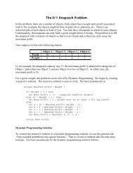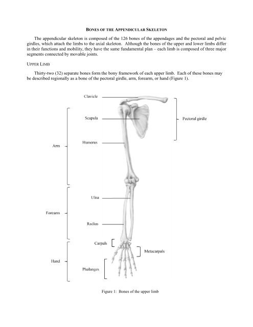
The appendicular skeleton is composed of the 126 bones of the ...
The appendicular skeleton is composed of the 126 bones of the ...
The appendicular skeleton is composed of the 126 bones of the ...

You also want an ePaper? Increase the reach of your titles
YUMPU automatically turns print PDFs into web optimized ePapers that Google loves.
BONES OF THE APPENDICULAR SKELETON<br />
<strong>The</strong> <strong>appendicular</strong> <strong>skeleton</strong> <strong>is</strong> <strong>composed</strong> <strong>of</strong> <strong>the</strong> <strong>126</strong> <strong>bones</strong> <strong>of</strong> <strong>the</strong> appendages and <strong>the</strong> pectoral and pelvic<br />
girdles, which attach <strong>the</strong> limbs to <strong>the</strong> axial <strong>skeleton</strong>. Although <strong>the</strong> <strong>bones</strong> <strong>of</strong> <strong>the</strong> upper and lower limbs differ<br />
in <strong>the</strong>ir functions and mobility, <strong>the</strong>y have <strong>the</strong> same fundamental plan – each limb <strong>is</strong> <strong>composed</strong> <strong>of</strong> three major<br />
segments connected by movable joints.<br />
UPPER LIMB<br />
Thirty-two (32) separate <strong>bones</strong> form <strong>the</strong> bony framework <strong>of</strong> each upper limb. Each <strong>of</strong> <strong>the</strong>se <strong>bones</strong> may<br />
be described regionally as a bone <strong>of</strong> <strong>the</strong> pectoral girdle, arm, forearm, or hand (Figure 1).<br />
Figure 1: Bones <strong>of</strong> <strong>the</strong> upper limb
Pectoral (Shoulder) Girdle (Marieb / Hoehn – Chapter 7; Pgs. 227 – 228)<br />
<strong>The</strong> paired pectoral girdles each cons<strong>is</strong>t <strong>of</strong> two <strong>bones</strong>, <strong>the</strong> anterior clavicle and <strong>the</strong> posterior scapula.<br />
<strong>The</strong> shoulder girdles function to attach <strong>the</strong> upper limbs to <strong>the</strong> axial <strong>skeleton</strong>. In addition, <strong>the</strong> <strong>bones</strong> <strong>of</strong> <strong>the</strong><br />
shoulder girdles serve as attachment points for many trunk and neck muscles.<br />
<strong>The</strong> pectoral girdle <strong>is</strong> exceptionally light and allows <strong>the</strong> upper limb a degree <strong>of</strong> mobility not seen<br />
anywhere else in <strong>the</strong> body. Th<strong>is</strong> <strong>is</strong> due to multiple factors including:<br />
1) <strong>The</strong> sternoclavicular joints are <strong>the</strong> only site <strong>of</strong> attachment <strong>of</strong> <strong>the</strong> shoulder girls to <strong>the</strong> axial <strong>skeleton</strong>.<br />
2) <strong>The</strong> relative looseness <strong>of</strong> <strong>the</strong> scapular attachment allows it to slide back and forth against <strong>the</strong> thorax<br />
with muscular activity.<br />
3) <strong>The</strong> bone connection between <strong>the</strong> humerus and scapula <strong>is</strong> shallow, and does little to stabilize <strong>the</strong><br />
shoulder joint.<br />
A. Clavicle: A slender, doubly-curved bone that joins <strong>the</strong> sternum to <strong>the</strong> scapula (Figure 2); serves as an<br />
anterior brace, or strut, to hold <strong>the</strong> arm away from <strong>the</strong> top <strong>of</strong> <strong>the</strong> thorax.<br />
Landmarks / Markings:<br />
Figure 2: Right clavicle, superior view<br />
Sternal end: Rounded terminus <strong>of</strong> clavicle; articulates with <strong>the</strong> sternal manubrium.<br />
Acromial end: Flattened terminus <strong>of</strong> clavicle; articulates with <strong>the</strong> scapula to form part <strong>of</strong> <strong>the</strong><br />
shoulder joint.<br />
B. Scapula: Thin, triangular flat bone; lies on <strong>the</strong> dorsal surface <strong>of</strong> <strong>the</strong> rib cage (Figure 3); serves as <strong>the</strong><br />
attachment point for <strong>the</strong> arm.<br />
Borders / angles:<br />
Superior border: Short, sharp border that forms <strong>the</strong> upper margin <strong>of</strong> <strong>the</strong> scapula when<br />
articulated with <strong>the</strong> axial <strong>skeleton</strong>.<br />
Medial (vertebral) border: Border which parallels <strong>the</strong> vertebral column when articulated with<br />
<strong>the</strong> axial <strong>skeleton</strong>; joins with superior border at <strong>the</strong> superior angle.<br />
Lateral (axillary) border: <strong>The</strong> thick border that abuts <strong>the</strong> armpit when articulated with <strong>the</strong><br />
axial <strong>skeleton</strong>; joins with superior border at <strong>the</strong> lateral angle and joins with medial border at <strong>the</strong><br />
inferior angle.<br />
2 BI 334 – Advanced Human Anatomy and Physiology<br />
Western Oregon University
Landmarks / Markings:<br />
Figure 3: Right scapula, anterior and posterior views<br />
Glenoid cavity (fossa): Small, shallow depression superior to <strong>the</strong> lateral border, near <strong>the</strong> lateral<br />
angle; articulates with humerus <strong>of</strong> <strong>the</strong> arm.<br />
Spine: Prominent ridge r<strong>is</strong>ing from <strong>the</strong> upper posterior surface <strong>of</strong> <strong>the</strong> scapula; site <strong>of</strong> muscle<br />
attachment.<br />
Acromion: Enlarged, roughened triangular structure projecting projection <strong>of</strong>f <strong>the</strong> lateral end <strong>of</strong><br />
<strong>the</strong> scapular spine; articulates with <strong>the</strong> acromial end <strong>of</strong> <strong>the</strong> clavicle.<br />
Coracoid process: Beak-like structure projecting anteriorly from <strong>the</strong> superior scapular border;<br />
site <strong>of</strong> muscle attachment.<br />
Suprascapular notch: Shallow groove in <strong>the</strong> superior border <strong>of</strong> <strong>the</strong> scapula at <strong>the</strong> base <strong>of</strong> <strong>the</strong><br />
coracoid process; passageway for nerves.<br />
Supraspinous fossa: Deep depression superior to <strong>the</strong> spine on <strong>the</strong> posterior surface <strong>of</strong> <strong>the</strong><br />
scapula; site <strong>of</strong> muscle attachment.<br />
Infraspinous fossa: Shallow depression inferior to <strong>the</strong> spine on <strong>the</strong> posterior surface <strong>of</strong> <strong>the</strong><br />
scapula; site <strong>of</strong> muscle attachment.<br />
Subscapular fossa: Shallow depression formed by <strong>the</strong> entire anterior scapular surface; site <strong>of</strong><br />
muscle attachment.<br />
3 BI 334 – Advanced Human Anatomy and Physiology<br />
Western Oregon University
Arm (Marieb / Hoehn – Chapter 7; Pgs. 228 – 230)<br />
<strong>The</strong> arm cons<strong>is</strong>ts <strong>of</strong> a single bone, <strong>the</strong> humerus (Figure 4). <strong>The</strong> largest and longest bone <strong>of</strong> <strong>the</strong> upper<br />
limb, it articulates with <strong>the</strong> scapula at <strong>the</strong> shoulder and with <strong>the</strong> radius and ulna (forearm <strong>bones</strong>) at <strong>the</strong> elbow.<br />
A. Humerus:<br />
Landmarks / Markings:<br />
Figure 4: Right humerus, anterior and posterior views<br />
Head: Smooth, hem<strong>is</strong>pherical projection at <strong>the</strong> proximal end <strong>of</strong> <strong>the</strong> humerus; articulates with<br />
glenoid cavity <strong>of</strong> scapula.<br />
Anatomical neck: Slight constriction just d<strong>is</strong>tal to <strong>the</strong> head <strong>of</strong> <strong>the</strong> humerus.<br />
Greater Tubercle: Large prominence just d<strong>is</strong>tal to <strong>the</strong> anatomical neck on <strong>the</strong> lateral surface <strong>of</strong><br />
<strong>the</strong> humerus; site <strong>of</strong> muscle attachment.<br />
Lesser Tubercle: Small prominence just d<strong>is</strong>tal to <strong>the</strong> anatomical neck on <strong>the</strong> medial surface <strong>of</strong><br />
<strong>the</strong> humerus; site <strong>of</strong> muscle attachment.<br />
4 BI 334 – Advanced Human Anatomy and Physiology<br />
Western Oregon University
Intertubercular sulcus: Shallow groove between lesser and greater tubercles; guides tendon.<br />
Surgical neck: Constricted region <strong>of</strong> <strong>the</strong> humerus just d<strong>is</strong>tal to <strong>the</strong> tubercles; common site <strong>of</strong><br />
bone fracture.<br />
Deltoid tuberosity: V-shaped rough region on <strong>the</strong> lateral aspect <strong>of</strong> <strong>the</strong> humerus about midway<br />
down <strong>the</strong> shaft; site <strong>of</strong> muscle attachment.<br />
Radial groove: A shallow depression running obliquely down <strong>the</strong> posterior aspect <strong>of</strong> <strong>the</strong><br />
humerus shaft; nerve passageway.<br />
Trochlea: Medial spool-shaped structure on <strong>the</strong> d<strong>is</strong>tal end <strong>of</strong> <strong>the</strong> humerus; articulates with ulna<br />
<strong>of</strong> <strong>the</strong> forearm.<br />
Capitulum: Lateral ball-like structure on <strong>the</strong> d<strong>is</strong>tal end <strong>of</strong> <strong>the</strong> humerus; articulates with radius<br />
<strong>of</strong> <strong>the</strong> forearm.<br />
Medial epicondyle: Small outward projection flanking trochlea; site <strong>of</strong> muscle attachment.<br />
Lateral epicondyle: Small outward projection flanking capitulum; site <strong>of</strong> muscle attachment.<br />
Coronoid fossa: Large, shallow depression superior to <strong>the</strong> trochlea on <strong>the</strong> anterior surface <strong>of</strong> <strong>the</strong><br />
humerus; receives corresponding process from ulna when elbow flexes / extends.<br />
Radial fossa: Small, shallow depression lateral to <strong>the</strong> coronoid fossa; receives head <strong>of</strong> radius<br />
when elbow flexes.<br />
Olecranon fossa: Large, deep depression superior to <strong>the</strong> trochlea on <strong>the</strong> posterior surface <strong>of</strong> <strong>the</strong><br />
humerus; anchors corresponding process from ulna to form elbow joint.<br />
5 BI 334 – Advanced Human Anatomy and Physiology<br />
Western Oregon University
Forearm (Marieb / Hoehn – Chapter 7; Pgs. 231 – 232)<br />
Two parallel <strong>bones</strong>, <strong>the</strong> radius and <strong>the</strong> ulna, form <strong>the</strong> forearm (Figure 5). <strong>The</strong>ir proximal ends articulate<br />
with <strong>the</strong> humerus and <strong>the</strong>ir d<strong>is</strong>tal ends articulate with <strong>the</strong> wr<strong>is</strong>t <strong>bones</strong>.<br />
Figure 5: Right ulna and radius, anterior and posterior views<br />
A. Ulna: Long, slender bone with a hook at <strong>the</strong> proximal end that forms <strong>the</strong> elbow joint with <strong>the</strong> humerus;<br />
lies medially in <strong>the</strong> forearm when <strong>the</strong> body <strong>is</strong> in anatomical position.<br />
Landmarks / Markings:<br />
Olecranon process: Large protuberance on <strong>the</strong> proximal end <strong>of</strong> <strong>the</strong> ulna; forms <strong>the</strong> upper<br />
portion <strong>of</strong> <strong>the</strong> hook that articulates with <strong>the</strong> trochlea <strong>of</strong> <strong>the</strong> humerus.<br />
Coronoid process: Small process located just d<strong>is</strong>tal to <strong>the</strong> olecranon process; forms <strong>the</strong> lower<br />
portion <strong>of</strong> <strong>the</strong> hook that articulates with <strong>the</strong> trochlea <strong>of</strong> <strong>the</strong> humerus.<br />
Trochlear notch: Deep concavity found between <strong>the</strong> olecranon process and coronoid process;<br />
‘grips’ trochlea to form elbow joint.<br />
6 BI 334 – Advanced Human Anatomy and Physiology<br />
Western Oregon University
Radial notch: Small depression on <strong>the</strong> lateral side <strong>of</strong> <strong>the</strong> coronoid process; articulates with head<br />
<strong>of</strong> radius.<br />
Head: Knob-like structure and <strong>the</strong> d<strong>is</strong>tal end <strong>of</strong> <strong>the</strong> ulna; articulates with wr<strong>is</strong>t bone.<br />
Styloid process: Pointed process medial to <strong>the</strong> head <strong>of</strong> <strong>the</strong> ulna; site <strong>of</strong> ligament attachment.<br />
B. Radius: Long bone that <strong>is</strong> thin at its proximal end and wide at its d<strong>is</strong>tal end; lies laterally in <strong>the</strong> forearm<br />
when <strong>the</strong> body <strong>is</strong> in anatomical position.<br />
Landmarks / Markings:<br />
Head: Wheel-shaped proximal end <strong>of</strong> radius; articulates with capitulum <strong>of</strong> humerus and radial<br />
notch <strong>of</strong> ulna.<br />
Radial tuberosity: Rough projection just inferior to <strong>the</strong> head <strong>of</strong> <strong>the</strong> radius; site <strong>of</strong> muscle<br />
attachment.<br />
Ulnar notch: Medial shallow depression on <strong>the</strong> d<strong>is</strong>tal end <strong>of</strong> <strong>the</strong> radius; articulates with <strong>the</strong><br />
ulna.<br />
Styloid process: Pointed process lateral to <strong>the</strong> ulnar notch; site <strong>of</strong> ligament attachment.<br />
7 BI 334 – Advanced Human Anatomy and Physiology<br />
Western Oregon University
Hand (Marieb / Hoehn – Chapter 7; Pgs. 232 – 234)<br />
<strong>The</strong> <strong>skeleton</strong> <strong>of</strong> <strong>the</strong> hand includes <strong>the</strong> <strong>bones</strong> <strong>of</strong> <strong>the</strong> carpus (wr<strong>is</strong>t); <strong>the</strong> <strong>bones</strong> <strong>of</strong> <strong>the</strong> metacarpus (palm),<br />
and <strong>the</strong> <strong>bones</strong> <strong>of</strong> <strong>the</strong> phalanges (fingers) (Figure 6).<br />
Figure 6: Right hand, anterior view<br />
A. Carpals (wr<strong>is</strong>t): Eight (8) marble-size short <strong>bones</strong> closely united by ligaments; quite flexible due to<br />
gliding movements between <strong>bones</strong>.<br />
1) Scaphoid<br />
2) Lunate<br />
3) Triquetrum<br />
4) P<strong>is</strong>iform<br />
Closely articulate<br />
with radius<br />
B. Metacarpals: Five (5) small long <strong>bones</strong> radiating from <strong>the</strong> wr<strong>is</strong>t like spokes; numbered 1 – 5 from <strong>the</strong><br />
thumb to <strong>the</strong> little finger.<br />
C. Phalanges: Fourteen (14) miniature long <strong>bones</strong> that form <strong>the</strong> fingers; numbered 1 – 5 from <strong>the</strong> thumb<br />
(pollex) to <strong>the</strong> little finger.<br />
1) Proximal phalange (1 – 5)<br />
2) Middle phalange (2 – 5)<br />
3) D<strong>is</strong>tal phalange (1 – 5)<br />
5) Trapezium<br />
6) Trapezoid<br />
7) Capitate<br />
8) Hamate<br />
8 BI 334 – Advanced Human Anatomy and Physiology<br />
Western Oregon University
CHECKLIST: BONES / MARKINGS OF THE UPPER LIMB<br />
BONES OF THE PECTORAL GIRDLE:<br />
Clavicle*<br />
Sternal end<br />
Acromial end<br />
Scapula*<br />
Superior / medial / lateral border<br />
Superior / lateral / inferior angle<br />
Glenoid cavity (fossa)<br />
Spine<br />
Acromion<br />
Coracoid process<br />
Suprascapular notch<br />
Supraspinous fossa<br />
Infraspinous fossa<br />
Subscapular fossa<br />
BONE OF THE ARM:<br />
Humerus*<br />
Head<br />
Anatomical / surgical neck<br />
Greater / lesser tubercle<br />
Intertubercular sulcus<br />
Deltoid tuberosity<br />
Radial groove<br />
Trochlea<br />
Capitulum<br />
Medial / lateral epicondyle<br />
Coronoid fossa<br />
Radial fossa<br />
Olecranon fossa<br />
* Need to be able to identify bone from right vs. left side <strong>of</strong> body<br />
BONES OF THE FOREARM:<br />
Ulna*<br />
Olecranon process<br />
Coronoid process<br />
Trochlear notch<br />
Radial notch<br />
Head<br />
Styloid process<br />
Radius*<br />
Head<br />
Radial tuberosity<br />
Ulnar notch<br />
Styloid process<br />
BONES OF THE WRIST / HAND:<br />
Scaphoid<br />
Lunate<br />
Triquetrum<br />
P<strong>is</strong>iform<br />
Trapezium<br />
Trapezoid<br />
Capitate<br />
Hamate<br />
Metacarpals (1 – 5)<br />
Phalanges<br />
Carpals (8)<br />
Proximal, middle, d<strong>is</strong>tal<br />
Pollex<br />
9 BI 334 – Advanced Human Anatomy and Physiology<br />
Western Oregon University