Distally based superficial Sural artery flap for foot - Ayub Medical ...
Distally based superficial Sural artery flap for foot - Ayub Medical ...
Distally based superficial Sural artery flap for foot - Ayub Medical ...

Create successful ePaper yourself
Turn your PDF publications into a flip-book with our unique Google optimized e-Paper software.
traumatic amputation of <strong>foot</strong>. Another had cut<br />
tendoachilles. One had <strong>foot</strong> degloving with exposed<br />
lateral melleolus. Nine children got their feet struck in<br />
the wheel of motorcycle; one patient had fracture of<br />
lateral malleolus. Heel injury was seen in 6 cases. Three<br />
patients got preoperative Vacuum Assisted Closure<br />
(VAC) treatment followed by <strong>flap</strong> coverage. Fasciocutaneous<br />
(FC) <strong>flap</strong>s were used in 9 cases and 7 patients<br />
were covered with fascia/adipofascial (AF) with skin<br />
graft. All cases were treated after 2 weeks because of<br />
delayed referral from other departments. One child<br />
underwent tendoachilles repair followed by fascia <strong>flap</strong><br />
cover. Fifteen <strong>flap</strong>s survived completely while one <strong>flap</strong><br />
had total necrosis. Another had distal necrosis which<br />
required debridement and wound healed with dressings.<br />
One patient developed donor site scar hypertrophy. One<br />
<strong>flap</strong> was bulky causing contour de<strong>for</strong>mity which was<br />
debulked after six months.<br />
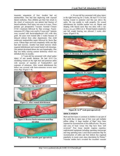
A 7-year-old boy presented with wheel spoke<br />
injury of one month duration. He had 10×8 Cm<br />
ranulating wound on the right heel and posterior ankle<br />
with necrosis of insertion of Tendoachille’s and<br />
exposure of calcaneus. After wound debridement the<br />
defect was covered with fasio-cutaneous reverse sural<br />
<strong>artery</strong> <strong>flap</strong>. (Figure-1–4).<br />
Figure-1: Pre-op exposed calcaneum<br />
Figure-2: After wound debridement<br />
Figure-3: Reverse fascio-cutanoeus sural <strong>flap</strong><br />
elevation<br />
Figure-4: Three months post operatively<br />
J <strong>Ayub</strong> Med Coll Abbottabad 2011;23(4)<br />
A 10-year-old boy presented with glass injury<br />
to the right lower leg <strong>for</strong> 2 weeks. He had 3×3 Cm non<br />
healing wound in posterior mid line just above the<br />
ankle. He was unable to walk properly. On wound<br />
debridement his Achilles tendon was cut. Repair and<br />
draping with fascial sural <strong>flap</strong> was done with a Split<br />
thickness skin graft (SSG) on it. Wound healed completely<br />
and full weight bearing was allowed 3 weeks after<br />
injury. (Figure-5–8).<br />
Figure-5: Division of Tendoachilles due to trauma<br />
Figure-6: TA repaired<br />
Figure-7: Fascia only <strong>flap</strong> inset over repaired TA<br />
Figure-8: At 4 th week post-operatively<br />
DISCUSSION<br />
Heel and <strong>foot</strong> injury is common in children in our part of<br />
the world due to open type of <strong>foot</strong> wear and multiple<br />
pillion riding. A large number of <strong>flap</strong>s 4 have been<br />
reported <strong>for</strong> reconstruction of this difficult area. The pros<br />
and cons of local versus free <strong>flap</strong> have been well<br />
reviewed. 8 Free tissue transfer requires expertise,<br />
sophisticated equipment including operating microscope<br />
and long operating time. Local fascio-cutaneous <strong>flap</strong> <strong>for</strong><br />
reconstruction of ankle and <strong>foot</strong> defects has regained<br />
popularity in recent years since description by Ponten. 9<br />
Vascular studies have revealed the excellent vascular<br />
http://www.ayubmed.edu.pk/JAMC/23-4/Farrukh.pdf 41