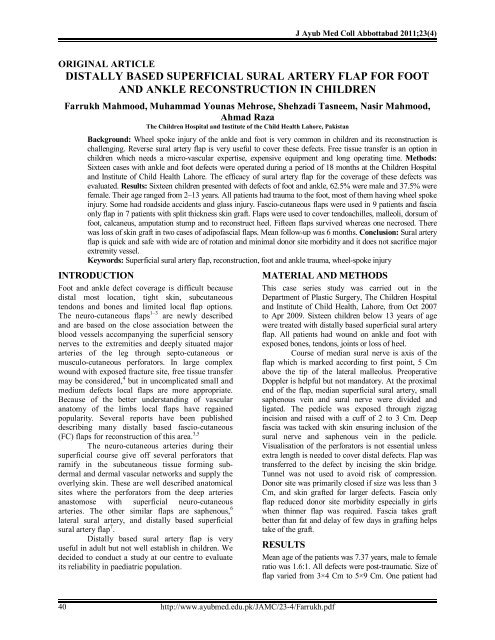
Distally based superficial Sural artery flap for foot - Ayub Medical ...
Distally based superficial Sural artery flap for foot - Ayub Medical ...
Distally based superficial Sural artery flap for foot - Ayub Medical ...

You also want an ePaper? Increase the reach of your titles
YUMPU automatically turns print PDFs into web optimized ePapers that Google loves.
40<br />
http://www.ayubmed.edu.pk/JAMC/23-4/Farrukh.pdf<br />
J <strong>Ayub</strong> Med Coll Abbottabad 2011;23(4)<br />
ORIGINAL ARTICLE<br />
DISTALLY BASED SUPERFICIAL SURAL ARTERY FLAP FOR FOOT<br />
AND ANKLE RECONSTRUCTION IN CHILDREN<br />
Farrukh Mahmood, Muhammad Younas Mehrose, Shehzadi Tasneem, Nasir Mahmood,<br />
Ahmad Raza<br />
The Children Hospital and Institute of the Child Health Lahore, Pakistan<br />
Background: Wheel spoke injury of the ankle and <strong>foot</strong> is very common in children and its reconstruction is<br />
challenging. Reverse sural <strong>artery</strong> <strong>flap</strong> is very useful to cover these defects. Free tissue transfer is an option in<br />
children which needs a micro-vascular expertise, expensive equipment and long operating time. Methods:<br />
Sixteen cases with ankle and <strong>foot</strong> defects were operated during a period of 18 months at the Children Hospital<br />
and Institute of Child Health Lahore. The efficacy of sural <strong>artery</strong> <strong>flap</strong> <strong>for</strong> the coverage of these defects was<br />
evaluated. Results: Sixteen children presented with defects of <strong>foot</strong> and ankle, 62.5% were male and 37.5% were<br />
female. Their age ranged from 2–13 years. All patients had trauma to the <strong>foot</strong>, most of them having wheel spoke<br />
injury. Some had roadside accidents and glass injury. Fascio-cutaneous <strong>flap</strong>s were used in 9 patients and fascia<br />
only <strong>flap</strong> in 7 patients with split thickness skin graft. Flaps were used to cover tendoachilles, malleoli, dorsum of<br />
<strong>foot</strong>, calcaneus, amputation stump and to reconstruct heel. Fifteen <strong>flap</strong>s survived whereas one necrosed. There<br />
was loss of skin graft in two cases of adipofascial <strong>flap</strong>s. Mean follow-up was 6 months. Conclusion: <strong>Sural</strong> <strong>artery</strong><br />
<strong>flap</strong> is quick and safe with wide arc of rotation and minimal donor site morbidity and it does not sacrifice major<br />
extremity vessel.<br />
Keywords: Superficial sural <strong>artery</strong> <strong>flap</strong>, reconstruction, <strong>foot</strong> and ankle trauma, wheel-spoke injury<br />
INTRODUCTION<br />
Foot and ankle defect coverage is difficult because<br />
distal most location, tight skin, subcutaneous<br />
tendons and bones and limited local <strong>flap</strong> options.<br />
The neuro-cutaneous <strong>flap</strong>s 1–3 are newly described<br />
and are <strong>based</strong> on the close association between the<br />
blood vessels accompanying the <strong>superficial</strong> sensory<br />
nerves to the extremities and deeply situated major<br />
arteries of the leg through septo-cutaneous or<br />
musculo-cutaneous per<strong>for</strong>ators. In large complex<br />
wound with exposed fracture site, free tissue transfer<br />
may be considered, 4 but in uncomplicated small and<br />
medium defects local <strong>flap</strong>s are more appropriate.<br />
Because of the better understanding of vascular<br />
anatomy of the limbs local <strong>flap</strong>s have regained<br />
popularity. Several reports have been published<br />
describing many distally <strong>based</strong> fascio-cutaneous<br />
(FC) <strong>flap</strong>s <strong>for</strong> reconstruction of this area. 3,5<br />
The neuro-cutaneous arteries during their<br />
<strong>superficial</strong> course give off several per<strong>for</strong>ators that<br />
ramify in the subcutaneous tissue <strong>for</strong>ming subdermal<br />
and dermal vascular networks and supply the<br />
overlying skin. These are well described anatomical<br />
sites where the per<strong>for</strong>ators from the deep arteries<br />
anastomose with <strong>superficial</strong> neuro-cutaneous<br />
arteries. The other similar <strong>flap</strong>s are saphenous, 6<br />
lateral sural <strong>artery</strong>, and distally <strong>based</strong> <strong>superficial</strong><br />
sural <strong>artery</strong> <strong>flap</strong> 7 .<br />
<strong>Distally</strong> <strong>based</strong> sural <strong>artery</strong> <strong>flap</strong> is very<br />
useful in adult but not well establish in children. We<br />
decided to conduct a study at our centre to evaluate<br />
its reliability in paediatric population.<br />
MATERIAL AND METHODS<br />
This case series study was carried out in the<br />
Department of Plastic Surgery, The Children Hospital<br />
and Institute of Child Health, Lahore, from Oct 2007<br />
to Apr 2009. Sixteen children below 13 years of age<br />
were treated with distally <strong>based</strong> <strong>superficial</strong> sural <strong>artery</strong><br />
<strong>flap</strong>. All patients had wound on ankle and <strong>foot</strong> with<br />
exposed bones, tendons, joints or loss of heel.<br />
Course of median sural nerve is axis of the<br />
<strong>flap</strong> which is marked according to first point, 5 Cm<br />
above the tip of the lateral malleolus. Preoperative<br />
Doppler is helpful but not mandatory. At the proximal<br />
end of the <strong>flap</strong>, median <strong>superficial</strong> sural <strong>artery</strong>, small<br />
saphenous vein and sural nerve were divided and<br />
ligated. The pedicle was exposed through zigzag<br />
incision and raised with a cuff of 2 to 3 Cm. Deep<br />
fascia was tacked with skin ensuring inclusion of the<br />
sural nerve and saphenous vein in the pedicle.<br />
Visualisation of the per<strong>for</strong>ators is not essential unless<br />
extra length is needed to cover distal defects. Flap was<br />
transferred to the defect by incising the skin bridge.<br />
Tunnel was not used to avoid risk of compression.<br />
Donor site was primarily closed if size was less than 3<br />
Cm, and skin grafted <strong>for</strong> larger defects. Fascia only<br />
<strong>flap</strong> reduced donor site morbidity especially in girls<br />
when thinner <strong>flap</strong> was required. Fascia takes graft<br />
better than fat and delay of few days in grafting helps<br />
take of the graft.<br />
RESULTS<br />
Mean age of the patients was 7.37 years, male to female<br />
ratio was 1.6:1. All defects were post-traumatic. Size of<br />
<strong>flap</strong> varied from 3×4 Cm to 5×9 Cm. One patient had
traumatic amputation of <strong>foot</strong>. Another had cut<br />
tendoachilles. One had <strong>foot</strong> degloving with exposed<br />
lateral melleolus. Nine children got their feet struck in<br />
the wheel of motorcycle; one patient had fracture of<br />
lateral malleolus. Heel injury was seen in 6 cases. Three<br />
patients got preoperative Vacuum Assisted Closure<br />
(VAC) treatment followed by <strong>flap</strong> coverage. Fasciocutaneous<br />
(FC) <strong>flap</strong>s were used in 9 cases and 7 patients<br />
were covered with fascia/adipofascial (AF) with skin<br />
graft. All cases were treated after 2 weeks because of<br />
delayed referral from other departments. One child<br />
underwent tendoachilles repair followed by fascia <strong>flap</strong><br />
cover. Fifteen <strong>flap</strong>s survived completely while one <strong>flap</strong><br />
had total necrosis. Another had distal necrosis which<br />
required debridement and wound healed with dressings.<br />
One patient developed donor site scar hypertrophy. One<br />
<strong>flap</strong> was bulky causing contour de<strong>for</strong>mity which was<br />
debulked after six months.<br />
A 7-year-old boy presented with wheel spoke<br />
injury of one month duration. He had 10×8 Cm<br />
ranulating wound on the right heel and posterior ankle<br />
with necrosis of insertion of Tendoachille’s and<br />
exposure of calcaneus. After wound debridement the<br />
defect was covered with fasio-cutaneous reverse sural<br />
<strong>artery</strong> <strong>flap</strong>. (Figure-1–4).<br />
Figure-1: Pre-op exposed calcaneum<br />
Figure-2: After wound debridement<br />
Figure-3: Reverse fascio-cutanoeus sural <strong>flap</strong><br />
elevation<br />
Figure-4: Three months post operatively<br />
J <strong>Ayub</strong> Med Coll Abbottabad 2011;23(4)<br />
A 10-year-old boy presented with glass injury<br />
to the right lower leg <strong>for</strong> 2 weeks. He had 3×3 Cm non<br />
healing wound in posterior mid line just above the<br />
ankle. He was unable to walk properly. On wound<br />
debridement his Achilles tendon was cut. Repair and<br />
draping with fascial sural <strong>flap</strong> was done with a Split<br />
thickness skin graft (SSG) on it. Wound healed completely<br />
and full weight bearing was allowed 3 weeks after<br />
injury. (Figure-5–8).<br />
Figure-5: Division of Tendoachilles due to trauma<br />
Figure-6: TA repaired<br />
Figure-7: Fascia only <strong>flap</strong> inset over repaired TA<br />
Figure-8: At 4 th week post-operatively<br />
DISCUSSION<br />
Heel and <strong>foot</strong> injury is common in children in our part of<br />
the world due to open type of <strong>foot</strong> wear and multiple<br />
pillion riding. A large number of <strong>flap</strong>s 4 have been<br />
reported <strong>for</strong> reconstruction of this difficult area. The pros<br />
and cons of local versus free <strong>flap</strong> have been well<br />
reviewed. 8 Free tissue transfer requires expertise,<br />
sophisticated equipment including operating microscope<br />
and long operating time. Local fascio-cutaneous <strong>flap</strong> <strong>for</strong><br />
reconstruction of ankle and <strong>foot</strong> defects has regained<br />
popularity in recent years since description by Ponten. 9<br />
Vascular studies have revealed the excellent vascular<br />
http://www.ayubmed.edu.pk/JAMC/23-4/Farrukh.pdf 41
connections between the <strong>superficial</strong> system and deep<br />
main arteries through musculo-cutaneous and septocutaneous<br />
per<strong>for</strong>ators. These vascular connections via<br />
per<strong>for</strong>ators make each of it a potential pivot point <strong>for</strong> <strong>flap</strong><br />
rotation. 10 Due to rich anastomosis between peroneal<br />
<strong>artery</strong> per<strong>for</strong>ators and longitudinally oriented median<br />
<strong>superficial</strong> sural <strong>artery</strong> both proximally 11 and distally<br />
<strong>based</strong> <strong>flap</strong>s can be elevated 3,5,12 . Masqualet et al named<br />
these <strong>flap</strong>s neuro-cutaneous in 1992 and several reports<br />
were published. 13–15 In our series of 16 patients we<br />
reconstructed defects around the ankle with good success<br />
rate. If the thinner <strong>flap</strong> is required the fascia alone was<br />
used to cover the defect with split skin graft on it. Fascia<br />
can be rotated to cover the defect or turned upside down<br />
like page of a book and its under surface takes the graft<br />
readily. Fascial <strong>flap</strong> also helps to reduce the donor site<br />
morbidity. Delay in grafting <strong>for</strong> couple of days enhances<br />
its take. <strong>Distally</strong> <strong>based</strong> <strong>superficial</strong> sural <strong>artery</strong> <strong>flap</strong> is a<br />
single stage procedure conserving the major extremity<br />
vessels. It is safe, quick to elevate with wide arc of<br />
rotation. Vascular anatomy is quite constant and<br />
preoperative Doppler is not mandatory if arc of rotation<br />
is kept above 5 Cm from tip of lateral malleolus.<br />
Similar <strong>flap</strong>s were also described <strong>based</strong> on<br />
anterior per<strong>for</strong>ators of the peroneal <strong>artery</strong> like the lateral<br />
supra malleolar <strong>flap</strong> 16 and saphenous <strong>flap</strong> 17 . Shalaby 18<br />
described a <strong>flap</strong> <strong>based</strong> on same posterior peroneal<br />
per<strong>for</strong>ators and island is taken more laterally overlying<br />
the posterior intermuscular septum. The vascular axis of<br />
the <strong>flap</strong> is an anastomotic arcade <strong>for</strong>med by longitudinal<br />
connections between neighbouring septo-cutaneous<br />
vessels in the same septum. Flap has a short pedicle and<br />
seems to be more suitable <strong>for</strong> above the ankle defect.<br />
Oberlin et al 7 described the posterolateral malleolar <strong>flap</strong><br />
with the pivot point at the tip of the lateral malleolus.<br />
This short <strong>flap</strong> depends on the communication between<br />
lateral calcaneal and lateral tarsal <strong>artery</strong> and is useful <strong>for</strong><br />
coverage of in-defects around lateral malleolus and<br />
posterior heel region.<br />
Main demerit of distal <strong>based</strong> sural <strong>artery</strong> <strong>flap</strong> is<br />
a scar on calf which can be reduced by making use of<br />
fascia <strong>flap</strong> wherever possible. Neurological deficit<br />
caused by division of nerve is minimal and tends to<br />
improve with time.<br />
CONCLUSION<br />
<strong>Distally</strong> <strong>based</strong> sural <strong>artery</strong> <strong>flap</strong> is reliable, safe, easy and<br />
quick to execute and durable having minimal donor site<br />
morbidity <strong>for</strong> the reconstruction of this challenging area<br />
in the children. It is a good alternative to free tissue<br />
transfer in suitable patients.<br />
42<br />
http://www.ayubmed.edu.pk/JAMC/23-4/Farrukh.pdf<br />
J <strong>Ayub</strong> Med Coll Abbottabad 2011;23(4)<br />
ACKNOWLEDGEMENTS<br />
Our sincere thanks to Dr. Moazzam Nazeer Tarar,<br />
Associate Professor and Head of Department of Plastic<br />
Surgery, Allama Iqbal <strong>Medical</strong> College/Jinnah Hospital<br />
<strong>for</strong> his vision and help. Thanks also to Dr. Waqas, <strong>for</strong><br />
his assistance in compiling data.<br />
REFERENCES<br />
1. Bertelli JA, Kaleli T. Retrograde flow neurocutaneous island<br />
<strong>flap</strong>s in the <strong>for</strong>earm. Anatomical basis and clinical results.<br />
Plast Reconst Surg 1995;95:851–9.<br />
2. Fachinelli A, Masquelet A, Restrepo J. The vascularized<br />
sural nerve. Int J Microsurg 1981;3:57–62.<br />
3. Masquelet AC, Romana MC, Wolf G. Skin island <strong>flap</strong>s<br />
supplied by the vascular axis of the sensitive <strong>superficial</strong><br />
nerves: anatomic study and clinical experience in the leg.<br />
Plast Reconstr Surg 1992;89:1115–20.<br />
4. Rajacic N, Lari AR, Khalaf ME, Kersnic M. Free <strong>flap</strong>s <strong>for</strong><br />
the treatment of avulsion injuries in the feet. J Pediatr Orthop<br />
1994;14:522–5.<br />
5. Donski PK, Fogdestam I. <strong>Distally</strong> <strong>based</strong> fasciocutaneous <strong>flap</strong><br />
from the sural region. Scand J Plast Reconstr Surg<br />
1983;17:191–6.<br />
6. Rajacic N, Gang RK, Krishnan J, Kojic S. Lower leg<br />
reconstruction using distally <strong>based</strong> saphenous island <strong>flap</strong>s.<br />
Eur J Plast Surg 2001;24:7–11.<br />
7. Oberlin C, Azoulay B, Bhatia A. the posterolateral malleolar<br />
<strong>flap</strong> of the ankle: a distally <strong>based</strong> sural neurocutaneous <strong>flap</strong>report<br />
of 14 cases. Plast Reconstr Surg 1995;96:400–5.<br />
8. Serafin D, Georgiade NG, Smith D. Comparison of free with<br />
pedicled <strong>flap</strong>s <strong>for</strong> coverage of defects of leg or <strong>foot</strong>. Plast<br />
Reconstr Surg 1977;59:492–9.<br />
9. Ponten B. The fasciocutaneous <strong>flap</strong>. Its use in soft tissue<br />
defects of the lower leg. Br J Plast Surg 1981;34:215–20.<br />
10. Masquelet A. The posterolateral malleolar <strong>flap</strong> of the ankle: a<br />
distally <strong>based</strong> sural neurocutaneous <strong>flap</strong>-Report of 14 cases.<br />
Plast Reconstr Surg 1995;96:406–7.<br />
11. Tolhurst DE, Haeseker B, Zeeman RJ. The development of<br />
the fasciocutaneous <strong>flap</strong> and its clinical applications. Plast<br />
Reconstr Surg 1983;71:597–60.<br />
12. Carriquiry CE. Heel coverage with a deepithelialized distally<br />
<strong>based</strong> fasciocutaneous <strong>flap</strong>. Plast Reconstr Surg<br />
1990;65:116–9.<br />
13. Hyakusoku H, Tonegawa H, Fumiiri M. Heel coverage with<br />
a T-shaped distally <strong>based</strong> sural island fasciocutaneous <strong>flap</strong>.<br />
Plast Reconstr Surg 1994;93:872–6.<br />
14. Hasegawa M, Torii S, Katooh H, Esaki S. The distally <strong>based</strong><br />
<strong>superficial</strong> sural <strong>artery</strong> <strong>flap</strong>. Plast Reconstr Surg<br />
1994;93:1012–20.<br />
15. Rajacic N, Darweesh M, Jayakrishnan K, Gang RK, Jojic S.<br />
The distally <strong>based</strong> <strong>superficial</strong> sural <strong>artery</strong> <strong>flap</strong> <strong>for</strong><br />
reconstruction of the lower leg and <strong>foot</strong>. Br J Plast Surg<br />
1996;49:383–9.<br />
16. Masquelet A, Beveridge J, Romana C, Gerber C. The lateral<br />
supramalleolar <strong>flap</strong>. Plast Reconstr Surg 1988;81:74–81.<br />
17. Rajacic N, Gang RK, Krishnan J, Kojic S. Lower leg<br />
reconstruction using distally <strong>based</strong> saphenous island <strong>flap</strong>s.<br />
Eur J Plast Surg 2001;24:7–11.<br />
18. Shalaby HA. The distally <strong>based</strong> peroneal island <strong>flap</strong>. Br J<br />
Plast Surg 1995;48:23–6.<br />
Address <strong>for</strong> Correspondence:<br />
Dr. Farrukh Mahmood, Department of Paediatric Plastic Surgery, The Children Hospital and Institute of Child<br />
Health, Lahore, Pakistan. Cell: +92-300-9483452<br />
Email: drfarrukhmahmood@gmail.com