The carotid bruit - Practical Neurology
The carotid bruit - Practical Neurology
The carotid bruit - Practical Neurology

Create successful ePaper yourself
Turn your PDF publications into a flip-book with our unique Google optimized e-Paper software.
INTRODUCTION<br />
When faced with a patient who may have had a<br />
stroke or transient ischaemic attack (TIA), one<br />
needs to ask oneself some simple questions: was<br />
the event vascular?; where was the brain lesion,<br />
and hence its vascular territory?; what was the<br />
cause? A careful history and focused physical<br />
examination are essential steps in getting the<br />
right answers. Although one can learn a great<br />
deal about the state of a patient’s arteries from<br />
expensive vascular imaging techniques, this<br />
does not make simple auscultation of the neck<br />
for <strong>carotid</strong> <strong>bruit</strong>s redundant. In this brief review,<br />
we will therefore defi ne the place of the <strong>bruit</strong> in<br />
the diagnosis and management of patients with<br />
suspected TIA or stroke.<br />
WHY ARE CAROTID BRUITS<br />
IMPORTANT?<br />
A <strong>bruit</strong> over the <strong>carotid</strong> region is important<br />
because it may indicate the presence of atherosclerotic<br />
plaque in the <strong>carotid</strong> arteries. Thromboembolism<br />
from atherosclerotic plaque at<br />
the <strong>carotid</strong> artery bifurcation is a major cause<br />
of TIA and ischaemic stroke. Plaques occur<br />
preferentially at the <strong>carotid</strong> bifurcation, usually<br />
fi rst on the posterior wall of the internal <strong>carotid</strong><br />
artery origin. <strong>The</strong> growth of these plaques and<br />
their subsequent disintegration, surface ulceration,<br />
and capacity to throw off emboli into the<br />
brain and eye determines the pattern of subsequent<br />
symptoms. <strong>The</strong> presence of an arterial<br />
<strong>bruit</strong> arising from stenosis at the origin of the<br />
internal <strong>carotid</strong> artery may therefore help to<br />
AUGUST 2002 221<br />
<strong>The</strong> <strong>carotid</strong> <strong>bruit</strong><br />
clarify whether an event was vascular or not,<br />
identify the cause as likely to be due to atheromatous<br />
stenosis, and the possibility that the<br />
stenosis may be severe enough to justify <strong>carotid</strong><br />
endarterectomy. However, not all noises in the<br />
neck indicate serious arterial disease. So, we<br />
need to review: how <strong>bruit</strong>s arise, how to identify<br />
arterial <strong>bruit</strong>s, to identify which noises in the<br />
neck matter, and what to do once you fi nd an<br />
arterial <strong>bruit</strong>.<br />
HOW BRUITS ARISE<br />
Carotid <strong>bruit</strong>s generally result from turbulent,<br />
non-laminar fl ow through a stenotic lesion,<br />
which causes arterial wall vibrations distal to<br />
the stenosis. <strong>The</strong> vibrations are transmitted to<br />
the body surface, where they can be detected<br />
with a stethoscope. A <strong>bruit</strong> can develop when<br />
the arterial lumen is reduced to less than 50% of<br />
its original cross-sectional diameter.<br />
HOW TO LISTEN FOR BRUITS<br />
Before you get out your stethoscope to listen for<br />
a <strong>bruit</strong>, you need to have a clear idea of what you<br />
will do if you fi nd one! Table 1 gives our view of<br />
when listening to the neck is likely to be useful in<br />
NEUROLOGICAL SIGN<br />
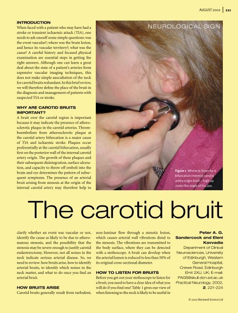
Figure 1 Where to listen for a<br />
bifurcation/internal <strong>carotid</strong><br />
artery origin <strong>bruit</strong> – high up<br />
under the angle of the jaw.<br />
Peter A. G.<br />
Sandercock and Eleni<br />
Kavvadia<br />
Department of Clinical<br />
Neurosciences, University<br />
of Edinburgh, Western<br />
General Hospital,<br />
Crewe Road, Edinburgh<br />
EH4 2XU, UK; E-mail:<br />
PAGS@skull.dcn.ed.ac.uk<br />
<strong>Practical</strong> <strong>Neurology</strong>, 2002,<br />
2, 221–224<br />
© 2002 Blackwell Science Ltd
222<br />
PRACTICAL NEUROLOGY<br />
Table 1 When is neck ausculation clinically useful?<br />
Situations when the presence or absence of a <strong>carotid</strong> <strong>bruit</strong> can be helpful<br />
In patients over 50 years of age with recent <strong>carotid</strong> territory TIA or stroke<br />
– a <strong>bruit</strong> over the symptomatic <strong>carotid</strong> artery suggests the possibility of an<br />
underlying severe stenosis, likely to benefi t from surgery<br />
– a <strong>bruit</strong> may indicate stroke or TIA is more likely due to atherosclerosis than some<br />
other nonatheromatosis pathophysiological process<br />
In patients with history of sudden onset of focal neurological signs (transient or<br />
persistent)<br />
– the presence of a <strong>bruit</strong> increases the probability that the symptoms were<br />
vascular in origin<br />
In younger patients with sudden focal symptoms (transient or persistent) where a<br />
vascular cause is thought unlikely (e.g. clinical syndrome of migraine aura without<br />
headache)<br />
– absence of a <strong>bruit</strong> makes underlying vascular disease even less likely.<br />
Situations where it may be better not to auscultate the neck<br />
Asymptomatic individuals<br />
Patients with nonfocal neurological symptoms<br />
– blackouts<br />
– dizzy turns<br />
– headaches<br />
Patients with pulsatile tinnitus<br />
Figure 2 <strong>The</strong> sites of maximal<br />
intensity of arterial <strong>bruit</strong>s in<br />
the neck. A <strong>bruit</strong> arising from<br />
the <strong>carotid</strong> bifurcation is high<br />
up under the angle of the jaw.<br />
Localized supraclavicular <strong>bruit</strong>s<br />
are caused either by subclavian<br />
or vertebral origin artery stenosis.<br />
Diffuse <strong>bruit</strong>s are transmitted<br />
from the arch of the aorta or the<br />
heart.<br />
© 2002 Blackwell Science Ltd<br />
neurological practice. <strong>The</strong>n, get the patient into<br />
a quiet room, in a relaxed and comfortable position.<br />
We use the diaphragm of the stethoscope,<br />
because it detects the higher frequency sounds<br />
of arterial <strong>bruit</strong>s rather better than the bell. Ask<br />
the patient to breathe in and hold their breath.<br />
Listen over an area beginning from just behind<br />
the upper end of the thyroid cartilage to just<br />
below the angle of the jaw, in other words over<br />
the line of the common <strong>carotid</strong> artery leading up<br />
to the bifurcation into the internal and external<br />
<strong>carotid</strong> arteries. Apply only suffi cient pressure<br />
to ensure the diaphragm rests squarely on the<br />
skin (Fig. 1). Excessive pressure can compress<br />
the underlying artery enough to cause a <strong>bruit</strong><br />
even when the artery is normal.<br />
THE BRUIT THAT MATTERS: THE<br />
ONE DUE TO CAROTID STENOSIS<br />
Bruits at the bifurcation of the common <strong>carotid</strong><br />
artery are best heard high up under the angle of<br />
the jaw (Fig. 2). At this level the common <strong>carotid</strong><br />
artery bifurcates and gives rise to its internal<br />
branch. If one hears a <strong>bruit</strong> only in the base of<br />
the neck, or along the course of the common<br />
<strong>carotid</strong> artery, it is referred to as ‘diffuse’. Diffuse<br />
<strong>bruit</strong>s are not a very specifi c indicator of internal<br />
<strong>carotid</strong> artery disease. Bruits heard only at the<br />
bifurcation are more specifi c for internal <strong>carotid</strong><br />
artery origin stenosis, but lack sensitivity. Unfortunately<br />
<strong>bruit</strong>s at this location can also arise<br />
from disease of the external <strong>carotid</strong> artery.<br />
OTHER NOISES IN THE NECK<br />
See Table 2. Bruits transmitted from the heart<br />
become attenuated as one moves the stethoscope<br />
up the neck towards the angle of the jaw. Thyroid<br />
<strong>bruit</strong>s are bilateral and more obviously located<br />
over the gland. A hyperdynamic circulation tends<br />
to cause a diffuse and bilateral <strong>bruit</strong>. Venous hums<br />
are caused by fl ow in the internal jugular vein.<br />
<strong>The</strong>y are continuous and roaring and are obliterated<br />
by light pressure over the ipsilateral jugular<br />
vein. <strong>The</strong>se are found in over 25% of young people<br />
but can be distinguished from <strong>bruit</strong>s by their disappearance<br />
with the Valsava manoeuvre. Venous<br />
hums are rarely heard with the patient lying down.<br />
An arterial <strong>bruit</strong> in the supraclavicular fossa suggests<br />
either subclavian or proximal vertebral arterial<br />
disease, but a transmitted <strong>bruit</strong> from aortic<br />
stenosis must also be considered. Normal young<br />
adults quite often have a short supraclavicular<br />
<strong>bruit</strong>; the reason is unknown.<br />
DEGREE OF CAROTID STENOSIS<br />
AND CHARACTER OF THE BRUIT<br />
With modest arterial stenosis or irregularity, any
Table 2 <strong>The</strong> source of neck <strong>bruit</strong>s<br />
Carotid bifurcation arterial <strong>bruit</strong><br />
Internal <strong>carotid</strong> artery stenosis<br />
External <strong>carotid</strong> artery stenosis<br />
Supraclavicular arterial <strong>bruit</strong><br />
Subclavian artery stenosis<br />
Vertebral artery origin stenosis<br />
Can be normal in young adults<br />
Diffuse neck <strong>bruit</strong><br />
Thyrotoxicosis<br />
Hyperdynamic circulation (pregnancy, anaemia, fever, haemodialysis)<br />
Transmitted <strong>bruit</strong> from the heart and great vessels<br />
Aortic valve stenosis<br />
Aortic arch atheroma<br />
Mitral valve regurgitation<br />
Patent ductus arteriosous<br />
Coarctation of the aorta<br />
<strong>bruit</strong> will be of short duration and heard just in<br />
mid-systole. As the degree of stenosis increases,<br />
the <strong>bruit</strong> is likely to become more audible and<br />
longer, expanding to be pan-systolic. Soft,<br />
long duration, high frequency <strong>bruit</strong>s represent<br />
haemodynamically-severe stenosis with a large<br />
pressure gradient throughout the cardiac cycle.<br />
<strong>The</strong> intensity of the <strong>bruit</strong> correlates with the degree<br />
of stenosis to some extent. A harsher <strong>bruit</strong><br />
implies greater stenosis, but remember that<br />
stenoses of more than 85% may be associated<br />
with low fl ow through the <strong>carotid</strong> artery, and<br />
hence no audible <strong>bruit</strong> at all.<br />
SENSITIVITY AND SPECIFICITY OF<br />
CAROTID BRUITS<br />
How reliable a sign is a <strong>carotid</strong> <strong>bruit</strong>? In symptomatic<br />
patients, Ziegler and colleagues found a<br />
sensitivity of only 0.29 and a specifi city of 0.61 for<br />
detecting stenosis greater than 50% (Ziegler et al.<br />
1971). <strong>The</strong> collaborators of the North American<br />
Symptomatic Carotid Endarterectomy Trial<br />
(NASCET) found that a focal <strong>carotid</strong> <strong>bruit</strong> had<br />
a sensitivity of 63% and a specifi city of 61% for<br />
high-grade stenosis (Sauve et al. 1994). In such<br />
patients when <strong>bruit</strong>s were absent, this only lowered<br />
the probability for high-grade stenosis from<br />
a pretest value of 52% to a post test probability<br />
of 40% (Sauve et al. 1994). When combined with<br />
four other clinical characteristics (infarction on<br />
CT brain scan, a <strong>carotid</strong> ultrasound scan suggesting<br />
more than 90% stenosis, a transient ischaemic<br />
attack rather than a minor stroke as a qualifying<br />
event, and a retinal rather than a hemispheric<br />
qualifying event), the predicted probabilities of<br />
high-grade stenosis ranged from a low of 18%<br />
(when none of the features was present) to a<br />
high of 94% (when all the features were present).<br />
Hankey and Warlow reported the most favourable<br />
of results, the presence of a <strong>bruit</strong> in patients<br />
with a symptomatic internal <strong>carotid</strong> artery had a<br />
sensitivity of 76% and a specifi city of 76% for the<br />
detection of <strong>carotid</strong> stenosis (defi ed as diameter<br />
stenosis of the ICA of 75–99%, as measured by<br />
the ECST method) (Hankey & Warlow 1990).<br />
So, in the right kind of patients, <strong>carotid</strong> <strong>bruit</strong>s<br />
are quite good (but not perfect) at identifying<br />
patients with signifi cant stenosis. A good going<br />
<strong>bruit</strong> is also a reasonably robust clinical sign.<br />
Among 55 patients examined independently by<br />
two neurologists (both of whom had normal<br />
audiograms), the agreement beyond chance for<br />
the presence of a <strong>bruit</strong> was good, with a kappa<br />
statistic of 0.67 (Chambers & Norris 1985).<br />
BRUITS IN SYMPTOMATIC<br />
PATIENTS WITH SUSPECTED TIA<br />
OR ISCHAEMIC STROKE<br />
In general, the presence or absence of a <strong>bruit</strong> is<br />
clinically most useful in symptomatic people.<br />
<strong>The</strong> most relevant intervention is <strong>carotid</strong> endarterectomy<br />
for patients with severe, recently<br />
symptomatic <strong>carotid</strong> stenosis. In the majority<br />
of such patients the benefi ts of surgery outweigh<br />
the risks (Cina et al. 2001). In our neurovascular<br />
clinic, if we have a patient with a recent <strong>carotid</strong><br />
territory nondisabling ischaemic stroke or TIA,<br />
who is fi t for surgery, and is prepared to consider<br />
having an endarterectomy, we aim to perform<br />
a <strong>carotid</strong> Doppler ultrasound on the same day,<br />
whether or not the patient has a <strong>bruit</strong>. In other<br />
words, the presence of a <strong>bruit</strong> is noteworthy, but<br />
AUGUST 2002 223<br />
© 2002 Blackwell Science Ltd
224 PRACTICAL NEUROLOGY<br />
© 2002 Blackwell Science Ltd<br />
does not have a major infl uence on our management<br />
On the other hand, a <strong>bruit</strong> becomes more valuable<br />
when the clinical features of the event are<br />
less clearly those of TIA. <strong>The</strong> two case vignettes<br />
below indicate the kind of patients where the<br />
presence of a <strong>bruit</strong> was helpful in steering us<br />
towards the correct diagnosis.<br />
A 50-year-old-man was referred to the neurovascular<br />
clinic with six episodes of blurring<br />
of vision in his right eye. <strong>The</strong> vision would<br />
blur for between 30 and 60 s. He had a history<br />
of migraine, but there was no headache with<br />
these attacks. <strong>The</strong>re were no vascular risk factors.<br />
He had a right <strong>carotid</strong> <strong>bruit</strong>. <strong>The</strong> clinical<br />
diagnosis lay between thromboembolic TIA<br />
and migraine without headache. However,<br />
Doppler ultrasound showed an 85% right internal<br />
<strong>carotid</strong> artery stenosis. He was started<br />
on aspirin, but continued to have attacks. <strong>The</strong><br />
attacks stopped after he had a right <strong>carotid</strong> endarterectomy,<br />
which suggests the attacks were<br />
indeed thromboembolic TIAs. <strong>The</strong> presence<br />
of the <strong>bruit</strong> increased the justifi cation and urgency<br />
for requesting a Doppler ultrasound.<br />
A woman aged 60 years was referred with four<br />
episodes of rather non-specifi c tingling of her<br />
left hand, each of sudden onset and lasting<br />
about 5–15 min. She was a smoker, but was<br />
otherwise in good health, with normal blood<br />
pressure. <strong>The</strong> only abnormality on examination<br />
was a left <strong>carotid</strong> <strong>bruit</strong>. As part of a research<br />
study, she had <strong>carotid</strong> ultrasound studies. <strong>The</strong><br />
operator was extremely surprised to fi nd that<br />
she had complete occlusion of the left and<br />
right internal <strong>carotid</strong> arteries. <strong>The</strong>re was also<br />
stenosis of the left external <strong>carotid</strong> artery,<br />
which was probably the source of her <strong>bruit</strong>. It<br />
later became clear that her episodes of tingling<br />
were precipitated by standing up, and hence<br />
were ‘haemodynamic’ TIAs due to low fl ow in<br />
the right middle cerebral artery territory. In her<br />
case, the <strong>bruit</strong> was a pointer to the presence of<br />
vascular disease as the cause of her symptoms.<br />
In places where noninvasive vascular imaging<br />
is not readily available, the presence of a <strong>bruit</strong> can<br />
help select which patients should have arterial<br />
imaging. However, it is important to remember<br />
that patients can have a haemodynamically-signifi<br />
cant stenosis yet no audible <strong>bruit</strong> at all.<br />
SHOULD ONE LISTEN FOR BRUITS<br />
IN ASYMPTOMATIC PEOPLE?<br />
In patients with asymptomatic <strong>carotid</strong> stenosis,<br />
the balance of risk and benefi t from <strong>carotid</strong><br />
endarterectomy is far from clear (Chambers et<br />
al. 2001). Because fi nding a stenosis in a patient<br />
without cerebrovascular symptoms will therefore<br />
not usually lead to surgical treatment, we<br />
do not listen for <strong>bruit</strong>s in asymptomatic people<br />
(or people with pulsatile tinnitus as their only<br />
symptom). Finding a <strong>bruit</strong> in an asymptomatic<br />
person can set in train an unstoppable series of<br />
events which engender a lot of anxiety.<br />
Nightmare scenario<br />
An anxious middle aged woman was referred<br />
to a medical clinic with tension headache. <strong>The</strong><br />
keen young trainee doctor listened to the neck<br />
and heard a <strong>bruit</strong>. He ordered a Doppler scan,<br />
which identifi ed an asymptomatic stenosis. He<br />
then told her that she was at risk of stroke and<br />
the patient’s anxiety rose further. To decide<br />
whether surgery was needed, an even more<br />
anxiety- provoking referral to a vascular surgeon<br />
followed. <strong>The</strong> surgeon wisely decided that<br />
the risks from <strong>carotid</strong> surgery outweighed the<br />
benefi ts and the stenosis was not operated on.<br />
Unfortunately, the patient continued to worry<br />
unnecessarily that she was about to drop dead<br />
from a stroke. Moral: in asymptomatic people it’s<br />
better not to listen to the neck in the fi rst place (‘if<br />
it ain’t broke, don’t fi x it’ is wise counsel!).<br />
SUMMARY<br />
Carotid <strong>bruit</strong>s, correctly identifi ed and sensibly<br />
employed in decision making, remain a useful<br />
part of the clinical assessment of patients with<br />
suspected TIA or ischaemic stroke.<br />
REFERENCES<br />
Chambers BR & Norris JW (1985) Clinical signifi cance<br />
of asymptomatic neck <strong>bruit</strong>s. <strong>Neurology</strong>, 35, 742–5.<br />
Chambers BR, You RX & Donnan GA (2001) Carotid<br />
endarterectomy for asymptomatic <strong>carotid</strong> stenosis<br />
(Cochrane systematic review). <strong>The</strong> Cochrane Library,<br />
Issue, Oxford, Update Software.<br />
Cina CS, Clase CM & Haynes RB (2001) Carotid endarterectomy<br />
for symptomatic <strong>carotid</strong> stenosis (Cochrane<br />
systematic review). <strong>The</strong> Cochrane Library, Issue<br />
4, Oxford, Update Software.<br />
Hankey GJ & Warlow CP (1990) Symptomatic <strong>carotid</strong><br />
ischaemic events: safest and most cost effective way of<br />
selecting patients for angiography, before <strong>carotid</strong> endarterectomy.<br />
British Medical Journal, 300, 1485–91.<br />
Sauve JS, Thorpe KE, Sackett DL et al. (1994) Can <strong>bruit</strong>s<br />
distinguish high-grade from moderate symptomatic<br />
<strong>carotid</strong> stenosis? <strong>The</strong> North American Symptomatic<br />
Carotid Endarterectomy Trial. Annals of Internal<br />
Medicine, 120, 633–7.<br />
Ziegler DR (1971) Zileli T. Dick A. Selbaugh JL. Correlation<br />
of <strong>bruit</strong>s over the <strong>carotid</strong> artery with angiographically<br />
demonstrated lesions. <strong>Neurology</strong>, 21, 860–5.