THE LENS - APPLIED ANATOMY AND EMBRYOLOGY
THE LENS - APPLIED ANATOMY AND EMBRYOLOGY
THE LENS - APPLIED ANATOMY AND EMBRYOLOGY

You also want an ePaper? Increase the reach of your titles
YUMPU automatically turns print PDFs into web optimized ePapers that Google loves.
Chapter - 1<br />
<strong>THE</strong> <strong>LENS</strong> - <strong>APPLIED</strong> <strong>ANATOMY</strong> <strong>AND</strong> <strong>EMBRYOLOGY</strong><br />
The Lens<br />
The lens of the eye is a transparent, biconvex, elliptical, semi solid, avascular<br />
body of crystalline appearance located between the iris and the vitreous. Laterally<br />
the equatorial zone of the lens projects into the posterior chamber and is attached by<br />
the zonules to the ciliary epithelium.<br />
The equatorial diameter of the adult lens is 9-10mm. By direct measurement<br />
its axial saggital width is about 3.5 - 4.0mm at birth, about 4mm at 40 years and<br />
increases slowly to 4.75 to 5.0 mm in extreme old age. It varies markedly with<br />
accommodation. In contrast its equatorial diameter is 6.5mm at birth, 9-10mm in the<br />
second decade and changes little thereafter.<br />
The lens has an anterior and posterior surface. The anterior surface, less<br />
convex than the posterior, is the segment of a sphere whose radius averages 10mm.<br />
The centre of the anterior surface is known as the anterior pole, and is about 3mm<br />
from the back of the cornea. The posterior surface is more curved than the anterior<br />
and presents a radius of about 6mm (4.5-7.5mm). It lies in a fossa lined by the<br />
hyaloid membrane on the front of the vitreous. The equator of the lens forms a circle<br />
lying 0.5 mm within the ciliary processes.<br />
The refractive index of the lens is 1.39 which is slightly more than that of the<br />
aqueous and vitreous humor (1.33) and hence, despite its smaller radii of curvature,<br />
it exerts much less dioptric effect than the cornea. The dioptric contribution of the<br />
lens is about 15, out of a total of about 55 diopters for the normal eye. At birth the<br />
accommodative power is 15-16 diopters, diminishing to half of this at about 25 years<br />
of age and to 2 diopters or less at age 50 years.<br />
When the pupil is dilated, in the slit beam a stratification of the lens into<br />
concentric layers may be made out. From the front backwards these are capsule,<br />
subcapsular clear zone (cortical zone) and a bright narrow scattering zone of<br />
discontinuity, the subcapsular zone of cortex. These make up the superficial cortex.<br />
There are two deep cortical or perinuclear zones. The nucleus, which follows,<br />
represents the prenatal part of the lens. The nucleus can be divided into various<br />
parts i.e. embryonal, fetal and infantile.<br />
4
Structure<br />
The lens consists of:<br />
1. The lens capsule<br />
2. The lens epithelium and<br />
3. The lens cells or fibers<br />
The characteristics of the lens capsule<br />
The capsule completely envelops the lens and the cells of origin are<br />
completely contained in it. The<br />
capsule is the basement<br />
membrane of the lens epithelium<br />
and is the thickest basement<br />
membrane of the body. It is much<br />
thicker in front than behind and the<br />
capsular bag<br />
anterior and posterior portions are thicker towards the periphery (equator) just within<br />
the attachment of the suspensory ligament than at the poles. The thickness at the<br />
posterior pole is 2.8- 4 µm and at anterior pole is 15.5 µm. Capsule thickness<br />
increases anteriorly with age but there is little change at the posterior pole. Under the<br />
light microscope the capsule appears transparent, homogenous and under polarized<br />
light, birefringent with an indication of a lamellar structure with fibers arranged<br />
parallel to its surface. Under electron microscope, the capsule appears to have a<br />
relatively amorphous appearance. There are upto 40 lamellae, each of which is<br />
40mm thick.<br />
The lens epithelium<br />
The lens epithelium consists of a single sheet of cuboidal cells spread over<br />
the front of the lens, deep to the capsule and extending outwards to the equator.<br />
There are about 500,000 cells in the mature lens. There is no corresponding<br />
posterior layer because the posterior epithelium of the embryogenic lens is involved<br />
in the formation of the primary lens fibers which occupy the centre of the lens<br />
nucleus.<br />
There are 3 zones in the lens epithelium. The central zone represents a stable<br />
population of cells. The intermediate zone is peripheral to the central zone and its<br />
cells are smaller. The germinative zone is the most peripheral and is located just preequatorially.<br />
5
Fiber elongation<br />
Following terminal cell division, one or both daughter cells pass into adjacent<br />
transitional zone in which the cells are organized into meridional rows. They<br />
differentiate into secondary lens fibres, rotating through 180 0 and elongating<br />
anteriorly and posteriorly.<br />
The lens fibres<br />
The lens fibers are laid down in concentric layers, the outermost of which lie in<br />
the cortex of the lens and the innermost in the core of the nucleus. The lens fibers<br />
are strap like or spindle shaped cells which arch over the lens in concentric layers<br />
from the front to the back. The average width is 10-12 µm and the average thickness<br />
is 1.5-2 µm at the adult equator. The tips of the fibers meet those of other fibres to<br />
from sutures.<br />
Lens sutures<br />
The suture arrangements of the lens become increasingly complex with the<br />
growth of the lens. In the fetal nucleus there is an anterior erect Y and a posterior<br />
inverted Y suture. After birth more branch points are added to the succeeding suture<br />
lines, so that in the adult nucleus the sutures have a stellate structure.<br />
Zonular apparatus<br />
The earliest fibres of the zonular apparatus<br />
are a continuation of the internal limiting membrane<br />
that thickens over the non pigmented epithelium of<br />
the developing ciliary processes. They begin to<br />
develop at about the tenth week of gestation (45mm<br />
stage). Later, zonular fibrils are synthesized by the<br />
ciliary epithelial cells, and the zonules increase in<br />
number, strength and coarseness. By the fifth month<br />
the zonules have reached the lens and merge with<br />
both the anterior and posterior capsule.<br />
6<br />
Zonular apparatus & anterior<br />
capsule<br />
A central 6.0 - 6.5mm area is free of<br />
zonular insertions. Hence a bigger<br />
sized rhexis will tend to run into<br />
zonules and escape peripherally
Changes in lens shape<br />
The lens undergoes several changes in shape during its embryogenesis.<br />
Initially, it is elongated in the anteroposterior direction. However, at the 18-24 mm<br />
stage, it is approximately spherical and, with the appearance of secondary lens<br />
fibres at the 26mm stage, the lens becomes wider in its equatorial diameter. The<br />
lens then increases rapidly in mass and volume as new fiberes are laid down and<br />
expands in both the sagittal and equatorial planes. At birth the lens is almost<br />
spheroidal, being slightly wider in the equatorial plane. The factors that govern these<br />
changes are unknown but they may be related in part to other factors such as the<br />
steady, but small, dehydration of the entire lens that begins in utero and continues<br />
postnatally.<br />
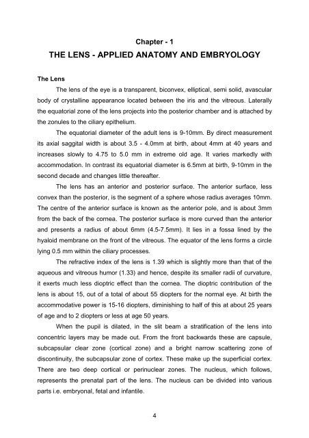
Applied anatomy<br />
Surgically, the lens is divided into four zones which have different<br />
characteristics and hence merit different attention. These are the capsular bag, the<br />
superficial cortex, the epinucleus, and the hard<br />
nucleus. The capsule has been described in detail<br />
above. The cortex is a soft thin layer present<br />
immediately beneath the capsule. It envelopes the<br />
epinucleus. It is aspirated or irrigated out of the<br />
capsular bag with ease. The epinucleus is a layer of<br />
semi-soft lens matter around the nucleus. This layer<br />
is difficult to aspirate and requires a large bore<br />
cannula for aspiration. It can be managed better by<br />
hydro- or visco-expression. The nucleus is a hard<br />
kernel forming the innermost layer of the lens. The<br />
cataractous lens can be minified down to the nucleus before extraction. This results<br />
in a smaller wound requirement for its exit as the nucleus is usually smaller than the<br />
lenticular opacity. The nucleus can be fragmented before its removal from the eye.<br />
Pre-operative nucleus grading gives us valuable input towards the surgical plan.<br />
7<br />
Surgical anatomy of the lens<br />
A - Anterior chamber; D - Capsule;<br />
C - Cortex ; E - Epinucleus;<br />
N-Nucleus
Anatomic considerations in IOL implantation<br />
The average central anterior chamber depth in the normal adult emmetropic<br />
eye in approximately 3 mm and becomes most narrow at the midperipheral iris. The<br />
anterior chamber deepens to approximately 4.5 mm after cataract extraction.<br />
The diameter of the anterior chamber angle is very important clinically for<br />
proper sizing of an angle fixated anterior chamber intraocular lens (ACIOL). A<br />
commonly used clinical rule is that the corneal diameter (white to white distance)<br />
plus 1 mm is approximately equal to the diameter of the anterior chamber angle.<br />
Exact measurement of the anterior chamber angle diameter is less crucial for the<br />
flexible open-loop lenses in common use today. The loops of angle-fixated lenses<br />
usually rest in the angle recess posterior to the scleral spur. Direct contact with<br />
delicate angle structures may causes irritation, with erosion of tissue and chronic<br />
inflammation. Glaucoma may result from direct contract or chronic inflammation.<br />
When the loops of footplates of the angle-fixated lens are misplaced anteriorly, the<br />
resulting direct contact with the peripheral corneal endothelium causes the death of<br />
these cells. Human corneal endothelial cells cannot divide; they can only enlarge<br />
and migrate to fill in the spaces left by the dead cells. As the central corneal<br />
endothelial cells spread and migrate to the periphery, the corneal endothelial cells<br />
count drops to a critically low level.<br />
Kelman-style flexible open loop ACIOLs were designed to minimize some<br />
of the complications associated with older ACIOLs. The complication rates with<br />
these lenses are clearly lower with respect to corneal pathology, inflammation,<br />
cystoid macular edema, decentration, glaucoma, and hemorrhage.<br />
The iris acts as a dynamic diaphragm that controls the amount of light<br />
entering the eye by regulating the size of its aperture, the pupil. The diameter of the<br />
iris is approximately 12 mm and its circumference is about 37.5 mm. The iris<br />
thickness is greatest at the collarette (2 mm from the edge of the pupil), where it is<br />
0.5 mm in anteroposterior dimensions. The iris is thinnest at its root where it is<br />
attached to the anterior surface of the ciliary body. Near the edge of the pupil, there<br />
is a ring of smooth muscle fibers about 1 mm wide, the sphincter pupillae. Peripheral<br />
to the sphincter pupillae is the dilator pupillae.<br />
8
Iris fixated IOLs were in vogue in the past. However, long term problems<br />
associated with these lenses have decreased their popularity. The highly mobile iris<br />
produced chafing and resultant inflammation. The only iris fixated lens in use today<br />
is the Singh - Worst lens which is fixated near the peripheral iris, which is relatively<br />
immobile. Recently there has been renewed interest in this lens design for use in<br />
phakic IOL implantation for the correction of the refractive errors.<br />
The posterior chamber is bounded anteriorly by the iris and posteriorly by<br />
the anterior hyaloid face of the vitreous. The circumference of the posterior chamber<br />
consists of the ciliary sulcus and the ciliary body. The lens equator is typically<br />
adjacent to the midpoint of the ciliary body. There is a space of 0.2-0.3 mm between<br />
the ciliary body and lens equator. After cataract removal, the capsular bag volume<br />
collapses to essentially zero as the anterior capsule comes into apposition with the<br />
posterior capsule. The equator of the evacuated bag extended laterally, causing the<br />
bag diameter to enlarge from the normal 9.5 mm to approximately 10.5mm. At this<br />
large diameter, the lens equator directly contacts the ciliary body and the normal<br />
psychological space is lost. The equator of the evacuated capsular bag also moves<br />
to a location posterior to the ciliary processes.<br />
When a 12mm PMMA PCIOL is implanted into the capsular bag, the bag<br />
becomes oval, with dimensions of 11.2 mm x 9.2 mm. The optics of almost all<br />
intracapsular IOLs will be positioned at a plane posterior to the ciliary body. One<br />
reason for this is that the angulation of the haptics in most IOLs produces direct<br />
contact of the optic with the posterior capsule and stretching of the posterior capsule.<br />
When the capsular bag is intact, in-the-bag fixation of both haptics is clearly the most<br />
optimal method of IOL fixation. Capsular bag fixation avoids direct contact between<br />
the haptics and uveal tissue and is associated with a lower likelihood of decentration<br />
and central posterior capsule opacification.<br />
The ciliary sulcus, also known as posterior chamber angle, is formed by the<br />
junction of the posterior iris with the ciliary body. The average diameter of the ciliary<br />
sulcus is 11.0mm. The ciliary sulcus is a common location for PCIOL implantation<br />
when the posterior capsule of does not provide sufficient support for the placement<br />
of an intracapsular lens and the anterior capsule is intact. When a continuous<br />
curvilinear capsulorrhexis has been performed and remains intact, the ciliary sulcus<br />
9
can be a very stable site of fixation for a posterior chamber lens. As the position of<br />
the lens will be slightly more anterior, an adjustment of approximately 0.5 diopter<br />
less power will be needed for lens implants to achieve the same intended refraction.<br />
Sulcus fixation of an IOL has the inherent disadvantage of direct contact<br />
between the implant and the uveal tissue, which may result in several complications.<br />
Pigment dispersion and glaucoma may occur if there is contact between the<br />
posterior iris and the implant. Erosion of the haptics into the sulcus will result in<br />
chronic inflammation. Sutured scleral fixation may be required where adequate<br />
anterior capsular support is unavailable. The haptics of sutured scleral-fixated<br />
PCIOLs ideally are meant to reside in the ciliary sulcus. A needle passed 1 mm<br />
posterior to surgical limbus closely corresponds to the ciliary sulcus. The long<br />
posterior ciliary arteries and nerves enter the ciliary body at the 3 and 9 o'clock<br />
meridians. The anterior ciliary arteries enter the ciliary body at the 3,6,9 and 12<br />
o'clock. Hence, it is most prudent to avoid suture fixation in the vertical and<br />
horizontal axes. A scleral fixated sutured PCIOL will most likely be safely and<br />
correctly placed in the ciliary sulcus when the needles are passed approximately<br />
0.87 mm - 1 mm posterior to the surgical limbus in the oblique meridians. A reduction<br />
of 0.5 diopter in the calculated power of IOL is recommended, as the IOL will rest<br />
more anterior as compared to that with intracapsular fixation.<br />
10