The Prime Focus Imaging Spectrograph for the Southern - Space ...
The Prime Focus Imaging Spectrograph for the Southern - Space ...
The Prime Focus Imaging Spectrograph for the Southern - Space ...

You also want an ePaper? Increase the reach of your titles
YUMPU automatically turns print PDFs into web optimized ePapers that Google loves.
<strong>The</strong> <strong>Prime</strong> <strong>Focus</strong> <strong>Imaging</strong> <strong>Spectrograph</strong> <strong>for</strong> <strong>the</strong><br />
Sou<strong>the</strong>rn African Large Telescope: Operational Modes<br />
Henry A. Kobulnicky a , Kenneth H. Nordsieck a , Eric B. Burgh a ,<br />
Michael P. Smith a , Jeffrey W. Percival a , Ted B. Williams b , and Darragh O’Donoghue c<br />
a University of Wisconsin, Madison, b Rutgers University, c South African Astrophysical Observatory<br />
ABSTRACT<br />
<strong>The</strong> <strong>Prime</strong> <strong>Focus</strong> <strong>Imaging</strong> <strong>Spectrograph</strong> (PFIS) will be <strong>the</strong> workhorse first-generation spectrograph on <strong>the</strong> Sou<strong>the</strong>rn African<br />
Large Telescope (SALT). Scheduled <strong>for</strong> commissioning in late 2004, PFIS is a versatile high-throughput imaging<br />
spectrograph with a complement of 5 volume-phase holographic gratings <strong>for</strong> spectroscopic programs from 3200 Å to 9000 Å<br />
at resolutions of R=1500 to R=10000. A magazine of 6 longslits and 30 custom laser-milled slitmasks enables single- or<br />
multi-object spectroscopy over an 8 arc minute diameter field. With <strong>the</strong> gratings stowed, a dual-etalon Fabry-Perot<br />
subsystem enables imaging spectroscopy at R=500, R=3000, and R=12,500. <strong>The</strong> polarization subsystem, consisting of a<br />
polarizing beam-splitter used in conjunction with half- and quarter-wave plates, allows linear or circular polarimetric<br />
measurements in ANY of <strong>the</strong> spectroscopic modes. Three mosaiced rapid-readout frame-transfer CCDs provide <strong>the</strong><br />
capability <strong>for</strong> time-resolved sampling at rates in excess of 10 Hz. Combinations of <strong>the</strong>se subsystems permit novel observing<br />
modes <strong>for</strong> specialized scientific programs. Examples include high time-resolution multi-object spectral polarimetry of<br />
accreting compact objects, and Fabry-Perot polarimetry or imaging spectral polarimetry of nebulae and stellar clusters. <strong>The</strong><br />
demands of queue-scheduled observing on a fixed-altitude telescope require that <strong>the</strong> instrument be capable of rapid<br />
reconfiguration between modes.<br />
Keywords: Astronomical spectrographs, optical design, near-ultraviolet spectroscopy, volume phase holographic gratings,<br />
Fabry-Perot interferometry, spectropolarimetry<br />
1. INSTRUMENT OVERVIEW<br />
<strong>The</strong> <strong>Prime</strong> <strong>Focus</strong> <strong>Imaging</strong> <strong>Spectrograph</strong> (PFIS) will be <strong>the</strong> workhorse first-generation, all-purpose spectrograph on <strong>the</strong><br />
Sou<strong>the</strong>rn African Large Telescope (SALT). <strong>The</strong> optical design of PFIS is described elsewhere in this volume 1 .<br />
<strong>The</strong> nominal field of view is 8’. <strong>The</strong> plate scale at <strong>the</strong> focal plane is 224 microns/arcsec.<br />
PFIS is designed as a facility-class, general-purpose spectrograph with many capabilities including medium-band imaging,<br />
Fabry-Perot narrow-band imaging, longslit grating spectroscopy and multi-slit grating spectroscopy. Linear and circular<br />
polarimetry may be per<strong>for</strong>med in any of <strong>the</strong> imaging or spectroscopic modes at <strong>the</strong> expense of halving <strong>the</strong> field of view.<br />
Fur<strong>the</strong>rmore, <strong>the</strong> frame-transfer CCDs 2 enable rapid readouts or charge shuffling so that very high time- resolutions in excess<br />
of 0.1 s are possible at <strong>the</strong> cost of restricted areal coverage. <strong>The</strong>se diverse capabilities, combined with <strong>the</strong> excellent shortwavelength<br />
throughput, enable specialized observational programs such as time-resolved spectropolarimetric studies of<br />
energetic phenomena like close binaries, active galactic nuclei, and gamma ray bursts. This report provides an overview of<br />
<strong>the</strong> operational capabilities of PFIS and some anticipated science drivers.<br />
<strong>The</strong> observational modes are most easily described in terms of <strong>the</strong> four major instrument subsystems: 1) <strong>the</strong> focal plane slits<br />
and masks, 2) <strong>the</strong> dispersive elements, consisting of ei<strong>the</strong>r <strong>the</strong> complement of volume-phase holographic gratings or Fabry-<br />
Perot Etalon, 3) Polarization optics consisting of a ½ wave plate, a ¼ wave plate, and a polarizing beamsplitter , and 4) <strong>the</strong><br />
array of 3 charge-transfer CCDs. Permutations of <strong>the</strong>se mechanisms combine to give PFIS <strong>the</strong> versatility to address a broad<br />
range of astrophysical research programs. Table 1 summarizes <strong>the</strong> observational modes permitted by combinations of <strong>the</strong>se<br />
four subsystems.<br />
a *chip@astro.wisc.edu, khn@sal.wisc.edu, ebb@sal.wisc.edu, smith@sal.wisc.edu, jwp@sal.wisc.edu, <strong>Space</strong><br />
Astronomy Laboratory, University of Wisconsin, Madison, 53706 b Department of Physics & Astronomy, Rutgers,<br />
Piscataway, NJ 08855, c dod@saao.ac.za, South African Astrophysical Observatory Observatory 7935, South Africa
CCD Readout<br />
Mode<br />
Normal Readout<br />
(
Table 2: Complement of Standard Longslits<br />
Reflective Slit Plate # Length Width (“)<br />
1 7.5’ 0.45”<br />
2 7.5’ 0.6”<br />
3 7.5’ 0.9”<br />
4 7.5’ 1.1”<br />
5 7.5’ 1.3”<br />
6 7.5’ 3.0”<br />
7 Central 4’ 1.1” adjustable <strong>for</strong> spectral polarimetry<br />
8 4’ above CTB 1.1” adjustable <strong>for</strong> high speed spectroscopy<br />
9 4’’ 1.1” w/ coronographic center<br />
<strong>The</strong> mean site seeing in<strong>for</strong>ms <strong>the</strong> distribution of longslit widths. A variety of slits should be available to cover <strong>the</strong> range of<br />
atmospheric seeing conditions expected at <strong>the</strong> site. For <strong>the</strong> SALT telescope, <strong>the</strong> median seeing is 0.9” at zenith. Seeing<br />
varies approximately as FWHM ∝ sec(z) 0.6 . For SALT, where <strong>the</strong> zenith distance is always 37±6 degrees, <strong>the</strong> factor by<br />
which seeing is degraded from zenith seeing is 1.14±0.05. Fur<strong>the</strong>rmore, imperfections in <strong>the</strong> telescope optical system<br />
(primary mirror plus spherical aberration corrector, SAC) deliver a finite-sized image, assumed here to be characterized by a<br />
Gaussian PSF with FWHM=0.6”. <strong>The</strong> focal plane image size is <strong>the</strong> zenith image size convolved with <strong>the</strong> effects of<br />
atmosphere at 37 degree zenith distance and <strong>the</strong> telescope optics. Figure 1 shows <strong>the</strong> fraction of energy from a point source<br />
which is transmitted by a focal plane slit <strong>for</strong> three different slit widths as a function of image FWHM.<br />
Figure 1. This Figure illustrates <strong>the</strong> fraction of energy from a point source which is transmitted by a focal plane slit <strong>for</strong> three different slit<br />
widths as a function of image FWHM. <strong>The</strong> solid line shows <strong>the</strong> enslitted energy fraction <strong>for</strong> a slit diameter of 2.0”. Open squares represent<br />
<strong>the</strong> case of a Gaussian PSF while filled circles represent <strong>the</strong> more realistic (i.e., long exposure) case when <strong>the</strong> PSF is given by <strong>the</strong><br />
<strong>the</strong>oretical atmospheric profile. 3 <strong>The</strong> dashed line illustrates <strong>the</strong> comparison between <strong>the</strong> enslitted energy fraction <strong>for</strong> <strong>the</strong> two profiles <strong>for</strong> a<br />
slit width of 1.0”. <strong>The</strong> dotted line illustrates <strong>the</strong> case of a slit width of 0.5”. Thin vertical lines indicate <strong>the</strong> seeing at zenith <strong>for</strong> <strong>the</strong> best<br />
(0.6”), median (0.9”) and worst 10% (1.8”) conditions at <strong>the</strong> SALT site in Sou<strong>the</strong>rland, South Africa.<br />
iii. Slitmask – Many science programs use custom laser-cut slitmasks to per<strong>for</strong>m multi-object spectroscopy or multi-object<br />
spectropolarimetry. <strong>The</strong> masks will be milled with a micromachining laser mill and placed in a slitmask magazine designed
to hold 30 masks. Mask insertion time will be < 30 s. User-designed slitmasks will be cut and stored on-site and labeled<br />
with a bar-code to identify <strong>the</strong> mask and mask holder (frame) to be used with each mask. <strong>The</strong> focal plane plate scale of 224<br />
microns/arcsec dictates that typical science slits (1.0 arcsec) will be very small (224 microns) and must be cut to extremely<br />
tight tolerances (few microns rms edge roughness). <strong>The</strong> slitmask manufacturing unit will be similar to <strong>the</strong> laser cutting<br />
systems adopted <strong>for</strong> <strong>the</strong> Gemini GMOS spectrograph 4 . Several standard masks will be available, including one which masks<br />
<strong>the</strong> upper and low quarter FOV <strong>for</strong> imaging polarimetry, and one which masks <strong>the</strong> lower half of <strong>the</strong> CCD <strong>for</strong> high timeresolution<br />
work. All high time-resolution imaging or Fabry-Perot modes require <strong>the</strong> use of a carbon-fiber mask in <strong>the</strong> focal<br />
plane to block <strong>the</strong> lower half of <strong>the</strong> field so that half of <strong>the</strong> CCD can be used as a storage buffer <strong>for</strong> frame-transfer operation<br />
(Mask 1). <strong>Imaging</strong> polarimetry and Fabry-Perot polarimetry require a mask to restrict <strong>the</strong> field of view to <strong>the</strong> central 4’<br />
(Mask 2).<br />
Table 3: Standard Focal Plane Masks<br />
Mask # Configuration Use<br />
1 Masks lower 4’ x 8’ High time-resolution imaging<br />
2 Masks upper and lower 2’ x 8’ (central 4’x8’ visible) <strong>Imaging</strong> and Fabry-Perot polarimetry<br />
3 1” pinholes spaced by 30” in cross-dispersion Calibration<br />
4+ Custom-cut science masks Multi-slit spectroscopy<br />
2048 2048 2048<br />
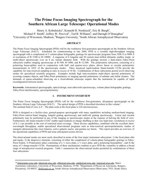
2.2 Dispersive Elements<br />
Image field<br />
8’<br />
Charge transfer region<br />
2048 2048 2048<br />
Figure 2. (Left) Schematic of <strong>the</strong> PFIS 8' field of view superimposed on <strong>the</strong> detector comprised of three 2048x4096<br />
CCDs. Shaded area shows how <strong>the</strong> standard Slitmask #1 covers half <strong>the</strong> field when CCDs are used in frame transfer<br />
(fast readout) mode. (Right) Schematic of standard Slitmask #2 <strong>for</strong> use with all polarimetric modes. Only <strong>the</strong> central<br />
4’ of <strong>the</strong> 8’ field is useable in polarimetric modes because <strong>the</strong> polarizing beamsplitter is used to image orthogonal<br />
polarizations onto <strong>the</strong> detector simultaneously.<br />
i. <strong>Imaging</strong> mode - No dispersive element is in <strong>the</strong> beam. This implies that <strong>the</strong> camera is in <strong>the</strong> unarticulated position. A<br />
special mode of spectropolarimetric imaging also uses this position, taking advantage of <strong>the</strong> lateral chromatic aberration in<br />
<strong>the</strong> camera to per<strong>for</strong>m low-resolution spectropolarimetric imaging.<br />
ii. Fabry-Perot <strong>Imaging</strong> - <strong>The</strong> etalon is <strong>the</strong> beam and <strong>the</strong> camera is at <strong>the</strong> unarticulated position. Estimated time <strong>for</strong> insertion<br />
of <strong>the</strong> etalons using a pneumatic piston is 30 s. Fabry-Perot imaging and Fabry-Perot polarimetry are modes that use this<br />
configuration. PFIS will have two F-P resolutions initially, a low-resolution (R=500-1000) tunable filter and a highresolution<br />
(R=12000) mode. Low-resolution mode will use a single etalon with an interference filter to select <strong>the</strong> desired<br />
interference order. <strong>The</strong> high-resolution mode will use two etalon in series with <strong>the</strong> low-resolution etalon serving as <strong>the</strong><br />
blocking filter to select <strong>the</strong> order <strong>for</strong> <strong>the</strong> high-resolution etalon. A third, medium-resolution etalon is planned as an upgrade<br />
path. Two of <strong>the</strong> three etalons will be installed in PFIS at any given time. <strong>The</strong> spectral range of <strong>the</strong> etalons will be 4300 Å<br />
to 8600 Å. Approximately 30 interference filters (R=50) will be required to isolate <strong>the</strong> FP orders over <strong>the</strong> entire spectral<br />
range. A subset of 15 of <strong>the</strong>se will be installed in <strong>the</strong> filter magazine at any given time.<br />
8’
iii. Grating Spectroscopy - <strong>The</strong> majority of spectroscopic modes employ one of 6 gratings as <strong>the</strong> dispersive element. Longslit<br />
spectroscopy, multi-object spectroscopy, and spectropolarimetry are per<strong>for</strong>med after selecting a grating from a magazine<br />
containing five volume-phase holographic (VPH) gratings and one conventional surface relief grating. Inserting time is<br />
designed to be
Figure 4. Tradeoff between spectral resolution and slit throughput <strong>for</strong> a point-like source as seen through <strong>the</strong> atmosphere and SALT<br />
telescope under excellent (0.7” at zenith) and poor (1.2” at zenith) seeing. Each panel shows a different PFIS VPH grating at <strong>the</strong> nominal<br />
wavelength of operation. Points in each graph denote slit widths of 0.3”, 0.5”, 0.7”, 1.0”, 1.5”, 2.0”, and 2.5”.<br />
2.3 Polarization Optics<br />
<strong>The</strong> PFIS collimator includes a quarter-wave plate and a half-wave plate which may be inserted into <strong>the</strong> beam separately, <strong>for</strong><br />
linear and circular polarization measurements, respectively. A polarizing beam splitter in <strong>the</strong> camera completes <strong>the</strong><br />
polarimetric optics. <strong>The</strong> waveplates may also be used in series <strong>for</strong> “all-Stokes” polarimetric measurements. In “linear”<br />
Polarimetric mode, <strong>the</strong> half-wave plate steps through 8 position angles separated by 22.5 degrees in order to per<strong>for</strong>m a<br />
complete linear polarization measurement. For circular polarization, two positions of <strong>the</strong> quarter-wave plate, in conjunction<br />
with 8 positions of <strong>the</strong> half-wave plate are required to complete a measurement. <strong>The</strong> waveplates may be used in conjunction<br />
with <strong>the</strong> Fabry-Perot etalon or <strong>the</strong> gratings and focal plane slits to per<strong>for</strong>m spectropolarimetry. Polarimetric optics <strong>for</strong> PFIS<br />
are described in more detail elsewhere in this volume. 5<br />
2.4 CCD Subsystem<br />
<strong>The</strong> detector is comprised of three mosaiced Marconi Applied Technologies 44-82 CCDs with 2048x4096 pixels and 15<br />
micron pixels. Figure 2 shows a depiction of <strong>the</strong> 8’ field of view projected onto <strong>the</strong> CCDs. <strong>The</strong> gap between CCDs is 15<br />
pixels (225 microns). At <strong>the</strong> detector, <strong>the</strong> plate scale is 117 microns/arcsec so that, in most common applications, <strong>the</strong> detector<br />
will be binned 2x2 and still provide critical sampling of <strong>the</strong> point spread function. <strong>The</strong> detector will operate in 3 readout<br />
modes depending on <strong>the</strong> time sampling required.<br />
i. Normal readout - <strong>The</strong> entire mini-mosaic, using 2x2 binning, can be read out in 3.6 s with 5e - /pix readout noise using 2<br />
amplifiers per chip. A reduced readout noise of 3 e - /pix can be achieved <strong>for</strong> lower readout rates, resulting in a read time of 11<br />
s. Most science operation will occur in this “normal” readout mode.<br />
ii. High Time-Resolution (fast readout) – <strong>The</strong> detectors may be operated in a “frame-transfer” mode whereby half of <strong>the</strong><br />
detector is used as a storage buffer <strong>for</strong> <strong>the</strong> o<strong>the</strong>r half of <strong>the</strong> chip. <strong>The</strong> contents of <strong>the</strong> upper half of <strong>the</strong> chip can be transferred<br />
to <strong>the</strong> lower half of <strong>the</strong> chip in only 0.205 s. A second exposure may begin while <strong>the</strong> lower half of <strong>the</strong> chip is read out at <strong>the</strong><br />
normal rate. In this mode, it is possible to obtain a continuous sequence of images with 1.8 s time resolution, but with <strong>the</strong><br />
spatial field of view reduced to 4’.
iii. Charge Shuffle (very fast) – Charge shuffling in may be used in conjunction with telescope nods in order to per<strong>for</strong>m highquality<br />
sky subtraction <strong>for</strong> slit spectroscopy or off-band subtraction <strong>for</strong> Fabry-Perot imaging. In this mode, <strong>the</strong> charge is<br />
shuffled between pixel rows in an arbitrary fashion ei<strong>the</strong>r during or between exposures. Alternatively, <strong>the</strong> chip may be<br />
windowed into arbitrary rectangular regions and read out at <strong>the</strong> maximum rate to achieve time resolutions as high as 0.1 s.<br />
Small groups of adjacent rows in <strong>the</strong> cross dispersion direction may be defined and used in conjunction with a longslit or slit<br />
mask to per<strong>for</strong>m very high time-resolution spectroscopy. For example, an object may be placed just above <strong>the</strong> charge transfer<br />
boundary on <strong>the</strong> CCDs and all but 32 rows (when on-chip prebinning of 2x2 is used) of <strong>the</strong> detector are masked by a custom<br />
focal plane mask. <strong>The</strong> 32 rows are clocked continuously along <strong>the</strong> CCD columns toward <strong>the</strong> amplifiers at a limiting rate<br />
which is set by <strong>the</strong> readout time of 0.07 s (roughly 0.0017 s/row). In this manner, imaging or spectroscopy of single objects<br />
may be per<strong>for</strong>med with time resolutions of 0.07 s. Images are smeared along <strong>the</strong> columns over time intervals of 0.0032 s, <strong>the</strong><br />
time required to transfer 32 rows across <strong>the</strong> charge transfer boundary. Figure 5 illustrates schematically how this very fast<br />
readout mode would occur. Table 3 summarizes <strong>the</strong> readout times <strong>for</strong> a range of image heights using <strong>the</strong> frame transfer<br />
operations. Finally, charge shuffling may also be used to per<strong>for</strong>m drift scan imaging by clocking <strong>the</strong> charge in <strong>the</strong> pixels at a<br />
sidereal rate.<br />
Table 3. PFIS readout times as a function of image height.<br />
Image Height<br />
(arcmin)<br />
Image Height<br />
(pixels)<br />
Transfer Time (s) Store Transfer Time<br />
(s)<br />
Readout Time<br />
(s)<br />
4.0 2048 0.102 0.102 1.595<br />
2.0 1024 0.051 0.051 0.797<br />
0.5 256 0.013 0.013 0.199<br />
0.125 64 0.003 0.003 0.050<br />
Figure 5. Schematic of very high speed spectroscopic operations using charge shuffling. An object is placed just above <strong>the</strong><br />
charge transfer boundary (horizontal dotted line) which bisects each CCD. All but 64 rows of <strong>the</strong> CCD are masked with a<br />
custom mask. A spectrum is transferred below <strong>the</strong> charge transfer boundary every 0.05 s and <strong>the</strong>n <strong>the</strong> CCD is read out at <strong>the</strong><br />
maximum rate as a series of spectra are recorded. Time resolutions of 0.07 s are possible.
3. EXAMPLES OF SCIENCE CASES AND OPERATIONAL CONFIGURATIONS<br />
Figure 6 shows <strong>the</strong> annulus of visibility <strong>for</strong> SALT as a function of declination and hour angle. Some common astronomical<br />
targets are labeled. Note that <strong>the</strong> Magellanic Clouds have visibility periods exceeding three hours, making <strong>the</strong>m prime<br />
observational targets.<br />
Figure 6. Annulus of total visibility <strong>for</strong> SALT as a function of declination and hour angle. Shaded ellipsoids mark <strong>the</strong> region of<br />
instantaneous visibility (6 degrees). <strong>The</strong> track at right indicates <strong>the</strong> relation between Galactic longitude and declination. It shows that<br />
much of <strong>the</strong> first quadrant and all of <strong>the</strong> fourth quadrant of <strong>the</strong> Galaxy is visible to SALT/PFIS.<br />
3.1 Multi-Object Spectroscopy<br />
Science Uses: Multi-object surveys in fields up to 8’ diameter. Redshift surveys. Stellar Galactic and Magellanic Cloud<br />
surveys.<br />
Target acquisition & guiding: By direct image with PFIS, <strong>the</strong>n multi-stage peak-up process similar to o<strong>the</strong>r multi-object<br />
spectrographs. No focal plane viewing system is available when using multi-slit masks. Guiding uses offset guide probes<br />
located on <strong>the</strong> perimeter of <strong>the</strong> focal plane which view an annular region outside <strong>the</strong> 8’ PFIS field.<br />
Operational Sequence:<br />
Afternoon<br />
Flatfields with continuum lamp <strong>for</strong> calibration and location of alignment star boxes on slitmask. Moving baffle at<br />
pupil of spherical aberration corrector mimics pupil illumination during actual track.<br />
Evening<br />
1 Slew telescope and tracker to desired field (120 s)<br />
2 Image of science field with SALTICAM or direct image with PFIS (10 s)<br />
3 Insert slitmask (30 s)<br />
4 Direct image with PFIS to verify field and mask alignment (10 s)<br />
5 Fine-tune pointing and begin guiding with guide probes (20 s)<br />
6 Insert and rotate grating to desired angle and articulate camera (30 s)<br />
7 Start science exposures<br />
8 Optional calibration lamp exposures using fiber-fed focal plane unit<br />
Data rate: 6154 spectral pixels by 4096 spatial pixels in as little as 3.6 s
Limitations: Wavelength coverage on <strong>the</strong> detector varies with position of object in <strong>the</strong> field in <strong>the</strong> dispersion direction (as<br />
with all multi-object spectrographs using focal plane masks. Fur<strong>the</strong>rmore, <strong>the</strong> blaze of VPH gratings varies with angle of<br />
incidence. This means that <strong>the</strong> efficiency of <strong>the</strong> grating also varies with field position in <strong>the</strong> dispersion direction. Figure 7<br />
illustrates this blaze shift <strong>for</strong> <strong>the</strong> case of <strong>the</strong> 780 l/mm grating and a 2’ field angle.<br />
Figure 7. (upper left) Efficiency of <strong>the</strong> 780 l/mm VPH grating at 8 grating angles. Heavy line denotes <strong>the</strong> superblaze. Three additional<br />
panels show <strong>the</strong> simultaneous wavelength coverage and efficiency <strong>for</strong> an on-axis source (solid line) and two sources with cross dispersion<br />
field angles of 2’ (dashed lines). Each panel illustrates a different grating angle corresponding to a different central wavelength.<br />
3.2 High Time-resolution Circular Spectropolarimetry<br />
Science Uses: Time-resolved single-object slit spectroscopy and spectropolarimetry of accreting objects, cataclysmic<br />
variables, active galactic nuclei, stellar flares, supernovae, gamma-ray bursts.<br />
Target acquisition & guiding: By SALTICAM functioning as an acquisition camera viewing <strong>the</strong> reflective longslit plate.<br />
Operational Sequence:<br />
Afternoon<br />
Flatfields with continuum lamp <strong>for</strong> calibration and location of alignment star boxes on slitmask. Moving baffle at<br />
pupil of spherical aberration corrector mimics pupil illumination during actual track.<br />
Evening<br />
1 Slew telescope and tracker to desired field (120 s)<br />
2 Insert and rotate grating to desired angle and articulate camera (30 s)<br />
3 Insert waveplates (20 s)<br />
4 Insert longslit plate #8 (or #7) (30 s)<br />
5 Visually select target & place it on slit while viewing slit with SALTICAM (10 s)<br />
6 Fine-tune pointing and begin guiding with guide probes or directly on slit with SALTICAM (20 s)<br />
7 Start science exposures<br />
8 For x=1 to 8 positions of <strong>the</strong> ½ wave plate
a. Set ½ wave plate to position x<br />
b. For y=1 to 2 positions of <strong>the</strong> ¼ wave plate<br />
i. Set ¼ waveplate to position y<br />
ii. Expose<br />
iii. Shuffle charge down by 32 rows<br />
c. Readout bottom 32 rows near amplifier<br />
9 Optional calibration lamp exposures using fiber-fed focal plane unit<br />
Data rate: 6154 spectral pixels by 32 spatial pixels in as little as 0.07 s<br />
Limitations: Multi-object capability is precluded in very high speed spectropolarimetric mode.<br />
4. SENSITIVITY AND CALIBRATION ISSUES<br />
PFIS is designed <strong>for</strong> extremely high throughput, especially shortward of 4000 Å, a region of <strong>the</strong> spectrum seldom exploited<br />
by today’s large telescopes and spectrographs. Figure 8 shows <strong>the</strong> efficiency <strong>for</strong> components of <strong>the</strong> SALT and PFIS system.<br />
Peak efficiencies exceed 20% from 4000 Å to 8000 Å.<br />
Figure 8. Total on-sky efficiency <strong>for</strong> PFIS on <strong>the</strong> SALT telescope as a function of wavelength. Due to <strong>the</strong> all-refractive optics and efficient<br />
VPH grating will allow PFIS to achieve superior sensitivity compared to existing 10 m class telescopes and spectrographs, especially<br />
shortward of 3500 Å.<br />
Calibration, especially flat fielding, will be challenging due to <strong>the</strong> varying pupil illumination as an astronomical source tracks<br />
across <strong>the</strong> spherical primary. Flat fielding will be achieved using flats from an illuminated screen in <strong>the</strong> pupil of <strong>the</strong> spherical<br />
aberration corrector (SAC) in conjunction with a moving baffle in <strong>the</strong> SAC. Design studies have shown that a moving baffle<br />
at this location can mimic <strong>the</strong> effects of varying pupil illumination. <strong>The</strong> baffle will move at several times <strong>the</strong> sidereal rate so<br />
that flat fields can be rapidly acquired during <strong>the</strong> afternoon, ei<strong>the</strong>r be<strong>for</strong>e or after a night’s science observations.<br />
Wavelength calibrations will be enabled by a fiber fed focal plane unit with a diffuser to uni<strong>for</strong>mly illuminate <strong>the</strong> focal plane.<br />
<strong>The</strong> observatory will supply a collection of arc lamps <strong>for</strong> wavelength calibration fed by fiber optic from a room below <strong>the</strong><br />
telescope structure.
Figure 9. Signal-to-noise <strong>for</strong> PFIS+SALT as a function of continuum magnitude <strong>for</strong> a stellar source at spectral resolutions up to R=12500.<br />
A single exposure of 3000 s is assumed.<br />
REFERENCES<br />
1. Eric B. Burgh, Kenneth H. Nordsieck, Henry A. Kobulnicky, Ted B. Williams, Darragh O’Donoghue, Michael P.<br />
Smith, and Jeffry W. Percival, ``<strong>The</strong> <strong>Prime</strong> <strong>Focus</strong> <strong>Imaging</strong> <strong>Spectrograph</strong> <strong>for</strong> <strong>the</strong> Sou<strong>the</strong>rn Afrtican Large Telescope:<br />
Optical Design,’’ in Instrument Design and Per<strong>for</strong>mance <strong>for</strong> Optical/Infreared Ground-Based Telescopes, Proc. SPIE,<br />
vol 4841-164<br />
2. Darragh O’Donoghue, D.B. Carter, G.P. Evans, W.P. Koorts, J.O’Conner, F. Osman, and S. can der Merwe,<br />
``SALTICAM: $0.5 M acquisition camera: every big telescope should have one,’’ in Instrument Design and<br />
Per<strong>for</strong>mance <strong>for</strong> Optical/Infrared Ground-Based Telescopes, Proc. SPIE, vol 4841, paper 52, 2002<br />
3. R.N. Wilson, in Reflecting Telescope Optics II, 2002, New York-Springer-Verlag, p. 390<br />
4. Kei Szeto, James R Stillburn, Tim Bond, Scott Roberts, Jerry Sebesta, and Leslie K. Saddlemyer, “Fabrication of<br />
Narrow Slit Masks <strong>for</strong> <strong>the</strong> Gemini Multi-Object <strong>Spectrograph</strong>, in Optical Telescopes of Today and Tomorrow, 1997,<br />
SPIE, 2871, p 1264<br />
5. Kenneth H. Nordsieck, Kurt P. Jaehnig, Eric B. Burgh, Henry A. Kobulnicky, Jeffrey W. Percival, and Michael P.<br />
Smith, ``Instrumentation <strong>for</strong> high-resolution spectropolarimetry in <strong>the</strong> visible and far-ultraviolet’’, in Instrument<br />
Design and Per<strong>for</strong>mance <strong>for</strong> Optical/Infrared Ground-Based Telescopes, Proc. SPIE, vol 4843- 24