Animal Diversity: Chordata
Animal Diversity: Chordata
Animal Diversity: Chordata

You also want an ePaper? Increase the reach of your titles
YUMPU automatically turns print PDFs into web optimized ePapers that Google loves.
Bones seen on the dorsal surface:<br />
Premaxillae, maxillae, quadratojugals, squamosals, septomaxillaries, nasals, fronto-parietals,<br />
prootics, exoccipitals and occipital condyles<br />
Bones seen on the ventral surface:<br />
Premaxillae, maxillae, quadratojugals, vomers, palatines, sphenethmoids, parasphenoid,<br />
pterygoids and exoccipitals<br />
The skull of urodela differs from that of the frog in many ways: The trabeculae do not meet<br />
either below the brain to form a basis cranii or above it to form a cranial roof. There is,<br />
above, a huge superior cranial fontanelle, and below an equally large basicranial fontanelle.<br />
The former is covered, in the complete skull, by the parietals and frontals, the latter by the<br />
parasphenoid. The parietals and frontals are separate. The parasphenoid is not T-shaped. A<br />
single bone, the vomeropalatine bearing teeth, represents the palatine and vomer. The hyoid<br />
arch is large and its dorsal end may be separated as a hyomandibular. There are three or four<br />
branchial arches. The stapes has no extra-columella and no tympanic cavity or membrane. In<br />
some anuran species, there is the presence of small supra and basi-occipitals. In others,<br />
investing bones of the roof are very strongly developed. In the apoda, the investing bones are<br />
very large and form a substantial structure.<br />
The vertebral column is divisible into: the cervical region, an abdominal / thoracolumbar<br />
region, a sacral region and the caudal region. The total number of vertebrae in urodeles and<br />
apoda may be as much as 250, while there are only 9 vertebrae and a single rod shaped<br />
caudal bone called the urostyle in anurans. In the lower urodela the centra are biconcave as in<br />
fishes. However, the neural arches are much better developed than in any fish and have well<br />
developed zygapophyses. The apoda also have biconcave vertebrae but in the higher<br />
urodeles, the anterior surface of the centrum has a convexity while the posterior surface<br />
retains its concavity, thus forming a ball and socket joint and the condition is known as<br />
opisthocoelous. In the anura the condition gets reversed with respect to the anterior and<br />
posterior surfaces and this condition is called as procoelous. A frog’s vertebral column<br />
includes the following bones:<br />
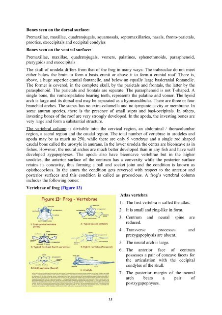
Vertebrae of frog (Figure 13)<br />
35<br />
Atlas vertebra<br />
1. The first vertebra is called the atlas.<br />
2. It is small and ring-like in form.<br />
3. Centrum and neural spine are<br />
reduced.<br />
4. Transverse processes and<br />
prezygapophysis are absent.<br />
5. The neural arch is large.<br />
6. The anterior face of centrum<br />
possesses a pair of concave facets for<br />
the articulation with the occipital<br />
condyles of the skull.<br />
7. The posterior margin of the neural<br />
arch bears a pair of<br />
postzygapophyses.