Kulkarni new fruit gall 1246.pmd - zoos' print
Kulkarni new fruit gall 1246.pmd - zoos' print
Kulkarni new fruit gall 1246.pmd - zoos' print

You also want an ePaper? Increase the reach of your titles
YUMPU automatically turns print PDFs into web optimized ePapers that Google loves.
NOTE ZOOS' PRINT JOURNAL 20(5): 1875-1876<br />
NEW RECORD OF FRUIT GALL ON<br />
STEREOSPERMUM CHELENOIDES (L.F.) DC. CAUSED<br />
BY A MITE (ERIOPHYIDAE: ACARINA)<br />
D.K. <strong>Kulkarni</strong> 1 , A.S. Upadhye 1 , M.R. Shindikar 1<br />
and R.M. Sharma 2<br />
1 Agharkar Research Institute, Gopal Ganesh Agarkar Road, Pune,<br />
Maharashtra 411004, India<br />
2 Zoological Survey of India, High Altitude Zoology Field Station,<br />
Saproon, Solan, Himachal Pradesh 173211, India<br />
Galls or plant tumors are structural deformities arising mostly<br />
by overgrowth (hypertrophy) and excessive cell division of<br />
tissues (hyperplasy). Gall formation is a product of interspecific<br />
association between a plant and other pathogenic organism or<br />
physiological changes in the plant body itself. The <strong>gall</strong>s induced<br />
by vegetative parasites are called ‘phytocecidia’ while animal<br />
parasites are termed ‘cecidozoa’ (Mani, 2000). There are a<br />
number of <strong>gall</strong> causing organisms found to induce <strong>gall</strong> in<br />
various parts and stages of plant (Mani, 1964). Some of these<br />
<strong>gall</strong>s are reported for their medicinal values particularly in<br />
Ayurvedic preparations (Upadhye et al., 1993) and traditional<br />
agricultural practices (Singhvi & Sharma, 1985).<br />
The <strong>fruit</strong> <strong>gall</strong>s are found to be initiated by cecidogenetic reaction<br />
on the plant in the immature <strong>fruit</strong> stage, after the pollination of<br />
the flower and fertilization of the ovary. In this connection,<br />
some outstanding observations on <strong>fruit</strong> <strong>gall</strong>s have been studied<br />
in the world. Some examples are reported in India like unilocular<br />
midge <strong>gall</strong> on the <strong>fruit</strong>s of Zizyphus xylopyra Willd and irregularly<br />
globose, rugose, multilocular solid <strong>gall</strong> on <strong>fruit</strong>s of Acacia<br />
concinna DC. Fruits of Acacia leucopholea Willd show <strong>gall</strong>s<br />
formed by the fungus Uromycladium. Narasimhan (1954)<br />
reported malformation of panicle in Mango by Eriophyes mite.<br />
Oval, solid, fibrous agglomerate, multilocular blisters arise on<br />
half mature <strong>fruit</strong>s of Terminalia paniculata Roth. caused by a<br />
psyllid, Trioza hirsute (Crawf.). Fruits of Terminalia arjuna<br />
W&A show subglobose, truncated swellings caused by an<br />
unknown cynipid (Hymenoptera), whereas normal development<br />
of <strong>fruit</strong> in Ludwigia adscendens (L.) has been observed to be<br />
arrested by an unknown weevil. Globose, fleshy, unilocular<br />
swellings with a short nipple-like and blunt apical process arise<br />
on <strong>fruit</strong>s of Maesa indica (Roxb.) caused by an unknown midge.<br />
However, during the literature survey it was noticed that <strong>gall</strong><br />
formation on <strong>fruit</strong>s of genus Stereospermum has not been<br />
reported so far.<br />
Genus Stereospermum belongs to family Bignoniaceae and plays<br />
an important part in the floristic composition as well as in<br />
ayurvedic medicines. Two species, Stereospermum<br />
chelenoides (L.f.) DC. (=S. suaveolens (Roxb.) DC.) and<br />
Stereospermum colais (Buch.-Ham. ex Dillw.) Mabb. (=S.<br />
chelenoides auct. non (L.f.) DC.) are mainly used since ancient<br />
times. Very little attention has been paid to study their<br />
distribution, germination and germplasm collection. During<br />
regular botanical excursions in Maharashtra, the authors<br />
collected the germplasm of above species from different<br />
localities, such as from remote forested hilly areas of Alapali<br />
forest in Gadchiroli district in December 1994 and collected 8-<br />
10 <strong>fruit</strong>s of Stereospermum chelenoides (L.f.) DC. The <strong>fruit</strong>s of<br />
Stereospermum were of capsule type (30-50cm by 2-3cm). These<br />
pods were straight, cylindrical, slightly ribbed somewhat rough<br />
with elevated whitish spikes and thick valve. Out of these,<br />
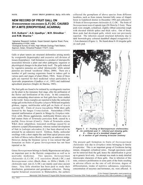
three pods had developed <strong>gall</strong>s, which were not previously<br />
reported. The infection caused structural deformity due to<br />
dark brownish-grey coloured dumbbell shaped overgrowth (1-<br />
1.5cm diameter) (Figure 1). We found about 8-10 irregular <strong>gall</strong>s<br />
on each pod.<br />
© Zoo Outreach Organisation; www.zoos<strong>print</strong>.org<br />
Manuscript 1246; Received 27 August 2004; Revised received 27 November 2004; Finally accepted 25 January 2005; Date of publication 21 April 2005<br />
May 2005 1875<br />
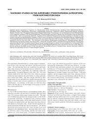
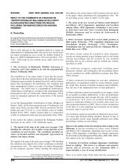
A<br />
D<br />
1 cm<br />
E<br />
C B<br />
Figure 1. A - Fruiting of Steriospermum chellenoides;<br />
B - An uninfected pod; C - Infected pod showing <strong>gall</strong>s;<br />
D - Close up of a dumbbell shaped <strong>gall</strong>;<br />
E - A section of <strong>gall</strong> showing bi-locular anatomy<br />
The present <strong>gall</strong> formation in <strong>fruit</strong>s of Stereospermum<br />
chelenoides was due to Eriophyes mites belonging to family<br />
Eriophyidae. This is an important group of Cecidozoa having<br />
minute, elongated, cylindrical mites. Enough specimens could<br />
not be retrieved from the <strong>gall</strong>, hence the identification is given<br />
up to generic level only. An attempt was made to study detailed<br />
microscopic characters of these <strong>gall</strong>s. Due to infection of this<br />
mite, extensive proliferation in some parts of the <strong>fruit</strong>s resulting<br />
in adverse effects on the rate of cell division, degree of<br />
differentiation, intake of nutritive substances, respiration,<br />
concentration of enzymes and hormones, etc. were affected. It<br />
5 cm
New record of <strong>fruit</strong> <strong>gall</strong> D.K.<strong>Kulkarni</strong> et al.<br />
also affected seed development in <strong>fruit</strong>s. The larvae of the<br />
cecidozoa may be situated in the septa or in the cavity of the<br />
<strong>fruit</strong>s. Horizontal section of <strong>gall</strong> showed Eriophyes spreading<br />
from the pierced cell. The effect of the reaction of the cell<br />
spread to other cells immediately in contact and only to a lesser<br />
extent to the cells away from the area of infection. The nuclear<br />
gigantism of the cell was attacked by the Eriophyes mites. This<br />
<strong>gall</strong> is used by tribals for medicinal purposes and thus<br />
constitutes an important record in the database of plant <strong>gall</strong>s.<br />
REFERENCE<br />
Mani, M.S. (1964). Ecology of plant <strong>gall</strong>s. Dr. W. Junk, Publishers,<br />
The Hague, Netherlands.<br />
Mani, M.S. (2000). Plant Galls of India (2 nd Edition) Oxford & IBH<br />
Publishing Co. Pvt. Ltd., New Delhi, 354pp.<br />
Narasimhan, M.J. (1954). Malformation of panicles in Mango incited<br />
by a species of Eriophyes. Current Science 23: 297-298.<br />
Singhvi, N.R. and K.D. Sharma (1985). A report on effect of aqueous<br />
extract of the <strong>gall</strong>s of Ficus racemosa L. on seed germination and<br />
seedling growth of pearl millet (Pennisetum typhoideum Rich.). Geo<br />
Bios 12: 153-154.<br />
Upadhye, A.S., V.D.Vartak and M.S. Kumbhojkar (1993). On the<br />
identity of market samples of the drug ‘Kakadshingi’. Bulletin of Medico-<br />
Ethno Botanical Research 15(1-2): 85-88.<br />
ACKNOWLEDGEMENTS<br />
We thank Dr. V.S. Rao, Director, Agharkar Research Institute, Pune for<br />
providing the necessary facilities. Discussion with Dr. V.G. Rao,<br />
Department of Mycology and Plant Pathology is gratefully<br />
acknowledged. Thanks are also due to the Director, Zoological Survey<br />
of India, Kolkata for encouragement.<br />
VET BRIEF ZOOS' PRINT JOURNAL 20(5): 1876<br />
ACARIASIS IN AN EMU (DROMAIUS<br />
NOVAELLANDIAE) - A CASE REPORT<br />
K. Senthil Kumar 1 , R. Thirumurugan 2 , K. Devaki 1<br />
and Pathan Nazrullah Khan 3<br />
1 Veterinary Assistant Surgeons, 2 Zoo Veterinarian, 3 Veterinary Officer,<br />
Arignar Anna Zoological Park, Vandalur, Chennai, Tamil Nadu 600048, India<br />
Birds serve as hosts for a wide variety of ectoparasites including<br />
ticks and mites (Greve, 1986). These not only cause annoyance<br />
to the birds but also spread many pathogens including blood<br />
protozoa. The infestation by ectoparasites may alter the birds’<br />
behavior by reducing their appetite and result in constant<br />
preening and pecking of the affected parts. The present report<br />
puts on record, a case of tick infestation and its treatment in a<br />
captive emu.<br />
The prestigious ratite a female Emu (Dromaius novaellandiae)<br />
in the Arignar Anna Zoological Park, Vandalur, was reported of<br />
having inappetance, constant preening and pecking and altered<br />
gait. The bird was active and responded to the keeper’s call. A<br />
thorough clinical examination was carried out by physically<br />
restraining it. To rule out any nutritional cause, the feed<br />
ingredients were checked and found to be normal. The<br />
droppings were examined and no ova or eggs of helminths<br />
were detected.<br />
The bird preened the feathers particularly over the back region,<br />
which was examined thoroughly for wound, growths,<br />
inflammation or ectoparasites; no abnormalities could be<br />
detected. Then the whole body was examined, which revealed<br />
numerous ticks over the skin around the ear canal and lateral<br />
sides of the neck resulting in the observed symptoms. The<br />
ticks were collected for identification and were identified as<br />
Heamaphysalis sp. (Soulsby, 1986).<br />
Treatment: The condition was treated by applying Deltamethrin<br />
solution (BUTOX liquid - Hoechst India Ltd.,) @ 2ml per liter of<br />
water sprayed on the affected areas taking care to avoid contact<br />
with eyes, left for twenty minutes and cleaned with water. The<br />
bird showed improvement from the next day and complete<br />
recovery in three days. The enclosure was also sprayed with<br />
the acaricide in higher concentration, after shifting the bird to a<br />
nearby enclosure. The treatment was repeated after a week to<br />
prevent reinfestation.<br />
In this case it was difficult to identify the ticks because of the<br />
colour of the feathers and skin. The infestated area seemed<br />
almost normal, with the ticks visible only on close and careful<br />
observation. The ticks were found localized in the head and<br />
neck regions. Greve (1986) opined that avain hard ticks prefer<br />
the head region where they are protected from being dislodged<br />
by preening. In this case, the bird was found preening the<br />
feathers over the back, but it was actually attempting to remove<br />
the ticks by rubbing the head and neck, which resulted in the<br />
altered gait.<br />
Greve (1986) stated that wild birds might be responsible for the<br />
introduction of ticks into bird exhibits. The zoological park<br />
contains numerous free ranging peafowls and, the emu enclosure<br />
being an open enclosure, they frequent the enclosure in search<br />
of feed. Subramanian et al. (2002) reported the occurrence of<br />
ticks belonging to Heamaphysalis sp. in free ranging peafowls.<br />
So the peafowls might be the source of introduction.<br />
The acaricide used in this case was found to be very effective<br />
in controlling the ticks. Greve (1986) suggested the use of<br />
rotenone, pyrethrin-piperonyl butoxide, malathion and carbaryl<br />
as safe and effective acaricides to be used on the birds.<br />
To prevent such occurrences in future, it was planned to conduct<br />
periodic clinical examination of the bird and the enclosure and<br />
to take measures to avoid contact with free ranging peafowls.<br />
REFERENCES<br />
Greve, J.H. (1986). Parasitic diseases. In: Fowler, M.E. Zoo and wild Animal<br />
medicine. 2 nd edition. W.B. Saunders Company, Philadelphia, 234 p.<br />
Soulsby, E.J.l. (1982). In: Helmints, Arthropods and Protozoa of Domestic<br />
Animals. 7 th edition. ELBS, London.<br />
Subramanian, K.S. and M. Raman (2002). Indian Veterinary Journal 79:<br />
276.<br />
ACKNOWLEDGEMENT<br />
The authors are thankful to the Director, Arignar Anna Zoological<br />
Park, Vandalur, Chennai for permitting to undertake the work.<br />
© Zoo Outreach Organisation; www.zoos<strong>print</strong>.org<br />
Manuscript 1097; Received 09 October 2003; Finally accepted 17 December 2004; Date of publication 21 April 2005<br />
1876 May 2005