Research in Action: - University of Calgary
Research in Action: - University of Calgary
Research in Action: - University of Calgary

You also want an ePaper? Increase the reach of your titles
YUMPU automatically turns print PDFs into web optimized ePapers that Google loves.
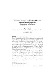
Southern Alberta Cancer <strong>Research</strong> Institute (SACRI)<br />
Cell Imag<strong>in</strong>g Facility<br />
The m<strong>in</strong>d’s eye has never had so much clarity. At SACRI’s Cell Imag<strong>in</strong>g Facility,<br />
Dr. Dallan Young studies three-dimensional images <strong>of</strong> cells us<strong>in</strong>g “the latest<br />
and greatest on the block.” He’s referr<strong>in</strong>g to the laboratory’s $700,000<br />
confocal microscope, a device that easily overcomes the flaws <strong>of</strong> regular<br />
microscopes and is be<strong>in</strong>g used by researchers work<strong>in</strong>g toward cures and<br />
new therapies for a wide range <strong>of</strong> cancers. A confocal microscope has the<br />
unique ability to visually elim<strong>in</strong>ate all <strong>of</strong> a cell’s material located beh<strong>in</strong>d and<br />
<strong>in</strong> front <strong>of</strong> the desired slice <strong>of</strong> the cell, tak<strong>in</strong>g pictures at different angles to<br />
produce these highly detailed images. “It gives you a three-dimensional, much<br />
crisper and more precise image <strong>of</strong> what’s go<strong>in</strong>g on <strong>in</strong> the cell without all the<br />
fuzz<strong>in</strong>ess,” says Young, a pr<strong>of</strong>essor <strong>in</strong> the <strong>University</strong> <strong>of</strong> <strong>Calgary</strong>’s Faculty <strong>of</strong><br />
Medic<strong>in</strong>e. A secondary high-powered microscope supplements the work <strong>of</strong> the<br />
confocal equipment, which graduate students and cancer researchers use to<br />
get a glimpse <strong>of</strong> th<strong>in</strong>gs they could never see before. It’s another valuable tool<br />
on the path to medical progress.<br />
sacri.ucalgary.ca<br />
ucalgary.ca/young/confocal<br />
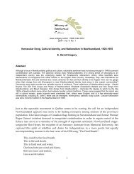
Microscopy and Imag<strong>in</strong>g Facility<br />
At first glance, it’s hard to tell the difference between all <strong>of</strong> the complex<br />
microscopes that fill the Microscopy and Imag<strong>in</strong>g Facility <strong>in</strong> the Health<br />
Sciences build<strong>in</strong>g. “The atomic force microscopes are the toys I use the<br />
most,” says Dr. Matthias Amre<strong>in</strong>, director <strong>of</strong> the facility, with a laugh. He’s<br />
talk<strong>in</strong>g about three microscopes which are so powerful they can scan the<br />
surface <strong>of</strong> a cell down to about half a nanometre, and hard crystall<strong>in</strong>e<br />
samples even at atomic resolution. The microscopes, together with the<br />
larger suite <strong>of</strong> transmission and scann<strong>in</strong>g electron microscopes, are be<strong>in</strong>g<br />
used to study diseases such as diabetes, multiple sclerosis and cancer,<br />
or to tackle basic questions <strong>in</strong> immunology to develop more effective<br />
vacc<strong>in</strong>es, to name just one <strong>of</strong> many examples. Prom<strong>in</strong>ent researchers<br />
work side-by-side with graduate students to obta<strong>in</strong> high-powered threedimensional<br />
images <strong>of</strong> a cell and its <strong>in</strong>ner work<strong>in</strong>gs. Amre<strong>in</strong> knows the<br />
impact these microscopes have goes far beyond the laboratory. “It’s a<br />
fantastic look at the <strong>in</strong>ternal work<strong>in</strong>gs <strong>of</strong> the cell,” he says. “Everybody<br />
here depends on this facility.”<br />
mif.ucalgary.ca/client/<br />
The Cave<br />
The world’s first complete object-oriented computer model <strong>of</strong> a human<br />
body, a k<strong>in</strong>d <strong>of</strong> four-dimensional human atlas dubbed the CAVEman, gives<br />
scientists the ability to translate medical and genomic data <strong>in</strong>to 4D<br />
images, and view the graphical representation <strong>of</strong> this data via the human<br />
form. Right before your eyes <strong>in</strong> this cube-shaped virtual reality room, the<br />
4D human model floats <strong>in</strong> space projected from three walls and the floor<br />
below. It allows medical researchers to <strong>in</strong>vestigate the genetics <strong>of</strong> various<br />
diseases and to develop new approaches to targeted treatments. A truly<br />
unique tool, the CAVE can size the data to any scale, says Dr. Christoph<br />
Sensen, director <strong>of</strong> the Sun Centre <strong>of</strong> Excellence for Visual Genomics<br />
<strong>in</strong> the Faculty <strong>of</strong> Medic<strong>in</strong>e at the U <strong>of</strong> C. While the CAVE is a cont<strong>in</strong>ual<br />
work <strong>in</strong> progress, this is a major breakthrough <strong>in</strong> medical <strong>in</strong>formatics and<br />
systems biology.<br />
Watch the CAVE live at youtube.com/watch?v=xF_4u-o6yPA<br />
Biomedical Eng<strong>in</strong>eer<strong>in</strong>g at the U <strong>of</strong> C: Technology + Facilities<br />
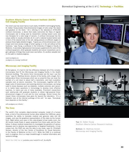
Top: Dr. Dallen Young<br />
Southern Alberta Cancer <strong>Research</strong> Institute<br />
Bottom: Dr. Matthias Amre<strong>in</strong><br />
Microscopy and Imag<strong>in</strong>g Facility<br />
33