Light-controlled growth of the maize seedling mesocotyl: Mechanical ...
Light-controlled growth of the maize seedling mesocotyl: Mechanical ...
Light-controlled growth of the maize seedling mesocotyl: Mechanical ...

You also want an ePaper? Increase the reach of your titles
YUMPU automatically turns print PDFs into web optimized ePapers that Google loves.
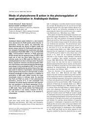
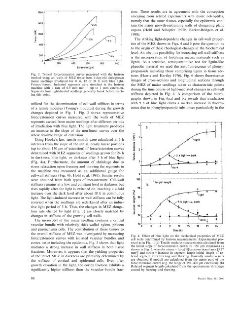
Fig. 3. Typical force/extension curves measured with <strong>the</strong> Instron<br />
method using cell walls <strong>of</strong> MEZ tissue from 4-day-old dark-grown<br />
<strong>maize</strong> <strong>seedling</strong>s irradiated for 0, 6, 12 or 24 h with blue light.<br />
Frozen/thawed, hydrated segments were stretched in <strong>the</strong> Instron<br />
machine with a rate <strong>of</strong> 0.5 mm min −1 up to 1 mm extension.<br />
Segments from light-treated <strong>seedling</strong>s generally break before reaching<br />
this point.<br />
utilized for <strong>the</strong> determination <strong>of</strong> cell-wall stiffness in terms<br />
<strong>of</strong> a tensile modulus (Young’s modulus) during <strong>the</strong> <strong>growth</strong><br />
changes depicted in Fig. 1. Fig. 3 shows representative<br />
force/extension curves measured with <strong>the</strong> walls <strong>of</strong> MEZ<br />
segments excised from <strong>maize</strong> <strong>seedling</strong>s after different periods<br />
<strong>of</strong> irradiation with blue light. The light treatment produces<br />
an increase in <strong>the</strong> slope <strong>of</strong> <strong>the</strong> non-linear curves over <strong>the</strong><br />
whole feasible range <strong>of</strong> extension.<br />
Using Hooke’s law, tensile moduli were calculated at 3-h<br />
intervals from <strong>the</strong> slope <strong>of</strong> <strong>the</strong> initial, nearly linear portions<br />
(up to about 150 m <strong>of</strong> extension) <strong>of</strong> force/extension curves<br />
determined with MEZ segments <strong>of</strong> <strong>seedling</strong>s grown for 24 h<br />
in darkness, blue light, or darkness after 3h<strong>of</strong>blue light<br />
(Fig. 4a). Fur<strong>the</strong>rmore, <strong>the</strong> amount <strong>of</strong> shrinkage due to<br />
stress relaxation upon freezing and thawing <strong>the</strong> segments in<br />
<strong>the</strong> machine was measured as an additional gauge for<br />
cell-wall stiffness (Fig. 4b, Hohl et al. 1995). Similar results<br />
were obtained from both types <strong>of</strong> measurement. Cell-wall<br />
stiffness remains at a low and constant level in darkness but<br />
rises rapidly after <strong>the</strong> light is switched on, reaching a 4-fold<br />
increase over <strong>the</strong> dark level after about 18 h in continuous<br />
light. The light-induced increase in wall stiffness can be fully<br />
reversed when <strong>the</strong> <strong>seedling</strong>s are redarkened after an inductive<br />
light period <strong>of</strong> 3 h. Thus, <strong>the</strong> changes in MEZ elongation<br />
rate elicited by light (Fig. 1) are closely matched by<br />
changes in stiffness <strong>of</strong> <strong>the</strong> growing cell walls.<br />
The <strong>mesocotyl</strong> <strong>of</strong> <strong>the</strong> <strong>maize</strong> <strong>seedling</strong> contains a central<br />
vascular bundle with relatively thick-walled xylem, phloem<br />
and parenchyma cells. The contribution <strong>of</strong> <strong>the</strong>se tissues to<br />
<strong>the</strong> overall stiffness <strong>of</strong> MEZ was investigated by measuring<br />
force/extension curves with isolated vascular bundles and<br />
cortex tissue including <strong>the</strong> epidermis. Fig. 5 shows that light<br />
mediates a strong increase in wall stiffness in both tissue<br />
fractions. Moreover, it appears that <strong>the</strong> yielding properties<br />
<strong>of</strong> <strong>the</strong> intact MEZ in darkness are primarily determined by<br />
<strong>the</strong> stiffness <strong>of</strong> cortical and epidermal cells. Even after<br />
<strong>growth</strong> cessation in <strong>the</strong> light, <strong>the</strong> cortex fraction exhibits a<br />
significantly higher stiffness than <strong>the</strong> vascular-bundle frac-<br />
86<br />
tion. These results are in agreement with <strong>the</strong> conception<br />
emerging from related experiments with <strong>maize</strong> coleoptiles,<br />
namely that <strong>the</strong> outer tissues, especially <strong>the</strong> epidermis, contain<br />
<strong>the</strong> major <strong>growth</strong>-restraining walls <strong>of</strong> elongating plant<br />
organs (Hohl and Schopfer 1992b, Barker-Bridgers et al.<br />
1998).<br />
The striking light-dependent changes in cell-wall properties<br />
<strong>of</strong> <strong>the</strong> MEZ shown in Figs. 4 and 5 pose <strong>the</strong> question as<br />
to <strong>the</strong> origin <strong>of</strong> <strong>the</strong>se rheological changes at <strong>the</strong> biochemical<br />
level. An obvious possibility for increasing cell-wall stiffness<br />
is <strong>the</strong> incorporation <strong>of</strong> fortifying matrix materials such as<br />
lignin. As a sensitive, semiquantitative test for lignin-like<br />
phenolic material we used <strong>the</strong> aut<strong>of</strong>luorescence <strong>of</strong> phenylpropanoids<br />
including those comprising lignin in tissue sections<br />
(Harris and Hartley 1976). Fig. 6 shows fluorescence<br />
images <strong>of</strong> cross-sections and longitudinal sections through<br />
<strong>the</strong> MEZ <strong>of</strong> <strong>maize</strong> <strong>seedling</strong>s taken at characteristic points<br />
during <strong>the</strong> time course <strong>of</strong> light-mediated changes in cell-wall<br />
stiffness depicted in Fig. 4. A comparison <strong>of</strong> <strong>the</strong> micrographs<br />
shown in Fig. 6a,d and b,e reveals that irradiation<br />
with9h<strong>of</strong>blue light elicits a marked increase in fluorescence<br />
due to phenylpropanoid substances particularly in <strong>the</strong><br />
Fig. 4. Effect <strong>of</strong> blue light on <strong>the</strong> mechanical properties <strong>of</strong> MEZ<br />
cell walls determined by Instron measurements. Experimental protocol<br />
as in Fig. 1. (a) Tensile modulus (stress/strain) calculated from<br />
<strong>the</strong> initial slope <strong>of</strong> force/extension curves (0–150 m extension) as<br />
shown in Fig. 3, whereby stress=force[N]/cross-sectional area [3.27<br />
mm 2 ] and strain=increase in segment length/initial length <strong>of</strong> relaxed<br />
segment after freezing and thawing. Basically similar results<br />
are obtained if moduli are calculated from <strong>the</strong> upper part <strong>of</strong> <strong>the</strong><br />
force/extension curves (e.g. <strong>the</strong> range <strong>of</strong> 350–420 m extension). (b)<br />
Relaxed segment length calculated from <strong>the</strong> spontaneous shrinkage<br />
caused by freezing and thawing.<br />
Physiol. Plant. 111, 2001