Module 4 Respiration - Pearson Schools
Module 4 Respiration - Pearson Schools
Module 4 Respiration - Pearson Schools

You also want an ePaper? Increase the reach of your titles
YUMPU automatically turns print PDFs into web optimized ePapers that Google loves.
UNIT<br />
<strong>Module</strong> 4<br />
<strong>Respiration</strong><br />
1<br />
Introduction<br />
All living organisms need energy to do work. This work includes movement, active<br />
transport, bulk transport, nerve conduction, synthesis of large molecules such as<br />
cellulose and proteins, and replication of DNA. Energy is also needed for synthesis of<br />
new organelles before a cell divides.<br />
When life developed on Earth around 3500 million years ago, the atmosphere did not<br />
contain any free oxygen. The earliest forms of life on Earth used various metabolic<br />
pathways to obtain energy from chemicals in their environment. After about 1500<br />
million years, cyanobacteria evolved that could trap sunlight energy for the process<br />
of photosynthesis. They used water as a source of electrons and protons, releasing<br />
free oxygen into the air. This changed the previously reducing atmosphere to an<br />
oxidising one and led eventually to vast biodiversity, as organisms that could use<br />
this oxygen for respiration evolved.<br />
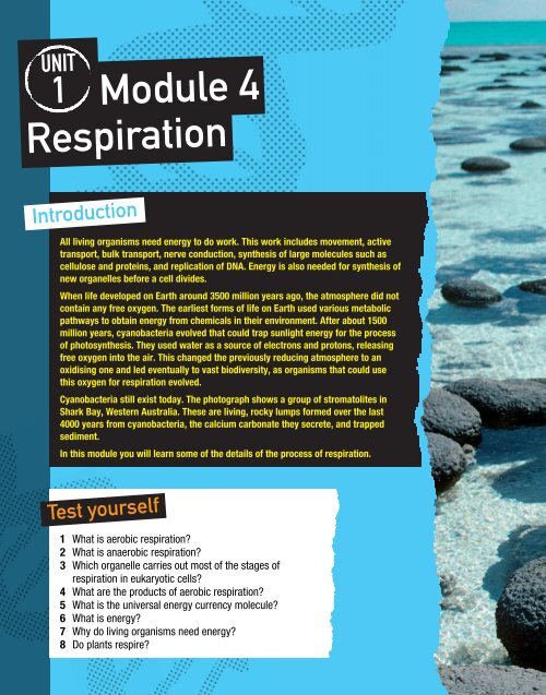
Cyanobacteria still exist today. The photograph shows a group of stromatolites in<br />
Shark Bay, Western Australia. These are living, rocky lumps formed over the last<br />
4000 years from cyanobacteria, the calcium carbonate they secrete, and trapped<br />
sediment.<br />
In this module you will learn some of the details of the process of respiration.<br />
Test yourself<br />
1 What is aerobic respiration?<br />
2 What is anaerobic respiration?<br />
3 Which organelle carries out most of the stages of<br />
respiration in eukaryotic cells?<br />
4 What are the products of aerobic respiration?<br />
5 What is the universal energy currency molecule?<br />
6 What is energy?<br />
7 Why do living organisms need energy?<br />
8 Do plants respire?
<strong>Module</strong> contents<br />
1 Why do living organisms need to<br />
respire?<br />
2 Coenzymes<br />
3 Glycolysis<br />
4 Structure and function of mitochondria<br />
5 The link reaction and Krebs cycle<br />
6 Oxidative phosphorylation and<br />
chemiosmosis<br />
7 Evaluating the evidence for<br />
chemiosmosis<br />
8 Anaerobic respiration in mammals and<br />
yeast<br />
9 Respiratory substrates
80<br />
1.4 1 Why do living organisms need to<br />
respire?<br />
Key defi nitions<br />
Energy is the ability to do work.<br />
ATP is a phosphorylated nucleotide and<br />
is the universal energy currency.<br />
Key defi nitions<br />
Anabolic reactions are biochemical<br />
reactions where large molecules are<br />
synthesised from smaller ones.<br />
In catabolic reactions larger molecules<br />
are hydrolysed to produce smaller<br />
molecules.<br />
By the end of this spread, you should be able to …<br />
✱ Outline why living organisms need to respire.<br />
✱ Describe the structure of ATP.<br />
✱ State that ATP provides the immediate source of energy for biological processes.<br />
What is respiration?<br />
<strong>Respiration</strong> is the process whereby energy stored in complex organic molecules<br />
(carbohydrates, fats and proteins) is used to make ATP. It occurs in living cells.<br />
What is energy?<br />
Energy exists as potential (stored) energy and kinetic energy (the energy of movement).<br />
Moving molecules have kinetic energy that allows them to diffuse down a concentration<br />
gradient. Large organic molecules contain chemical potential energy.<br />
Energy:<br />
• cannot be created or destroyed but can be converted from one form to another<br />
• is measured in joules or kilojoules<br />
• has many forms, e.g. sound (mechanical), light, heat, electrical, chemical and atomic.<br />
Why do we need it?<br />
All living organisms need energy to drive their biological processes. All the reactions that<br />
take place within organisms are known collectively as metabolism. Metabolic reactions<br />
that build large molecules are described as anabolic and those that break large<br />
molecules into smaller ones are catabolic.<br />
Metabolic processes that need energy include:<br />
• Active transport – moving ions and molecules across a membrane against a<br />
concentration gradient. Much of an organism’s energy is used for this. All cell<br />
membranes have sodium–potassium pumps and these maintain the resting potential.<br />
When this pump momentarily stops in neurone membranes, sodium ions enter the<br />
neurone and an action potential occurs.<br />
• Secretion – large molecules made in some cells are exported by exocytosis.<br />
• Endocytosis – bulk movement of large molecules into cells.<br />
• Synthesis of large molecules from smaller ones, such as proteins from amino acids,<br />
steroids from cholesterol and cellulose from β-glucose. These are all examples of<br />
anabolism.<br />
• Replication of DNA and synthesis of organelles before a cell divides.<br />
• Movement – such as movement of bacterial flagella, eukaryotic cilia and undulipodia,<br />
muscle contraction and microtubule motors that move organelles around inside cells.<br />
• Activation of chemicals – glucose is phosphorylated at the beginning of respiration so<br />
that it is more unstable and can be broken down to release energy.<br />
Some of the energy from catabolic reactions is released in the form of heat. This is useful<br />
as metabolic reactions are controlled by enzymes, so organisms need to maintain a<br />
suitable temperature that allows enzyme action to proceed at a speed that will sustain<br />
life.
Where does the energy come from?<br />
Plants, some protoctists and some bacteria<br />
are photoautotrophs. They use sunlight<br />
energy in photosynthesis to make large,<br />
organic molecules that contain chemical<br />
potential energy, which they and consumers<br />
and decomposers can then use. <strong>Respiration</strong><br />
releases the energy, which is used to<br />
phosphorylate (add inorganic phosphate to)<br />
ADP, making ATP. This phosphorylation also<br />
transfers energy to the ATP molecule.<br />
The role of ATP<br />
ATP is a phosphorylated nucleotide. It is a<br />
high-energy intermediate compound, found in<br />
both prokaryotic and eukaryotic cells. Each<br />
molecule consists of adenosine (adenine and<br />
ribose sugar) plus three phosphate (more<br />
correctly, phosphoryl) groups. It can be<br />
hydrolysed to ADP and P i (inorganic phosphate),<br />
releasing 30.6 kJ energy per mol. So, energy is<br />
immediately available to cells in small,<br />
manageable amounts that will not damage the<br />
cell and will not be wasted. ATP is described as<br />
the universal energy currency.<br />
Photosynthesis<br />
Photoautotrophs<br />
e.g. plants,<br />
some protoctists,<br />
some bacteria<br />
Figure 1 Energy transfer<br />
Chemical potential<br />
energy in organic<br />
molecules, e.g.<br />
carbohydrates, lipids,<br />
proteins<br />
Light<br />
energy<br />
Thermal<br />
energy<br />
(heat)<br />
Helps<br />
maintain<br />
suitable<br />
temperature<br />
<strong>Respiration</strong><br />
Chemical<br />
potential<br />
in ATP<br />
Enables<br />
living<br />
organisms<br />
to do work<br />
<strong>Respiration</strong> occurs in many small steps. The energy released at each stage joins ADP<br />
and P i to make ATP. You probably use 25–50 kg ATP each day, depending on your level<br />
of activity, but you will only have about 5 g of ATP in your body at any one point in time.<br />
It is continually being hydrolysed and resynthesised.<br />
The hydrolysis of ATP is coupled with a synthesis reaction, such as DNA replication or<br />
protein synthesis, in cells. Such synthesis reactions require energy. The energy released<br />
from ATP hydrolysis is an immediate source of energy for these biological processes.<br />
30.6 kJ mol 1<br />
ATP H 2O ADP H 2O<br />
30.6 kJ mol 1<br />
CO 2 H 2 O<br />
AMP H 2O<br />
Figure 3 The energy released from hydrolysis of ATP<br />
14.2 kJ mol 1<br />
P i P i P i<br />
Adenosine<br />
Questions<br />
1 Fireflies can produce light in a process called bioluminescence. Outline the energy<br />
transformations that occur in fireflies as they use energy from their food to produce<br />
bioluminescence.<br />
2 ATP is a nucleic acid/nucleotide derivative. Do you think it is derived from DNA or RNA<br />
nucleotides? Give reasons for your answer.<br />
3 Decide whether each of the following is an anabolic or catabolic reaction:<br />
(a) synthesis of spindle microtubules during mitosis<br />
(b) digestion of starch to maltose<br />
(c) formation of insulin in cells of the pancreas<br />
(d) conversion of glycogen to glucose in liver cells<br />
(e) digestion of a pathogen inside a phagolysosome of a macrophage.<br />
4 Explain why ATP is known as the universal energy currency.<br />
Heterotrophs<br />
Consumers and<br />
decomposers, e.g.<br />
animals, fungi and<br />
most bacteria<br />
P<br />
P<br />
Figure 2 The structure of ATP<br />
Figure 4 The ATP cycle<br />
P<br />
Ribose<br />
Adenine<br />
Adenosine<br />
Adenosine monophosphate<br />
Energy released for use<br />
by cells to do work<br />
Hydrolysis<br />
Adenosine diphosphate<br />
Adenosine triphosphate<br />
ATP ATPsynthase ADP Pi Condensation<br />
Energy released from<br />
organic substrate, during<br />
respiration<br />
Examiner tip<br />
<strong>Module</strong> 4<br />
<strong>Respiration</strong><br />
Why do living organisms need to<br />
respire?<br />
Remember that energy cannot be<br />
created or destroyed. So, never refer to<br />
energy being produced. <strong>Respiration</strong><br />
releases energy to produce ATP.<br />
81
82<br />
1.4 2 Coenzymes<br />
Key defi nitions<br />
Oxidation reactions involve loss of<br />
electrons.<br />
Reduction reactions involve addition of<br />
electrons.<br />
These reactions are coupled – one<br />
substrate becomes oxidised and<br />
another becomes reduced. In the<br />
reactions of respiration where<br />
coenzymes are involved, the coenzymes<br />
become reduced as substrate becomes<br />
oxidised. Later the reduced coenzyme<br />
becomes reoxidised so that it can be<br />
used again.<br />
By the end of this spread, you should be able to …<br />
✱ Explain the importance of coenzymes in respiration, with reference to NAD and coenzyme A.<br />
The stages of respiration<br />
Glycolysis<br />
Glucose Pyruvate<br />
Anaerobic<br />
Aerobic<br />
Figure 1 The stages of respiration<br />
Lactate<br />
fermentation<br />
Ethanol<br />
fermentation<br />
Link reaction Krebs<br />
Pyruvate Acetyl<br />
CoA<br />
cycle<br />
Carbon dioxide Carbon dioxide<br />
Lactate<br />
Ethanol<br />
<br />
carbon dioxide<br />
Oxidative<br />
phosphorylation<br />
Oxygen<br />
H e <br />
<strong>Respiration</strong> of glucose can be described in four stages:<br />
• Glycolysis – this happens in the cytoplasm of all cells. It is an ancient biochemical<br />
pathway. It doesn’t need oxygen and can take place in aerobic or anaerobic<br />
conditions. During glycolysis, glucose (a 6-carbon sugar) is broken down to two<br />
molecules of pyruvate (a 3-carbon compound).<br />
• The link reaction – this happens in the matrix of mitochondria. Pyruvate is<br />
dehydrogenated (hydrogen removed) and decarboxylated (carboxyl removed) and<br />
converted to acetate.<br />
• Krebs cycle – also takes place in the matrix of mitochondria. Acetate is<br />
decarboxylated and dehydrogenated.<br />
• Oxidative phosphorylation – takes place on the folded inner membranes (cristae) of<br />
mitochondria. This is where ADP is phosphorylated to ATP.<br />
The last three stages will only take place under aerobic conditions. Under anaerobic<br />
conditions, pyruvate is converted to either ethanol or lactate.<br />
Why are coenzymes needed?<br />
During glycolysis, the link reaction and Krebs cycle, hydrogen atoms are removed from<br />
substrate molecules in oxidation reactions. These reactions are catalysed by<br />
dehydrogenase enzymes. Although enzymes catalyse a wide variety of metabolic<br />
reactions, they are not very good at catalysing oxidation or reduction reactions.<br />
Coenzymes are needed to help them carry out the oxidation reactions of respiration. The<br />
hydrogen atoms are combined with coenzymes such as NAD. These carry the hydrogen<br />
atoms, which can later be split into hydrogen ions and electrons, to the inner<br />
mitochondrial membranes. Here, they will be involved in the process of oxidative<br />
phosphorylation (see spread 1.4.6), which produces a lot of ATP. Delivery of the<br />
hydrogens to the cristae reoxidises the coenzymes so they can combine with (or ‘pick<br />
up’) more hydrogen atoms from the fi rst three stages of respiration.<br />
Water
NAD<br />
This is an organic, non-protein molecule that helps dehydrogenase enzymes to carry out<br />
oxidation reactions. Nicotinamide adenine dinucleotide (NAD) is made of two linked<br />
nucleotides. It is made in the body from nicotinamide (Vitamin B 3), the 5-carbon sugar<br />
ribose, adenine and two phosphate (or, more accurately, phosphoryl) groups. One<br />
nucleotide contains the nitrogenous base adenine. The other contains a nicotinamide<br />
ring that can accept hydrogen atoms – each of which can later be split into a hydrogen<br />
ion and an electron.<br />
When a molecule of NAD has accepted two hydrogen atoms with their electrons, it is<br />
reduced. When it loses the electrons it is oxidised. NAD operates during glycolysis (see<br />
spread 1.4.3), the link reaction (see spread 1.4.5), Krebs cycle (see spread 1.4.5) and<br />
during the anaerobic ethanol and lactate pathways (see spread 1.4.8).<br />
Coenzyme A (CoA)<br />
This coenzyme is made from pantothenic acid (a B-group vitamin), adenosine (ribose and<br />
adenine), three phosphate (phosphoryl) groups and cysteine (an amino acid). Its function<br />
is to carry ethanoate (acetate) groups, made from pyruvate during the link reaction, onto<br />
Krebs cycle. It can also carry acetate groups that have been made from fatty acids or<br />
from some amino acids (see spread 1.4.9) onto Krebs cycle.<br />
P P<br />
Ribose<br />
P<br />
Pantothenic<br />
acid<br />
Cysteine<br />
Figure 3 The structure of coenzyme A<br />
STRETCH and CHALLENGE<br />
In the early part of the twentieth century, a dietary defi ciency disease called pellagra<br />
(diarrhoea, dermatitis and dementia) was endemic in rural parts of the Southern United<br />
States of America, where the diet consisted mainly of corn products. It could be treated<br />
with nicotinamide (vitamin B 3). Humans, like many animals, can synthesise<br />
nicotinamide from the amino acid tryptophan. Corn contains very little tryptophan. It<br />
contains a lot of nicotinamide but in a form that needs to be treated with a base<br />
(alkaline substance) before it can be absorbed from the intestine.<br />
Mexican Indians are thought to have domesticated the corn plant and their diet has<br />
always contained a lot of corn. They soak the corn in limewater (calcium hydroxide<br />
solution) before using it to make tortillas. They do not suffer from pellagra.<br />
Question<br />
A Explain why Mexican Indians do not suffer from pellagra, whilst people living in rural<br />
Southern states of the US did, although both ate a diet rich in corn.<br />
Questions<br />
1 Explain why living organisms do not have very much NAD or CoA in their cells.<br />
2 Alcohol is metabolised in the liver. It is oxidised to ethanal by dehydrogenation, and<br />
then to ethanoate (acetate). Suggest why people who drink large amounts of alcohol<br />
may be deficient in NAD.<br />
3 Explain why NAD is called a nucleic acid/nucleotide derivative.<br />
P<br />
P<br />
Ribose<br />
Ribose<br />
Adenine<br />
<strong>Module</strong> 4<br />
<strong>Respiration</strong><br />
Coenzymes<br />
Nicotinamide<br />
Figure 2 Molecular structure of NAD<br />
Examiner tips<br />
Don’t confuse NAD and NADP. We have<br />
met NADP in photosynthesis (think of<br />
the P in NADP as a reminder of<br />
photosynthesis!).<br />
We have met NAD involved in<br />
respiration.<br />
When these coenzymes become<br />
reduced they carry hydrogen atoms<br />
(which later become protons and<br />
electrons). Don’t say that they carry<br />
hydrogen ions or molecules.<br />
83
84<br />
Key defi nitions<br />
Glycolysis is a metabolic pathway<br />
where each glucose molecule is broken<br />
down to two molecules of pyruvate. It<br />
occurs in the cytoplasm of all living cells<br />
and is common to anaerobic (without<br />
oxygen) and aerobic (with oxygen)<br />
respiration.<br />
Hexose sugars have six carbon atoms<br />
in each molecule.<br />
Hydrolysis is the breaking down of<br />
large molecules to smaller molecules by<br />
the addition of water.<br />
Triose sugars have three carbon atoms<br />
in each molecule.<br />
ATP<br />
ATP<br />
1.4 3 Glycolysis<br />
Glucose (6C)<br />
Glucose-6-P<br />
Fructose-1-P<br />
Hexose 1,6-bisphosphate<br />
2 Triose phosphate (3C)<br />
2 Intermediate compound (3C)<br />
2 Pyruvate (3C)<br />
Figure 1 Summary of glycolysis<br />
By the end of this spread, you should be able to …<br />
✱ State that glycolysis occurs in the cytoplasm of cells.<br />
✱ Outline the process of glycolysis.<br />
✱ State that in aerobic respiration, pyruvate is actively transported into mitochondria.<br />
Glycolysis is a very ancient biochemical pathway, occurring in the cytoplasm of all living<br />
cells that respire. This means it happens in prokaryotic and eukaryotic cells. It has been<br />
studied extensively by biochemists and is probably the best-understood metabolic<br />
pathway.<br />
This pathway involves a sequence of ten reactions, each catalysed by a different<br />
enzyme. The coenzyme NAD (see spread 1.4.2) is also involved. You only need to know<br />
this pathway in outline so we will consider it as just four stages.<br />
Stage 1: Phosphorylation<br />
Glucose is a hexose sugar – it contains six carbon atoms. Its molecules are stable and<br />
need to be activated before they can be split into two.<br />
• One ATP molecule is hydrolysed and the phosphate group released is attached to<br />
the glucose molecule at carbon 6.<br />
• Glucose 6-phosphate is changed to fructose 6-phosphate.<br />
• Another ATP is hydrolysed and the phosphate group released is attached to fructose<br />
6-phosphate at carbon 1. This activated hexose sugar is now called fructose<br />
1,6-bisphosphate.<br />
• The energy from the hydrolysed ATP molecules activates the hexose sugar and prevents<br />
it from being transported out of the cell. We can refer to the activated, phosphorylated<br />
sugar as hexose 1,6-bisphosphate. (This name tells us that it is a hexose sugar with<br />
two phosphates attached, one at carbon 1 and the other at carbon 6.)<br />
• Note that this stage has used two molecules of ATP for each molecule of glucose.<br />
2ATP<br />
2 reduced NAD<br />
2ATP<br />
Stage 1<br />
Stage 2<br />
Stage 3<br />
Stage 4<br />
Stage 2: Splitting of hexose 1,6-bisphosphate<br />
• Each molecule of hexose bisphosphate is split into two<br />
molecules of triose phosphate (3-carbon sugar molecules<br />
each with one phosphate group attached).<br />
Stage 3: Oxidation of triose phosphate<br />
• Although this process is anaerobic, it involves oxidation.<br />
• Two hydrogen atoms (with their electrons) are removed<br />
from each triose phosphate molecule (the substrate).<br />
• This involves dehydrogenase enzymes.<br />
• These are aided by the coenzyme NAD (nicotinamide<br />
adenine dinucleotide), which is a hydrogen acceptor (see<br />
spread 1.4.2). NAD combines with the hydrogen atoms,<br />
becoming reduced NAD.<br />
• So, at this stage of glycolysis, two molecules of NAD are<br />
reduced per molecule of glucose.<br />
• Also, at this stage, two molecules of ATP are formed. This<br />
is called substrate-level phosphorylation.
Stage 4: Conversion of triose phosphate to pyruvate<br />
• Four enzyme-catalysed reactions convert each triose phosphate molecule to a<br />
molecule of pyruvate. Pyruvate is also a 3-carbon compound.<br />
• In the process another two molecules of ADP are phosphorylated (an inorganic<br />
phosphate group, P i, is added) to two molecules of ATP (by substrate-level<br />
phosphorylation).<br />
What are the products of glycolysis?<br />
From each molecule of glucose at the beginning of this pathway, at the end of glycolysis<br />
there are:<br />
• two molecules of ATP. Four have been made but two were used to ‘kick-start’ the<br />
process, so the net gain is two molecules of ATP<br />
• two molecules of reduced NAD. These will carry hydrogen atoms, indirectly via a shunt<br />
mechanism, to the inner mitochondrial membranes and be used to generate more<br />
ATP during oxidative phosphorylation (see spread 1.4.6)<br />
• two molecules of pyruvate. This will normally be actively transported into the<br />
mitochondrial matrix for the next stage of aerobic respiration (see spread 1.4.4). In the<br />
absence of oxygen it will be changed, in the cytoplasm, to lactate or ethanol (see<br />
spread 1.4.8).<br />
Fermentation and glycolysis<br />
For thousands of years humans used the process of fermentation of glucose to ethanol,<br />
by yeast, without understanding that glycolysis was involved. In the second half of the<br />
nineteenth century, scientists investigated the mechanism. Pasteur established that<br />
alcoholic fermentation is caused by microorganisms. Buchner showed that extracts<br />
from yeast cells could also cause fermentation. He used the word enzyme – the word<br />
means ‘in yeast’. By 1940 many biochemists had helped to analyse and work out the<br />
pathway. They had used cells and tissues from many living organisms in their studies<br />
and found that, with very few exceptions (such as some Archaea), all living things have<br />
this metabolic pathway.<br />
STRETCH and CHALLENGE<br />
Enzymes that cause the shape of a molecule to change (without changing the<br />
proportions of atoms in that molecule) are called isomerases.<br />
Questions<br />
A At which stage of glycolysis are isomerase enzymes involved?<br />
B How does the fact that nearly all living things use the glycolysis pathway support the<br />
theory of evolution?<br />
Questions<br />
1 What was in Buchner’s cell-free extract (made from yeast) that enabled the<br />
fermentation of glucose to alcohol?<br />
2 Outline the role of coenzymes (spread 1.4.2) in the glycolysis pathway.<br />
3 Explain why the net gain of ATP during glycolysis is two, not four, molecules.<br />
4 Explain how oxidation occurs during glycolysis, although no oxygen is involved.<br />
Examiner tip<br />
<strong>Module</strong> 4<br />
<strong>Respiration</strong><br />
Glycolysis<br />
Learn the stages of glycolysis where:<br />
ATP is used<br />
ATP is produced<br />
NAD is reduced<br />
85
86<br />
Key defi nition<br />
Mitochondria are organelles found in<br />
eukaryote cells. They are the sites of<br />
the link reaction, Krebs cycle and<br />
oxidative phosphorylation – the aerobic<br />
stages of respiration.<br />
Figure 1 Electron micrograph of a<br />
mitochondrion from an intestinal cell<br />
(×32 000)<br />
Inner<br />
membrane<br />
Matrix<br />
1.4 4 Structure and function of<br />
mitochondria<br />
Envelope Intermembrane space<br />
Figure 2 Structure of a mitochondrion<br />
Examiner tip<br />
Outer<br />
membrane<br />
Stalked particles<br />
(ATP synthase)<br />
Cristae<br />
Always say that protons fl ow down the<br />
(electrochemical/pH/proton) gradient<br />
through ATP synthase enzyme. Don’t<br />
say they fl ow along the gradient.<br />
By the end of this spread, you should be able to …<br />
✱ Explain, with the aid of diagrams and electron micrographs, how the structure of mitochondria<br />
enables them to carry out their functions.<br />
Mitochondrial ultrastructure<br />
Mitochondria were fi rst identifi ed in animal cells, using light microscopy, in 1840. Plant<br />
mitochondria were observed about 60 years later. In 1953 the fi rst extensive electron<br />
microscope studies of mitochondria were made.<br />
• All mitochondria have an inner and outer phospholipid membrane. These two<br />
membranes make up the envelope.<br />
• The outer membrane is smooth and the inner membrane is folded into cristae<br />
(singular crista) that give the inner membrane a large surface area.<br />
• The two membranes enclose and separate the two compartments within the<br />
mitochondrion. Between the inner and outer membranes is the intermembrane<br />
space.<br />
• The matrix is enclosed by the inner membrane. It is semi-rigid and gel-like, consisting<br />
of a mixture of proteins and lipids. It also contains looped mitochondrial DNA,<br />
mitochondrial ribosomes and enzymes.<br />
Shape, size and distribution<br />
Mitochondria may be rod-shaped or thread-like. Their shape can change but most are<br />
between 0.5–1.0 µm in diameter and 2–5 µm long, although some can be 10 µm long.<br />
A trained athlete may have larger mitochondria in his/her muscle tissue. Metabolically<br />
active cells (large demand for ATP) have more mitochondria. These mitochondria usually<br />
have longer and more densely packed cristae to house more electron transport chains<br />
and more ATP synthase enzymes. Mammalian liver cells may each contain up to 2500<br />
mitochondria, occupying up to 20% of the cell’s volume.<br />
Mitochondria can be moved around within cells by the cytoskeleton (microtubules). In<br />
some types of cells the mitochondria are permanently positioned near a site of high ATP<br />
demand, for example at the synaptic knobs of nerve cells. However, they have been<br />
moved to that position by microtubules.<br />
How does their structure enable them to carry out their functions?<br />
The matrix<br />
The matrix is where the link reaction and Krebs cycle take place. It contains:<br />
• the enzymes that catalyse the stages of these reactions<br />
• molecules of coenzyme NAD<br />
• oxaloacetate – the 4-carbon compound that accepts acetate from the link reaction<br />
• mitochondrial DNA, some of which codes for mitochondrial enzymes and other<br />
proteins<br />
• mitochondrial ribosomes (structurally the same as prokaryote ribosomes) where these<br />
proteins are assembled.<br />
The outer membrane<br />
The phospholipid composition of the outer membrane is similar to membranes around<br />
other organelles. It contains proteins, some of which form channels or carriers that allow<br />
the passage of molecules such as pyruvate. Other proteins in this membrane are enzymes.
The inner membrane<br />
The inner membrane:<br />
• has a different lipid composition from the outer membrane and is<br />
impermeable to most small ions, including hydrogen ions (protons)<br />
• is folded into many cristae to give a large surface area<br />
• has embedded in it many electron carriers and ATP synthase<br />
enzymes.<br />
The electron carriers are protein complexes, arranged in electron<br />
transport chains.<br />
• Each electron carrier is an enzyme. Each is associated with a<br />
cofactor. The cofactors are non-protein groups. They are haem<br />
groups and contain an iron atom.<br />
• The cofactors can accept and donate electrons because the<br />
iron atoms can become reduced (to Fe2+ ) by accepting an<br />
electron and oxidised (to Fe3+ ) by donating an electron to the<br />
next electron carrier.<br />
• They are oxidoreductase enzymes as they are involved in<br />
oxidation and reduction reactions.<br />
• Some of the electron carriers also have a coenzyme that pumps<br />
(using energy released from the passage of electrons) protons<br />
from the matrix to the intermembrane space.<br />
• Because the inner membrane is impermeable to small ions,<br />
protons accumulate in the intermembrane space, building up a<br />
proton gradient – a source of potential energy.<br />
The ATP synthase enzymes:<br />
• are large and protrude from the inner membrane into the matrix<br />
• are also known as stalked particles<br />
• allow protons to pass through them.<br />
Protons flow down a proton gradient, through the ATP synthase enzymes, from the<br />
intermembrane space into the matrix. This flow is called chemiosmosis. The force of this<br />
flow (the proton motive force) drives the rotation of part of the enzyme and allows ADP<br />
and Pi (inorganic phosphate) to be joined to make ATP.<br />
The coenzyme FAD, which becomes reduced during one stage of Krebs cycle, is tightly<br />
bound to a dehydrogenase enzyme that is embedded in the inner membrane. The<br />
hydrogen atoms accepted by FAD do not get pumped into the inert membrane space.<br />
Instead they pass back into the mitochondrial matrix.<br />
FAD is flavine adenine dinucleotide, derived from vitamin B2 (riboflavin), adenine, ribose<br />
and two phosphate groups.<br />
1 ADP+ Pi 2<br />
ATP<br />
Figure 5 ATPsynthesis occurs in three steps. The axle (stalk) rotates the<br />
head, shown here. ADP and Pi join to form ATP, which is then released as<br />
that section of the headpiece undergoes a conformational (shape) change<br />
Questions<br />
1 Suggest how the structure of a mitochondrion from a skin cell would differ from that of<br />
a mitochondrion from heart muscle tissue.<br />
2 Suggest why synaptic knobs of nerve cells have many mitochondria.<br />
3 Explain the following terms: chemiosmosis; proton motive force; oxidoreductase enzyme.<br />
ATP<br />
3<br />
Matrix<br />
Outer membrane<br />
Hydrogen atoms<br />
from Krebs cycle<br />
and link reaction<br />
2e <br />
2H<br />
<br />
<br />
2e Inner membrane<br />
Intermembrane<br />
space<br />
2e 2e <br />
Fe<br />
Fe<br />
H <br />
H <br />
Build-up of protons in the intermembrane space,<br />
producing a proton gradient across the inner membrane<br />
F 0 – the fraction of the<br />
molecule that binds to<br />
oligomycin (spread 1.4.7)<br />
Base piece (F 0)<br />
Stalk or axle<br />
Headpiece<br />
(F 1)<br />
H H H <br />
H <br />
<strong>Module</strong> 4<br />
<strong>Respiration</strong><br />
Structure and function of<br />
mitochondria<br />
ADPP i<br />
<br />
2H 2e ATP ATP synthase<br />
Figure 3 Diagram showing the structure of the inner membrane<br />
and the flow of electrons between electron carriers and the flow of<br />
protons into the intermembrane space<br />
Fe<br />
H <br />
H <br />
Fe<br />
1<br />
2 O2 Proton channel<br />
Inner<br />
mitochondrial<br />
membrane<br />
Stator<br />
Figure 4 The structure of ATP synthase<br />
STRETCH and<br />
CHALLENGE<br />
It has been suggested that<br />
mitochondria are derived from<br />
prokaryotes.<br />
Question<br />
A What features of their structure<br />
support this suggestion?<br />
H 2O<br />
87
88<br />
Key defi nitions<br />
The link reaction converts pyruvate to<br />
acetate. NAD is reduced.<br />
Krebs cycle oxidises acetate to carbon<br />
dioxide. NAD and FAD are reduced. ATP<br />
is made by substrate-level<br />
phosphorylation.<br />
Both of these reactions occur in the<br />
mitochondrial matrix.<br />
Reduced<br />
NAD<br />
Reduced<br />
FAD<br />
ATP<br />
1.4 5 The link reaction and Krebs cycle<br />
Reduced<br />
NAD<br />
2H<br />
4C compound<br />
Pyruvate (3C)<br />
Figure 1 Summary of the link reaction and Krebs cycle<br />
2H<br />
Oxaloacetate (4C)<br />
5<br />
6<br />
4C compound<br />
4<br />
4C compound<br />
2H<br />
Reduced<br />
NAD<br />
Acetyl CoA (2C)<br />
3<br />
CO 2<br />
Acetate (2C)<br />
By the end of this spread, you should be able to …<br />
✱ Outline the link reaction, with reference to decarboxylation of pyruvate to acetate and the<br />
reduction of NAD, and state that it takes place in the mitochondrial matrix.<br />
✱ Explain that coenzyme A carries acetate from the link reaction to Krebs cycle.<br />
✱ Outline the Krebs cycle, including the roles of NAD and FAD, and substrate-level<br />
phosphorylation, and state that it takes place in the mitochondrial matrix.<br />
Pyruvate produced during glycolysis is transported across the inner and outer<br />
mitochondrial membranes to the matrix. It is changed into a 2-carbon compound,<br />
acetate, during the link reaction. Acetate is then oxidised during Krebs cycle.<br />
The link reaction<br />
Decarboxylation and dehydrogenation of pyruvate to acetate are enzyme-catalysed<br />
reactions.<br />
• Pyruvate dehydrogenase removes hydrogen atoms from pyruvate.<br />
• Pyruvate decarboxylase removes a carboxyl group, which eventually becomes<br />
carbon dioxide, from pyruvate.<br />
• The coenzyme NAD (spread 1.4.2) accepts the hydrogen atoms.<br />
• Coenzyme A (CoA) (spread 1.4.2) accepts acetate, to become acetyl coenzyme A.<br />
The function of CoA is to carry acetate to Krebs cycle.<br />
The following equation summarises the link reaction:<br />
2pyruvate + 2NAD + + 2CoA → 2CO 2 + 2reduced NAD + 2acetyl CoA<br />
NAD + indicates NAD in the oxidised state. Two molecules of pyruvate are considered in<br />
the equation as two molecules of pyruvate are derived from each molecule of glucose.<br />
CO 2<br />
1<br />
5C compound<br />
CoA<br />
Citrate (6C)<br />
2<br />
2H<br />
CO 2<br />
Link<br />
reaction<br />
Krebs<br />
cycle<br />
Reduced<br />
NAD<br />
Note that no ATP is produced. However each reduced<br />
NAD will take a pair of hydrogen atoms to the inner<br />
mitochondrial membrane and they will be used to make<br />
ATP during oxidative phosphorylation (spread 1.4.6).<br />
The Krebs cycle<br />
The Krebs cycle also takes place in the mitochondrial<br />
matrix. It is a series of enzyme-catalysed reactions that<br />
oxidise the acetyl group of acetyl CoA to two molecules of<br />
carbon dioxide. It also produces one molecule of ATP by<br />
substrate-level phosphorylation, and reduces three<br />
molecules of NAD and one molecule of FAD. These<br />
reduced coenzymes have the potential to produce more<br />
ATP during oxidative phosphorylation.<br />
1 The acetate is offloaded from coenzyme A (which is then<br />
free to collect more acetate) and joins with a 4-carbon<br />
compound, called oxaloacetate, to form a 6-carbon<br />
compound, called citrate.<br />
2 Citrate is decarboxylated (one molecule of carbon<br />
dioxide removed) and dehydrogenated (a pair of<br />
hydrogen atoms removed) to form a 5-carbon<br />
compound. The pair of hydrogen atoms is accepted by<br />
a molecule of NAD, which becomes reduced.
3 The 5-carbon compound is decarboxylated and dehydrogenated to form a 4-carbon<br />
compound and another molecule of reduced NAD.<br />
4 The 4-carbon compound is changed into another 4-carbon compound. During this<br />
reaction a molecule of ADP is phosphorylated to produce a molecule of ATP. This is<br />
substrate-level phosphorylation.<br />
5 The second 4-carbon compound is changed into another 4-carbon compound. A pair of<br />
hydrogen atoms is removed and accepted by the coenzyme FAD, which is reduced.<br />
6 The third 4-carbon compound is further dehydrogenated and regenerates<br />
oxaloacetate. Another molecule of NAD is reduced.<br />
How many turns of the cycle?<br />
There is one turn of the cycle for each molecule of acetate, which was made from one<br />
molecule of pyruvate. Therefore there are two turns of the cycle for each molecule of<br />
glucose.<br />
What are the products of the link reaction and Krebs cycle?<br />
For each molecule of glucose (i.e. two turns of the cycle):<br />
Product per molecule of glucose Link reaction Krebs cycle<br />
Reduced NAD 2 6<br />
Reduced FAD 0 2<br />
Carbon dioxide 2 4<br />
ATP 0 2<br />
Table 1 The products of the link reaction and Krebs cycle<br />
Although oxygen is not used in these stages of respiration, they won’t occur in the<br />
absence of oxygen so they are aerobic.<br />
• Other food substrates besides glucose can be respired.<br />
• Fatty acids are broken down to acetates and can enter Krebs cycle via coenzyme A.<br />
• Amino acids can be deaminated (NH 2 group removed) and the rest of the molecule<br />
may enter Krebs cycle directly or be changed to pyruvate or acetate, depending on<br />
the type of amino acid (spread 1.4.9).<br />
STRETCH and CHALLENGE<br />
Questions<br />
A Explain why mature erythrocytes (red blood cells) cannot carry out the link reaction<br />
or Krebs cycle.<br />
B The inner mitochondrial membranes are impermeable to reduced NAD. For this<br />
reason a shunt mechanism moves hydrogen atoms from reduced NAD made during<br />
glycolysis, to the matrix side of the inner mitochondrial membrane. The hydrogens<br />
are carried in by another chemical that then becomes reoxidised, reducing NAD that<br />
is already in the mitochondrial matrix. Explain why such a shunt mechanism is not<br />
needed for NAD reduced during the link reaction and Krebs cycle.<br />
C Aerobic prokaryotes can carry out the link reaction, Krebs cycle and oxidative<br />
phosphorylation. Suggest where in the prokaryotic cell these reactions take place.<br />
Questions<br />
1 Suggest why living organisms have only small amounts of oxaloacetate in their cells.<br />
2 Explain why each stage of Krebs cycle needs to be catalysed by its own specific<br />
enzyme.<br />
3 State the role of pyruvate dehydrogenase.<br />
4 Describe how amino acids that are converted to pyruvate enter Krebs cycle.<br />
Examiner tip<br />
<strong>Module</strong> 4<br />
<strong>Respiration</strong><br />
The link reaction and Krebs cycle<br />
You may be asked why an enzyme has<br />
a particular name. The answer is that<br />
the name describes its role. For<br />
example pyruvate decarboxylase is so<br />
called because it removes carboxyl<br />
groups from its substrate, pyruvate.<br />
89
90<br />
Key defi nition<br />
Oxidative phosphorylation is the<br />
formation of ATP by adding a phosphate<br />
group to ADP, in the presence of<br />
oxygen, which is the fi nal electron<br />
acceptor.<br />
Matrix<br />
Reduced<br />
NAD<br />
Inner<br />
mitochondrial<br />
membrane<br />
Intermembrane<br />
space<br />
1.4 6 Oxidative phosphorylation and<br />
chemiosmosis<br />
2H<br />
NAD<br />
2e <br />
2e <br />
2H <br />
Figure 1 The electron transport chain and chemiosmosis<br />
H<br />
2H from<br />
Krebs<br />
2e <br />
FAD<br />
2e <br />
By the end of this spread, you should be able to …<br />
✱ Outline the process of oxidative phosphorylation, with reference to the roles of electron<br />
carriers, oxygen and mitochondrial cristae.<br />
✱ Outline the process of chemiosmosis, with reference to the electron transport chain, proton<br />
gradients and ATP synthase.<br />
✱ State that oxygen is the fi nal electron acceptor in aerobic respiration.<br />
✱ Explain that the theoretical yield of ATP per glucose molecule is rarely, if ever, achieved.<br />
The fi nal stage of aerobic respiration<br />
• The final stage of aerobic respiration involves electron carriers embedded in the inner<br />
mitochondrial membranes (spread 1.4.4).<br />
• These membranes are folded into cristae, increasing the surface area for electron<br />
carriers and ATP synthase enzymes.<br />
• Reduced NAD and reduced FAD are reoxidised when they donate hydrogen atoms,<br />
which are split into protons and electrons, to the electron carriers.<br />
• The first electron carrier to accept electrons from reduced NAD is a protein complex,<br />
complex I, called NADH – coenzyme Q reductase (also known as NADH<br />
dehydrogenase).<br />
• The protons go into solution in the matrix.<br />
ATP synthase The electron transport chain<br />
The electrons are passed along a<br />
ATP chain of electron carriers and then<br />
donated to molecular oxygen, the<br />
ADP + Pi 2H fi nal electron acceptor.<br />
Chemiosmosis<br />
• As electrons flow along the<br />
electron transport chain, energy<br />
is released and used, by<br />
coenzymes associated with<br />
some of the electron carriers<br />
(complexes I, III and IV), to pump<br />
the protons across to the<br />
intermembrane space.<br />
• This builds up a proton gradient,<br />
which is also a pH gradient and<br />
an electrochemical gradient.<br />
• Thus, potential energy builds up in the intermembrane space.<br />
• The hydrogen ions cannot diffuse through the lipid part of the inner membrane but can<br />
diffuse through ion channels in it. These channels are associated with the enzyme<br />
ATP synthase. This flow of hydrogen ions (protons) is chemiosmosis.<br />
Oxidative phosphorylation<br />
Oxidative phosphorylation is the formation of ATP by the addition of inorganic phosphate<br />
to ADP in the presence of oxygen. This is how it happens:<br />
• As protons flow through an ATP synthase enzyme, they drive the rotation of part of<br />
the enzyme and join ADP and Pi (inorganic phosphate) to form ATP.<br />
• The electrons are passed from the last electron carrier in the chain to molecular<br />
oxygen, which is the fi nal electron acceptor.<br />
<br />
O2 2H 2H 2H2O 2e 2e 2e 2e 4e I II III IV<br />
H H H
• Hydrogen ions also join so that oxygen is reduced to water.<br />
4H + + 4 e – + O 2 → 2H 2O<br />
How much ATP is made before oxidative phosphorylation?<br />
So far, for each glucose molecule:<br />
• two molecules of ATP have been gained, during glycolysis, by substrate-level<br />
phosphorylation<br />
• two molecules of ATP have been made, during Krebs cycle, by substrate-level<br />
phosphorylation.<br />
How much ATP is made during oxidative phosphorylation?<br />
• More ATP will be made during oxidative phosphorylation, where the reduced NAD and<br />
FAD molecules are reoxidised.<br />
The number of molecules made from one molecule of glucose<br />
Name of molecule produced Stage of respiration<br />
Glycolysis Link Krebs cycle<br />
Reduced NAD 2 2 6<br />
Reduced FAD 0 0 2<br />
Table 1 Number of molecules of reduced NAD and FAD per molecule of glucose<br />
• The reduced NAD and reduced FAD will both provide electrons to the electron<br />
transport chain, to be used in oxidative phosphorylation.<br />
• Reduced NAD also provides hydrogen ions that contribute to the build-up of the<br />
proton gradient for chemiosmosis. The hydrogens from reduced FAD stay in the<br />
matrix but can combine with oxygen to form water.<br />
• The 10 molecules of reduced NAD can theoretically produce 26 molecules of ATP<br />
during oxidative phosphorylation.<br />
• Therefore for each molecule of reduced NAD that is reoxidised, up to 2.6 molecules of<br />
ATP should be made.<br />
• Together with the ATP made during glycolysis and Krebs cycle, the total yield of ATP<br />
molecules, per molecule of glucose respired, should be 30.<br />
However this is rarely achieved for the following reasons:<br />
• Some protons leak across the mitochondrial membrane, reducing the number of<br />
protons to generate the proton motive force.<br />
• Some ATP produced is used to actively transport pyruvate into the mitochondria.<br />
• Some ATP is used for the shuttle to bring hydrogen from reduced NAD made during<br />
glycolysis, in the cytoplasm, into the mitochondria.<br />
STRETCH and CHALLENGE<br />
In the cytoplasm, reduced NAD from glycolysis reduces oxaloacetate to malate. In the<br />
process the coenzyme NAD is reoxidised. Malate passes into the mitochondria, through<br />
the outer and inner membranes, to the matrix.<br />
Malate dehydrogenase catalyses the oxidation of malate back to oxaloacetate, with the<br />
formation of reduced NAD, which goes to the inner membrane. The oxaloacetate is<br />
changed to aspartate which can pass from the mitochondria back into the cytoplasm,<br />
where it is converted to oxaloacetate.<br />
Question<br />
A Suggest why malate and aspartate can pass through the inner mitochondrial<br />
membrane but oxaloacetate and reduced NAD cannot.<br />
Examiner tip<br />
<strong>Module</strong> 4<br />
<strong>Respiration</strong><br />
Oxidative phosphorylation and<br />
chemiosmosis<br />
Always refer to protons being pumped<br />
into the intermembrane space. Don’t<br />
say that they are actively transported as<br />
this implies that ATP is used and, in this<br />
case, the energy is from the electron<br />
fl ow, not from ATP.<br />
Questions<br />
1 Explain why oxygen is known as<br />
the fi nal electron acceptor.<br />
2 Explain why the proton gradient<br />
across the inner membrane is a<br />
source of potential energy.<br />
3 Describe the pathway taken by<br />
an oxygen molecule from a red<br />
blood cell in a capillary to the<br />
matrix of a mitochondrion in a<br />
respiring cell.<br />
4 Suggest how the formation of<br />
water from hydrogen ions, from<br />
reduced FAD, and oxygen in the<br />
matrix can indirectly contribute<br />
to the proton gradient across the<br />
inner mitochondrial membrane.<br />
91
1.4 7 Evaluating the evidence for<br />
How Science Works<br />
chemiosmosis<br />
Key defi nition<br />
Chemiosmosis is the diffusion of ions<br />
through a partially permeable<br />
membrane. It relates specifi cally to the<br />
fl ow of hydrogen ions (protons) across a<br />
membrane, which is coupled to the<br />
generation of ATP during respiration. In<br />
eukaryotic cells the membrane is the<br />
inner mitochondrial membrane and in<br />
prokaryotes it is the cell surface<br />
membrane, which may be invaginated<br />
to increase surface area.<br />
92<br />
By the end of this spread, you should be able to …<br />
✱ Evaluate the experimental evidence for the theory of chemiosmosis.<br />
Early studies<br />
By the early 1940s the link between oxidation of sugars and the formation of ATP, the universal<br />
energy currency of cells, was made. By the end of that decade, scientists knew that reduced<br />
NAD linked metabolic pathways, such as Krebs cycle, with the production of ATP.<br />
However, they did not know the biochemical mechanism by which the ATP was made and<br />
thought that the energy associated with reduced NAD was fi rst stored in a high-energy<br />
intermediate chemical before being used to make ATP. Investigations did not fi nd such a<br />
high-energy intermediate.<br />
By the early 1960s, research teams were extracting mitochondria from cells and examining<br />
them, using electron microscopes and special staining techniques. They could identify an outer<br />
and inner membrane with a space between them, and could see that the inner membrane was<br />
folded into cristae covered on the inner surface with many small (9 nm diameter), mushroomshaped<br />
particles.<br />
Peter Mitchell’s theory<br />
In 1961, Peter Mitchell realised that the build-up of hydrogen ions on one side of a membrane<br />
would be a source of potential energy and that the movement of ions across the membrane,<br />
down an electrochemical gradient, could provide the energy needed to power the formation of<br />
ATP from ADP and P i. He called this chemiosmosis theory.<br />
The inner mitochondrial membrane is therefore an energy-transducing membrane. He<br />
postulated that the energy released from the transfer of electrons along the electron transport<br />
chain was used to pump hydrogen ions from the matrix to the intermembrane space and that<br />
these protons then fl owed through protein channels, attached to enzymes. The kinetic energy or<br />
the force of this fl ow, the proton motive force, drove the formation of ATP.<br />
At fi rst this theory was greeted with great scepticism as it was radically different from the idea<br />
of a high-energy intermediate compound. However, by 1978 there was much evidence<br />
Matrix<br />
Reduced<br />
NAD<br />
Inner<br />
mitochondrial<br />
membrane<br />
Intermembrane<br />
space<br />
2H<br />
NAD<br />
2e <br />
2e <br />
2H <br />
Figure 1 The electron transport chain and chemiosmosis<br />
H<br />
2H from<br />
Krebs<br />
2e <br />
FAD<br />
2e <br />
2H <br />
2e <br />
2H <br />
4e <br />
2e <br />
2e 2e <br />
I II III IV<br />
O 2<br />
2H <br />
H H H <br />
2H 2O<br />
ATP synthase<br />
ATP<br />
ADP + P i
supporting the theory and Mitchell was awarded the Nobel Prize for chemistry. Since then<br />
scientists have established that the stalked particles are ATP synthase enzymes and have<br />
discovered how they function. It is also now known that some of the complexes in the electron<br />
transport chain have coenzymes that can use the energy released from electron transport to<br />
pump hydrogen ions across the membrane, into the intermembrane space, where a proton or<br />
electrochemical gradient builds up.<br />
Evidence from other studies<br />
Some researchers treated isolated mitochondria by placing them in solutions of very low water<br />
potential so that the outer membrane ruptured, releasing the contents of the intermembrane space.<br />
By further treating the resulting mitoblasts (mitochondria stripped of their outer membranes) with<br />
strong detergent, they could rupture the inner membrane and release the contents of the matrix.<br />
All this allowed them to identify where various enzymes are in the mitochondria, and to work<br />
out that the link reaction and Krebs cycle take place in the matrix, whilst the electron transfer<br />
chain enzymes are embedded in the inner mitochondrial membrane.<br />
Electron transfer in mitoblasts did not produce any ATP, so they concluded that the<br />
intermembrane space was also involved. ATP was not made if the mushroom-shaped parts of<br />
the stalked particles were removed from the inner membrane of intact mitochondria. ATP was<br />
not made in the presence of oligomycin, an antibiotic, now known to block the fl ow of protons<br />
through the ion channel part of the stalked particles.<br />
In intact mitochondria:<br />
• the potential difference across the inner membrane was –200 mV, being more negative on<br />
the matrix side of the membrane than on the intermembrane space side of the membrane<br />
• the pH of the intermembrane space was also lower than that of the matrix.<br />
1 ADP+ Pi 2<br />
Figure 3 ATP synthesis occurs in three steps. The axle (stalk) rotates the<br />
head. ADP and P i join to form ATP, which is then released as that section<br />
of the headpiece undergoes a conformational (shape) change<br />
Notice that there is quite a large time lag between making a discovery and being awarded the Nobel<br />
Prize. During this time other scientists repeat the work or carry out further research, gathering more<br />
evidence to support the theory. The more the studies are replicated, with other scientists coming to<br />
the same conclusion, the more reliable the evidence is. By this time, the scientifi c community is able<br />
to accept a new theory and to judge just how signifi cant the discovery is.<br />
Question<br />
1 Explain how each of the following pieces of evidence supports the chemiosmosis<br />
theory:<br />
(a) lower pH in intermembrane space than in mitochondrial matrix<br />
(b) the more negative potential on the matrix side of the inner mitochondrial membrane<br />
(c) no ATP made in mitoblasts<br />
(d) no ATP made if headpieces are removed from the stalked particles<br />
(e) no ATP made in the presence of oligomycin<br />
(f) coenzymes within complexes I, III and IV can use energy released from the<br />
transfer of electrons to pump hydrogen ions across the inner mitochondrial<br />
membrane to the intermembrane space.<br />
ATP<br />
ATP<br />
3<br />
F 0 – the fraction of the<br />
molecule that binds to<br />
oligomycin<br />
Base piece (F 0)<br />
Stalk or axle<br />
Headpiece<br />
(F 1)<br />
<strong>Module</strong> 4<br />
<strong>Respiration</strong><br />
Evaluating the evidence for<br />
chemiosmosis<br />
Proton channel<br />
Inner<br />
mitochondrial<br />
membrane<br />
Stator<br />
Figure 2 The structure of ATP synthase<br />
Figure 4 Molecular structure of ATP<br />
synthase<br />
93
94<br />
1.4 8 Anaerobic respiration in mammals<br />
and yeast<br />
Key defi nition<br />
Anaerobic respiration is the release of<br />
energy from substrates, such as<br />
glucose, in the absence of oxygen.<br />
Figure 1 Zebra running from a predator<br />
By the end of this spread, you should be able to …<br />
✱ Explain why anaerobic respiration produces a much lower yield of ATP than aerobic respiration.<br />
✱ Compare and contrast anaerobic respiration in mammals and in yeast.<br />
What happens if there is no oxygen?<br />
We have seen that oxygen acts as the fi nal electron acceptor in oxidative<br />
phosphorylation. If oxygen is absent, the electron transport chain cannot function, so<br />
Krebs cycle and the link reaction also stop. This leaves only the anaerobic process of<br />
glycolysis as a source of ATP. The reduced NAD, generated during the oxidation of<br />
glucose, has to be reoxidised so that glycolysis can keep operating. This increases the<br />
chances of the organism surviving under temporary adverse conditions.<br />
For eukaryote cells there are two pathways to reoxidise NAD:<br />
• Fungi, such as yeast, use ethanol (alcohol) fermentation (plant cells, such as root cells<br />
under waterlogged conditions, can also use this pathway).<br />
• Animals use lactate fermentation.<br />
Neither of these pathways produce any ATP but two molecules of ATP, per molecule of<br />
glucose, are made by substrate-level phosphorylation during glycolysis.<br />
Glycolysis (see spread 1.4.3) produces two molecules of ATP, two molecules of reduced<br />
NAD and two molecules of pyruvate per molecule of glucose.<br />
Lactate fermentation<br />
Pyruvate (CH 3COCOOH)<br />
Reduced<br />
NAD<br />
Figure 2 The fate of pyruvate under anaerobic conditions in mammals –<br />
the lactate pathway. Pyruvate accepts hydrogen atoms from reduced<br />
NAD, which is reoxidised. Pyruvate is reduced to lactate<br />
2H<br />
Lactate<br />
dehydrogenase<br />
Lactate (CH 3CHOHCOOH)<br />
Lactate fermentation occurs in mammalian muscle tissue during vigorous activity, such<br />
as when running to escape a predator, when the demand for ATP (for muscle<br />
contraction) is high and there is an oxygen defi cit.<br />
• Reduced NAD must be reoxidised to NAD + .<br />
• Pyruvate is the hydrogen acceptor.<br />
• It accepts hydrogen atoms from reduced NAD.<br />
• NAD is now reoxidised and is available to accept more hydrogen atoms from glucose.<br />
• Glycolysis can continue, generating enough ATP to sustain muscle contraction.<br />
• The enzyme lactate dehydrogenase catalyses the oxidation of reduced NAD, together<br />
with the reduction of pyruvate to lactate.<br />
The lactate is carried in the blood away from muscles, to the liver. When more oxygen is<br />
available the lactate can be converted back to pyruvate, which may then enter Krebs<br />
cycle via the link reaction, or it may be recycled to glucose and glycogen. It is not a buildup<br />
of lactate that causes muscle fatigue (muscles can still function in the presence of<br />
lactate if their pH is kept constant by buffers), but it is specifi cally the reduction in pH that<br />
will reduce enzyme activity in the muscles.<br />
NAD
Alcoholic fermentation<br />
Under anaerobic conditions in yeast cells:<br />
• each pyruvate molecule loses a carbon dioxide molecule;<br />
it is decarboxylated and becomes ethanal<br />
• this reaction is catalysed by the enzyme pyruvate<br />
decarboxylase (not present in animals), which has a<br />
coenzyme (thiamine diphosphate) bound to it<br />
• ethanal accepts hydrogen atoms from reduced NAD, which<br />
becomes reoxidised as ethanal is reduced to ethanol<br />
(catalysed by ethanol dehydrogenase)<br />
• the reoxidised NAD can now accept more hydrogen atoms<br />
from glucose, during glycolysis.<br />
Pyruvate<br />
(CH 3 COCOOH)<br />
Pyruvate<br />
decarboxylase<br />
Yeast is a facultative anaerobe – it can live without oxygen,<br />
although it is killed when the concentration of ethanol builds up<br />
to around 15%. However, the rate of growth is faster under aerobic conditions<br />
(with equal concentrations of glucose). At the beginning of the brewing process, yeast is<br />
grown under aerobic conditions and then placed in anaerobic conditions to undergo<br />
alcoholic fermentation.<br />
STRETCH and CHALLENGE<br />
Pasteur observed that yeast consumes far more glucose when growing under anaerobic<br />
conditions than when growing under aerobic conditions. Scientists now know that the<br />
rate of ATP production by anaerobic glycolysis can be up to 100 times faster than that<br />
of oxidative phosphorylation, but a lot of glucose is consumed and the end product,<br />
ethanol, still has a lot of potential chemical energy.<br />
Question<br />
A When mammalian muscle tissues are rapidly using ATP, they can regenerate it almost<br />
entirely by anaerobic glycolysis and lactate fermentation. A great deal of glucose is<br />
used but this process is not as wasteful as ethanol fermentation. Suggest why this is.<br />
Questions<br />
1 Complete the table, comparing anaerobic respiration in yeast and mammals.<br />
Hydrogen acceptor<br />
Is carbon dioxide produced?<br />
Is ATP produced?<br />
Is NAD reoxidised?<br />
End products<br />
Enzymes involved<br />
Yeast Mammals<br />
2 Aerobic respiration can theoretically produce a maximum of 30 molecules of ATP per<br />
molecule of glucose. How many molecules of ATP are produced per molecule of<br />
glucose during anaerobic respiration?<br />
3 Why can’t mammalian tissues carry out alcoholic fermentation of pyruvate under<br />
anaerobic conditions?<br />
4 Explain how a build-up of acid during glycolysis leads to muscle fatigue in mammals.<br />
5 Suggest how diving mammals, such as seals, whales and dolphins can swim below<br />
water without suffering muscle fatigue.<br />
CO 2<br />
CH 3 CHO<br />
ethanal<br />
<strong>Module</strong> 4<br />
<strong>Respiration</strong><br />
Anaerobic respiratin in mammals<br />
and yeast<br />
Reduced<br />
NAD<br />
Ethanol<br />
dehydrogenase<br />
CH 3 CH 2 OH<br />
ethanol<br />
Figure 3 The fate of pyruvate under anaerobic conditions in yeast –<br />
ethanol fermentation. Pyruvate is decarboxylated to ethanal. Ethanal<br />
accepts hydrogen atoms from reduced NAD, which is reoxidised.<br />
Ethanal is reduced to ethanol<br />
2H<br />
NAD<br />
Figure 4 Scanning electron micrograph<br />
of yeast cells, Saccharomyces cerevisiae,<br />
×3000<br />
Examiner tip<br />
Remember that the main signifi cance or<br />
purpose of the anaerobic pathways in<br />
mammals and yeast is to reoxidise NAD<br />
and allow glycolysis to continue,<br />
thereby generating some ATP.<br />
95
96<br />
Key defi nitions<br />
A respiratory substrate is an organic<br />
substance that can be used for<br />
respiration.<br />
One mole is the gram molecular mass<br />
of a substance. 180 g glucose is one<br />
mole of glucose (mol for short).<br />
Phenylalanine,<br />
tyrosine<br />
1.4 9 Respiratory substrates<br />
Oxaloacetate<br />
(4C)<br />
4C<br />
compound<br />
4C<br />
compound<br />
Pyruvate (3C)<br />
Acetyl CoA (2C)<br />
Acetate<br />
5C<br />
compound<br />
Figure 1 How amino acids enter the Krebs cycle<br />
By the end of this spread, you should be able to …<br />
✱ Defi ne the term respiratory substrate.<br />
✱ Explain the diff erence in relative energy values of carbohydrate, lipid and protein respiratory<br />
substrates.<br />
Energy values of diff erent respiratory substrates<br />
We have seen that the majority of ATP made during respiration is produced during<br />
oxidative phosphorylation when hydrogen ions (protons) flow through channels<br />
associated with ATP synthase enzymes, on the inner mitochondrial membranes. The<br />
hydrogen ions and electrons then combine with oxygen to produce water.<br />
The more protons, the more ATP is produced. It follows, then, that the more hydrogen<br />
atoms there are in a molecule of respiratory substrate, the more ATP can be generated<br />
when that substrate is respired. It also follows that if there are more hydrogen atoms per<br />
mole of respiratory substrate, then more oxygen is needed to respire that substance.<br />
Carbohydrate<br />
You may remember from AS that the general formula for carbohydrate is C n(H 2O) n.<br />
Glucose is the chief respiratory substrate and some mammalian cells, e.g. brain cells and<br />
red blood cells, can use only glucose for respiration. Animals store glucose as glycogen<br />
and plants store it as starch. Both can be hydrolysed to glucose for respiration.<br />
Other monosaccharides, such as fructose and galactose, are changed to glucose for<br />
respiration.<br />
• The theoretical maximum energy yield for glucose is 2870 kJ mol –1 .<br />
• It takes 30.6 kJ to produce 1 mol ATP.<br />
• So, theoretically the respiration of 1 mol of glucose should produce nearly 94 mol<br />
ATP.<br />
• The actual yield is more like 30 mol ATP, an efficiency of about 32%.<br />
• The remaining energy is released as heat, which helps maintain a suitable body<br />
temperature, thus allowing enzyme-controlled reactions to proceed.<br />
Threonine, glycine,<br />
serine, cysteine,<br />
alanine, tryptophan<br />
Lysine, tryptophan,<br />
leucine, isoleucine<br />
Citrate<br />
(6C)<br />
Glutamate, proline,<br />
histidine, arginine<br />
Protein<br />
Excess amino acids, released after protein digestion, may be<br />
deaminated. This involves removal of the amine group and its<br />
conversion to urea – see spread 1.2.3. The rest of the molecule<br />
is changed into glycogen or fat. These can be stored and later<br />
respired to release energy.<br />
• When an organism is undergoing fasting, starvation or<br />
prolonged exercise, protein from muscle can be hydrolysed to<br />
amino acids, which can be respired.<br />
• Some can be converted to pyruvate, or to acetate, and be<br />
carried to Krebs cycle.<br />
• Some enter Krebs cycle directly.<br />
• The number of hydrogen atoms per mole accepted by NAD<br />
and then used in oxidative phosphorylation is slightly more<br />
than the number of hydrogen atoms per mole of glucose, so<br />
proteins release slightly more energy than equivalent masses<br />
of carbohydrate.
Lipids<br />
Lipids are an important respiratory substrate for many tissues, particularly muscle.<br />
Triglycerides are hydrolysed by lipase to fatty acids and glycerol. Glycerol can be<br />
converted to glucose, and then respired, but fatty acids cannot.<br />
Triglyceride<br />
3H 20<br />
Glycerol 3 fatty acids<br />
Figure 2 Hydrolysis of triglyceride to fatty<br />
acids and glycerol<br />
H<br />
H H H H H H H H H H H H H H H<br />
C C C C C C C C C C C C C C C C<br />
H H H H H H H H H H H H H H H<br />
Figure 3 Palmitic acid, a fatty acid<br />
Fatty acids are long-chain hydrocarbons with a carboxylic acid group. Hence, in each<br />
molecule there are many carbon atoms and even more hydrogen atoms. These<br />
molecules are a source of many protons for oxidative phosphorylation so they produce a<br />
lot of ATP.<br />
• Each fatty acid is combined with CoA. This requires energy from the hydrolysis of a<br />
molecule of ATP to AMP (adenosine monophosphate) and two inorganic phosphate<br />
groups.<br />
• The fatty acid–CoA complex is transported into the mitochondrial matrix where it is<br />
broken down into 2-carbon acetyl groups that are attached to CoA.<br />
• During this breakdown, by the β-oxidation pathway, reduced NAD and reduced FAD<br />
are formed.<br />
• The acetyl groups are released from CoA and enter Krebs cycle, where three<br />
molecules of reduced NAD, one molecule of reduced FAD and one molecule of ATP<br />
(by substrate-level phosphorylation) are formed for each acetate.<br />
• The large amount of reduced NAD is reoxidised at the electron transport chain, during<br />
oxidative phosphorylation, producing large amounts of ATP by chemiosmosis.<br />
Respiratory substrate Mean energy value/kJ g –1<br />
Carbohydrate 15.8<br />
Lipid 39.4<br />
Protein 17.0<br />
Table 1 Energy values per gram of different respiratory substrates. These are mean values as lipids<br />
and proteins vary in their compositions of fatty acids or amino acids respectively<br />
STRETCH and CHALLENGE<br />
Palmitic acid produces eight 2-carbon fragments. This requires seven turns of the<br />
β-oxidation cycle.<br />
For each turn of the β-oxidation cycle one reduced NAD and one reduced FAD are<br />
produced. The seven FAD and seven NAD are reoxidised via oxidative phosphorylation;<br />
the hydrogen atoms from reduced NAD are involved in chemiosmosis and ATP synthesis.<br />
Each acetyl group enters the Krebs cycle and produces one reduced FAD and three<br />
reduced NAD, as well as one ATP, by substrate-level phosphorylation. Eight turns of the<br />
Krebs cycle are needed to deal with the eight fragments produced during β-oxidation.<br />
The energy equivalent to the hydrolysis of two ATP molecules is used to combine the<br />
fatty acid with acetyl CoA.<br />
Question<br />
A Calculate the net gain of ATP for one molecule of palmitic acid, oxidised via<br />
β-oxidation and Krebs cycle.<br />
O<br />
O<br />
H<br />
Examiner tip<br />
<strong>Module</strong> 4<br />
<strong>Respiration</strong><br />
Respiratory substrates<br />
Remember that fats and proteins can<br />
only be respired aerobically. They<br />
cannot undergo glycolysis.<br />
Questions<br />
1 Explain why a diet high in fat is<br />
also high in energy content.<br />
2 Explain why palmitic acid, a large<br />
molecule, can pass into the<br />
matrix of the mitochondria.<br />
3 Explain why children whose diet<br />
does not contain enough fat or<br />
carbohydrate can suffer from<br />
muscle wastage.<br />
4 If a respiratory substrate<br />
contains more hydrogen atoms<br />
per mole, then it needs more<br />
oxygen to respire it and<br />
consequently it produces more<br />
metabolic water per mole.<br />
Camels’ humps contain stored<br />
lipid.<br />
(a) Explain why lipid produces<br />
more metabolic water per<br />
mole than glucose.<br />
(b) Explain why the lipid in<br />
camels’ humps is respired<br />
aerobically.<br />
97
98<br />
1.4 <strong>Respiration</strong> summary<br />
Mitochondrial<br />
matrix<br />
Reduced FAD<br />
ATP<br />
Amino<br />
acids<br />
Reduced FAD<br />
Reduced NAD<br />
ATP<br />
Lactate<br />
Ethanol<br />
+ CO 2<br />
Fatty<br />
acids<br />
Glycolysis y y<br />
Glucose<br />
Pyruvate<br />
Link reaction<br />
Pyruvate<br />
Acetyl CoA<br />
The Krebs<br />
cycle<br />
Oxidative<br />
phosphorylation<br />
Reduced NAD<br />
ATP<br />
Amino<br />
acids<br />
CO 2<br />
Reduced NAD<br />
Amino<br />
acids<br />
Reduced NAD<br />
CO 2<br />
Reduced NAD<br />
CO 2<br />
ATP + H2O Cytosol<br />
Amino<br />
acids<br />
Inner<br />
mitochondrial<br />
membrane
Practice questions<br />
1 The main product from respiration is ATP. List four uses of<br />
ATP within cells. [4]<br />
2 Where do the following stages of respiration take place in<br />
eukaryotic cells?<br />
(a) glycolysis<br />
(b) link reaction<br />
(c) Krebs cycle<br />
(d) oxidative phosphorylation. [4]<br />
3 How many molecules of ATP are produced during the<br />
glycolysis of one molecule of glucose? [1]<br />
4 How is pyruvate transported into the mitochondria in<br />
eukaryote cells? [1]<br />
5 (a) Describe the structure of ATP. [3]<br />
(b) Explain the roles of each of the following during<br />
respiration:<br />
(i) ATP<br />
(ii) NAD<br />
(iii) electron carriers<br />
(iv) cristae<br />
(v) acetyl coenzyme A<br />
(vi) oxygen. [18]<br />
6 Explain how the structure of a mitochondrion enables it to<br />
carry out its functions. [6]<br />
7 Discuss how proton gradients and ATP synthase enzymes<br />
contribute to the formation of ATP. [10]<br />
<strong>Module</strong> 4<br />
<strong>Respiration</strong><br />
Practice questions<br />
8 (a) What is meant by the term ‘respiratory substrate’? [1]<br />
(b) Explain why more ATP is produced during the<br />
respiration of lipids than during the respiration of<br />
sugars. [3]<br />
9 Describe three ways in which anaerobic respiration in yeast<br />
cells is different from anaerobic respiration in mammalian<br />
muscle cells. [3]<br />
10 Under normal circumstances the brain cells can only use<br />
glucose as their respiratory substrate. However, during<br />
prolonged starvation, ketone bodies become the brain’s<br />
major respiratory substrate. Liver cells convert stored fats<br />
to fatty acids and then to acetate. They then convert acetyl<br />
coenzyme A to the water-soluble ketone bodies. These are<br />
carried in the blood to the brain. There, they are converted<br />
back to acetyl coenzyme A, which can enter Krebs cycle.<br />
Heart and skeletal muscle use ketone bodies as respiratory<br />
substrates under normal circumstances.<br />
(a) Explain the significance of ketone bodies being watersoluble.<br />
(b) Explain why the oxygen supply to brain cells is crucial<br />
to survival.<br />
(c) Name the type of reaction used to change fats to fatty<br />
acids.<br />
(d) What molecules are produced when fats are broken<br />
down in this way?<br />
(e) Name the pathway used to convert fatty acids to<br />
acetate.<br />
(f) Explain why fats are used as a source of energy during<br />
prolonged starvation. [6]<br />
99
100<br />
1.4 Examination questions<br />
1 Figure 1.1 shows the relationship between various metabolic<br />
processes.<br />
Glucose<br />
C<br />
B<br />
CO + H O Pyruvate Lactate<br />
2 2<br />
Figure 1.1<br />
(a) (i) Identify the three metabolic processes A, B and C.<br />
[3]<br />
(ii) State the letter of the pathway in which acetyl<br />
coenzyme A is involved. [1]<br />
(iii) State the letter of the pathway in which ATP is<br />
utilised. [1]<br />
In an investigation, mammalian liver cells were<br />
homogenised (broken up) and the resulting homogenate<br />
centrifuged. Portions containing only nuclei, ribosomes,<br />
mitochondria and cytosol (residual cytoplasm) were each<br />
isolated. Samples of each portion, and of the complete<br />
homogenate, were incubated in four ways:<br />
1 with glucose<br />
2 with pyruvate<br />
3 with glucose plus cyanide<br />
4 with pyruvate plus cyanide.<br />
Cyanide inhibits carriers in the electron transport chain.<br />
After incubation the presence or absence of carbon dioxide<br />
and lactate in each sample was determined. The results are<br />
summarised in Table 1.1.<br />
(b) (i) With reference to this investigation, name two<br />
organelles not involved in respiration. [1]<br />
(ii) Explain why carbon dioxide is produced when<br />
mitochondria are incubated with pyruvate but not<br />
when incubated with glucose. [3]<br />
(iii) Explain why, in the presence of cyanide, lactate<br />
production does occur, but not carbon dioxide<br />
production. [3]<br />
A<br />
This investigation may be repeated using yeast cells instead<br />
of liver cells.<br />
(c) State the products that would be formed by the<br />
incubation of glucose with cytosol from yeast. [1]<br />
[Total: 13]<br />
(OCR 2804 Jan02)<br />
2 Figure 2.1 is a diagram of a section through a mitochondrion.<br />
Circular DNA<br />
T<br />
Figure 2.1<br />
S R Q<br />
(a) In each case, state the letter which indicates the site of:<br />
the Krebs cycle<br />
oxidative phosphorylation<br />
decarboxylation. [3]<br />
(b) Suggest one function of the loop of DNA shown in<br />
Figure 2.1. [1]<br />
Figure 2.2 is a diagrammatic representation of a section<br />
through the inner mitochondrial membrane showing the<br />
processes leading to ATP formation.<br />
Intermembranal<br />
space<br />
Inner<br />
membrane<br />
Matrix<br />
Figure 2.2<br />
Reduced<br />
hydrogen<br />
carrier<br />
2H + 2H + 2H + 2H +<br />
2e – 2e – 2e –<br />
2H<br />
Oxidised<br />
hydrogen<br />
carrier<br />
+<br />
2H +<br />
P<br />
ATP synthase<br />
ATP<br />
ADP + Pi 2e – + 2H + + ½O H O<br />
2 2<br />
Samples of homogenate<br />
Complete Nuclei only Ribosomes only Mitochondria only Cytosol<br />
Carbon Lactate Carbon Lactate Carbon Lactate Carbon Lactate Carbon<br />
dioxide<br />
dioxide<br />
dioxide<br />
dioxide<br />
dioxide<br />
1. glucose ✓ ✓ ✗ ✗ ✗ ✗ ✗ ✗ ✗ ✓<br />
2. pyruvate ✓ ✓ ✗ ✗ ✗ ✗ ✓ ✗ ✗ ✓<br />
3. glucose and<br />
cyanide<br />
4. pyruvate and<br />
cyanide<br />
✗ = absent ✓ = present<br />
Table 1.1<br />
✗ ✓ ✗ ✗ ✗ ✗ ✗ ✗ ✗ ✓<br />
✗ ✓ ✗ ✗ ✗ ✗ ✗ ✗ ✗ ✓<br />
Lactate
Answers to examination questions will be found<br />
on the Exam Café CD.<br />
(c) Name a hydrogen carrier that links the Krebs cycle to<br />
the electron transport chain. [1]<br />
(d) Explain how oxidative phosphorylation results in the<br />
production of ATP. [5]<br />
(e) The poison cyanide binds with one of the electron<br />
carriers. When this happens, the flow of electrons stops.<br />
Suggest how ingestion of cyanide by humans leads to<br />
death by muscle failure. [3]<br />
[Total: 13]<br />
(OCR 2804 Jun05)<br />
3 Figure 3.1 is an outline of the glycolytic pathway.<br />
A<br />
B<br />
C<br />
Glucose<br />
Fructose 1,6-bisphosphate<br />
Triose phosphate<br />
Pyruvate<br />
Figure 3.1<br />
(a) With reference to Figure 3.1, state the letter, A, B, or C,<br />
in the glycolytic pathway where the following processes<br />
occur:<br />
phosphorylation using ATP<br />
dehydrogenation<br />
formation of ATP<br />
splitting of hexose. [4]<br />
(b) State where glycolysis occurs in a cell. [1]<br />
(c) State the net gain in ATP molecules when one<br />
molecule of glucose is broken down to pyruvate in<br />
glycolysis. [1]<br />
(d) Describe what would happen to the pyruvate molecules<br />
formed under anaerobic conditions in mammalian<br />
muscle tissue. [3]<br />
(e) Explain why, under aerobic conditions, lipids have a<br />
greater energy value per unit mass than carbohydrates<br />
or proteins. [2]<br />
(f) Many chemicals will ‘uncouple’ oxidation from<br />
phosphorylation. In this situation, the energy released<br />
by oxidation of food materials is converted into heat<br />
instead of being used to form ATP. One such compound<br />
is dinitrophenol, which was used in munitions factories<br />
for the manufacture of explosives during the First<br />
World War. People working in these factories were<br />
exposed to high levels of dinitrophenol.<br />
Suggest and explain why people working in munitions<br />
factories during the First World War became very thin<br />
regardless of how much they ate. [3]<br />
[Total: 14]<br />
(OCR 2804 Jan06)<br />
<strong>Module</strong> 4<br />
<strong>Respiration</strong><br />
Examination questions<br />
4 (a) ATP is often described as the immediate source of<br />
energy for all living cells. Figure 4.1 is a diagram of the<br />
structure of an ATP molecule.<br />
X<br />
Figure 4.1<br />
Y<br />
P<br />
P<br />
P<br />
P<br />
= Phosphate<br />
(i) Name the base labelled X.<br />
(ii) Name the sugar labelled Y. [2]<br />
(b) In a liver cell, ATP is formed during the<br />
respiratory pathway either directly (substrate level<br />
phosphorylation) or by oxidative phosphorylation.<br />
Indicate at which stage of respiration these two<br />
mechanisms occur by placing a tick (✓) or a cross (✗)<br />
in the appropriate box in Table 4.1. The glycolysis line<br />
has been completed for you.<br />
Stage of<br />
respiratory<br />
pathway<br />
Substrate level<br />
phosphorylation<br />
Glycolysis ✓ ✗<br />
Link<br />
reaction<br />
Krebs cycle<br />
Electron<br />
transport<br />
chain<br />
Oxidative<br />
phosphorylation<br />
Table 4.1 [3]<br />
(c) A photosynthetic plant cell can also make ATP by<br />
photophosphorylation.<br />
(i) Name the organelle in which photophosphorylation<br />
occurs. [1]<br />
(ii) Describe the similarities between the mechanisms<br />
of photophosphorylation and oxidative<br />
phosphorylation. [5]<br />
(d) ATP is used by nerve cells so that they are able to<br />
transmit nerve impulses.<br />
Explain how ATP enables nerve cells to transmit<br />
impulses. [4]<br />
[Total: 15]<br />
(OCR 2804 Jan05)<br />
101