Growth and morphogenesis at the vegetative shoot apex of ...
Growth and morphogenesis at the vegetative shoot apex of ...
Growth and morphogenesis at the vegetative shoot apex of ...

You also want an ePaper? Increase the reach of your titles
YUMPU automatically turns print PDFs into web optimized ePapers that Google loves.
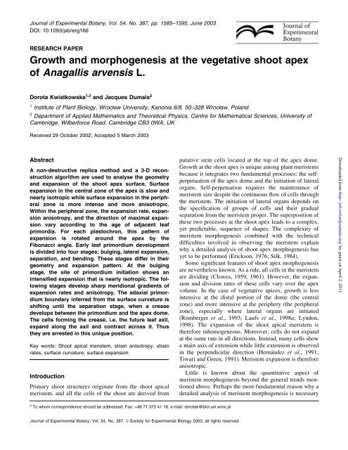
Journal <strong>of</strong> Experimental Botany, Vol. 54, No. 387, pp. 1585±1595, June 2003<br />
DOI: 10.1093/jxb/erg166<br />
RESEARCH PAPER<br />
<strong>Growth</strong> <strong>and</strong> <strong>morphogenesis</strong> <strong>at</strong> <strong>the</strong> veget<strong>at</strong>ive <strong>shoot</strong> <strong>apex</strong><br />
<strong>of</strong> Anagallis arvensis L.<br />
Dorota Kwi<strong>at</strong>kowska 1,3 <strong>and</strong> Jacques Dumais 2<br />
1 Institute <strong>of</strong> Plant Biology, Wrocøaw University, Kanonia 6/8, 50±328 Wrocøaw, Pol<strong>and</strong><br />
2 Department <strong>of</strong> Applied M<strong>at</strong>hem<strong>at</strong>ics <strong>and</strong> Theoretical Physics, Centre for M<strong>at</strong>hem<strong>at</strong>ical Sciences, University <strong>of</strong><br />
Cambridge, Wilberforce Road, Cambridge CB3 0WA, UK<br />
Received 29 October 2002; Accepted 5 March 2003<br />
Abstract<br />
A non-destructive replica method <strong>and</strong> a 3-D reconstruction<br />
algorithm are used to analyse <strong>the</strong> geometry<br />
<strong>and</strong> expansion <strong>of</strong> <strong>the</strong> <strong>shoot</strong> <strong>apex</strong> surface. Surface<br />
expansion in <strong>the</strong> central zone <strong>of</strong> <strong>the</strong> <strong>apex</strong> is slow <strong>and</strong><br />
nearly isotropic while surface expansion in <strong>the</strong> peripheral<br />
zone is more intense <strong>and</strong> more anisotropic.<br />
Within <strong>the</strong> peripheral zone, <strong>the</strong> expansion r<strong>at</strong>e, expansion<br />
anisotropy, <strong>and</strong> <strong>the</strong> direction <strong>of</strong> maximal expansion<br />
vary according to <strong>the</strong> age <strong>of</strong> adjacent leaf<br />
primordia. For each plastochron, this p<strong>at</strong>tern <strong>of</strong><br />
expansion is rot<strong>at</strong>ed around <strong>the</strong> <strong>apex</strong> by <strong>the</strong><br />
Fibonacci angle. Early leaf primordium development<br />
is divided into four stages: bulging, l<strong>at</strong>eral expansion,<br />
separ<strong>at</strong>ion, <strong>and</strong> bending. These stages differ in <strong>the</strong>ir<br />
geometry <strong>and</strong> expansion p<strong>at</strong>tern. At <strong>the</strong> bulging<br />
stage, <strong>the</strong> site <strong>of</strong> primordium initi<strong>at</strong>ion shows an<br />
intensi®ed expansion th<strong>at</strong> is nearly isotropic. The following<br />
stages develop sharp meridional gradients <strong>of</strong><br />
expansion r<strong>at</strong>es <strong>and</strong> anisotropy. The adaxial primordium<br />
boundary inferred from <strong>the</strong> surface curv<strong>at</strong>ure is<br />
shifting until <strong>the</strong> separ<strong>at</strong>ion stage, when a crease<br />
develops between <strong>the</strong> primordium <strong>and</strong> <strong>the</strong> <strong>apex</strong> dome.<br />
The cells forming <strong>the</strong> crease, i.e. <strong>the</strong> future leaf axil,<br />
exp<strong>and</strong> along <strong>the</strong> axil <strong>and</strong> contract across it. Thus<br />
<strong>the</strong>y are arrested in this unique position.<br />
Key words: Shoot apical meristem, strain anisotropy, strain<br />
r<strong>at</strong>es, surface curv<strong>at</strong>ure, surface expansion.<br />
Introduction<br />
Primary <strong>shoot</strong> structures origin<strong>at</strong>e from <strong>the</strong> <strong>shoot</strong> apical<br />
meristem, <strong>and</strong> all <strong>the</strong> cells <strong>of</strong> <strong>the</strong> <strong>shoot</strong> are derived from<br />
3 To whom correspondence should be addressed. Fax: +48 71 375 41 18. e-mail: dorotak@biol.uni.wroc.pl<br />
Journal <strong>of</strong> Experimental Botany, Vol. 54, No. 387, ã Society for Experimental Biology 2003; all rights reserved<br />
put<strong>at</strong>ive stem cells loc<strong>at</strong>ed <strong>at</strong> <strong>the</strong> top <strong>of</strong> <strong>the</strong> <strong>apex</strong> dome.<br />
<strong>Growth</strong> <strong>at</strong> <strong>the</strong> <strong>shoot</strong> <strong>apex</strong> is unique among plant meristems<br />
because it integr<strong>at</strong>es two fundamental processes: <strong>the</strong> selfperpetu<strong>at</strong>ion<br />
<strong>of</strong> <strong>the</strong> <strong>apex</strong> dome <strong>and</strong> <strong>the</strong> initi<strong>at</strong>ion <strong>of</strong> l<strong>at</strong>eral<br />
organs. Self-perpetu<strong>at</strong>ion requires <strong>the</strong> maintenance <strong>of</strong><br />
meristem size despite <strong>the</strong> continuous ¯ow <strong>of</strong> cells through<br />
<strong>the</strong> meristem. The initi<strong>at</strong>ion <strong>of</strong> l<strong>at</strong>eral organs depends on<br />
<strong>the</strong> speci®c<strong>at</strong>ion <strong>of</strong> groups <strong>of</strong> cells <strong>and</strong> <strong>the</strong>ir gradual<br />
separ<strong>at</strong>ion from <strong>the</strong> meristem proper. The superposition <strong>of</strong><br />
<strong>the</strong>se two processes <strong>at</strong> <strong>the</strong> <strong>shoot</strong> <strong>apex</strong> leads to a complex,<br />
yet predictable, sequence <strong>of</strong> shapes. The complexity <strong>of</strong><br />
meristem <strong>morphogenesis</strong> combined with <strong>the</strong> technical<br />
dif®culties involved in observing <strong>the</strong> meristem explain<br />
why a detailed analysis <strong>of</strong> <strong>shoot</strong> <strong>apex</strong> <strong>morphogenesis</strong> has<br />
yet to be performed (Erickson, 1976; Silk, 1984).<br />
Some signi®cant fe<strong>at</strong>ures <strong>of</strong> <strong>shoot</strong> <strong>apex</strong> <strong>morphogenesis</strong><br />
are never<strong>the</strong>less known. As a rule, all cells in <strong>the</strong> meristem<br />
are dividing (Clowes, 1959, 1961). However, <strong>the</strong> expansion<br />
<strong>and</strong> division r<strong>at</strong>es <strong>of</strong> <strong>the</strong>se cells vary over <strong>the</strong> <strong>apex</strong><br />
volume. In <strong>the</strong> case <strong>of</strong> veget<strong>at</strong>ive apices, growth is less<br />
intensive <strong>at</strong> <strong>the</strong> distal portion <strong>of</strong> <strong>the</strong> dome (<strong>the</strong> central<br />
zone) <strong>and</strong> more intensive <strong>at</strong> <strong>the</strong> periphery (<strong>the</strong> peripheral<br />
zone), especially where l<strong>at</strong>eral organs are initi<strong>at</strong>ed<br />
(Romberger et al., 1993; Laufs et al., 1998a; Lyndon,<br />
1998). The expansion <strong>of</strong> <strong>the</strong> <strong>shoot</strong> apical meristem is<br />
<strong>the</strong>refore inhomogeneous. Moreover, cells do not exp<strong>and</strong><br />
<strong>at</strong> <strong>the</strong> same r<strong>at</strong>e in all directions. Instead, many cells show<br />
a main axis <strong>of</strong> extension while little extension is observed<br />
in <strong>the</strong> perpendicular direction (HernaÂndez et al., 1991;<br />
Tiwari <strong>and</strong> Green, 1991). Meristem expansion is <strong>the</strong>refore<br />
anisotropic.<br />
Little is known about <strong>the</strong> quantit<strong>at</strong>ive aspect <strong>of</strong><br />
meristem <strong>morphogenesis</strong> beyond <strong>the</strong> general trends mentioned<br />
above. Perhaps <strong>the</strong> most fundamental reason why a<br />
detailed analysis <strong>of</strong> meristem <strong>morphogenesis</strong> is necessary<br />
Downloaded from<br />
http://jxb.oxfordjournals.org/ by guest on April 2, 2013
1586 Kwi<strong>at</strong>kowska <strong>and</strong> Dumais<br />
is th<strong>at</strong> a particular sequence <strong>of</strong> shapes can result from a<br />
wide range <strong>of</strong> growth p<strong>at</strong>terns <strong>at</strong> <strong>the</strong> cell level (Hejnowicz<br />
<strong>and</strong> Nakielski, 1979; Nakielski, 1982, 1987). The only way<br />
to address <strong>the</strong> role <strong>of</strong> individual cells in <strong>morphogenesis</strong> is<br />
to quantify <strong>the</strong>ir contribution directly. Such quantit<strong>at</strong>ive<br />
d<strong>at</strong>a about meristem <strong>morphogenesis</strong> would be an important<br />
addition to <strong>the</strong> molecular knowledge <strong>of</strong> this process. They<br />
would help us to underst<strong>and</strong> how known cytohistological<br />
zones (Clowes, 1961; Romberger et al., 1993) <strong>and</strong> gene<br />
expression domains (Bowman <strong>and</strong> Eshed, 2000; Br<strong>and</strong><br />
et al., 2001; Traas <strong>and</strong> Doonan, 2001) are maintained, even<br />
if <strong>the</strong> cells th<strong>at</strong> compose <strong>the</strong>se zones <strong>and</strong> domains are<br />
continually changing. In particular, quantit<strong>at</strong>ive d<strong>at</strong>a on <strong>the</strong><br />
r<strong>at</strong>e <strong>of</strong> cell migr<strong>at</strong>ion from one expression domain to <strong>the</strong><br />
next would indic<strong>at</strong>e how quickly cells need to acquire new<br />
`identities'.<br />
With <strong>the</strong>se applic<strong>at</strong>ions in mind, wh<strong>at</strong> would constitute<br />
an adequ<strong>at</strong>e quantit<strong>at</strong>ive description <strong>of</strong> <strong>morphogenesis</strong> <strong>at</strong><br />
<strong>the</strong> <strong>shoot</strong> apical meristem? Ideally, <strong>morphogenesis</strong> should<br />
be described in 3-D <strong>and</strong> with high sp<strong>at</strong>ial <strong>and</strong> temporal<br />
resolution. The two fundamental variables for any quantit<strong>at</strong>ive<br />
analysis <strong>of</strong> <strong>morphogenesis</strong> are <strong>the</strong> surface curv<strong>at</strong>ure<br />
<strong>and</strong> <strong>the</strong> surface strain r<strong>at</strong>es. Curv<strong>at</strong>ure is a measure <strong>of</strong><br />
<strong>the</strong> local geometry <strong>of</strong> <strong>the</strong> surface, while <strong>the</strong> strain r<strong>at</strong>es<br />
measure its rel<strong>at</strong>ive r<strong>at</strong>e <strong>of</strong> expansion. If surface curv<strong>at</strong>ure<br />
<strong>and</strong> strain r<strong>at</strong>es are to be compared with gene expression<br />
d<strong>at</strong>a, cellular resolution <strong>of</strong> <strong>the</strong>se variables is needed since<br />
<strong>the</strong> transition between gene expression domains can occur<br />
over one cell length. Morphogenesis <strong>at</strong> <strong>the</strong> <strong>shoot</strong> apical<br />
meristem repe<strong>at</strong>s itself <strong>at</strong> a regular time interval, <strong>the</strong><br />
plastochron, corresponding to <strong>the</strong> time required for <strong>the</strong><br />
initi<strong>at</strong>ion <strong>of</strong> successive leaves or groups <strong>of</strong> leaves <strong>at</strong><br />
<strong>the</strong> <strong>apex</strong> (Erickson <strong>and</strong> Michelini, 1957). While, for <strong>the</strong><br />
root meristem, a description <strong>of</strong> geometry <strong>at</strong> one instant is<br />
suf®cient to apprehend <strong>the</strong> entire morphogenetic process<br />
(Silk et al., 1989), for <strong>the</strong> <strong>shoot</strong> apical meristem <strong>the</strong><br />
description must span <strong>the</strong> whole plastochron. Therefore,<br />
protocols to investig<strong>at</strong>e <strong>morphogenesis</strong> <strong>at</strong> <strong>the</strong> <strong>shoot</strong> <strong>apex</strong><br />
must be able to resolve <strong>morphogenesis</strong> within a plastochron,<br />
which, in apices producing rel<strong>at</strong>ively large leaf<br />
primordia, usually lasts for a couple <strong>of</strong> days (Dale <strong>and</strong><br />
Milthorpe, 1983; Lyndon, 1998).<br />
This paper presents an analysis <strong>of</strong> surface expansion <strong>at</strong><br />
<strong>the</strong> <strong>shoot</strong> apical meristem. The protocol uses a noninvasive<br />
replica method (Williams <strong>and</strong> Green, 1988) <strong>and</strong> a<br />
new comput<strong>at</strong>ional approach th<strong>at</strong> includes 3-D reconstruction<br />
<strong>of</strong> <strong>the</strong> meristem surface (Dumais <strong>and</strong> Kwi<strong>at</strong>kowska,<br />
2002). This protocol allows <strong>the</strong> quanti®c<strong>at</strong>ion <strong>of</strong> <strong>the</strong><br />
geometry <strong>and</strong> expansion <strong>of</strong> <strong>the</strong> <strong>apex</strong> with rel<strong>at</strong>ively high<br />
temporal <strong>and</strong> sp<strong>at</strong>ial resolution. The d<strong>at</strong>a collected are used<br />
to address two fundamental questions concerning <strong>morphogenesis</strong><br />
<strong>at</strong> <strong>the</strong> <strong>shoot</strong> <strong>apex</strong>: (i) wh<strong>at</strong> is <strong>the</strong> surface<br />
expansion p<strong>at</strong>tern leading to <strong>the</strong> self-perpetu<strong>at</strong>ion <strong>of</strong> <strong>the</strong><br />
apical dome; <strong>and</strong> (ii) wh<strong>at</strong> is <strong>the</strong> surface expansion p<strong>at</strong>tern<br />
leading to partitioning <strong>of</strong> new leaf primordia from <strong>the</strong><br />
meristem proper. The investig<strong>at</strong>ion is performed on<br />
veget<strong>at</strong>ive <strong>shoot</strong>s <strong>of</strong> Anagallis arvensis L. (Primulaceae)<br />
exhibiting spiral Fibonacci phyllotaxis.<br />
M<strong>at</strong>erials <strong>and</strong> methods<br />
Plant m<strong>at</strong>erial<br />
Four <strong>shoot</strong> apices <strong>of</strong> A. arvensis were used in this investig<strong>at</strong>ion. With<br />
this m<strong>at</strong>erial, meristem <strong>morphogenesis</strong> could be followed over seven<br />
plastochrons. Two apices (<strong>the</strong> ®rst <strong>and</strong> <strong>the</strong> second <strong>apex</strong>) came from a<br />
plant collected on <strong>the</strong> Stanford University campus <strong>and</strong> kept in a<br />
growth chamber under a 16/8 h day/night cycle, a 15 W m ±2<br />
illumin<strong>at</strong>ion, <strong>and</strong> a temper<strong>at</strong>ure <strong>of</strong> 20±21 °C (Dumais <strong>and</strong><br />
Kwi<strong>at</strong>kowska, 2002). These conditions were non-inductive despite<br />
<strong>the</strong> fact th<strong>at</strong> A. arvensis is a long day plant (Brulfert et al., 1985),<br />
presumably because <strong>the</strong> light spectrum did not include <strong>the</strong> inductive<br />
wavelengths. The o<strong>the</strong>r two apices (including <strong>the</strong> third <strong>apex</strong><br />
presented here) origin<strong>at</strong>ed from a clone derived by means <strong>of</strong><br />
veget<strong>at</strong>ive propag<strong>at</strong>ion from a wild plant growing in <strong>the</strong> Sudety Mts.,<br />
Pol<strong>and</strong> (<strong>the</strong> same clone as used by Kwi<strong>at</strong>kowska, 1997). These<br />
plants were kept <strong>at</strong> a temper<strong>at</strong>ure ranging from 18±24 °C, in short<br />
days (10/14 h day/night) to prevent ¯owering, with an illumin<strong>at</strong>ion<br />
<strong>of</strong> 9 W m ±2 .<br />
D<strong>at</strong>a collection <strong>and</strong> analysis<br />
A non-destructive replica method (Williams <strong>and</strong> Green, 1988; Green<br />
et al., 1991; HernaÂndez et al., 1991) was used to obtain a<br />
developmental sequence for each <strong>apex</strong>. To expose <strong>the</strong> meristem<br />
surface, thin threads were glued to <strong>the</strong> tip <strong>of</strong> <strong>the</strong> young leaves<br />
overarching <strong>the</strong> meristem so th<strong>at</strong> <strong>the</strong>se leaves could be temporarily<br />
pulled away. The leaves were returned to <strong>the</strong>ir original position once<br />
<strong>the</strong> replica was completed. Replicas were made <strong>at</strong> 12±24 h time<br />
intervals <strong>and</strong> examined with scanning electron microscopy (Philips<br />
SEM505 <strong>and</strong> LEO435VP).<br />
Computer tools developed earlier (Dumais <strong>and</strong> Kwi<strong>at</strong>kowska,<br />
2002) were used to collect <strong>and</strong> analyse <strong>the</strong> morphogenetic d<strong>at</strong>a. The<br />
cell vertices, i.e. <strong>the</strong> points where three cells come into contact, were<br />
used as surface markers. For each member <strong>of</strong> a developmental<br />
sequence, two scanning electron micrographs were taken, one tilted<br />
by 10° with respect to <strong>the</strong> o<strong>the</strong>r. The x±y position <strong>of</strong> vertices was<br />
collected manually from one <strong>of</strong> <strong>the</strong> images, while for <strong>the</strong> o<strong>the</strong>r image<br />
<strong>the</strong> vertex loc<strong>at</strong>ions were found with an area m<strong>at</strong>ching protocol.<br />
Since <strong>the</strong> two images <strong>of</strong>fer slightly different perspectives <strong>of</strong> <strong>the</strong> same<br />
structure, <strong>the</strong> distances between vertices differed in <strong>the</strong>se images.<br />
This shift in <strong>the</strong> x±y position <strong>of</strong> vertices was used to determine <strong>the</strong><br />
elev<strong>at</strong>ion (<strong>the</strong> z position) <strong>of</strong> <strong>the</strong> vertices.<br />
The curv<strong>at</strong>ure <strong>of</strong> <strong>the</strong> meristem surface was computed from <strong>the</strong><br />
reconstructed meristem. A quadr<strong>at</strong>ic surface was ®rst ®tted to <strong>the</strong><br />
vertices <strong>of</strong> a given cell <strong>and</strong> its contacting neighbours. The local<br />
curv<strong>at</strong>ure could <strong>the</strong>n be computed from <strong>the</strong> coef®cients <strong>of</strong> this<br />
quadr<strong>at</strong>ic surface. The comput<strong>at</strong>ion <strong>of</strong> strain r<strong>at</strong>es requires <strong>the</strong><br />
identi®c<strong>at</strong>ion <strong>of</strong> corresponding vertices on consecutive replicas <strong>of</strong><br />
<strong>the</strong> growing meristem. This was done with a clonal analysis in which<br />
cells <strong>and</strong> <strong>the</strong>ir progeny were identi®ed in consecutive images, like<br />
<strong>the</strong> exemplary cells highlighted in Fig. 1A±E. The strain r<strong>at</strong>es <strong>at</strong> a<br />
given vertex were <strong>the</strong>n computed from <strong>the</strong> deform<strong>at</strong>ion <strong>of</strong> <strong>the</strong><br />
triangle de®ned by <strong>the</strong> three vertices in direct contact with th<strong>at</strong><br />
vertex. The overall strain r<strong>at</strong>es for <strong>the</strong> cell were computed as <strong>the</strong><br />
weighted sum <strong>of</strong> <strong>the</strong> strain r<strong>at</strong>es <strong>at</strong> <strong>the</strong> cell's vertices. A graphical<br />
user interface <strong>and</strong> computer programs written in M<strong>at</strong>lab (The<br />
M<strong>at</strong>hworks, N<strong>at</strong>ick, MA, USA) can be downloaded <strong>at</strong>: http://<br />
culex.biol.uni.wroc.pl/instbot/dorotak.<br />
Downloaded from<br />
http://jxb.oxfordjournals.org/ by guest on April 2, 2013
Results<br />
Similarity <strong>of</strong> <strong>the</strong> developmental sequences con®rms<br />
<strong>the</strong> validity <strong>of</strong> quantit<strong>at</strong>ive d<strong>at</strong>a<br />
Although <strong>the</strong> manipul<strong>at</strong>ions <strong>of</strong> <strong>the</strong> meristem required to<br />
make replicas do not involve any wounding, <strong>the</strong>y could<br />
never<strong>the</strong>less disrupt <strong>morphogenesis</strong>. Inspection <strong>of</strong> <strong>the</strong><br />
m<strong>at</strong>erial collected indic<strong>at</strong>es th<strong>at</strong> <strong>the</strong> geometry <strong>of</strong> <strong>the</strong><br />
meristem has not been affected. For example, casts <strong>of</strong><br />
apices <strong>at</strong> <strong>the</strong> same plastochron stage are strikingly similar.<br />
This is true whe<strong>the</strong>r replicas from <strong>the</strong> same <strong>apex</strong>, fur<strong>the</strong>r<br />
referred to as members <strong>of</strong> <strong>the</strong> same developmental<br />
sequence (e.g. Fig. 1A, E), or from different apices (e.g.<br />
Figs 1A, 2C) are compared. The meristem manipul<strong>at</strong>ions<br />
could also have slowed down <strong>the</strong> r<strong>at</strong>e <strong>of</strong> growth, which<br />
would lead to a longer plastochron. Such an effect on<br />
growth was indeed observed in some sequences excluded<br />
from this investig<strong>at</strong>ion. An increase in <strong>the</strong> length <strong>of</strong> <strong>the</strong><br />
plastochron can also be seen in <strong>the</strong> sequence published in<br />
Fig. 5 <strong>of</strong> Green et al. (1991). A careful comparison <strong>of</strong> <strong>the</strong><br />
observed plastochron is <strong>the</strong>refore in order. The estim<strong>at</strong>ed<br />
plastochrons for <strong>the</strong> sequences presented in this paper fall<br />
between 40 h <strong>and</strong> 45 h, irrespective <strong>of</strong> <strong>the</strong> origin <strong>of</strong> <strong>the</strong><br />
plant m<strong>at</strong>erial <strong>and</strong> <strong>the</strong> growth conditions. The m<strong>at</strong>erial<br />
presented is <strong>the</strong>refore consistent <strong>at</strong> th<strong>at</strong> level. Precise<br />
measurements <strong>of</strong> <strong>the</strong> plastochron were also published by<br />
o<strong>the</strong>r authors (Ballard <strong>and</strong> Grant Lipp, 1964; Brulfert et al.,<br />
1985), for decuss<strong>at</strong>e veget<strong>at</strong>ive <strong>shoot</strong>s <strong>of</strong> A. arvensis kept<br />
in <strong>the</strong> growth conditions similar to <strong>the</strong> plants used here.<br />
Again, this plastochron is consistent with <strong>the</strong> measured<br />
plastochrons for decuss<strong>at</strong>e <strong>shoot</strong>s from which replicas<br />
were made (results not shown). The plastochron for <strong>shoot</strong>s<br />
with decuss<strong>at</strong>e phyllotaxis is more than twice as long as<br />
<strong>shoot</strong>s with spiral phyllotaxis, but not surprisingly since in<br />
decuss<strong>at</strong>e phyllotaxis two leaf primordia are initi<strong>at</strong>ed<br />
simultaneously. Therefore, half r<strong>at</strong>her than <strong>the</strong> whole<br />
plastochron characteristic for decuss<strong>at</strong>e <strong>shoot</strong>s should be<br />
compared with th<strong>at</strong> <strong>of</strong> spiral ones (Rutishauser, 1998).<br />
Variables describing <strong>the</strong> geometry <strong>and</strong> expansion <strong>of</strong><br />
<strong>the</strong> <strong>shoot</strong> <strong>apex</strong> surface<br />
The geometrical analysis yields <strong>the</strong> principal curv<strong>at</strong>ures<br />
<strong>and</strong> <strong>the</strong> Gaussian curv<strong>at</strong>ure for each cell on <strong>the</strong> reconstructed<br />
meristem surface. It should be emphasized th<strong>at</strong> <strong>the</strong><br />
curv<strong>at</strong>ure values reported do not refer to individual outer<br />
walls <strong>of</strong> <strong>the</strong> cells but r<strong>at</strong>her to <strong>the</strong> curv<strong>at</strong>ure <strong>of</strong> <strong>the</strong> smooth<br />
surface de®ning <strong>the</strong> overall geometry <strong>of</strong> <strong>the</strong> meristem. The<br />
principal curv<strong>at</strong>ures are <strong>the</strong> extreme curv<strong>at</strong>ure values,<br />
<strong>at</strong>tained on a surface along principal curv<strong>at</strong>ure directions.<br />
In plots <strong>of</strong> <strong>the</strong> meristem curv<strong>at</strong>ure (<strong>the</strong> second column in<br />
Figs 1±3), <strong>the</strong> principal curv<strong>at</strong>ure directions for each cell<br />
are illustr<strong>at</strong>ed by <strong>the</strong> two arms <strong>of</strong> a cross. The length <strong>of</strong><br />
<strong>the</strong>se arms is proportional to <strong>the</strong> curv<strong>at</strong>ure in <strong>the</strong>ir<br />
direction. If in <strong>the</strong> arm direction <strong>the</strong> surface is convex,<br />
<strong>the</strong> cross arm appears in black; if <strong>the</strong> surface is concave,<br />
<strong>Growth</strong> <strong>of</strong> <strong>the</strong> <strong>shoot</strong> <strong>apex</strong> surface 1587<br />
<strong>the</strong> arm is red. The Gaussian curv<strong>at</strong>ure is <strong>the</strong> product <strong>of</strong> <strong>the</strong><br />
principal curv<strong>at</strong>ures <strong>and</strong> shows how much a surface differs<br />
from being ¯<strong>at</strong>. If a surface is ¯<strong>at</strong> or can be ¯<strong>at</strong>tened<br />
without any tears or folds, its Gaussian curv<strong>at</strong>ure is zero. A<br />
concave or convex surface th<strong>at</strong> tears when ¯<strong>at</strong>tened, for<br />
example, a hemisphere, has a positive Gaussian curv<strong>at</strong>ure.<br />
A surface shaped like a saddle, which will form folds, if<br />
¯<strong>at</strong>tened, has a neg<strong>at</strong>ive Gaussian curv<strong>at</strong>ure. In curv<strong>at</strong>ure<br />
plots, <strong>the</strong> Gaussian curv<strong>at</strong>ure is indic<strong>at</strong>ed by <strong>the</strong> colour<br />
assigned to each cell (Figs 1±3). The warmer <strong>the</strong> colour,<br />
<strong>the</strong> higher is <strong>the</strong> curv<strong>at</strong>ure.<br />
The growth analysis yields <strong>the</strong> principal strain r<strong>at</strong>es ( : e1<br />
<strong>and</strong> : e2) <strong>and</strong> directions, areal strain r<strong>at</strong>e, <strong>and</strong> strain r<strong>at</strong>e<br />
anisotropy. The strain r<strong>at</strong>es are <strong>the</strong> rel<strong>at</strong>ive r<strong>at</strong>es <strong>of</strong><br />
extension (Erickson, 1966, 1976; Hejnowicz <strong>and</strong><br />
Romberger, 1984; Silk, 1984; Goodall <strong>and</strong> Green, 1986;<br />
Dumais <strong>and</strong> Kwi<strong>at</strong>kowska, 2002). If surface expansion is<br />
anisotropic, <strong>the</strong>se r<strong>at</strong>es assessed in different directions are<br />
not <strong>the</strong> same. There are two unique mutually orthogonal<br />
directions on <strong>the</strong> surface, in which strain r<strong>at</strong>es <strong>at</strong>tain<br />
extremal, minimal <strong>and</strong> maximal, values. These directions<br />
are <strong>the</strong> principal strain r<strong>at</strong>e directions. The extremal values<br />
are called principal strain r<strong>at</strong>es. The crosses plotted for<br />
each cell in <strong>the</strong> third column in Figs 1±3 indic<strong>at</strong>e <strong>the</strong><br />
principal strain r<strong>at</strong>e directions. The length <strong>of</strong> cross arms is<br />
proportional to <strong>the</strong> corresponding principal strain r<strong>at</strong>e.<br />
Positive strain r<strong>at</strong>es (extension) are indic<strong>at</strong>ed with black<br />
cross arms, neg<strong>at</strong>ive strain r<strong>at</strong>es (contraction) are in red.<br />
The principal strain r<strong>at</strong>e directions used in <strong>the</strong> present paper<br />
are <strong>the</strong> same as directions pointed by <strong>the</strong> `strain crosses'<br />
introduced by Goodall <strong>and</strong> Green (1986), <strong>and</strong> <strong>the</strong>oretically<br />
de®ned principal directions <strong>of</strong> growth (Hejnowicz <strong>and</strong><br />
Romberger, 1984). The areal strain r<strong>at</strong>e is computed as <strong>the</strong><br />
sum <strong>of</strong> <strong>the</strong> principal strain r<strong>at</strong>es <strong>and</strong> is indic<strong>at</strong>ed in <strong>the</strong><br />
strain r<strong>at</strong>e plots by <strong>the</strong> cell colour. The warmer <strong>the</strong> colour,<br />
<strong>the</strong> higher is <strong>the</strong> areal strain r<strong>at</strong>e. The strain r<strong>at</strong>e anisotropy,<br />
given by <strong>the</strong> equ<strong>at</strong>ion ( : :<br />
e1 e2†=… : e1 ‡ : e2), shows how much<br />
<strong>the</strong> two principal strain r<strong>at</strong>es differ from each o<strong>the</strong>r.<br />
Geometry <strong>and</strong> growth <strong>of</strong> <strong>the</strong> apical dome are not<br />
uniform <strong>and</strong> change during <strong>the</strong> plastochron<br />
The apical dome is always in contact with three leaf<br />
primordia <strong>of</strong> various ages. The Gaussian curv<strong>at</strong>ure <strong>of</strong> <strong>the</strong><br />
central zone <strong>of</strong> <strong>the</strong> dome is positive but close to zero<br />
(<strong>the</strong> second column in Figs 1, 2). The curv<strong>at</strong>ure <strong>of</strong> <strong>the</strong><br />
peripheral zone depends on <strong>the</strong> plastochron stage <strong>and</strong> <strong>the</strong><br />
age <strong>of</strong> <strong>the</strong> contacting leaf primordium. At <strong>the</strong> beginning <strong>of</strong><br />
<strong>the</strong> plastochron, a new leaf primordium becomes discernible<br />
within <strong>the</strong> peripheral zone as a region where <strong>the</strong><br />
Gaussian curv<strong>at</strong>ure is locally higher <strong>and</strong> minimal <strong>and</strong><br />
maximal curv<strong>at</strong>ures are very similar (e.g. P1 in Fig. 2F).<br />
Leaf initi<strong>at</strong>ion sites become apparent before any signs <strong>of</strong><br />
bulging can be seen on scanning electron micrographs <strong>of</strong><br />
<strong>the</strong> <strong>apex</strong> surface (compare P1 in micrograph in Fig. 2C<br />
with <strong>the</strong> curv<strong>at</strong>ure plot in Fig. 2F). During <strong>the</strong> second half<br />
Downloaded from<br />
http://jxb.oxfordjournals.org/ by guest on April 2, 2013
1588 Kwi<strong>at</strong>kowska <strong>and</strong> Dumais<br />
Downloaded from<br />
http://jxb.oxfordjournals.org/<br />
by guest on April 2, 2013
<strong>of</strong> <strong>the</strong> plastochron, a narrow b<strong>and</strong>, one to three cells wide,<br />
becomes visible within <strong>the</strong> peripheral zone along <strong>the</strong><br />
recently initi<strong>at</strong>ed primordium (e.g. along P3 in Fig. 1G).<br />
<strong>Growth</strong> <strong>of</strong> <strong>the</strong> <strong>shoot</strong> <strong>apex</strong> surface 1589<br />
These cells are curved almost exclusively in <strong>the</strong> direction<br />
<strong>of</strong> <strong>the</strong> primordium margin. Thus a typical cross <strong>of</strong> principal<br />
curv<strong>at</strong>ures is reduced to a l<strong>at</strong>itudinal line segment. At <strong>the</strong><br />
Fig. 2. Scanning electron micrographs (A±C), curv<strong>at</strong>ure (D±F), <strong>and</strong> strain r<strong>at</strong>e plots (G±H) for <strong>the</strong> second <strong>apex</strong>. Labelling as in Fig. 1. This<br />
sequence covers 36 h, which is slightly less than one plastochron. Primordia are initi<strong>at</strong>ed in a counter-clockwise order. The sequence begins<br />
during <strong>the</strong> ®rst half <strong>of</strong> <strong>the</strong> plastochron.<br />
Fig. 1. Scanning electron micrographs (A±E), curv<strong>at</strong>ure plots (F±J), <strong>and</strong> strain r<strong>at</strong>e plots (K±N) for <strong>the</strong> developmental sequence <strong>of</strong> <strong>the</strong> ®rst <strong>shoot</strong><br />
<strong>apex</strong>. The time <strong>at</strong> which <strong>the</strong> replica was made is given in <strong>the</strong> lower right corner <strong>of</strong> each micrograph. Leaf primordia are numbered from <strong>the</strong> youngest<br />
primordium observed in <strong>the</strong> sequence to <strong>the</strong> oldest. Each primordium is labelled as soon as it becomes discernible as a region <strong>of</strong> locally elev<strong>at</strong>ed<br />
Gaussian curv<strong>at</strong>ure. Cells included in <strong>the</strong> analysis are outlined in black on <strong>the</strong> micrographs. An exemplary cell <strong>and</strong> its progeny are outlined in red in<br />
all <strong>the</strong> sequence members. The colour map for <strong>the</strong> curv<strong>at</strong>ure plots represents <strong>the</strong> Gaussian curv<strong>at</strong>ure measured for a cell <strong>and</strong> its adjacent neighbours.<br />
Note th<strong>at</strong> all <strong>the</strong> measurements were made in 3-D but are here presented as 2-D projections for clarity. The Gaussian curv<strong>at</strong>ure on <strong>the</strong> scale bar is<br />
given in 10 ±4 mm ±2 . The orient<strong>at</strong>ion <strong>and</strong> length <strong>of</strong> <strong>the</strong> cross arms indic<strong>at</strong>e <strong>the</strong> direction <strong>and</strong> magnitude <strong>of</strong> <strong>the</strong> principal curv<strong>at</strong>ures, respectively. The<br />
arm appears in red if <strong>the</strong> surface in its direction bends upward (concave). Areal strain r<strong>at</strong>es <strong>and</strong> strain r<strong>at</strong>e crosses are plotted for each cell on <strong>the</strong><br />
plot <strong>of</strong> cell outlines as <strong>the</strong>y appeared <strong>at</strong> <strong>the</strong> beginning <strong>of</strong> <strong>the</strong> considered time interval. The colour map represents <strong>the</strong> areal strain r<strong>at</strong>e measured in<br />
units <strong>of</strong> 10 ±2 h ±1 . Cross arms are oriented along principal strain r<strong>at</strong>e directions; <strong>the</strong>ir length is proportional to <strong>the</strong> corresponding principal strain r<strong>at</strong>es.<br />
The arms appear in red if neg<strong>at</strong>ive strain r<strong>at</strong>e, i.e. contraction, took place in <strong>the</strong>ir direction. Regions th<strong>at</strong> will give rise to a leaf primordium during<br />
<strong>the</strong> following time interval are outlined in black. Additional lines are added along <strong>the</strong> primordium margin to deline<strong>at</strong>e <strong>the</strong> future leaf axil. The<br />
sequence covers 83 h, which is about two plastochrons, <strong>and</strong> starts during <strong>the</strong> ®rst half <strong>of</strong> <strong>the</strong> plastochron. The second plastochron begins <strong>at</strong> <strong>the</strong> third<br />
sequence member. Primordia are formed in a clockwise order. Two <strong>of</strong> <strong>the</strong>m, P1 <strong>and</strong> P0, are initi<strong>at</strong>ed within <strong>the</strong> time <strong>of</strong> observ<strong>at</strong>ion.<br />
Downloaded from<br />
http://jxb.oxfordjournals.org/ by guest on April 2, 2013
1590 Kwi<strong>at</strong>kowska <strong>and</strong> Dumais<br />
Fig. 3. Developmental sequence <strong>of</strong> a primordium formed on <strong>the</strong> ¯anks <strong>of</strong> <strong>the</strong> third <strong>shoot</strong> <strong>apex</strong>. Scanning electron micrographs (A±D), curv<strong>at</strong>ure<br />
(E±H), <strong>and</strong> strain r<strong>at</strong>e (I±K) plots are labelled as in Fig. 1. The sequence covers 70 h, starting from <strong>the</strong> l<strong>at</strong>eral expansion stage <strong>of</strong> P1 up to <strong>the</strong><br />
bending stage. Slightly less than <strong>the</strong> ®rst two plastochrons <strong>of</strong> <strong>the</strong> primordium development can be followed.<br />
same time a part <strong>of</strong> <strong>the</strong> peripheral zone where <strong>the</strong> next<br />
primordium will be initi<strong>at</strong>ed <strong>at</strong>tains a slightly higher<br />
curv<strong>at</strong>ure (e.g. <strong>the</strong> upper right portion <strong>of</strong> <strong>the</strong> dome in<br />
Fig. 2E). During <strong>the</strong> following plastochron <strong>the</strong> cells<br />
forming <strong>the</strong> b<strong>and</strong> <strong>of</strong> zero Gaussian curv<strong>at</strong>ure give rise to<br />
<strong>the</strong> primordium axil (compare cells loc<strong>at</strong>ed along <strong>the</strong> P3<br />
margin in Fig. 2D, E). In <strong>the</strong> present paper <strong>the</strong> term axil<br />
cells is referring to cells, which have <strong>the</strong> most neg<strong>at</strong>ive<br />
meridional curv<strong>at</strong>ure <strong>of</strong> all <strong>the</strong> cells loc<strong>at</strong>ed near <strong>the</strong><br />
primordium margin. In <strong>the</strong> curv<strong>at</strong>ure plots <strong>the</strong>y have <strong>the</strong><br />
longest red arms, such as <strong>the</strong> cells on <strong>the</strong> margin <strong>of</strong><br />
primordium P3 in Fig. 2F. The parts <strong>of</strong> <strong>the</strong> peripheral zone<br />
contacting <strong>the</strong> axil (e.g. <strong>at</strong> <strong>the</strong> contact with P4 in Fig. 1F<br />
<strong>and</strong> G) are usually saddle-shaped: convex along <strong>the</strong> dome<br />
circumference (black cross arms), <strong>and</strong> slightly concave<br />
along its meridian (red cross arms). Between <strong>the</strong> regions<br />
contacting leaf primordia, <strong>the</strong> peripheral zone cells are<br />
curved predominantly in <strong>the</strong> meridional direction (between<br />
P2 <strong>and</strong> P3 in Fig. 1H).<br />
The expansion <strong>of</strong> <strong>the</strong> <strong>apex</strong> dome surface is non-uniform<br />
<strong>and</strong> generally anisotropic (<strong>the</strong> third column in Fig. 1 <strong>and</strong><br />
2). In terms <strong>of</strong> surface expansion, <strong>the</strong> central zone <strong>and</strong> <strong>the</strong><br />
peripheral zone merge into each o<strong>the</strong>r gradually. Areal<br />
strain r<strong>at</strong>es are <strong>the</strong> lowest <strong>and</strong> strain is nearly isotropic in<br />
<strong>the</strong> central zone, while in <strong>the</strong> peripheral zone both strain<br />
anisotropy <strong>and</strong> areal strain r<strong>at</strong>es are generally higher.<br />
Downloaded from<br />
http://jxb.oxfordjournals.org/ by guest on April 2, 2013
Fig. 4. Leaf primordia deline<strong>at</strong>ed on plots <strong>of</strong> cell outlines <strong>of</strong> <strong>the</strong> last<br />
sequence member in which a given primordium was observed. The<br />
lines <strong>of</strong> various colours are primordium boundaries as recognized in<br />
consecutive members <strong>of</strong> <strong>the</strong> sequence. Corresponding member<br />
numbers are: red line ± member 1; blue ± 2; green ± 3; black ± 4;<br />
orange ± 5. Primordia labels are <strong>the</strong> same as in <strong>the</strong> micrographs <strong>of</strong> <strong>the</strong><br />
sequences. (A) P1 <strong>and</strong> P2 <strong>of</strong> <strong>the</strong> ®rst <strong>apex</strong> (refer to Fig. 1) plotted on<br />
cell outlines <strong>of</strong> member 5 <strong>of</strong> <strong>the</strong> ®rst developmental sequence. No<br />
crease has yet been formed on P1 margins <strong>and</strong> its deline<strong>at</strong>ion is not<br />
stable. The boundary <strong>of</strong> P2 is stable starting from member 4 (black<br />
line), where shifts occur only on <strong>the</strong> axil sides. (B) Outlines <strong>of</strong><br />
primordia P2 <strong>and</strong> P3 shown on member 3 <strong>of</strong> <strong>the</strong> sequence for <strong>the</strong><br />
second <strong>apex</strong> (refer to Fig. 2). The boundary <strong>of</strong> P3 is stable starting<br />
from member 2 (blue line). P2 has not yet been deline<strong>at</strong>ed from <strong>the</strong><br />
meristem by a crease. (C) Outlines <strong>of</strong> primordium P1 shown on<br />
member 4 <strong>of</strong> <strong>the</strong> third sequence (refer to Fig. 3). The boundary <strong>of</strong> P1<br />
is unstable in members 1±2 (red <strong>and</strong> blue lines), <strong>and</strong> become stable<br />
when <strong>the</strong> crease appears in member 2.<br />
However, expansion <strong>of</strong> <strong>the</strong> peripheral zone is not uniform.<br />
It is slow <strong>and</strong> not strongly anisotropic in <strong>the</strong> region<br />
adjacent to <strong>the</strong> recently formed primordium (e.g. <strong>the</strong> cells<br />
adjacent to P2 in Fig. 1L±M), where maximum strain r<strong>at</strong>es<br />
<strong>Growth</strong> <strong>of</strong> <strong>the</strong> <strong>shoot</strong> <strong>apex</strong> surface 1591<br />
are l<strong>at</strong>itudinal, i.e. in <strong>the</strong> direction parallel to <strong>the</strong><br />
primordium margin. The above-mentioned b<strong>and</strong> <strong>of</strong> cells<br />
having nearly zero Gaussian curv<strong>at</strong>ure gives rise to <strong>the</strong> leaf<br />
axil exp<strong>and</strong>ing along <strong>the</strong> primordium margin (l<strong>at</strong>itudinally)<br />
<strong>and</strong> contracting across it (meridionally, like <strong>the</strong> cells<br />
along P3 in Fig. 1L). The expansion <strong>of</strong> <strong>the</strong> formed axil<br />
cells is similar (like cells <strong>of</strong> <strong>the</strong> axil <strong>of</strong> P3 in Fig. 1M or P3<br />
in Fig. 2H). The area <strong>of</strong> <strong>the</strong>se cells is almost not changing,<br />
or slightly diminishing. Where <strong>the</strong> dome ¯anks are<br />
rebuilding <strong>at</strong> <strong>the</strong> contact with <strong>the</strong> primordium axil (for<br />
example, adjacent to <strong>the</strong> axil <strong>of</strong> P3 in Fig. 1M), areal strain<br />
r<strong>at</strong>es <strong>and</strong> anisotropy are high. There <strong>the</strong> maximal strain<br />
r<strong>at</strong>e is in <strong>the</strong> meridional direction. This direction is<br />
perpendicular to <strong>the</strong> maximal strain r<strong>at</strong>e direction <strong>of</strong> <strong>the</strong><br />
adjacent axil cells. The region contacting <strong>the</strong> axil is <strong>the</strong><br />
fastest growing portion <strong>of</strong> <strong>the</strong> peripheral zone during<br />
<strong>the</strong> ®rst half <strong>of</strong> <strong>the</strong> plastochron (Fig. 1M). In <strong>the</strong> second<br />
half <strong>of</strong> <strong>the</strong> plastochron, an additional region <strong>of</strong> increased<br />
areal strain r<strong>at</strong>e appears <strong>at</strong> <strong>the</strong> initi<strong>at</strong>ion site <strong>of</strong> <strong>the</strong> next<br />
primordium. This region covers a portion <strong>of</strong> <strong>the</strong> peripheral<br />
zone larger than is actually contributing to <strong>the</strong> form<strong>at</strong>ion <strong>of</strong><br />
a new primordium (e.g. around P1 in Fig. 1L). When <strong>the</strong><br />
next plastochron begins, <strong>the</strong> cycle is repe<strong>at</strong>ed but rot<strong>at</strong>ed<br />
by <strong>the</strong> Fibonacci angle around <strong>the</strong> <strong>apex</strong>, e.g. by about<br />
137.5° clockwise between members <strong>of</strong> <strong>the</strong> ®rst sequence<br />
shown in Fig. 1K <strong>and</strong> M.<br />
Emerging leaf primordia show increasing gradients <strong>of</strong><br />
curv<strong>at</strong>ure <strong>and</strong> strain r<strong>at</strong>e<br />
Four developmental stages can be distinguished in <strong>the</strong><br />
course <strong>of</strong> <strong>the</strong> primordium form<strong>at</strong>ion covered in this study.<br />
These are: (i) <strong>the</strong> bulging stage; (ii) <strong>the</strong> l<strong>at</strong>eral expansion<br />
stage; (iii) <strong>the</strong> separ<strong>at</strong>ion stage; <strong>and</strong> (iv) <strong>the</strong> bending stage.<br />
The ®rst two stages cover about one plastochron. The third<br />
one lasts nearly half <strong>of</strong> a plastochron; whereas <strong>the</strong> fourth<br />
one extends beyond <strong>the</strong> period covered in this study.<br />
Primordium P2 from <strong>the</strong> ®rst sequence (Fig. 1) <strong>and</strong><br />
primordium P1 from <strong>the</strong> third one (Fig. 3) can illustr<strong>at</strong>e all<br />
<strong>the</strong> stages. In <strong>the</strong> ®rst stage, <strong>the</strong> leaf buttress is<br />
characterized by locally elev<strong>at</strong>ed Gaussian curv<strong>at</strong>ure (P2<br />
in Fig. 1F±H). The buttress surface is exp<strong>and</strong>ing with high<br />
areal strain r<strong>at</strong>es <strong>and</strong> nearly isotropically (P2 in Fig. 1K,<br />
L). The region <strong>of</strong> increased expansion sometimes covers<br />
more than <strong>the</strong> surface <strong>of</strong> <strong>the</strong> emerging primordium (e.g. P2<br />
in Fig. 1L). During <strong>the</strong> second stage, <strong>the</strong> buttress <strong>at</strong>tains an<br />
oval shape (P2 in Fig. 1I; P1 in Fig. 3E). Cells on <strong>the</strong><br />
primordium surface exp<strong>and</strong> mostly l<strong>at</strong>itudinally, while<br />
some contraction may take place in <strong>the</strong> meridional<br />
direction (P2 in Fig. 1M). The cell area, however, is still<br />
increasing since <strong>the</strong> l<strong>at</strong>itudinal expansion exceeds <strong>the</strong><br />
meridional contraction. During <strong>the</strong> third stage (P2 in<br />
Fig. 1J; P1 in Fig. 3F), <strong>the</strong> buttress becomes separ<strong>at</strong>ed from<br />
<strong>the</strong> dome by <strong>the</strong> axil. At <strong>the</strong> same time, <strong>the</strong> primordium<br />
surface exp<strong>and</strong>s similarly to <strong>the</strong> preceding stage. Finally in<br />
<strong>the</strong> fourth stage (P1 in Fig. 3G, H), a ¯<strong>at</strong>tened primordium<br />
Downloaded from<br />
http://jxb.oxfordjournals.org/ by guest on April 2, 2013
1592 Kwi<strong>at</strong>kowska <strong>and</strong> Dumais<br />
is formed with an adaxial surface <strong>of</strong> neg<strong>at</strong>ive Gaussian<br />
curv<strong>at</strong>ure <strong>and</strong> an upper portion <strong>of</strong> high positive curv<strong>at</strong>ure.<br />
The adaxial surface is concave meridionally <strong>and</strong> convex<br />
l<strong>at</strong>itudinally. At this stage <strong>the</strong> primordium starts to<br />
overarch <strong>the</strong> <strong>apex</strong> (like P1 in Fig. 3G, H). The fourth<br />
stage is characterized by differential growth. The expansion<br />
<strong>of</strong> <strong>the</strong> upper leaf portion where <strong>the</strong> Gaussian curv<strong>at</strong>ure<br />
becomes rel<strong>at</strong>ively high is intensive <strong>and</strong> not strongly<br />
anisotropic (for example, <strong>the</strong> outermost cells <strong>of</strong> P1 in<br />
Fig. 3J, K). The adaxial surface <strong>of</strong> <strong>the</strong> primordium bends<br />
towards <strong>the</strong> dome. Its cells exp<strong>and</strong> rel<strong>at</strong>ively slowly <strong>and</strong><br />
with high strain r<strong>at</strong>e anisotropy (see remaining cells <strong>of</strong> <strong>the</strong><br />
above-mentioned primordium). Some meridional contraction<br />
is observed on <strong>the</strong> adaxial side <strong>of</strong> <strong>the</strong> primordium,<br />
while <strong>the</strong> maximal strain r<strong>at</strong>e direction is parallel to <strong>the</strong><br />
leaf axil.<br />
Leaf primordium boundaries inferred from <strong>the</strong> surface<br />
curv<strong>at</strong>ure are unstable with respect to cells during early<br />
leaf development, i.e. during <strong>the</strong> bulging <strong>and</strong> l<strong>at</strong>eral<br />
expansion stages. The boundaries are shifting during <strong>the</strong>se<br />
two stages by one or two rows <strong>of</strong> cells. Both an<br />
incorpor<strong>at</strong>ion <strong>of</strong> new cells into <strong>the</strong> primordium (e.g. P1<br />
in Fig. 4A) <strong>and</strong>, less <strong>of</strong>ten, a reduction <strong>of</strong> <strong>the</strong> primordium<br />
area can take place (P2 in Fig. 4A). Starting from <strong>the</strong> third<br />
developmental stage, <strong>the</strong> displacements <strong>of</strong> <strong>the</strong> primordium<br />
boundaries are signi®cantly reduced. This corresponds to<br />
<strong>the</strong> time when <strong>the</strong> leaf axil is formed between <strong>the</strong> apical<br />
dome <strong>and</strong> <strong>the</strong> leaf primordium. Subsequently, shifting <strong>of</strong><br />
<strong>the</strong> primordium boundaries is restricted to <strong>the</strong> l<strong>at</strong>eral sides<br />
<strong>of</strong> <strong>the</strong> primordium (right side <strong>of</strong> P1 in Fig. 4C), where <strong>the</strong><br />
Gaussian curv<strong>at</strong>ure is neg<strong>at</strong>ive but closer to zero than in<br />
<strong>the</strong> middle <strong>of</strong> <strong>the</strong> axil. Sometimes, starting from <strong>the</strong><br />
separ<strong>at</strong>ion stage, <strong>the</strong> boundaries do not move <strong>at</strong> all (P3 in<br />
Fig. 4B).<br />
Discussion<br />
A non-invasive replica method <strong>and</strong> a 3-D reconstruction<br />
algorithm were used to quantify <strong>the</strong> geometry <strong>and</strong> surface<br />
expansion <strong>of</strong> <strong>the</strong> <strong>shoot</strong> <strong>apex</strong> <strong>of</strong> A. arvensis. The results<br />
were analysed in <strong>the</strong> light <strong>of</strong> two fundamental processes<br />
occurring <strong>at</strong> <strong>the</strong> <strong>shoot</strong> <strong>apex</strong>: <strong>the</strong> self-perpetu<strong>at</strong>ion <strong>of</strong> <strong>the</strong><br />
apical dome <strong>and</strong> <strong>the</strong> form<strong>at</strong>ion <strong>of</strong> leaf primordia. The<br />
observed changes in <strong>the</strong> geometry <strong>and</strong> expansion <strong>of</strong> a leaf<br />
primordium surface were characteristic enough to differenti<strong>at</strong>e<br />
early leaf development into distinct stages. These<br />
stages could be regarded as represent<strong>at</strong>ive for <strong>the</strong> early<br />
development <strong>of</strong> dicot leaves, <strong>of</strong> which <strong>the</strong> leaf <strong>of</strong> A.<br />
arvensis is a typical example.<br />
Self-perpetu<strong>at</strong>ion <strong>of</strong> <strong>the</strong> veget<strong>at</strong>ive <strong>shoot</strong> apical dome<br />
The overall p<strong>at</strong>tern <strong>of</strong> expansion in <strong>the</strong> <strong>shoot</strong> <strong>apex</strong> is<br />
characterized by a gradual increase in <strong>the</strong> r<strong>at</strong>e <strong>of</strong> expansion<br />
<strong>and</strong> expansion anisotropy as one goes from <strong>the</strong> central<br />
zone to <strong>the</strong> peripheral zone. These observ<strong>at</strong>ions are in<br />
agreement with earlier studies <strong>of</strong> <strong>shoot</strong> <strong>apex</strong> growth<br />
(Newman, 1956; Soma <strong>and</strong> Ball, 1963; Hussey, 1971;<br />
Green et al., 1991; Lyndon, 1998; Laufs et al., 1998a).<br />
This particular p<strong>at</strong>tern <strong>of</strong> expansion, however, is not<br />
unique in its ability to maintain <strong>the</strong> size <strong>and</strong> dome-like<br />
shape <strong>of</strong> <strong>the</strong> <strong>shoot</strong> apical meristem (Hejnowicz <strong>and</strong><br />
Nakielski, 1979; Nakielski, 1987). Opposite gradients <strong>of</strong><br />
expansion were observed on <strong>the</strong> dome <strong>of</strong> tip-growing cells<br />
(Hejnowicz et al., 1977; Shaw et al., 2000) <strong>and</strong> <strong>the</strong>re is no<br />
geometrical reason why a similar expansion p<strong>at</strong>tern would<br />
not work in <strong>the</strong> <strong>shoot</strong> apical meristem. A possible<br />
biological explan<strong>at</strong>ion for <strong>the</strong> slow growth <strong>of</strong> <strong>the</strong> central<br />
zone is th<strong>at</strong> it helps <strong>the</strong> plant preserve <strong>the</strong> genetic m<strong>at</strong>erial<br />
<strong>of</strong> reproductive cells which will origin<strong>at</strong>e from this region<br />
(Hejnowicz <strong>and</strong> Nakielski, 1979; Klekowski, 1988). If <strong>the</strong><br />
r<strong>at</strong>e <strong>of</strong> expansion is low, <strong>the</strong> cells will divide less<br />
frequently thus reducing <strong>the</strong> likelihood th<strong>at</strong> replic<strong>at</strong>ion<br />
errors will occur. An altern<strong>at</strong>ive explan<strong>at</strong>ion is th<strong>at</strong> <strong>the</strong><br />
high r<strong>at</strong>e <strong>of</strong> growth in <strong>the</strong> peripheral zone results from <strong>the</strong><br />
rapid initi<strong>at</strong>ion <strong>of</strong> l<strong>at</strong>eral organs. This explan<strong>at</strong>ion is<br />
incomplete since in <strong>the</strong> pin1 mutant <strong>of</strong> A. thaliana <strong>the</strong><br />
in¯orescence <strong>apex</strong> does not initi<strong>at</strong>e l<strong>at</strong>eral organs <strong>and</strong> yet<br />
<strong>the</strong> mitotic index remains rel<strong>at</strong>ively high in <strong>the</strong> peripheral<br />
zone (Vernoux et al., 2000).<br />
The peripheral zone is differenti<strong>at</strong>ed into regions whose<br />
distribution is rel<strong>at</strong>ed to <strong>the</strong> youngest three leaf primordia.<br />
Similar sectors, comprising <strong>the</strong> peripheral zone <strong>of</strong> <strong>the</strong><br />
<strong>shoot</strong> <strong>apex</strong> <strong>of</strong> Clethra barbinervis Sieb. et Zucc. exhibiting<br />
spiral phyllotaxis, were reported by Hara (1971) who<br />
determined <strong>the</strong>m based on <strong>the</strong> anticlinal cell walls p<strong>at</strong>tern.<br />
In <strong>the</strong> <strong>apex</strong> <strong>of</strong> A. arvensis, <strong>the</strong> geometry <strong>and</strong> growth <strong>of</strong><br />
<strong>the</strong>se sectors were shown to depend on <strong>the</strong> age <strong>of</strong> <strong>the</strong><br />
adjacent leaf primordium. The sectors differ in <strong>the</strong>ir<br />
Gaussian curv<strong>at</strong>ure <strong>and</strong> in <strong>the</strong>ir expansion r<strong>at</strong>es <strong>and</strong><br />
anisotropy. Even <strong>the</strong> direction <strong>of</strong> maximal strain r<strong>at</strong>e in<br />
adjacent sectors may be entirely different. The site <strong>of</strong> leaf<br />
primordium initi<strong>at</strong>ion is characterized by locally intensi-<br />
®ed growth. Interestingly, <strong>the</strong> region <strong>of</strong> high strain r<strong>at</strong>e<br />
also includes cells th<strong>at</strong> will not contribute to <strong>the</strong> new<br />
primordium. For each plastochron, <strong>the</strong> growth p<strong>at</strong>tern is<br />
rot<strong>at</strong>ed around <strong>the</strong> <strong>apex</strong> by a ®xed angle (<strong>the</strong> Fibonacci<br />
angle). The temporal <strong>and</strong> sp<strong>at</strong>ial complexity <strong>of</strong> <strong>the</strong> dome<br />
expansion is a consequence <strong>of</strong> <strong>the</strong> fact th<strong>at</strong> <strong>the</strong> continuous<br />
initi<strong>at</strong>ion <strong>of</strong> leaf primordia in a regular but asymmetric<br />
p<strong>at</strong>tern is superimposed on <strong>the</strong> self-perpetu<strong>at</strong>ion <strong>of</strong> <strong>the</strong><br />
dome.<br />
Form<strong>at</strong>ion <strong>of</strong> <strong>the</strong> leaf axilÐpartitioning <strong>of</strong> <strong>the</strong> <strong>shoot</strong><br />
<strong>apex</strong><br />
The leaf axil is unique among <strong>the</strong> different regions <strong>of</strong> <strong>the</strong><br />
meristem, both in its geometry <strong>and</strong> growth. The meridional<br />
gradients <strong>of</strong> curv<strong>at</strong>ure, areal strain r<strong>at</strong>e, <strong>and</strong> strain r<strong>at</strong>e<br />
anisotropy are especially sharp across this region. The<br />
expansion observed in <strong>the</strong> leaf axil is similar to <strong>the</strong><br />
expansion observed during <strong>the</strong> form<strong>at</strong>ion <strong>of</strong> <strong>the</strong> radial<br />
Downloaded from<br />
http://jxb.oxfordjournals.org/ by guest on April 2, 2013
creases between stamens in A. arvensis ¯owers<br />
(HernaÂndez et al., 1991), <strong>and</strong> also <strong>the</strong> creases delimiting<br />
organs in adventitious <strong>shoot</strong>s <strong>of</strong> Graptopetalum paraguayense<br />
E. Wal<strong>the</strong>r (Tiwari <strong>and</strong> Green, 1991). The<br />
expansion <strong>of</strong> <strong>the</strong> future axil cells is very slow, <strong>and</strong> takes<br />
place only along <strong>the</strong> axil as observed also by Green et al.<br />
(1991). Thus, <strong>the</strong> cells forming <strong>the</strong> axil are arrested in this<br />
particular position. This observ<strong>at</strong>ion may explain why<br />
Soma <strong>and</strong> Ball (1963), as well as Hussey (1971), noted th<strong>at</strong><br />
some <strong>of</strong> <strong>the</strong> markers applied to <strong>the</strong> <strong>apex</strong> surface were not<br />
moving out <strong>of</strong> <strong>the</strong> young leaf axils. Ano<strong>the</strong>r consequence<br />
<strong>of</strong> such a p<strong>at</strong>tern <strong>of</strong> cell expansion is th<strong>at</strong> its appearance on<br />
<strong>the</strong> adaxial margins <strong>of</strong> a leaf primordium signi®cantly<br />
reduces <strong>the</strong> displacement <strong>of</strong> <strong>the</strong> primordium boundary, i.e.<br />
it partitions <strong>the</strong> <strong>shoot</strong> <strong>apex</strong> surface into <strong>the</strong> dome <strong>and</strong> new<br />
leaf primordium.<br />
Early development <strong>of</strong> <strong>the</strong> leaf primordium<br />
In this study, <strong>the</strong> course <strong>of</strong> leaf primordium form<strong>at</strong>ion is<br />
divided into four stages. These stages cover <strong>the</strong> ®rst<br />
(`organogenesis') stage <strong>and</strong> a part <strong>of</strong> <strong>the</strong> second (`early<br />
growth <strong>of</strong> <strong>the</strong> primordium') stage <strong>of</strong> leaf development<br />
de®ned by Sylvester et al. (1996). Distinct changes in<br />
primordium geometry <strong>and</strong> growth allow <strong>the</strong> ®rst, `organogenesis'<br />
stage to be resolved. It comprises <strong>the</strong> bulging,<br />
<strong>the</strong> l<strong>at</strong>eral expansion, <strong>and</strong> <strong>the</strong> separ<strong>at</strong>ion stages <strong>of</strong> <strong>the</strong><br />
present paper, while <strong>the</strong> bending stage is <strong>at</strong> <strong>the</strong> beginning<br />
<strong>of</strong> <strong>the</strong> stage <strong>of</strong> `early growth'. During <strong>the</strong> bulging stage,<br />
growth <strong>of</strong> <strong>the</strong> primordium is rel<strong>at</strong>ively intensive <strong>and</strong><br />
isotropic. The cytoplasm <strong>of</strong> cells comprising <strong>the</strong> primordium<br />
<strong>at</strong> this stage, visible in longitudinal sections <strong>of</strong> <strong>the</strong><br />
<strong>apex</strong>, stains more intensely than <strong>the</strong> cytoplasm <strong>of</strong> o<strong>the</strong>r<br />
meristem cells (Fontaine, 1972). The intense staining is<br />
ano<strong>the</strong>r manifest<strong>at</strong>ion <strong>of</strong> <strong>the</strong> high meristem<strong>at</strong>ic activity <strong>of</strong><br />
<strong>the</strong>se cells. By contrast with <strong>the</strong> bulging stage, l<strong>at</strong>eral<br />
expansion <strong>of</strong> <strong>the</strong> primordium surface relies on rel<strong>at</strong>ively<br />
low areal strain r<strong>at</strong>es, but high strain r<strong>at</strong>e anisotropy. L<strong>at</strong>er,<br />
during primordium development, sharp meridional gradients<br />
<strong>of</strong> strain r<strong>at</strong>es <strong>and</strong> anisotropy appear, <strong>and</strong> <strong>the</strong><br />
primordium surface becomes differenti<strong>at</strong>ed into regions<br />
<strong>of</strong> various expansion <strong>and</strong> geometry.<br />
The control <strong>of</strong> areal strain r<strong>at</strong>es <strong>and</strong> strain r<strong>at</strong>e<br />
anisotropy<br />
One important conclusion from this work is th<strong>at</strong> <strong>morphogenesis</strong><br />
<strong>at</strong> <strong>the</strong> <strong>shoot</strong> <strong>apex</strong> depends on sharp gradients <strong>of</strong><br />
areal strain r<strong>at</strong>es as well as on anisotropy <strong>of</strong> strain r<strong>at</strong>es.<br />
One should <strong>the</strong>refore ask how <strong>the</strong>se gradients <strong>and</strong><br />
anisotropy are cre<strong>at</strong>ed <strong>and</strong> maintained <strong>at</strong> <strong>the</strong> cell <strong>and</strong><br />
molecular levels.<br />
Areal strain r<strong>at</strong>e is scalar, th<strong>at</strong> is, any given point <strong>of</strong> <strong>the</strong><br />
meristem surface has a single areal strain r<strong>at</strong>e value. Its<br />
level could be directly regul<strong>at</strong>ed by a scalar factor, such as<br />
<strong>the</strong> concentr<strong>at</strong>ion <strong>of</strong> gene products or growth regul<strong>at</strong>ors<br />
(Hejnowicz <strong>and</strong> Romberger, 1984). In <strong>the</strong> apical dome, <strong>the</strong><br />
<strong>Growth</strong> <strong>of</strong> <strong>the</strong> <strong>shoot</strong> <strong>apex</strong> surface 1593<br />
distribution <strong>of</strong> such factors can be regul<strong>at</strong>ed by genes<br />
whose expression is speci®c to <strong>the</strong> peripheral or central<br />
zone (Nishimura et al., 1999; Zondlo <strong>and</strong> Irish, 1999).<br />
Some <strong>of</strong> <strong>the</strong>se genes, such as CLAVATA <strong>and</strong> MGOUN, are<br />
known to regul<strong>at</strong>e <strong>the</strong> size <strong>of</strong> <strong>the</strong>se meristem zones (Laufs<br />
et al., 1998a, b; Traas <strong>and</strong> Doonan, 2001). The primordium<br />
initi<strong>at</strong>ion site, in turn, shows an elev<strong>at</strong>ed expression <strong>of</strong> <strong>the</strong><br />
expansin gene (Reinhardt et al., 1998). Also, exogenous<br />
applic<strong>at</strong>ion <strong>of</strong> expansin to <strong>the</strong> <strong>apex</strong> surface can induce leaflike<br />
outgrowths (Fleming et al., 1999), while local<br />
induction <strong>of</strong> expansin expression can induce <strong>the</strong> entire<br />
process <strong>of</strong> leaf form<strong>at</strong>ion (Pien et al., 2001). The<br />
intensi®ed areal strain r<strong>at</strong>e observed <strong>at</strong> leaf initi<strong>at</strong>ion<br />
may result from a local loosening <strong>of</strong> cell walls on <strong>the</strong> <strong>apex</strong><br />
surface by secreted expansin proteins (Fleming et al.,<br />
1999).<br />
Future axil cells presumably also express speci®c genes,<br />
although <strong>the</strong> exact sp<strong>at</strong>ial <strong>and</strong> temporal rel<strong>at</strong>ionships<br />
between gene expression domains <strong>and</strong> future axil cells<br />
remain unknown. The expression domains <strong>of</strong> CUP-<br />
SHAPED COTYLEDON in Arabidopsis thaliana (L.)<br />
Heynh. (Aida et al., 1999; Br<strong>and</strong> et al., 2001; Traas <strong>and</strong><br />
Doonan, 2001), NO APICAL MERISTEM in Petunia sp.<br />
hybrids (Souer et al., 1996), <strong>and</strong> various OSH genes in<br />
Oryza s<strong>at</strong>iva L. (Sentoku et al., 1999) are correl<strong>at</strong>ed<br />
with primordium boundaries. The CUP-SHAPED<br />
COTYLEDON2 gene is thought to act as a local growth<br />
suppressor (Traas <strong>and</strong> Doonan, 2001). Theoretically, a<br />
crease can be formed by locally lowering <strong>the</strong> areal strain<br />
(Todd, 1986). Therefore, some <strong>of</strong> <strong>the</strong> above-mentioned<br />
genes may be directly involved in <strong>the</strong> control <strong>of</strong> axil<br />
form<strong>at</strong>ion by locally inhibiting cell expansion.<br />
Unlike areal strain r<strong>at</strong>es, <strong>the</strong> strain r<strong>at</strong>e anisotropy must<br />
be regul<strong>at</strong>ed by factors <strong>of</strong> a similar, anisotropic n<strong>at</strong>ure<br />
(Hejnowicz <strong>and</strong> Romberger, 1984). The signi®cance <strong>of</strong><br />
anisotropic factors in <strong>morphogenesis</strong> is supported by <strong>the</strong><br />
fact th<strong>at</strong> dividing plant cells seem to `perceive' stress or<br />
growth anisotropy existing in <strong>the</strong>ir environment (Miller,<br />
1980; Hejnowicz, 1984; Lintilhac <strong>and</strong> Vesecky, 1984;<br />
Lynch <strong>and</strong> Lintilhac, 1997). Nei<strong>the</strong>r <strong>the</strong> concentr<strong>at</strong>ion <strong>of</strong><br />
gene products (scalars), nor <strong>the</strong> concentr<strong>at</strong>ion gradients <strong>of</strong><br />
such gene products (vectors) are anisotropic factors. The<br />
l<strong>at</strong>ter have only a single direction de®ned toge<strong>the</strong>r with a<br />
corresponding scalar value. By contrast, cellular structures<br />
such as <strong>the</strong> cytoskeleton or <strong>the</strong> cell wall <strong>of</strong>ten show<br />
anisotropy. A connection between cytoskeletal arrangement,<br />
cell wall reinforcement, <strong>and</strong> anisotropic cell expansion<br />
has been shown (see for example Selker <strong>and</strong> Green,<br />
1984; Tomos et al., 1989; Green, 1992; Bichet et al.,<br />
2001). In <strong>the</strong> case <strong>of</strong> cell walls, a non-r<strong>and</strong>om alignment <strong>of</strong><br />
cellulose micro®brils results in differences in mechanical<br />
properties <strong>of</strong> walls along different directions. Cell walls<br />
are mechanically reinforced in <strong>the</strong> direction <strong>of</strong> cellulose<br />
alignment. Since <strong>the</strong> outer walls <strong>of</strong> tunica cells in <strong>the</strong> <strong>shoot</strong><br />
apical meristem are <strong>the</strong> thickest <strong>of</strong> all <strong>the</strong> meristem cell<br />
Downloaded from<br />
http://jxb.oxfordjournals.org/ by guest on April 2, 2013
1594 Kwi<strong>at</strong>kowska <strong>and</strong> Dumais<br />
walls, <strong>the</strong> reinforcement <strong>of</strong> <strong>the</strong>se particular walls is a good<br />
c<strong>and</strong>id<strong>at</strong>e for <strong>the</strong> direct control <strong>of</strong> <strong>the</strong> <strong>apex</strong> surface growth<br />
(Green, 1986; Green <strong>and</strong> Selker, 1991). Combining <strong>the</strong><br />
study <strong>of</strong> cell wall mechanics with quantit<strong>at</strong>ive d<strong>at</strong>a on <strong>the</strong><br />
<strong>morphogenesis</strong> <strong>at</strong> <strong>the</strong> <strong>shoot</strong> apical meristem may help to<br />
underst<strong>and</strong> how meristem growth is controlled.<br />
Acknowledgements<br />
The authors thank Dr Zygmunt Hejnowicz (Silesian University,<br />
Pol<strong>and</strong>) for critical reading <strong>of</strong> <strong>the</strong> manuscript <strong>and</strong> Drs Zo®a Czarna<br />
<strong>and</strong> Krystyna Heller (Electron Microscopy Labor<strong>at</strong>ory, Wrocøaw<br />
University <strong>of</strong> Agricultural Sciences, Pol<strong>and</strong>) for help in preparing<br />
some <strong>of</strong> <strong>the</strong> scanning electron micrographs used in this work. Part <strong>of</strong><br />
this research was ®nanced by a research grant No. 3P04C 015 22<br />
from <strong>the</strong> Polish Committee for Scienti®c Research to DK <strong>and</strong> a NSF<br />
grant to <strong>the</strong> l<strong>at</strong>e Paul Green. DK acknowledges additional support<br />
from <strong>the</strong> Fulbright Found<strong>at</strong>ion while visiting Stanford University.<br />
JD acknowledges support from <strong>the</strong> Center for Comput<strong>at</strong>ional<br />
Genetics <strong>and</strong> Biological Modeling (Stanford University).<br />
References<br />
Aida M, Ishida T, Tasaka M. 1999. Shoot apical meristem <strong>and</strong><br />
cotyledon form<strong>at</strong>ion during Arabidopsis embryogenesis:<br />
interaction among <strong>the</strong> CUP-SHAPED COTYLEDON <strong>and</strong><br />
SHOOT MERISTEMLESS genes. Development 126, 1563±1570.<br />
Ballard LAT, Grant Lipp AE. 1964. Juvenile photoperiodic<br />
sensitivity in Anagallis arvensis L. subsp. feomina (Mill.) Schinz<br />
<strong>and</strong> Thell. Australian Journal <strong>of</strong> Biological Sciencess 17, 323±<br />
337.<br />
Bichet A, Desnos T, Turner S, Gr<strong>and</strong>jean O, HoÈfte H. 2001.<br />
BOTERO1 is required for normal orient<strong>at</strong>ion <strong>of</strong> cortical<br />
microtubules <strong>and</strong> anisotropic cell expansion in Arabidopsis. The<br />
Plant Journal 25, 137±148.<br />
Bowman JL, Eshed Y. 2000. Form<strong>at</strong>ion <strong>and</strong> maintenance <strong>of</strong> <strong>the</strong><br />
<strong>shoot</strong> apical meristem. Trends in Plant Science 5, 110±115.<br />
Br<strong>and</strong> U, Hobe M, Simon R. 2001. Functional domains in plant<br />
<strong>shoot</strong> meristems. BioEssays 23, 134±141.<br />
Brulfert J, Fontaine D, Imh<strong>of</strong>f C. 1985. Anagallis arvensis. In:<br />
Halevy AH, ed. CRC h<strong>and</strong>book <strong>of</strong> ¯owering. Boca R<strong>at</strong>on: CRC<br />
Press, 334±349.<br />
Clowes FAL. 1959. Adenine incorpor<strong>at</strong>ion <strong>and</strong> cell division in<br />
<strong>shoot</strong> apices. New Phytologist 58, 16±19.<br />
Clowes FAL. 1961. Apical meristems. Oxford: Blackwell Scienti®c<br />
Public<strong>at</strong>ions.<br />
Dale JE, Milthorpe FL. 1983. General fe<strong>at</strong>ures <strong>of</strong> <strong>the</strong> production<br />
<strong>and</strong> growth <strong>of</strong> leaves. In: Dale JE, Milthorpe FL, eds. The growth<br />
<strong>and</strong> functioning <strong>of</strong> leaves. Cambridge: Cambridge University<br />
Press, 151±178.<br />
Dumais J, Kwi<strong>at</strong>kowska D. 2002. Analysis <strong>of</strong> surface growth in<br />
<strong>shoot</strong> apices. The Plant Journal 31, 229±241.<br />
Erickson RO. 1966. Rel<strong>at</strong>ive elemental r<strong>at</strong>es <strong>and</strong> anisotropy <strong>of</strong><br />
growth in area: a computer program. Journal <strong>of</strong> Experimental<br />
Botany 17, 390±403.<br />
Erickson RO. 1976. Modeling <strong>of</strong> plant growth. Annual Review <strong>of</strong><br />
Plant Physiology 27, 407±434.<br />
Erickson RO, Michelini FJ. 1957. The plastochron index.<br />
American Journal <strong>of</strong> Botany 44, 297±305.<br />
Fleming AJ, Caderas D, Wehrli E, McQueen-Mason S,<br />
Kuhlemeier C. 1999. Analysis <strong>of</strong> expansin-induced<br />
<strong>morphogenesis</strong> on <strong>the</strong> apical meristem <strong>of</strong> tom<strong>at</strong>o. Planta 208,<br />
166±174.<br />
Fontaine D. 1972. Diagnostic morphologique du stade de<br />
deÂveloppement, au cours du plastochrone, du point veÂgeÂt<strong>at</strong>if de<br />
l'Anagallis arvensis L. plante aÁ feuilles opposeÂes et deÂcusseÂes.<br />
Comptes Rendus hebdomadaires des SeÂances de l'AcadeÂmie des<br />
Sciences, Paris 274 SeÂrie D, 58±61.<br />
Goodall CR, Green PB. 1986. Quantit<strong>at</strong>ive analysis <strong>of</strong> surface<br />
growth. Botanical Gazette 147, 1±15.<br />
Green PB. 1986. Plasticity in <strong>shoot</strong> development: a biophysical<br />
view. In: Jennings DH, Trewavas AJ, eds. Plasticity in plants.<br />
Cambridge: Society for Experimental Biology, 211±232.<br />
Green PB. 1992. P<strong>at</strong>tern form<strong>at</strong>ion in <strong>shoot</strong>s: a likely role for<br />
minimal energy con®gur<strong>at</strong>ions <strong>of</strong> <strong>the</strong> tunica. Intern<strong>at</strong>ional<br />
Journal <strong>of</strong> Plant Sciences 153, 59±75.<br />
Green PB, Havelange A, Bernier G. 1991. Floral <strong>morphogenesis</strong><br />
in Anagallis: Scanning-electron-micrograph sequences from<br />
individual growing meristems before, during, <strong>and</strong> after <strong>the</strong><br />
transition to ¯owering. Planta 185, 502±512.<br />
Green PB, Selker JML. 1991. Mutual alignments <strong>of</strong> cell walls,<br />
cellulose, <strong>and</strong> cytoskeletons: <strong>the</strong>ir role in meristems. In: Lloyd<br />
CW, ed. The cytoskeletal basis <strong>of</strong> plant growth <strong>and</strong> form. London:<br />
Academic Press, 303±322.<br />
Hara N. 1971. Structure <strong>of</strong> <strong>the</strong> veget<strong>at</strong>ive <strong>shoot</strong> <strong>apex</strong> <strong>of</strong> Clethra<br />
barbinervis. III. Longisectional view, summary analysis <strong>and</strong><br />
discussion. Botanical Magazine, Tokyo 84, 283±292.<br />
Hejnowicz Z. 1984. Trajectories <strong>of</strong> principal directions <strong>of</strong> growth,<br />
n<strong>at</strong>ural coordin<strong>at</strong>e system in growing plant organ. Acta Societ<strong>at</strong>is<br />
Botanicorum Poloniae 53, 301±316.<br />
Hejnowicz Z, Nakielski J. 1979. Modeling <strong>of</strong> growth in <strong>shoot</strong><br />
apical dome. Acta Societ<strong>at</strong>is Botanicorum Poloniae 48, 423±442.<br />
Hejnowicz Z, Romberger JA. 1984. <strong>Growth</strong> tensor <strong>of</strong> plant<br />
organs. Journal <strong>of</strong> Theoretical Biology 110, 93±114.<br />
Hejnowicz Z, Heinemann B, Sievers A. 1977. Tip growth:<br />
p<strong>at</strong>terns <strong>of</strong> growth r<strong>at</strong>e <strong>and</strong> stress in <strong>the</strong> Chara rhizoid. Zeitschrift<br />
fuÈr P¯anzenphysiologie 81, 409±424.<br />
HernaÂndez LF, Havelange A, Bernier G, Green PB. 1991.<br />
<strong>Growth</strong> behavior <strong>of</strong> single epidermal cells during ¯ower<br />
form<strong>at</strong>ion: sequential scanning electron micrographs provide<br />
kinem<strong>at</strong>ic p<strong>at</strong>terns for Anagallis. Planta 185, 139±147.<br />
Hussey G. 1971. Cell division <strong>and</strong> expansion <strong>and</strong> resultant tissue<br />
tension in <strong>the</strong> <strong>shoot</strong> <strong>apex</strong> during <strong>the</strong> form<strong>at</strong>ion <strong>of</strong> a leaf<br />
primordium in <strong>the</strong> tom<strong>at</strong>o. Journal <strong>of</strong> Experimental Botany 22,<br />
702±714.<br />
Klekowski EJ. 1988. Mut<strong>at</strong>ion, developmental selection, <strong>and</strong> plant<br />
evolution. New York: Columbia University Press.<br />
Kwi<strong>at</strong>kowska D. 1997. Intraspeci®c vari<strong>at</strong>ion <strong>of</strong> phyllotactic<br />
stability in Anagallis arvensis. Acta Societ<strong>at</strong>is Botanicorum<br />
Poloniae 66, 259±271.<br />
Laufs P, Dockx J, Kronenberger J, Traas J. 1998b. MGOUN1<br />
<strong>and</strong> MGOUN2: two genes required for primordium initi<strong>at</strong>ion <strong>at</strong><br />
<strong>the</strong> <strong>shoot</strong> apical <strong>and</strong> ¯oral meristems in Arabidopsis thaliana.<br />
Development 125, 1253±1260.<br />
Laufs P, Gr<strong>and</strong>jean O, Jonak C, Kieu K, Traas J. 1998a.<br />
Cellular parameters <strong>of</strong> <strong>the</strong> <strong>shoot</strong> apical meristem in Arabidopsis.<br />
The Plant Cell 10, 1375±1389.<br />
Lintilhac PM, Vesecky TB. 1984. Stress-induced alignment <strong>of</strong><br />
division plane in plant tissues grown in vitro. N<strong>at</strong>ure 307, 363±<br />
364.<br />
Lynch TM, Lintilhac PM. 1997. Mechanical signals in plant<br />
development: a new method for single cell studies.<br />
Developmental Biology 181, 246±256.<br />
Lyndon RF. 1998. The <strong>shoot</strong> apical meristem. Cambridge:<br />
Cambridge University Press.<br />
Miller JH. 1980. Orient<strong>at</strong>ion <strong>of</strong> <strong>the</strong> plane <strong>of</strong> cell division in fern<br />
gametophytes: <strong>the</strong> roles <strong>of</strong> cell shape <strong>and</strong> stress. American<br />
Journal <strong>of</strong> Botany 67, 534±542.<br />
Nakielski J. 1982. Geometry <strong>of</strong> <strong>shoot</strong> apical dome <strong>and</strong> distribution<br />
Downloaded from<br />
http://jxb.oxfordjournals.org/ by guest on April 2, 2013
<strong>of</strong> growth r<strong>at</strong>es. Acta Societ<strong>at</strong>is Botanicorum Poloniae 51, 389±<br />
402.<br />
Nakielski J. 1987. Sp<strong>at</strong>ial vari<strong>at</strong>ion <strong>of</strong> growth within domes having<br />
different p<strong>at</strong>terns <strong>of</strong> principal growth directions. Acta Societ<strong>at</strong>is<br />
Botanicorum Poloniae 56, 611±623.<br />
Newman IV. 1956. P<strong>at</strong>tern in meristems <strong>of</strong> vascular plants. 1. Cell<br />
partition in living apices <strong>and</strong> in <strong>the</strong> cambial zone in rel<strong>at</strong>ion to <strong>the</strong><br />
concepts <strong>of</strong> initial cells <strong>and</strong> apical cells. Phytomorphology 6,<br />
1±19.<br />
Nishimura A, Tamaoki M, S<strong>at</strong>o Y, M<strong>at</strong>suoka M. 1999. The<br />
expression <strong>of</strong> tobacco knotted1-type class 1 homeobox genes<br />
corresponds to regions predicted by <strong>the</strong> cytohistological zon<strong>at</strong>ion<br />
model. The Plant Journal 18, 337±347.<br />
Pien S, Wyrzykowska J, McQueen-Mason S, Smart C, Fleming<br />
A. 2001. Local expression <strong>of</strong> expansin induces <strong>the</strong> entire process<br />
<strong>of</strong> leaf development <strong>and</strong> modi®es leaf shape. Proceedings <strong>of</strong> <strong>the</strong><br />
N<strong>at</strong>ional Academy <strong>of</strong> Sciences, USA 98, 11812±11817.<br />
Reinhardt D, Wittwer F, M<strong>and</strong>el T, Kuhlemeier C. 1998.<br />
Localized upregul<strong>at</strong>ion <strong>of</strong> a new expansion gene predicts <strong>the</strong> site<br />
<strong>of</strong> leaf form<strong>at</strong>ion in <strong>the</strong> tom<strong>at</strong>o meristem. The Plant Cell 10,<br />
1427±1437.<br />
Romberger JA, Hejnowicz Z, Hill JF. 1993. Plant structure:<br />
function <strong>and</strong> development. Berlin, New York: Springer Verlag.<br />
Rutishauser R. 1998. Plastochron r<strong>at</strong>io <strong>and</strong> leaf arc as parameters<br />
<strong>of</strong> a quantit<strong>at</strong>ive phyllotaxis analysis in vascular plants. In: Jean<br />
RV, Barabe D, eds. Symmetry in plants. Singapore: World<br />
Scienti®c Public<strong>at</strong>ions, 171±212.<br />
Selker JML, Green PB. 1984. Organogenesis in Graptopetalum<br />
paraguayense E. Wal<strong>the</strong>r: shifts in orient<strong>at</strong>ion <strong>of</strong> cortical<br />
microtubule arrays are associ<strong>at</strong>ed with periclinal divisions.<br />
Planta 160, 289±297.<br />
Sentoku N, S<strong>at</strong>o Y, Kur<strong>at</strong>a N, Ito Y, Kitano H, M<strong>at</strong>suoka M.<br />
1999. Regional expression <strong>of</strong> <strong>the</strong> rice KN1-type homeobox gene<br />
family during embryo, <strong>shoot</strong>, <strong>and</strong> ¯ower development. The Plant<br />
Cell 11, 1651±1663.<br />
Shaw SL, Dumais J, Long SR. 2000. Cell surface expansion in<br />
<strong>Growth</strong> <strong>of</strong> <strong>the</strong> <strong>shoot</strong> <strong>apex</strong> surface 1595<br />
polarly growing root hairs <strong>of</strong> Medicago trunc<strong>at</strong>ula. Plant<br />
Physiology 124, 959±969.<br />
Silk WK. 1984. Quantit<strong>at</strong>ive descriptions <strong>of</strong> development. Annual<br />
Review <strong>of</strong> Plant Physiology 35, 479±518.<br />
Silk WK, Lord EM, Eckard KJ. 1989. <strong>Growth</strong> p<strong>at</strong>terns inferred<br />
from an<strong>at</strong>omical records: empirical tests using longisections <strong>of</strong><br />
roots <strong>of</strong> Zea mays L. Plant Physiology 90, 708±713.<br />
Soma K, Ball E. 1963. Studies <strong>of</strong> <strong>the</strong> surface growth <strong>of</strong> <strong>the</strong> <strong>shoot</strong><br />
<strong>apex</strong> <strong>of</strong> Lupinus albus. Brookhaven Symposia in Biology 16,<br />
13±45.<br />
Souer E, van Houwelingen A, Kloos D, Nol J, Koes R. 1996. The<br />
no apical meristem gene <strong>of</strong> Petunia is required for p<strong>at</strong>tern<br />
form<strong>at</strong>ion in embryos <strong>and</strong> ¯owers <strong>and</strong> is expressed <strong>at</strong> meristem<br />
<strong>and</strong> primordia boundaries. Cell 85, 159±170.<br />
Sylvester AW, Smith L, Freeling M. 1996. Acquisition <strong>of</strong> identity<br />
in <strong>the</strong> developing leaf. Annual Review <strong>of</strong> Cell Developmental<br />
Biology 12, 257±304.<br />
Tiwari SC, Green PB. 1991. Shoot initi<strong>at</strong>ion on a Graptopetalum<br />
leaf: sequential scanning electron microscopic analysis for<br />
epidermal division p<strong>at</strong>terns <strong>and</strong> quanti®c<strong>at</strong>ion <strong>of</strong> surface growth<br />
(kinem<strong>at</strong>ics). Canadian Journal <strong>of</strong> Botany 69, 2302±2319.<br />
Todd PH. 1986. Intrinsic geometry <strong>of</strong> biological surface growth.<br />
Berlin: Springer-Verlag.<br />
Tomos AD, Malone M, Pritchard J. 1989. The biophysics <strong>of</strong><br />
differential growth. In: Barlow PW, ed. Differential growth in<br />
plants. Oxford, New York: Pergamon Press, 7±23.<br />
Traas J, Doonan JH. 2001. Cellular basis <strong>of</strong> <strong>shoot</strong> apical meristem<br />
development. Intern<strong>at</strong>ional Review <strong>of</strong> Cytology 208, 161±206.<br />
Vernoux T, Kronenberger J, Gr<strong>and</strong>jean O, Laufs P, Traas J.<br />
2000. PIN-FORMED 1 regul<strong>at</strong>es cell f<strong>at</strong>e <strong>at</strong> <strong>the</strong> periphery <strong>of</strong> <strong>the</strong><br />
<strong>shoot</strong> apical meristem. Development 127, 5157±5165.<br />
Williams MH, Green PB. 1988. Sequential scanning electron<br />
microscopy <strong>of</strong> a growing plant meristem. Protoplasma 147,<br />
77±79.<br />
Zondlo SC, Irish VF. 1999. CYP78A5 encodes a cytochrome P450<br />
th<strong>at</strong> marks <strong>the</strong> <strong>shoot</strong> apical meristem boundary in Arabidopsis.<br />
The Plant Journal 19, 259±268.<br />
Downloaded from<br />
http://jxb.oxfordjournals.org/ by guest on April 2, 2013