Analysis of Diopside in Lapis Lazuli for Provenance Study by Means ...
Analysis of Diopside in Lapis Lazuli for Provenance Study by Means ...
Analysis of Diopside in Lapis Lazuli for Provenance Study by Means ...

You also want an ePaper? Increase the reach of your titles
YUMPU automatically turns print PDFs into web optimized ePapers that Google loves.
<strong>Analysis</strong> <strong>of</strong> <strong>Diopside</strong> <strong>in</strong> <strong>Lapis</strong> <strong>Lazuli</strong> <strong>for</strong> <strong>Provenance</strong> <strong>Study</strong> <strong>by</strong> <strong>Means</strong> <strong>of</strong> Micro-PIXE<br />
A. Re 1 , D. Angelici 1 , L. La Torre 2 , A. Lo Giudice 1 , G. Pratesi 3 , V. Rigato 2 .<br />
1 Dipartimento di Fisica Sperimentale, Università di Tor<strong>in</strong>o and INFN, sezione di Tor<strong>in</strong>o, Tor<strong>in</strong>o, Italy.<br />
2 INFN-LNL, Laboratori Nazionali di Legnaro, Legnaro (Padova), Italy.<br />
3 Dipartimento di Scienze della Terra and Museo di Storia Naturale, Università di Firenze, Firenze, Italy.<br />
INTRODUCTION<br />
<strong>Lapis</strong> lazuli is one <strong>of</strong> the oldest semi-precious stone,<br />
be<strong>in</strong>g used <strong>for</strong> glyptic as early as 7000 years ago. It was<br />
diffused throughout the Ancient East and Egypt <strong>by</strong> the end<br />
<strong>of</strong> the fourth millennium B.C. and cont<strong>in</strong>ued to be used<br />
dur<strong>in</strong>g the follow<strong>in</strong>g millennia: jewels, amulets, seals and<br />
<strong>in</strong>lays are examples <strong>of</strong> objects produced us<strong>in</strong>g this<br />
material. In more recent times, from sixth century B.C.<br />
until last centuries, f<strong>in</strong>ely gr<strong>in</strong>ded lapis lazuli was also<br />
used as pigment. Only few sources <strong>of</strong> this stone exist <strong>in</strong> the<br />
world due to the low probability <strong>of</strong> geological conditions<br />
<strong>in</strong> which it can be <strong>for</strong>med [1,2], so that the possibility to<br />
associate the raw material to man-made objects is helpful<br />
to reconstruct trade routes. This is especially true <strong>for</strong><br />
ancient contexts where there is an absence or scarceness <strong>of</strong><br />
written evidences. Although the Badakhshan m<strong>in</strong>es <strong>in</strong><br />
Afghanistan (the more famous be<strong>in</strong>g Sar-e-Sang) are<br />
widely considered as now as the only sources <strong>of</strong> the lapis<br />
lazuli <strong>in</strong> ancient times, other sources have been taken <strong>in</strong><br />
consideration: Pamir mounta<strong>in</strong>s (Lyadzhuar Dara,<br />
Tajikistan) [3,4,5], Pakistan (Chagai Hills) [4,5,6], Siberia<br />
(Irkutsk, near Lake Baikal) [3], Iran [3] and S<strong>in</strong>ai [7]. The<br />
last two possibilities are not geologically confirmed and<br />
their <strong>in</strong>terpretations are still debated. However a systematic<br />
and exhaustive provenance study <strong>of</strong> the raw material<br />
utilized <strong>in</strong> works <strong>of</strong> art is still lack<strong>in</strong>g.<br />
In this work we report about the micro-PIXE<br />
characterization <strong>of</strong> lapis lazuli rocks, <strong>for</strong> a provenance<br />
study. The f<strong>in</strong>al aim is to f<strong>in</strong>d markers permitt<strong>in</strong>g to<br />
identify the orig<strong>in</strong> <strong>of</strong> the raw material com<strong>in</strong>g from three<br />
quarries <strong>in</strong> regions <strong>of</strong> historical importance: Afghanistan,<br />
Pamir Mounta<strong>in</strong>s and Siberia. Due to the heterogeneity <strong>of</strong><br />
lapis lazuli we concentrate our attention on s<strong>in</strong>gle phases<br />
<strong>in</strong>stead <strong>of</strong> the whole stone; <strong>in</strong> particular we focused on the<br />
diopside, the more frequent accessory m<strong>in</strong>eral <strong>of</strong> lazurite,<br />
the ma<strong>in</strong> phase <strong>of</strong> lapis lazuli.<br />
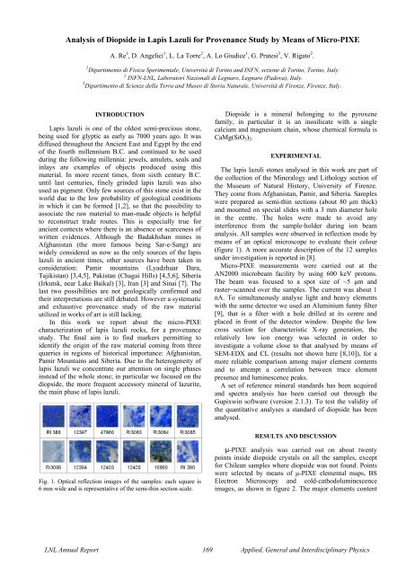
Fig. 1. Optical reflection images <strong>of</strong> the samples: each square is<br />
6 mm wide and is representative <strong>of</strong> the semi-th<strong>in</strong> section scale.<br />
<strong>Diopside</strong> is a m<strong>in</strong>eral belong<strong>in</strong>g to the pyroxene<br />
family, <strong>in</strong> particular it is an <strong>in</strong>osilicate with a s<strong>in</strong>gle<br />
calcium and magnesium cha<strong>in</strong>, whose chemical <strong>for</strong>mula is<br />
CaMg(SiO3)2.<br />
EXPERIMENTAL<br />
The lapis lazuli stones analysed <strong>in</strong> this work are part <strong>of</strong><br />
the collection <strong>of</strong> the M<strong>in</strong>eralogy and Lithology section <strong>of</strong><br />
the Museum <strong>of</strong> Natural History, University <strong>of</strong> Firenze.<br />
They come from Afghanistan, Pamir, and Siberia. Samples<br />
were prepared as semi-th<strong>in</strong> sections (about 80 μm thick)<br />
and mounted on special slides with a 3 mm diameter hole<br />
<strong>in</strong> the centre. The holes were made to avoid any<br />
<strong>in</strong>terference from the sample-holder dur<strong>in</strong>g ion beam<br />
analysis. All samples were observed <strong>in</strong> reflection mode <strong>by</strong><br />
means <strong>of</strong> an optical microscope to evaluate their colour<br />
(figure 1). A more accurate description <strong>of</strong> the 12 samples<br />
under <strong>in</strong>vestigation is reported <strong>in</strong> [8].<br />
Micro-PIXE measurements were carried out at the<br />
AN2000 microbeam facility <strong>by</strong> us<strong>in</strong>g 600 keV protons.<br />
The beam was focused to a spot size <strong>of</strong> ~5 μm and<br />
raster−scanned over the samples. The current was about 1<br />
nA. To simultaneously analyse light and heavy elements<br />
with the same detector we used an Alum<strong>in</strong>ium funny filter<br />
[9], that is a filter with a hole drilled at its centre and<br />
placed <strong>in</strong> front <strong>of</strong> the detector w<strong>in</strong>dow. Despite the low<br />
cross section <strong>for</strong> characteristic X-ray generation, the<br />
relatively low ion energy was selected <strong>in</strong> order to<br />
<strong>in</strong>vestigate a volume close to that analysed <strong>by</strong> means <strong>of</strong><br />
SEM-EDX and CL (results not shown here [8,10]), <strong>for</strong> a<br />
more reliable comparison among major element contents<br />
and to attempt a correlation between trace element<br />
presence and lum<strong>in</strong>escence peaks.<br />
A set <strong>of</strong> reference m<strong>in</strong>eral standards has been acquired<br />
and spectra analysis has been carried out through the<br />
Gupixw<strong>in</strong> s<strong>of</strong>tware (version 2.1.3). To test the validity <strong>of</strong><br />
the quantitative analyses a standard <strong>of</strong> diopside has been<br />
analysed.<br />
RESULTS AND DISCUSSION<br />
µ-PIXE analysis was carried out on about twenty<br />
po<strong>in</strong>ts <strong>in</strong>side diopside crystals on all the samples, except<br />
<strong>for</strong> Chilean samples where diopside was not found. Po<strong>in</strong>ts<br />
were selected <strong>by</strong> means <strong>of</strong> μ-PIXE elemental maps, BS<br />
Electron Microscopy and cold-cathodolum<strong>in</strong>escence<br />
images, as shown <strong>in</strong> figure 2. The major elements content<br />
LNL Annual Report 169 Applied, General and Interdiscipl<strong>in</strong>ary Physics
is compatible with SEM-EDX measurements [10], but<br />
does not show any useful <strong>in</strong>dication <strong>for</strong> a provenance<br />
study.<br />
Fig. 2. μ-PIXE elemental maps (the <strong>in</strong>clusion <strong>in</strong> the map is<br />
composed <strong>by</strong> calcium, magnesium and silicon that are the ma<strong>in</strong><br />
elements <strong>of</strong> diopside), BS Electron Microscopy and coldcathodolum<strong>in</strong>escence<br />
images.<br />
In figure 3 the trace elements detected and their content<br />
are presented. Identified trace elements are common <strong>in</strong><br />
almost all diopside crystals, but there are many differences<br />
<strong>in</strong> quantity, if we compare samples from Pamir Mounta<strong>in</strong>s<br />
and from Afghanistan. In fact, all the samples com<strong>in</strong>g from<br />
Pamir Mounta<strong>in</strong>s show contents <strong>in</strong> titanium, vanadium and<br />
chromium lower than those found <strong>in</strong> Afghan samples, but<br />
on the other hand a higher mean quantity <strong>of</strong> iron is<br />
observed. The Siberian sample does not show any evident<br />
difference with respect to the other samples.<br />
Fig. 3. M<strong>in</strong>or and trace elements <strong>in</strong> diopside. The columns<br />
<strong>in</strong>dicates the average elemental content and the po<strong>in</strong>ts represent<br />
the <strong>in</strong>dividual measurements [10].<br />
Compar<strong>in</strong>g these results with lum<strong>in</strong>escence spectra, no<br />
evident correlations between lum<strong>in</strong>escence peaks and trace<br />
elements were found. Ti, Mn and Fe are the ma<strong>in</strong><br />
responsible <strong>for</strong> lum<strong>in</strong>escence <strong>in</strong> diopside but due to a<br />
competition process between activators and quenchers a<br />
direct proportionality does not always exist between the<br />
<strong>in</strong>tensity <strong>of</strong> a lum<strong>in</strong>escence peak and element contents.<br />
CONCLUSIONS<br />
The elemental analysis <strong>of</strong> the s<strong>in</strong>gle crystals <strong>of</strong> diopside<br />
<strong>in</strong> lapis lazuli <strong>by</strong> means <strong>of</strong> μ-PIXE showed to be useful <strong>for</strong><br />
a provenance study <strong>of</strong> this stone. Even tough the ma<strong>in</strong><br />
elements do not give any <strong>in</strong><strong>for</strong>mation about the orig<strong>in</strong> <strong>of</strong> a<br />
sample, the analysis <strong>of</strong> trace elements content permits to<br />
have some <strong>in</strong>dication useful to dist<strong>in</strong>guish among three<br />
Asian provenances <strong>of</strong> lapis lazuli.<br />
ACKNOWLEDGMENTS<br />
The authors gratefully acknowledge the “Compagnia di<br />
San Paolo” <strong>for</strong> the fund<strong>in</strong>g <strong>of</strong> a PhD scholarship on<br />
Physics applied to Cultural Heritage.<br />
[1] N. Voskobo<strong>in</strong>ikova, The m<strong>in</strong>eralogy <strong>of</strong> the Slyudyanka<br />
deposits <strong>of</strong> lazurite. Zapiski Vserossiyskogo<br />
M<strong>in</strong>eralogicheskogo Obshchestva: 67 (1938) 601-622 (<strong>in</strong><br />
Russian).<br />
[2] J. Wyart et al., <strong>Lapis</strong> lazuli from Sar-i-Sang, Badakhshan,<br />
Afghanistan. Gems and Gemmology, 17 (1981) 184-190.<br />
[3] G. Herrmann, <strong>Lapis</strong> <strong>Lazuli</strong>: the early phases <strong>of</strong> its trade,<br />
Iraq, Vol. 30, No. 1 (Spr<strong>in</strong>g 1968) 21-57.<br />
[4] M. Casanova, The Sources <strong>of</strong> the <strong>Lapis</strong> <strong>Lazuli</strong> found <strong>in</strong> Iran,<br />
South Asian Archaeology, (1989) 49-56.<br />
[5] A.B. Delmas, M. Casanova, The <strong>Lapis</strong> <strong>Lazuli</strong> sources <strong>in</strong> the<br />
ancient east. South Asian Archaeology, 1987 (1990) 493-<br />
505.<br />
[6] P. Ballirano, A. Maras, Amer. M<strong>in</strong>eral., 91 (2006) 997-<br />
1005.<br />
[7] A. Nibbi, Ancient Egypt and some Eastern neightbours,<br />
Park Ridge, Noyes Press, 1981.<br />
[8] A. Re et al., Nucl. Instr. and Meth. B, <strong>in</strong> press<br />
(DOI: 10.1016/j.nimb.2011.02.070)<br />
[9] S. Gama et al., Nucl. Instr. and Meth. B, 181<br />
(2001) 150-156.<br />
[10] A. Lo Giudice et al., Anal. and Bioanal. Chem., 395 (2009)<br />
2211-2217.<br />
LNL Annual Report 170 Applied, General and Interdiscipl<strong>in</strong>ary Physics