MCDB 4650 Developmental Biology Problem Set 2 Answers 1) (1 ...
MCDB 4650 Developmental Biology Problem Set 2 Answers 1) (1 ...
MCDB 4650 Developmental Biology Problem Set 2 Answers 1) (1 ...
Create successful ePaper yourself
Turn your PDF publications into a flip-book with our unique Google optimized e-Paper software.
<strong>MCDB</strong> <strong>4650</strong> <strong>Developmental</strong> <strong>Biology</strong><br />
<strong>Problem</strong> <strong>Set</strong> 2 <strong>Answers</strong><br />
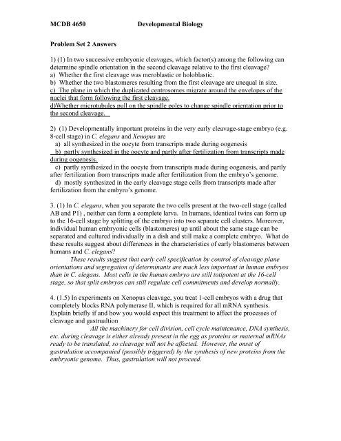
1) (1) In two successive embryonic cleavages, which factor(s) among the following can<br />
determine spindle orientation in the second cleavage relative to the first cleavage?<br />
a) Whether the first cleavage was meroblastic or holoblastic.<br />
b) Whether the two blastomeres resulting from the first cleavage are unequal in size.<br />
c) The plane in which the duplicated centrosomes migrate around the envelopes of the<br />
nuclei that form following the first cleavage.<br />
d)Whether microtubules pull on the spindle poles to change spindle orientation prior to<br />
the second cleavage.<br />
2) (1) <strong>Developmental</strong>ly important proteins in the very early cleavage-stage embryo (e.g.<br />
8-cell stage) in C. elegans and Xenopus are<br />
a) all synthesized in the oocyte from transcripts made during oogenesis<br />
b) partly synthesized in the oocyte and partly after fertilization from transcripts made<br />
during oogenesis.<br />
c) partly synthesized in the oocyte from transcripts made during oogenesis, and partly<br />
after fertilization from transcripts made after fertilization from the embryo’s genome.<br />
d) mostly synthesized in the early cleavage stage cells from transcripts made after<br />
fertilization from the embyro’s genome.<br />
3. (1) In C. elegans, when you separate the two cells present at the two-cell stage (called<br />
AB and P1) , neither can form a complete larva. In humans, identical twins can form up<br />
to the 16-cell stage by splitting of the embryo into two separate cell clusters. Moreover,<br />
individual human embryonic cells (blastomeres) up until about the same stage can be<br />
separated and cultured individually in a dish and still make a complete embryo. What do<br />
these results suggest about differences in the characteristics of early blastomeres between<br />
humans and C. elegans?<br />
These results suggest that early cell specification by control of cleavage plane<br />
orientations and segregation of determinants are much less important in human embryos<br />
than in C. elegans. Most cells in the human embryo are still totipotent at the 16-cell<br />
stage, so that split embryos can still regulate cell commitments and develop normally.<br />
4. (1.5) In experiments on Xenopus cleavage, you treat 1-cell embryos with a drug that<br />
completely blocks RNA polymerase II, which is required for all mRNA synthesis.<br />
Explain briefly if and how you would expect this treatment to affect the processes of<br />
cleavage and gastrualtion<br />
All the machinery for cell division, cell cycle maintenance, DNA synthesis,<br />
etc. during cleavage is either already present in the egg as proteins or maternal mRNAs<br />
ready to be translated, so cleavage will not be affected. However, the onset of<br />
gastrulation accompanied (possibly triggered) by the synthesis of new proteins from the<br />
embryonic genome. Thus, gastrulation will not proceed.
<strong>MCDB</strong> <strong>4650</strong> <strong>Developmental</strong> <strong>Biology</strong><br />
5. (1)<br />
Almost all animals form a blastocoel (a fluid-filled cavity in the middle of the embryo) of<br />
some sort in the middle of the developing embryo, usually quite early in<br />
development. The diagram below shows the progression of 8 cells in a<br />
tight ball-shape to a ring of cells around the blastocoel. Describe what must<br />
be happening at each of the two steps in order to generate these changes<br />
within the embryo.<br />
The first change must require a change in adhesivity, as the cells are no<br />
longer adherent to each other for their entire extent. The second change is<br />
impacted by the plane of division. In this case, all the cells divided the<br />
same way (radially), so that no cells were invading the blastocoel space,<br />
but rather were creating a larger space. If the cells had divided<br />
perpendicularly to the way shown, some cells would be obscuring the<br />
blastocoel. Lastly,<br />
6. (.5) Which of the following are possible reasons for an embryo to have a<br />
blastocoel? Select all that are correct.<br />
a. space for future movement of cells<br />
b. keeps cells separated that will signal each other later<br />
c. pre-formed endodermal cavity<br />
d. to allow for storage of nutrients<br />
7. (1) Imagine that gastrulation is blocked in a Xenopus embryo. Predict one<br />
consequence for the embryo regarding the differentiation of cell types.<br />
Cells that normally come into contact for important signaling events will not do so.<br />
Thus, not only will the embryo be disorganized, but specific cell types will be missing.—<br />
Neurulation will not take place because the neural cells will not organize into a flat sheet<br />
bounded by epithelial cells, because the signaling required for those two cell types to<br />
form will not be present. Without these junction points, and the underlying notochord,<br />
the neural tube cannot fold. There are certainly other possibilities.<br />
8. (1) A cell that migrates through the dorsal lip of the blastopore at the very end of<br />
gastrulation in Xenopus is likely to differentiate into what kind of cell type (endoderm,<br />
ectoderm or mesoderm) and be located in what general region of the embryo? Mesoderm,<br />
because after endoderm initially migrates in, that is the only cell type that migrates<br />
during gastrulation, and at the end of gastrulation, cells are populating what will be the<br />
posterior end of the embryo (specifically, ventral posterior mesoderm).<br />
9. (1) In the figure below (A, B) are the results of an experiment showing the effects of<br />
levels of Sonic hedgehog (Shh), secreted from notochord cells, on the differentiation of<br />
neurons in the intact neural tube. In panel C, cells from chick neural tube were cultured<br />
in vitro in the presence of different concentrations of Shh, then immunostained for
<strong>MCDB</strong> <strong>4650</strong> <strong>Developmental</strong> <strong>Biology</strong><br />
proteins characteristic of the different types of neurons shown in the picture (e.g. floor<br />
plate=FP, Motor, etc.). The left diagram in (C) shows the positions of cells that stain for<br />
these proteins in the normal intact neural tube.<br />
Based on the data shown in the figure, describe the role of Shh. How is it functioning in<br />
specification of neural tube cell fates?<br />
a. Shh works as an autocrine signaling molecule<br />
b. Shh works as a juxtacrine signaling molecule<br />
c. Shh works as a paracrine signaling molecule<br />
d. Shh works as a morphogen<br />
e. Shh works to determine only the floorplate cells<br />
10. (1) The diagram below shows a cartoon drawing of the developing neural tube (b) and<br />
the overlying non-neuronal epithelium (a). If these cells are dissociated with trypsin into<br />
single cells and put into a cell culture system called a “drop culture”, they first associate<br />
loosely together as shown in (c). A drop culture is literally a small drop of culture<br />
solution that allows the cells to associate with each other. After some time, the two cell<br />
populations separate from each other (d and e).<br />
Explain what is most likely responsible for the segregation of these two cell types.<br />
The cells lose their external receptors when they are dissociated, thus they will<br />
loosely associate together initially when in this kind of a culture system. However, over<br />
the course of a few hours, the neural cells will make n-cadherin, while the epidermal<br />
cells will make E-cadherin; thus the two types of cells will segregate.<br />
During closure of the neural tube the two epithelia must segregate. The neural<br />
tube expresses N-cadherin and the overlying epithelium on the dorsal surface expressess<br />
E-cadherin. These are homophilic cell adhesion receptors and thus, the two tissues will<br />
segregate and only interact with like receptors, allowing the neural tissue to segregate<br />
from the non-neural tissue.


