01 Epithelium
01 Epithelium
01 Epithelium

You also want an ePaper? Increase the reach of your titles
YUMPU automatically turns print PDFs into web optimized ePapers that Google loves.
HISTOLOGY BIO 542<br />
EPITHELIUM<br />
<strong>Epithelium</strong>, along with connective tissue, muscle, and nerve, is one of the four primary tissue types. It is<br />
commonly found as a covering or lining for other types of tissues, as in the case of the epidermis and the<br />
lining of the entire gastrointestinal tract, and as a major component of numerous multicellular glands, such as<br />
the liver and pancreas.<br />
Epithelial tissues, and the epithelial portions of mixed tissues and organs, are characterized by being<br />
composed of closely-packed epithelial cells with little or no intercellular space resting on a basal lamina. A<br />
basal lamina may or may not be part of a basement membrane attaching epithelium to connective tissue.<br />
Basal laminae are not visible in the light microscope (too thin) and basement membranes are just barely<br />
visible in good, specially-stained sections.<br />
Histological differences among the various epithelia are based on the shapes of the cells (squamous,<br />
cuboidal, columnar), the number of cell layers present (simple [one layer], stratified [two or more],<br />
pseudostratified [irregular]), various modifications to the surfaces of the cells that can be seen with the light<br />
microscope (microvilli, stereocilia, cilia), and characteristic patterns of reaction with particular dyes<br />
(acidophilia, basophilia, special stains).<br />
Epithelial tissues have numerous particular functions which you will learn in detail but, in general, their<br />
functions are to mediate absorption, excretion, protection, secretion, sensory reception, and transport.<br />
Use these first two lab periods to become familiar with the epithelia. You will find that almost every slide<br />
you examine in this lab will contain one or more epithelia. Get in the habit of identifying all the epithelia you<br />
come across in all sections; it is always a good idea to pick slides at random out of your slide box<br />
and examine them for, in this case, epithelia. You should do the same with the other primary<br />
tissue types after you learn their characteristics. Don't limit yourself to those slides listed and assigned each<br />
week; there are LOTS of good examples of various cell types on other slides.<br />
EPITHELIUM SLIDES<br />
SIMPLE SQUAMOUS:<br />
Amphibian skin. (slide 75). This is a "spread" preparation in which the entire skin is attached<br />
to a slide and stained. Note the cell outlines ("paving stone" appearance) and the somewhat<br />
darkly stained nuclei.<br />
Endothelium of blood vessels (slide 3, any other slide). Examine the inner edge (lumen) of<br />
the medium size blood vessels. You will find (I hope) a single layer of very thin cells. About all<br />
you can identify with certainty is the cells' nuclei. Next try to find endothelial cells in other, bigger<br />
or smaller, vessels.
Ovary (slide 53, Triarch HN1-21T). The ovaries are covered on their outer surface with a<br />
simple squamous endothelium known as a mesothelium. Again, look for cells whose nuclei<br />
might be prominent. The mesothelial cells in this example are relatively “high” squamous. The<br />
trichrome stain accents the connective tissue in the section.<br />
Kidney (slide 34, Triarch HL1-21). The inner covering of Bowman's capsule is a simple<br />
squamous epithelium made up of thin, flat cells. Look for nuclei that bulge into the empty space of<br />
Bowman’s capsule that surrounds the renal corpuscle, a large, round structure.<br />
SIMPLE CUBOIDAL:<br />
Kidney (slide 34). The kidney tubules (proximal, distal, collecting) are composed of simple<br />
cuboidal epithelia arranged in tube form. The actual appearance of cells in such tubes will<br />
depend on the angle of sectioning. When sectioned, a tube can present circles, ovals, U-shapes,<br />
or linear arrays. Think about what the plane of section must have been relative to the various<br />
tube shapes you find in the kidney slide.<br />
Liver (slide 37, HK10-2). The parenchymal cells of the liver (hepatocytes) are cuboidal and<br />
arranged as plates or "walls" of cells; sheets of cells one cell thick. It is not a simple planar sheet,<br />
however; it has folds and branches.<br />
SIMPLE COLUMNAR:<br />
Stomach (slides 76, 77, or 78). Any stomach slide will do. The inner covering of the gastric<br />
mucosa (stomach lining) is simple columnar, as are the tubular gastric glands that extend down<br />
into connective tissue from the inner surface of the stomach.<br />
Small intestine (slides 31, 32). The epithelial covering of the intestinal mucosa is simple<br />
columnar and the cells have a thick microvillus coat at their apical ends that appears as a brush<br />
border. It takes a little practice to see the brush border. Fiddling with the condenser iris can<br />
often increase contrast and make the brush border clearer.<br />
Large intestine (slide 33). Find columnar cells with brush border. This BB may be<br />
significantly shorter than that in the small intestine. Goblet cells are present in large<br />
numbers.<br />
Rectoanal junction (slide 64). At the junction between the rectum and the anus, the mucosa<br />
changes from simple columnar to stratified squamous. Find the rectum portion and identify<br />
simple columnar cells with brush borders. The rectum is essentially the same as the large<br />
intestine.<br />
Testis and Epididymis (slide 81). Find stereocilia at the apical ends of tall columnar cells.<br />
After you examine stratified columnar epithelium, come back to this slide and decide of it’s<br />
simple or stratified. What are the problems in identification? Don’t necessarily believe what your
textbooks say – there are differences between species in especially the exact kind of epithelium<br />
found in a particular tissue. Always identify what you are actually seeing, not what you<br />
expect to see!!<br />
Gall bladder (slide 28). Tall simple columnar epithelial cells with apical microvilli.<br />
SIMPLE COLUMNAR WITH CILIA:<br />
Oviduct (Uterine tube) (slide 55). Be aware that the plane of section can often cause you<br />
to confuse simple with stratified epithelium. One way to decide fairly confidently is to<br />
examine the entire extent of a particular epithelium and see if there is any portion of it<br />
that is clearly simple. If one portion is simple it’s a good bet that the whole thing is<br />
simple and its appearance as stratified is due mostly to an odd plane of section. Again,<br />
what sort of plane of section would cause simple columnar or cuboidal to look stratified?<br />
STRATIFIED SQUAMOUS, NON-KERATINIZED:<br />
Vagina (slide 98). The lining of the vagina is stratified squamous. Note the different cell<br />
shapes as you travel from basal to apical cell layers.<br />
Soft palate (slide 70). Examine the epithelial covering of the oral side of the palate; the<br />
respiratory side is covered by columnar cells. Also look for simple squamous epithelium<br />
and simple columnar. Where do you expect to find these two types?<br />
Esophagus (slide 26). The esophagus and vagina are famous "look alikes" in histology.<br />
Rectoanal junction (slide 64). Examine the anus portion this time. Note there is a transition<br />
from non-keratinized to keratinized epithelium as you go from the junction outwards to the<br />
epidermal skin.<br />
STRATIFIED SQUAMOUS, KERATINIZED:<br />
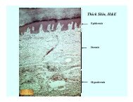
Thin skin (Caucasian and Negroid), Thick skin, Scalp (primate), Recto-Anal Junction<br />
(slides 67, 68, 64). These are all sections of skin and can be treated the same. Find the<br />
stratified squamous covering (epidermal epithelium) and note the varying thicknesses of<br />
keratinization of the individual examples; i.e., some kinds of skin have thin keratinized layers,<br />
some have thick.<br />
STRATIFIED CUBOIDAL:<br />
Thin skin (slide 68), Thick skin (67). Sweat glands show some stratified cuboidal. Look for<br />
cross-sections of tubes (circles) that clearly have two cell layers. The portion of the sweat gland<br />
that is stratified tends to be located in the deeper regions of the dermis.<br />
STRATIFIED COLUMNAR:<br />
Sublingual gland, Submandibular gland (slides 79, 80). The epithelium comprising the walls<br />
of the large ducts in these glands is often stratified columnar. Look around until you find an
example.<br />
Liver/Salivary gland (DEMO slide). Good examples of stratified columnar epithelium can be<br />
found in the sublingual salivary gland portion.<br />
Penis (slide 59). The penile urethra is lined by an epithelium that ranges from stratified<br />
columnar to simple columnar. The urethra is the urinary passage. Now go back and examine<br />
epididymis and decide between simple and stratified columnar.<br />
PSEUDOSTRATIFIED COLUMNAR:<br />
Trachea (slide 90). The surface cells of the airway are ciliated. Fiddle with the condenser iris<br />
to see the cilia.<br />
Soft palate (slide 70). Often ciliated on the respiratory (airway) side.<br />
Testis/Epididymis (slide 81). Stereocilia can be found on the inner surface cells of the<br />
epididymis.<br />
TRANSITIONAL:<br />
Ureter (slide 91) and Bladder (contracted and distended) (slide 5). The ureter has<br />
transitional epithelium on its inner surface. Distended (full) and contracted (empty) bladders<br />
show the two states of transitional. In the section of distended bladder the “blebs” or “bubbles”<br />
on the surface layer cells is an artefact of preparation and is not the “dome” shape of the cells in<br />
contracted bladder. Check the DEMO contracted bladder slides, too.