Vidyavathi et al., J Cytol Histol 2012, 3 - OMICS Publishing Group
Vidyavathi et al., J Cytol Histol 2012, 3 - OMICS Publishing Group
Vidyavathi et al., J Cytol Histol 2012, 3 - OMICS Publishing Group
Create successful ePaper yourself
Turn your PDF publications into a flip-book with our unique Google optimized e-Paper software.
Citation: <strong>Vidyavathi</strong> K, Udayakumar M, Suresh TN, Sreeramulu PN (<strong>2012</strong>) Granulomatous Mastitis: A <strong>Cytol</strong>ogic<strong>al</strong> Dilemma. J <strong>Cytol</strong> <strong>Histol</strong> 3:137.<br />
doi:10.4172/2157-7099.1000137<br />
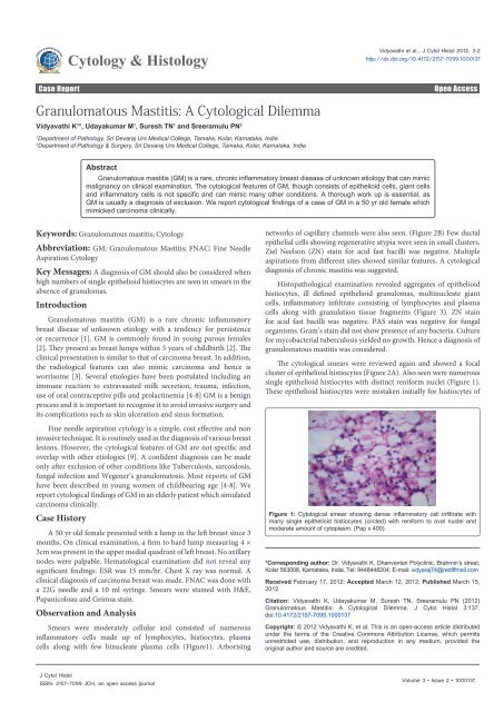
Figure 2: A. <strong>Cytol</strong>ogic<strong>al</strong> smear showing an aggregate of epithelioid histiocytes<br />
(arrow) within a mixed inflammatory background (Pap x 400). B. Smear showing<br />
arborising n<strong>et</strong>work of capillary fragments (double arrow) (Pap x400).<br />
Figure 3: Histopathology showing non caseating epithelioid granulomas <strong>al</strong>ong<br />
with Langhan’s type giant cells (arrow) and foreign body type (double arrow)<br />
giant cells (H & E x 400).<br />
a chronic infiltrate. Hence a cytologic<strong>al</strong> diagnosis of granulomatous<br />
mastitis was possible on review.<br />
Discussion<br />
GM is an uncommon breast lesion that was first described by Kessler<br />
and Wolloch in 1972 [10]. It is seen in women of child bearing age<br />
and usu<strong>al</strong>ly present within 5 yrs of childbirth [2]. However it has been<br />
reported in patients as young as 11 years and as old as 80 years [11,12].<br />
The most common clinic<strong>al</strong> presentation is a unilater<strong>al</strong> firm discr<strong>et</strong>e<br />
breast lump, often associated with inflammation of overlying skin [2].<br />
It can be seen in any quadrant of the breast except in subareolar region<br />
[13]. It can <strong>al</strong>so show nipple r<strong>et</strong>raction or peau de orange appearance,<br />
thus simulating carcinoma [14].<br />
The histologic<strong>al</strong> features are characterized by non caseating<br />
granulomas within the breast parenchyma and lobulitis with or<br />
without neutrophilic microabscesss [15]. The cytologic<strong>al</strong> features are<br />
characterized by aggregates of epithelioid histiocytes, multinucleate<br />
giant cells lymphocytes, plasma cells and a variable number of<br />
neutrophils [1]. Presence of single epithelioid histiocytes having a<br />
reniform to plump nuclei and a moderate to abundant p<strong>al</strong>e pink<br />
cytoplasm has <strong>al</strong>so been reported by many authors [4,9,16].<br />
Though, most studies have shown the presence of neutrophils<br />
as the predominant inflammatory infiltrate, these were significantly<br />
absent in the present case. The presence of numerous lymphocytes and<br />
J <strong>Cytol</strong> <strong>Histol</strong><br />
ISSN: 2157-7099 JCH, an open access journ<strong>al</strong><br />
Page 2 of 3<br />
plasma cells, <strong>al</strong>ong with binucleate plasma cells, significant arborising<br />
vascular n<strong>et</strong>work <strong>al</strong>ong with absence of multinucleate giant cells, led us<br />
to give an initi<strong>al</strong> diagnosis of chronic mastitis. However, a review of the<br />
slide showed a foc<strong>al</strong> cluster of epithelioid histiocytes <strong>al</strong>ong with single<br />
epithelioid histiocytes with a distinct reniform or ov<strong>al</strong> nuclei, which<br />
was missed on initi<strong>al</strong> examination. Tse GM <strong>et</strong> <strong>al</strong>. found epithelioid<br />
granulomas only in h<strong>al</strong>f of their case series thus, suggesting that they<br />
are not pathognomic for GM [9]. They opined that presence of these<br />
single epithelioid histiocytes in the absence of well defined granulomas<br />
should <strong>al</strong>ert the pathologist to the possibility of a granulomatous<br />
inflammation [9]. Most studies have <strong>al</strong>so described presence of<br />
granulation tissue fragments in GM [9,16].<br />
Accurate cytologic<strong>al</strong> diagnosis still remains a ch<strong>al</strong>lenge, because<br />
the features overlap with other <strong>et</strong>iologies like tuberculosis (TB), fung<strong>al</strong><br />
infections, fat necrosis, sarcodoisis <strong>et</strong>c. The single most important<br />
differenti<strong>al</strong> diagnosis is with tuberculosis especi<strong>al</strong>ly in endemic<br />
countries like India [17]. Treating TB with steroids would aggravate<br />
the infection, whereas giving unnecessary anti tubercular drugs may<br />
cause numerous side effects. The absence of caseous necrosis and<br />
a predominantly neutrophilic infiltrate in the background favour<br />
a diagnosis of GM [9]. Langhans giant cells, epithelioid cells and<br />
caseation are features of TB. However, acid fast stains and culture is<br />
<strong>al</strong>so essenti<strong>al</strong> in confirming diagnosis of TB.<br />
Demonstration of fungi by speci<strong>al</strong> stains like PAS and culture is<br />
necessary to diagnose fung<strong>al</strong> mastitis. In fat necrosis, the presence<br />
of abundant foamy cells is a classic feature, whereas in GM foamy<br />
cells are seen only occasion<strong>al</strong>ly. In addition, epitheli<strong>al</strong> cells which are<br />
seen in GM are not seen in fat necrosis [16]. In sarcodoisis, smears<br />
show abundant lymphocytes with neutrophils or necrosis <strong>al</strong>ong with<br />
epithelioid granulomas [16]. As the cytologic<strong>al</strong> features are not specific,<br />
GM is usu<strong>al</strong>ly a diagnosis of exclusion. The definite diagnosis depends<br />
on clinic<strong>al</strong> correlation, histopathologic<strong>al</strong> picture and a negative<br />
microbiologic<strong>al</strong> investigation.<br />
The cause of idiopathic GM remains unclear. Autoimmune disease,<br />
infection, trauma have been implicated by some authors [2]. Sever<strong>al</strong><br />
theories about the mechanisms of idiopathic GM have been proposed.<br />
Miller <strong>et</strong> <strong>al</strong>. [18] suggested that squamous m<strong>et</strong>aplasia in the ducts can<br />
initiate the process as a response to keratin. Murthy [7] reasoned that<br />
or<strong>al</strong> contraceptive pills increase the amount of secr<strong>et</strong>ion in the ducts<br />
and cause the inflammatory response. Others suggested that increased<br />
prolactin levels or loc<strong>al</strong>ised immune response to extravasated milk<br />
secr<strong>et</strong>ion can cause mastitis [5,8]. An association with loc<strong>al</strong> infection<br />
by Corynebacterium Kroppenstedtii has recently been suggested [5].<br />
In conclusion, the cytologic<strong>al</strong> diagnosis of GM is difficult because<br />
there are no specific features. A high index of suspicion and awareness<br />
of this entity by the cytopathologist is needed, to make a diagnosis and<br />
to prevent unnecessary mastectomies. A diagnosis of GM should <strong>al</strong>so be<br />
considered when numerous epithelioid histiocytes are seen in smears,<br />
even in the absence of granulomas [9]. A definite diagnosis depends<br />
on histopathologic<strong>al</strong> examination and negative microbiologic<strong>al</strong><br />
investigations.<br />
References<br />
1. Gupta RK (2010) Fine Needle Aspiration <strong>Cytol</strong>ogy of Granulomatous Mastitis.<br />
A study of 18 cases. Acta <strong>Cytol</strong> 54: 138-141.<br />
2. Al-Khaffaf B, Knox F, Bundred NJ (2008) Idiopathic granulomatous mastitis: a<br />
25 year experience. J Am Coll Surg 206: 269-273.<br />
Volume 3 • Issue 2 • 1000137
Citation: <strong>Vidyavathi</strong> K, Udayakumar M, Suresh TN, Sreeramulu PN (<strong>2012</strong>) Granulomatous Mastitis: A <strong>Cytol</strong>ogic<strong>al</strong> Dilemma. J <strong>Cytol</strong> <strong>Histol</strong> 3:137.<br />
doi:10.4172/2157-7099.1000137<br />
3. Heer R, Shrimankar J, Griffith CD (2003) Granulomatous mastitis can mimic<br />
breast cancer on clinic<strong>al</strong>, radiologic<strong>al</strong> or cytologic<strong>al</strong> examination: a cautionary<br />
t<strong>al</strong>e. Breast 12: 283-286.<br />
4. Poniecka AW, Krasuski P, G<strong>al</strong> E, Lubin J, Howard L, <strong>et</strong> <strong>al</strong>. (2001) Granulomatous<br />
inflammation of the breast in a pregnant woman: report of a case with fine<br />
needle aspiration diagnosis. Acta <strong>Cytol</strong> 45: 797-801.<br />
5. Taylor GB, Paviour SD, Musaad S, Jones WO, Holland DJ (2003) A<br />
clinicopathologic<strong>al</strong> review of 34 cases of inflammatory breast disease showing<br />
an association b<strong>et</strong>ween corynebacteria infection and granulomatous mastitis.<br />
Pathology 35: 109-119.<br />
6. Cserni G, Szajki K (1999) Granulomatous lobular mastitis following drug<br />
induced g<strong>al</strong>actorrhea and blunt trauma. Breast J 5: 398-403.<br />
7. Murthy MS (1973) Granulomatous mastitis and lipogranuloma of the breast.<br />
Am J Clin Pathol 60: 432-433.<br />
8. Rowe PH (1984) Granulomatous mastitis associated with pituitary prolactinoma.<br />
Br J Clin Pract 38: 32-34.<br />
9. Tse GM, Poon CS, Law BK, Pang LM, Chu WC, <strong>et</strong> <strong>al</strong>. (2003) Fine needle<br />
aspiration cytology of granulomatous mastitis. J Clin Pathol 56: 519-521.<br />
10. Kessler E, Wolloch Y (1972) Granulomatous mastitis : A lesion clinic<strong>al</strong>ly<br />
simulating carcinoma. Am J Clin Pathol 58: 642-646.<br />
11. Bani-Hani KE, Yaghan RJ, Mat<strong>al</strong>ka II, Shatnawi NJ (2004) Idiopathic<br />
J <strong>Cytol</strong> <strong>Histol</strong><br />
ISSN: 2157-7099 JCH, an open access journ<strong>al</strong><br />
Page 3 of 3<br />
granulomatous mastitis: time to avoid unnecessary mastectomies. Breast J 10:<br />
318-322.<br />
12. Lai EC, Chan WC, Ma TK, Tang AP, Poon CS, <strong>et</strong> <strong>al</strong>. (2005) The role of<br />
conservative treatment in idiopathic granulomatous mastitis. Breast J 11: 454-<br />
456.<br />
13. Akcan A, Akyildiz H, Deneme MA, Akgun H, Aritas Y (2006) Granulomatous<br />
lobular mastitis: A complex diagnostic and therapeutic problem. World J Surg<br />
30: 1403-1409.<br />
14. Baslaim MM, Khayat HA, Al-Amoudi SA (2007) Idiopathic granulomatous<br />
mastitis: A h<strong>et</strong>erogenous disease with variable clinic<strong>al</strong> presentation. World J<br />
Surg 31: 1677-1681.<br />
15. Ellis IO, Elston CW, Goulding H (1987) Inflammatory conditions. In: Elston<br />
CW,Ellis IO,eds.Systemic pathology,Vol 13,(3 rd ed.) The Breast .Edinburg:<br />
Churchill Livingstone.<br />
16. Kumarasinghe MP (1997) <strong>Cytol</strong>ogy of granulomatous mastitis. Acta <strong>Cytol</strong> 41:<br />
727-730.<br />
17. Kishore B, Khare P, Gupta RJ, Bisht SP (2007) Fine needle aspiration cytology<br />
in the diagnosis of inflammatotry lesions of the breast with emphasis on<br />
tuberculous mastitis. J <strong>Cytol</strong> 24: 155-156.<br />
18. Miller F, Seidman I, Smith CA (1971) Granulomatous mastitis. N Y State J Med<br />
71: 2194-2195.<br />
Submit your next manuscript and g<strong>et</strong> advantages of <strong>OMICS</strong><br />
<strong>Group</strong> submissions<br />
Unique features:<br />
• User friendly/feasible website-translation of your paper to 50 world’s leading languages<br />
• Audio Version of published paper<br />
• Digit<strong>al</strong> articles to share and explore<br />
Speci<strong>al</strong> features:<br />
• 200 Open Access Journ<strong>al</strong>s<br />
• 15,000 editori<strong>al</strong> team<br />
• 21 days rapid review process<br />
• Qu<strong>al</strong>ity and quick editori<strong>al</strong>, review and publication processing<br />
• Indexing at PubMed (parti<strong>al</strong>), Scopus, DOAJ, EBSCO, Index Copernicus and Google Scholar <strong>et</strong>c<br />
• Sharing Option: Soci<strong>al</strong> N<strong>et</strong>working Enabled<br />
• Authors, Reviewers and Editors rewarded with online Scientific Credits<br />
• B<strong>et</strong>ter discount for your subsequent articles<br />
Submit your manuscript at: http://www.omicsonline.org/submission<br />
Volume 3 • Issue 2 • 1000137


