Effects of insulin insufficiency on forelimb and tail ... - Development
Effects of insulin insufficiency on forelimb and tail ... - Development
Effects of insulin insufficiency on forelimb and tail ... - Development

You also want an ePaper? Increase the reach of your titles
YUMPU automatically turns print PDFs into web optimized ePapers that Google loves.
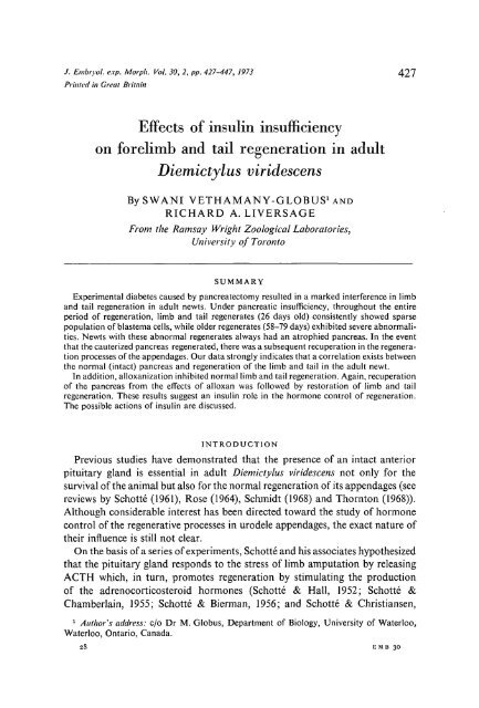
. Embryol. exp. Morph. Vol. 30, 2, pp. 427-447, 1973 427<br />
Printed in Great Britain<br />
<str<strong>on</strong>g>Effects</str<strong>on</strong>g> <str<strong>on</strong>g>of</str<strong>on</strong>g> <str<strong>on</strong>g>insulin</str<strong>on</strong>g> <str<strong>on</strong>g>insufficiency</str<strong>on</strong>g><br />
<strong>on</strong> <strong>forelimb</strong> <strong>and</strong> <strong>tail</strong> regenerati<strong>on</strong> in adult<br />
Diemictylus viridescens<br />
BySWANI VETHAMANY-GLOBUS^ND<br />
RICHARD A. LIYERSAGE<br />
From the Ramsay Wright Zoological Laboratories,<br />
University <str<strong>on</strong>g>of</str<strong>on</strong>g> Tor<strong>on</strong>to<br />
SUMMARY<br />
Experimental diabetes caused by pancreatectomy resulted in a marked interference in limb<br />
<strong>and</strong> <strong>tail</strong> regenerati<strong>on</strong> in adult newts. Under pancreatic <str<strong>on</strong>g>insufficiency</str<strong>on</strong>g>, throughout the entire<br />
period <str<strong>on</strong>g>of</str<strong>on</strong>g> regenerati<strong>on</strong>, limb <strong>and</strong> <strong>tail</strong> regenerates (26 days old) c<strong>on</strong>sistently showed sparse<br />
populati<strong>on</strong> <str<strong>on</strong>g>of</str<strong>on</strong>g> blastema cells, while older regenerates (58-79 days) exhibited severe abnormalities.<br />
Newts with these abnormal regenerates always had an atrophied pancreas. In the event<br />
that the cauterized pancreas regenerated, there was a subsequent recuperati<strong>on</strong> in the regenerati<strong>on</strong><br />
processes <str<strong>on</strong>g>of</str<strong>on</strong>g> the appendages. Our data str<strong>on</strong>gly indicates that a correlati<strong>on</strong> exists between<br />
the normal (intact) pancreas <strong>and</strong> regenerati<strong>on</strong> <str<strong>on</strong>g>of</str<strong>on</strong>g> the limb <strong>and</strong> <strong>tail</strong> in the adult newt.<br />
In additi<strong>on</strong>, alloxanizati<strong>on</strong> inhibited normal limb <strong>and</strong> <strong>tail</strong> regenerati<strong>on</strong>. Again, recuperati<strong>on</strong><br />
<str<strong>on</strong>g>of</str<strong>on</strong>g> the pancreas from the effects <str<strong>on</strong>g>of</str<strong>on</strong>g> alloxan was followed by restorati<strong>on</strong> <str<strong>on</strong>g>of</str<strong>on</strong>g> limb <strong>and</strong> <strong>tail</strong><br />
regenerati<strong>on</strong>. These results suggest an <str<strong>on</strong>g>insulin</str<strong>on</strong>g> role in the horm<strong>on</strong>e c<strong>on</strong>trol <str<strong>on</strong>g>of</str<strong>on</strong>g> regenerati<strong>on</strong>.<br />
The possible acti<strong>on</strong>s <str<strong>on</strong>g>of</str<strong>on</strong>g> <str<strong>on</strong>g>insulin</str<strong>on</strong>g> are discussed.<br />
INTRODUCTION<br />
Previous studies have dem<strong>on</strong>strated that the presence <str<strong>on</strong>g>of</str<strong>on</strong>g> an intact anterior<br />
pituitary gl<strong>and</strong> is essential in adult Diemictylus viridescens not <strong>on</strong>ly for the<br />
survival <str<strong>on</strong>g>of</str<strong>on</strong>g> the animal but also for the normal regenerati<strong>on</strong> <str<strong>on</strong>g>of</str<strong>on</strong>g> its appendages (see<br />
reviews by Schotte (1961), Rose (1964), Schmidt (1968) <strong>and</strong> Thornt<strong>on</strong> (1968)).<br />
Although c<strong>on</strong>siderable interest has been directed toward the study <str<strong>on</strong>g>of</str<strong>on</strong>g> horm<strong>on</strong>e<br />
c<strong>on</strong>trol <str<strong>on</strong>g>of</str<strong>on</strong>g> the regenerative processes in urodele appendages, the exact nature <str<strong>on</strong>g>of</str<strong>on</strong>g><br />
their influence is still not clear.<br />
On the basis <str<strong>on</strong>g>of</str<strong>on</strong>g> a series <str<strong>on</strong>g>of</str<strong>on</strong>g> experiments, Schotte <strong>and</strong> his associates hypothesized<br />
that the pituitary gl<strong>and</strong> resp<strong>on</strong>ds to the stress <str<strong>on</strong>g>of</str<strong>on</strong>g> limb amputati<strong>on</strong> by releasing<br />
ACTH which, in turn, promotes regenerati<strong>on</strong> by stimulating the producti<strong>on</strong><br />
<str<strong>on</strong>g>of</str<strong>on</strong>g> the adrenocorticosteroid horm<strong>on</strong>es (Schotte & Hall, 1952; Schotte &<br />
Chamberlain, 1955; Schotte & Bierman, 1956; <strong>and</strong> Schotte & Christiansen,<br />
1<br />
Author's address: c/o Dr M. Globus, Department <str<strong>on</strong>g>of</str<strong>on</strong>g> Biology, University <str<strong>on</strong>g>of</str<strong>on</strong>g> Waterloo,<br />
Waterloo, Ontario, Canada.<br />
28 EMB 30
428 SWANI VETHAMANY-GLOBUS AND R. A. LIVERSAGE<br />
1957). They also postulated that the pituitary-adrenal synergism is probably<br />
active in: (a) preventing dermal invasi<strong>on</strong> under the wound epidermis, <strong>and</strong><br />
(b) supporting tissue dedifferentiati<strong>on</strong>.<br />
However, Wilkers<strong>on</strong> (1963) obtained limb regenerati<strong>on</strong> when he injected<br />
bovine somatotropin into hypophysectomized newts, beginning as late as 15 days<br />
post-amputati<strong>on</strong>. He c<strong>on</strong>cluded that the loss <str<strong>on</strong>g>of</str<strong>on</strong>g> regenerative ability in the adult<br />
newt limb following hypophysectomy was due to the absence <str<strong>on</strong>g>of</str<strong>on</strong>g> growth horm<strong>on</strong>e.<br />
Several workers have shown that limb regenerati<strong>on</strong> is thyroxine-dependent.<br />
Following thyroidectomy normal limb regenerati<strong>on</strong> is retarded but not completely<br />
inhibited. Early removal <str<strong>on</strong>g>of</str<strong>on</strong>g> the thyroid gl<strong>and</strong> interferes with dedifferentiati<strong>on</strong><br />
<strong>and</strong> late removal interferes with the growth <str<strong>on</strong>g>of</str<strong>on</strong>g> the regenerate (Walter,<br />
1910; Richards<strong>on</strong>, 1940; Schotte & Washburn, 1954). Antuitrin G (a commercially<br />
prepared mammalian anterior pituitary extract) in combinati<strong>on</strong> with<br />
thyroxine was more effective in promoting regenerati<strong>on</strong> in hypophysectomized<br />
adult newts than Antuitrin G al<strong>on</strong>e (Richards<strong>on</strong>, 1945).<br />
Recently, prolactin has been shown to have an effect <strong>on</strong> limb regenerati<strong>on</strong><br />
(C<strong>on</strong>nelly, Tassava & Thornt<strong>on</strong>, 1968; Tassava, 1969). They showed that prolactin<br />
al<strong>on</strong>e increased survival <strong>and</strong> limb regenerati<strong>on</strong>, but less effectively than<br />
did prolactin in combinati<strong>on</strong> with thyroxine. From this evidence Tassava (1969)<br />
postulated a 'prolactin-thyroxine' synergism operating in adult newt limb<br />
regenerati<strong>on</strong>.<br />
All <str<strong>on</strong>g>of</str<strong>on</strong>g> these horm<strong>on</strong>es that have been associated with regenerati<strong>on</strong> in D.<br />
viridescens, namely prolactin, cortis<strong>on</strong>e, thyroxine <strong>and</strong> growth horm<strong>on</strong>e, are<br />
also known to induce the producti<strong>on</strong> <str<strong>on</strong>g>of</str<strong>on</strong>g> <str<strong>on</strong>g>insulin</str<strong>on</strong>g> from the islets <str<strong>on</strong>g>of</str<strong>on</strong>g> Langerhans<br />
(Haist, 1959; Lazarus & Volk, 1962; Tepperman, 1965; Bern & Nicoll, 1968).<br />
Prol<strong>on</strong>ged treatment <str<strong>on</strong>g>of</str<strong>on</strong>g> mammals with these horm<strong>on</strong>es can exhaust the islets<br />
<strong>and</strong>, ultimately, cause diabetes; hence, these horm<strong>on</strong>es are called 'diabetogenic<br />
horm<strong>on</strong>es' (review by Tepperman, 1965). In this c<strong>on</strong>nexi<strong>on</strong>, Genuth & Lebovitz<br />
(1965) observed that additi<strong>on</strong> <str<strong>on</strong>g>of</str<strong>on</strong>g> ACTH to culture medium induced the producti<strong>on</strong><br />
<str<strong>on</strong>g>of</str<strong>on</strong>g> <str<strong>on</strong>g>insulin</str<strong>on</strong>g> in the islets <str<strong>on</strong>g>of</str<strong>on</strong>g> mouse pancreas in vitro. In c<strong>on</strong>siderati<strong>on</strong> <str<strong>on</strong>g>of</str<strong>on</strong>g> these<br />
findings, the horm<strong>on</strong>es thus far related to limb regenerati<strong>on</strong> in the adult newt<br />
may c<strong>on</strong>ceivably act by stimulating the producti<strong>on</strong> <str<strong>on</strong>g>of</str<strong>on</strong>g> <str<strong>on</strong>g>insulin</str<strong>on</strong>g>. The following<br />
experiments have therefore been designed to try to determine whether or not<br />
<str<strong>on</strong>g>insulin</str<strong>on</strong>g> is involved in the horm<strong>on</strong>e c<strong>on</strong>trol <str<strong>on</strong>g>of</str<strong>on</strong>g> regenerati<strong>on</strong>.<br />
MATERIALS AND METHODS<br />
Adult newts, Diemictylus viridescens used in this investigati<strong>on</strong>, were obtained<br />
from central Massachusetts, U.S.A. Medium sized animals <str<strong>on</strong>g>of</str<strong>on</strong>g> both sexes,<br />
weighing approximately 1-8 g, were used. Prior to experimentati<strong>on</strong> the animals<br />
were allowed a 2-week acclimatizati<strong>on</strong> period <strong>and</strong> were kept in dechlorinated<br />
tap water at 20 ± 1 °C <strong>and</strong> fed lean ground beef twice weekly.
Insulin <strong>and</strong> regenerati<strong>on</strong> 429<br />
Pancreatectomy<br />
Adult newts, previously deprived <str<strong>on</strong>g>of</str<strong>on</strong>g> food for 3 days, were anaesthetized in<br />
M.S. 222 (1 g: 1000 ml aqueous soluti<strong>on</strong>, S<strong>and</strong>oz), prior to pancreatectomy. An<br />
oblique incisi<strong>on</strong>, about 3 mm in length, was made in the left ventro-lateral regi<strong>on</strong><br />
<str<strong>on</strong>g>of</str<strong>on</strong>g> the abdomen in order to expose the pancreas. This diffuse gl<strong>and</strong> lies between<br />
the duodenum <strong>and</strong> the stomach <strong>and</strong> is enclosed in the hepato-gastric <strong>and</strong> dorsal<br />
ligaments.<br />
Pancreatectomy was achieved by cauterizing the gl<strong>and</strong> with a Radiotom<br />
(Siemens-Model 614) electro-cautery unit. The pancreas was cauterized until<br />
the tissue became opaque; great care was exercised to avoid extensive damage<br />
to the blood vessels supplying the gut <strong>and</strong> the spleen.<br />
Following pancreatectomy, the operated area was rinsed with sterile amphibian<br />
Ringer's soluti<strong>on</strong>, <strong>and</strong> the organs which were previously exteriorized<br />
were returned to the body cavity; the incisi<strong>on</strong> was sutured using no. 6-0 Ethic<strong>on</strong><br />
eye sutures. Both sham pancreatectomized <strong>and</strong> c<strong>on</strong>trol animals served to compare<br />
the effects <str<strong>on</strong>g>of</str<strong>on</strong>g> surgery. All animals were allowed to recuperate for 2 days<br />
post-operatively at 15 °C <strong>and</strong> then returned to 20 ± 1 °C. Their regular feeding<br />
schedule was not resumed until 15 days post-pancreatectom.y. The water level<br />
was maintained at a depth <str<strong>on</strong>g>of</str<strong>on</strong>g> about 0-5 cm which was sufficient to keep the<br />
animals' skin moist until the incisi<strong>on</strong> had healed.<br />
Alloxan treatment<br />
Following the method <str<strong>on</strong>g>of</str<strong>on</strong>g> Falkmer (1961), alloxan was administered to adult<br />
newts in an attempt to bring about beta cell destructi<strong>on</strong> <strong>and</strong> ultimately induce<br />
a state <str<strong>on</strong>g>of</str<strong>on</strong>g> <str<strong>on</strong>g>insulin</str<strong>on</strong>g> <str<strong>on</strong>g>insufficiency</str<strong>on</strong>g>. Adult newts, <str<strong>on</strong>g>of</str<strong>on</strong>g> uniform size (weighing approximately<br />
2 g), were deprived <str<strong>on</strong>g>of</str<strong>on</strong>g> food for 3 days prior to the injecti<strong>on</strong> <str<strong>on</strong>g>of</str<strong>on</strong>g> alloxan<br />
m<strong>on</strong>ohydrate (a 5 % soluti<strong>on</strong>, freshly prepared in citrate-phosphate buffer at<br />
10 °C <strong>and</strong> pH 4-0). One group <str<strong>on</strong>g>of</str<strong>on</strong>g> animals received two intramuscular injecti<strong>on</strong>s<br />
<str<strong>on</strong>g>of</str<strong>on</strong>g> alloxan (each 0-3 mg per g <str<strong>on</strong>g>of</str<strong>on</strong>g> b.w.) into the midback regi<strong>on</strong> with a 2-day<br />
interval between injecti<strong>on</strong>s. The sec<strong>on</strong>d group <str<strong>on</strong>g>of</str<strong>on</strong>g> animals received two injecti<strong>on</strong>s<br />
<str<strong>on</strong>g>of</str<strong>on</strong>g> 0-2 mg acid alloxan per g b.w. at intervals <str<strong>on</strong>g>of</str<strong>on</strong>g> 27 days between injecti<strong>on</strong>s. The<br />
c<strong>on</strong>trols received an equal volume <str<strong>on</strong>g>of</str<strong>on</strong>g> buffer soluti<strong>on</strong>. The l<strong>on</strong>ger interval between<br />
injecti<strong>on</strong>s in the sec<strong>on</strong>d group was chosen in an attempt to minimize the<br />
possible toxic effects <str<strong>on</strong>g>of</str<strong>on</strong>g> alloxan <strong>on</strong> the newt. All animals were kept at 10 °C,<br />
for 24 h prior to injecti<strong>on</strong> <strong>and</strong> then for 2 additi<strong>on</strong>al days following the administrati<strong>on</strong><br />
<str<strong>on</strong>g>of</str<strong>on</strong>g> alloxan, whereup<strong>on</strong> they were returned to 20+1 °C (see Falkmer,<br />
1961). Urine-sugar analyses were performed <strong>on</strong> all animals commencing <strong>on</strong> the<br />
fourth day following alloxan treatment.<br />
Daily urine-sugar analyses were made <strong>on</strong> pancreatectomized, alloxanized <strong>and</strong><br />
c<strong>on</strong>trol animals, using Testape (Lilly <strong>and</strong> Co. Canada Ltd., Tor<strong>on</strong>to) as a<br />
method <str<strong>on</strong>g>of</str<strong>on</strong>g> indicating the amount <str<strong>on</strong>g>of</str<strong>on</strong>g> glucose (range = 0-1-2 %) in the urine.<br />
Thus, the <strong>on</strong>set <strong>and</strong> durati<strong>on</strong> <str<strong>on</strong>g>of</str<strong>on</strong>g> the diabetic state <str<strong>on</strong>g>of</str<strong>on</strong>g> the experimental animals<br />
28-2
430 SWANI VETHAMANY-GLOBUS AND R. A. LIVERSAGE<br />
was detected <strong>and</strong> recorded. The right <strong>forelimb</strong>s <strong>and</strong> <strong>tail</strong>s <str<strong>on</strong>g>of</str<strong>on</strong>g> the pancreatectomized<br />
animals were amputated at the <strong>on</strong>set <str<strong>on</strong>g>of</str<strong>on</strong>g> diabetes.<br />
Up<strong>on</strong> terminati<strong>on</strong> <str<strong>on</strong>g>of</str<strong>on</strong>g> the experiments limb <strong>and</strong> <strong>tail</strong> regenerates <strong>and</strong> the pancreatic<br />
tissues <str<strong>on</strong>g>of</str<strong>on</strong>g> the experimental <strong>and</strong> c<strong>on</strong>trol animals were fixed in Bouin's<br />
fluid. Limb <strong>and</strong> <strong>tail</strong> regenerates were decalcified in Jenkins' soluti<strong>on</strong>, secti<strong>on</strong>ed<br />
at 8 /an <strong>and</strong> stained with hematoxylin <strong>and</strong> counterstained with orange G-eosin.<br />
Pancreatic tissue was stained with aldehyde fuchsin, P<strong>on</strong>ceau de xylidine-acid<br />
fuchsin <strong>and</strong> counterstained with fast green (Epple, 1967).<br />
RESULTS<br />
<str<strong>on</strong>g>Effects</str<strong>on</strong>g> <str<strong>on</strong>g>of</str<strong>on</strong>g> pancreatectomy <strong>on</strong> <strong>forelimb</strong> <strong>and</strong> <strong>tail</strong> regenerati<strong>on</strong><br />
Pancreatectomized animals excreting 0-1 % glucose in the urine survived for<br />
periods <str<strong>on</strong>g>of</str<strong>on</strong>g> up to 52 days, but those excreting 0-25 % glucose died within 15 days<br />
after pancreatectomy. It was difficult to keep animals alive in a prol<strong>on</strong>ged state<br />
<str<strong>on</strong>g>of</str<strong>on</strong>g> diabetes; nevertheless, a minimum period <str<strong>on</strong>g>of</str<strong>on</strong>g> 25 days from the time <str<strong>on</strong>g>of</str<strong>on</strong>g> amputati<strong>on</strong><br />
is required to observe changes in limb regenerati<strong>on</strong>. Therefore, the majority<br />
<str<strong>on</strong>g>of</str<strong>on</strong>g> cases were fixed 25 days post-pancreactectomy. The results are summarized in<br />
Table 1. The regenerates will be c<strong>on</strong>sidered in two groups: Group A is composed<br />
<str<strong>on</strong>g>of</str<strong>on</strong>g> regenerates fixed 25-35 days post-pancreatectomy; whereas Group B c<strong>on</strong>sists<br />
<str<strong>on</strong>g>of</str<strong>on</strong>g> regenerates fixed 58-79 days after pancreatectomy.<br />
Complete pancreatectomy in the newt resulted in death about 4 days after<br />
surgery (Table 1, Group A, Series I). Cauterizati<strong>on</strong> <str<strong>on</strong>g>of</str<strong>on</strong>g> approximately 75 % <str<strong>on</strong>g>of</str<strong>on</strong>g><br />
the pancreas did not result in an observable difference between the c<strong>on</strong>trol regenerates<br />
<strong>and</strong> those <str<strong>on</strong>g>of</str<strong>on</strong>g> depancreatized animals, <strong>and</strong> also glucose was not detected<br />
in the urine during the experiment (see Table 1, Group A, Series II). However,<br />
when approximately 90 % <str<strong>on</strong>g>of</str<strong>on</strong>g> the pancreas was cauterized, inhibiti<strong>on</strong> <str<strong>on</strong>g>of</str<strong>on</strong>g> normal<br />
limb <strong>and</strong> <strong>tail</strong> regenerati<strong>on</strong> was observed (Series III, IV <strong>and</strong> V).<br />
Limb regenerates<br />
Group A: Series III <strong>and</strong> IV, Table 1, 25-28 days. By 25 days post-amputati<strong>on</strong>,<br />
the c<strong>on</strong>trol regenerates showed advanced c<strong>on</strong>e-shaped blastemata (Fig. 1). B<strong>on</strong>e<br />
dedifferentiati<strong>on</strong> had occurred <strong>and</strong> a dense populati<strong>on</strong> <str<strong>on</strong>g>of</str<strong>on</strong>g> blastema cells had<br />
accumulated distal to the stump b<strong>on</strong>e. Mitoses were frequently observed <strong>and</strong><br />
procartilage c<strong>on</strong>densati<strong>on</strong> was apparent in the blastema regi<strong>on</strong> (see reviews by<br />
Schotte (1961), Rose (1964), Schmidt (1968) <strong>and</strong> Thornt<strong>on</strong> (1968)).<br />
A total <str<strong>on</strong>g>of</str<strong>on</strong>g> 110 adult animals (c<strong>on</strong>trols = 50; experimentals = 60) was included<br />
in series III <str<strong>on</strong>g>of</str<strong>on</strong>g> which 31 experimental animals survived. Limb <strong>and</strong> <strong>tail</strong> regenerates<br />
from these experimental animals were fixed 25-28 days following pancreatectomy.<br />
Histological examinati<strong>on</strong> <str<strong>on</strong>g>of</str<strong>on</strong>g> the regenerates from these pancreatectomized<br />
animals showed 11/31 cases with b<strong>on</strong>e protrusi<strong>on</strong> through the wound epidermis,<br />
5/31 cases with epidermal blisters <strong>and</strong> 28/31 cases exhibiting sparse populati<strong>on</strong>s<br />
<str<strong>on</strong>g>of</str<strong>on</strong>g> cells in the regenerati<strong>on</strong> area. The remaining three cases showed near normal
Table 1. <str<strong>on</strong>g>Effects</str<strong>on</strong>g> <str<strong>on</strong>g>of</str<strong>on</strong>g> pancreatectomy <strong>on</strong> <strong>forelimb</strong> <strong>and</strong> <strong>tail</strong> regenerati<strong>on</strong> in adult newt at 20 °C<br />
Total<br />
no. <str<strong>on</strong>g>of</str<strong>on</strong>g><br />
Total no. <str<strong>on</strong>g>of</str<strong>on</strong>g> Survival at end days<br />
animals <str<strong>on</strong>g>of</str<strong>on</strong>g> expt. <str<strong>on</strong>g>of</str<strong>on</strong>g> re-<br />
,<br />
Group<br />
K s / K Limb regenerates Tail regenerates<br />
C<strong>on</strong>trol C<strong>on</strong>trol Experimental<br />
^ genera-<br />
Experimental<br />
C<strong>on</strong>- C<strong>on</strong>- ti<strong>on</strong> at<br />
Abnor-<br />
Nor- De- Nor- De-<br />
<strong>and</strong> series trols Exptls trols Exptls fixati<strong>on</strong> Normal mal Normal Abnormal Delayed mal layed mal layed<br />
Complete pancreatectomy followed by amputati<strong>on</strong><br />
— N<strong>on</strong>e recovered 20 — N<strong>on</strong>e fixed<br />
Partial pancreatectomy (75 %) followed by amputati<strong>on</strong><br />
— 12 = D3 — — 5 — 1 2 —<br />
— 10 = CO — — 2 0 — 1 0 —<br />
Partial pancreatectomy (90 %) followed by amputati<strong>on</strong><br />
— 3 = CO 12 = Bp 10 = RC 48 — 3 28<br />
20<br />
4<br />
0<br />
20<br />
40<br />
20<br />
D3<br />
CO<br />
5 =<br />
20 =<br />
35<br />
25<br />
12<br />
10<br />
5<br />
20<br />
20<br />
10<br />
5<br />
20<br />
Gr. A. Ser. I<br />
Ser. II<br />
(fl)<br />
(b)<br />
25-28 48 = CO<br />
31<br />
48<br />
50 60<br />
6 = EB<br />
Partial pancreatectomy (90 %) 4 days post amputati<strong>on</strong><br />
_ 3 = s 9 = RC 19 — — 14<br />
2 = Bp<br />
Partial pancreatectomy (90 %) followed by amputati<strong>on</strong><br />
8 = D4; 17 = PAT 6 = CO 20 — 8 = PNR17 = RC;<br />
PNR 9 = Dab AF;PAT<br />
2 = S<br />
Ser. Ill<br />
3<br />
26 19 = CO —<br />
14<br />
19<br />
20 30<br />
Ser. IV<br />
20 40 20 25 58-79 20 = D4 —<br />
Gr. B. Ser. V<br />
Abbreviati<strong>on</strong>s (included in Table 1): AF = abnormal fin; Bp = b<strong>on</strong>e protrusi<strong>on</strong>; CO = c<strong>on</strong>e stage; D3 = early three-digit regenerate; D4 = fourdigit<br />
regenerate; Dab = digital abnormalities; EB = epidermal blister; PAT = atrophied pancreas; PNR = pancreas regenerati<strong>on</strong>; RC = sparse<br />
blastema cell populati<strong>on</strong>; S = stump (no regenerati<strong>on</strong>).
432 SWANI VETHAMANY-GLOBUS AND R. A. LIYERSAGE<br />
FIGURES 1, 2. The arrows indicate the level <str<strong>on</strong>g>of</str<strong>on</strong>g> amputati<strong>on</strong><br />
Fig. 1. A l<strong>on</strong>gitudinal secti<strong>on</strong> through a 26-day <strong>forelimb</strong> regenerate <str<strong>on</strong>g>of</str<strong>on</strong>g> a c<strong>on</strong>trol<br />
adult newt showing a normal advanced c<strong>on</strong>e-shaped blastema: b, blastema cells,<br />
e, epidermis.<br />
Fig. 2. A similar secti<strong>on</strong> through an experimental 26-day <strong>forelimb</strong> regenerate <str<strong>on</strong>g>of</str<strong>on</strong>g><br />
a partially pancreatectomized adult newt. The blastema cells (b) are sparse; growth<br />
is inhibited as compared to the c<strong>on</strong>trol. The stump b<strong>on</strong>e lies close to the epidermis.<br />
Magnificati<strong>on</strong> about x 75.
Insulin <strong>and</strong> regenerati<strong>on</strong> 433<br />
c<strong>on</strong>e regenerates when fixed at 28 days post-amputati<strong>on</strong>. These latter three<br />
cases excreted sugar for the first 10-15 days post-amputati<strong>on</strong>, <strong>and</strong> then stopped.<br />
Neither b<strong>on</strong>e protrusi<strong>on</strong> nor epidermal blisters were observed in the c<strong>on</strong>trol<br />
animals. Procartilage cell c<strong>on</strong>densati<strong>on</strong> was not detected in any <str<strong>on</strong>g>of</str<strong>on</strong>g> the experimental<br />
cases, indicating a delay in the rate <str<strong>on</strong>g>of</str<strong>on</strong>g> regenerati<strong>on</strong> (Fig. 2).<br />
In series IV partial pancreatectomy (90 %) was performed 4 days following<br />
amputati<strong>on</strong> <strong>on</strong> 30 animals; by this time the epidermis had completely covered the<br />
amputati<strong>on</strong> surface. Out <str<strong>on</strong>g>of</str<strong>on</strong>g> the 14 animals that survived for 26 days, two cases<br />
had regenerates in which the humerus protruded through the epidermis; three<br />
cases exhibited stumping (no regenerati<strong>on</strong>); <strong>and</strong> nine showed markedly reduced<br />
blastema cell populati<strong>on</strong>s compared to the c<strong>on</strong>trols.<br />
Group A: reamputati<strong>on</strong> experiments. Reamputati<strong>on</strong> <str<strong>on</strong>g>of</str<strong>on</strong>g> 10 pancreatectomized<br />
animals from series III was performed <strong>and</strong> subsequent regenerati<strong>on</strong> over the<br />
additi<strong>on</strong>al 25 days was compared with the original 25-day regenerates. The<br />
diabetic state <str<strong>on</strong>g>of</str<strong>on</strong>g> these animals was m<strong>on</strong>itored by daily urine-sugar analyses, as<br />
previously described. Although the survival rate am<strong>on</strong>g these re-amputees<br />
amounted to <strong>on</strong>ly two cases, interesting results were obtained. The first <strong>and</strong><br />
sec<strong>on</strong>d limb regenerates from <strong>on</strong>e diabetic animal were similar in their morphological<br />
<strong>and</strong> histological appearance <strong>and</strong> in their rate <str<strong>on</strong>g>of</str<strong>on</strong>g> regenerati<strong>on</strong>. That is to<br />
say, both regenerates exhibited reduced blastema cell populati<strong>on</strong>s compared to<br />
c<strong>on</strong>trol regenerates <strong>and</strong> the diabetic state persisted throughout the experiment<br />
(total <str<strong>on</strong>g>of</str<strong>on</strong>g> 50 days). However, the sec<strong>on</strong>d regenerate <str<strong>on</strong>g>of</str<strong>on</strong>g> the other case showed<br />
a c<strong>on</strong>siderably advanced limb morphogenesis over the first regenerate. Interestingly<br />
enough this was preceded by the recovery <str<strong>on</strong>g>of</str<strong>on</strong>g> the animal from diabetes.<br />
This animal excreted sugar in the urine for 35 days <strong>and</strong> thereafter the urine was<br />
free <str<strong>on</strong>g>of</str<strong>on</strong>g> sugar. The first regenerate showed a sparse populati<strong>on</strong> <str<strong>on</strong>g>of</str<strong>on</strong>g> cells <strong>and</strong> resembled<br />
the delayed regenerate <str<strong>on</strong>g>of</str<strong>on</strong>g> the diabetic animals previously described;<br />
cartilage c<strong>on</strong>densati<strong>on</strong> was not apparent. This was in sharp c<strong>on</strong>trast to the<br />
sec<strong>on</strong>d regenerate <str<strong>on</strong>g>of</str<strong>on</strong>g> this case which showed cartilage differentiati<strong>on</strong>, dense<br />
blastema cell aggregati<strong>on</strong> <strong>and</strong> numerous mitotic figures. (Figs. 3-5). Restorati<strong>on</strong><br />
<str<strong>on</strong>g>of</str<strong>on</strong>g> normal limb regenerati<strong>on</strong> in the sec<strong>on</strong>d regenerate was correlated with<br />
a correcti<strong>on</strong> in the diabetic c<strong>on</strong>diti<strong>on</strong> <str<strong>on</strong>g>of</str<strong>on</strong>g> the animal, as evidenced by the sugarfree<br />
urine <strong>and</strong> the regenerated pancreas.<br />
Group B: series V {Table T), 58- to 79-day regenerates. C<strong>on</strong>trol limb regenerates<br />
from all 20 newts exhibited four, well-formed cartilaginous digits by 79 days<br />
post-amputati<strong>on</strong> <strong>and</strong> the morphological pattern <str<strong>on</strong>g>of</str<strong>on</strong>g> the regenerates resembled<br />
that <str<strong>on</strong>g>of</str<strong>on</strong>g> normal <strong>forelimb</strong>s.<br />
Twenty-five pancreatectomized animals that stopped excreting sugar within<br />
10 days post-pancreatectomy were allowed to regenerate for 58-79 days. Out<br />
<str<strong>on</strong>g>of</str<strong>on</strong>g> the 25 experimental animals, eight regenerated normal limbs <strong>and</strong> <strong>tail</strong>s. These<br />
eight animals appeared healthy <strong>and</strong> accepted food readily. An examinati<strong>on</strong> <str<strong>on</strong>g>of</str<strong>on</strong>g><br />
their viscera revealed a partial regenerati<strong>on</strong> <str<strong>on</strong>g>of</str<strong>on</strong>g> the pancreas. The histological<br />
preparati<strong>on</strong>s showed the presence <str<strong>on</strong>g>of</str<strong>on</strong>g> normal acinar <strong>and</strong> islet tissues.
434 SWANI VETHAMANY-GLOBUS AND R. A. LIVERSAGE
Insulin <strong>and</strong> regenerati<strong>on</strong> 435<br />
The remaining 17 pancreatectomized animals, however, exhibited either<br />
abnormal or no regenerates. These animals refused food, lost weight <strong>and</strong> became<br />
extremely thin. Limb regenerati<strong>on</strong> was totally absent in two cases, even after<br />
58 days post-amputati<strong>on</strong> <strong>and</strong> abnormal in nine others. Of these nine cases four<br />
exhibited spike-like regenerates, whereas five showed a lack <str<strong>on</strong>g>of</str<strong>on</strong>g> digital comp<strong>on</strong>ents<br />
<strong>and</strong> c<strong>on</strong>siderable retardati<strong>on</strong> <str<strong>on</strong>g>of</str<strong>on</strong>g> regenerati<strong>on</strong>. Histological examinati<strong>on</strong> <str<strong>on</strong>g>of</str<strong>on</strong>g> these<br />
four spike-like regenerates revealed complete absence <str<strong>on</strong>g>of</str<strong>on</strong>g> the radius <strong>and</strong> the<br />
regenerates had <strong>on</strong>ly <strong>on</strong>e digit giving a spike-like form to the new limb (see<br />
Figs. 6, 7). Also, muscle <strong>and</strong> c<strong>on</strong>nective tissue development was c<strong>on</strong>siderably<br />
reduced. The five cases that exhibited a lack <str<strong>on</strong>g>of</str<strong>on</strong>g> digital comp<strong>on</strong>ents were short,<br />
thin <strong>and</strong> stunted. Histologically they showed <strong>on</strong>e digital cartilage c<strong>on</strong>densati<strong>on</strong><br />
in <strong>on</strong>ly <strong>on</strong>e <str<strong>on</strong>g>of</str<strong>on</strong>g> the five regenerates; the others were digitless. In additi<strong>on</strong> to the<br />
above-menti<strong>on</strong>ed abnormal cases, extremely delayed regenerati<strong>on</strong> was observed<br />
in three cases exhibiting <strong>on</strong>ly a c<strong>on</strong>e stage at 60 days. The remaining three<br />
animals showed no signs <str<strong>on</strong>g>of</str<strong>on</strong>g> limb regenerati<strong>on</strong> up to 32 days post-amputati<strong>on</strong><br />
whereup<strong>on</strong> small c<strong>on</strong>e-like limb regenerates began to appear, 58 days after<br />
amputati<strong>on</strong>.<br />
The cauterized pancreas <str<strong>on</strong>g>of</str<strong>on</strong>g> the animals that exhibited abnormal limb <strong>and</strong><br />
<strong>tail</strong> regenerati<strong>on</strong> was c<strong>on</strong>siderably reduced in bulk <strong>and</strong> severe atrophy was<br />
observed in the acinar as well as in the islet tissue. The nuclei were pycnotic<br />
<strong>and</strong> surrounded by <strong>on</strong>ly scanty amounts <str<strong>on</strong>g>of</str<strong>on</strong>g> cytoplasm; the latter was significantly<br />
lacking in zymogen granules.<br />
Tail regenerates<br />
Group A: Series III <strong>and</strong> IV {Table 1), 25- to 28-day <strong>tail</strong> regenerates. The c<strong>on</strong>trol<br />
regenerates were approximately 2-2 mm l<strong>on</strong>g by 25 days post-amputati<strong>on</strong>, <strong>and</strong><br />
showed well-differentiated dorsal <strong>and</strong> ventral fins, spinal cord <strong>and</strong> muscles.<br />
Maturing cartilage <strong>and</strong> vertebral segmentati<strong>on</strong> were evident. A comparis<strong>on</strong><br />
between the c<strong>on</strong>trol <strong>and</strong> experimental <strong>tail</strong> regenerates revealed c<strong>on</strong>siderable<br />
delay in the rate <str<strong>on</strong>g>of</str<strong>on</strong>g> regenerati<strong>on</strong> in the pancreatectomized animals. Mitotic<br />
figures were infrequent in the sparse populati<strong>on</strong>s <str<strong>on</strong>g>of</str<strong>on</strong>g> blastema cells.<br />
FIGURES 3-8. The arrows indicate the level <str<strong>on</strong>g>of</str<strong>on</strong>g> amputati<strong>on</strong><br />
Fig. 3. A l<strong>on</strong>gitudinal secti<strong>on</strong> through a 25-day <strong>forelimb</strong> regenerate <str<strong>on</strong>g>of</str<strong>on</strong>g> a c<strong>on</strong>trol<br />
adult newt, showing a normal advanced c<strong>on</strong>e stage regenerate; procartilage c<strong>on</strong>densati<strong>on</strong><br />
(c) is initiated. Magnificati<strong>on</strong> about x 90.<br />
Fig. 4. A l<strong>on</strong>gitudinal secti<strong>on</strong> through a 25-day <strong>forelimb</strong> regenerate <str<strong>on</strong>g>of</str<strong>on</strong>g> a partially<br />
pancreatectomized newt showing a delay in regenerati<strong>on</strong> as compared to the c<strong>on</strong>trol<br />
(Fig. 3). Sugar excreti<strong>on</strong> was observed throughout the entire period (25 days) <str<strong>on</strong>g>of</str<strong>on</strong>g><br />
regenerati<strong>on</strong>, b, blastema. Magnificati<strong>on</strong> about x 90.<br />
Fig. 5. A similar secti<strong>on</strong> through a 25-day <strong>forelimb</strong> regenerate from the reamputee<br />
<str<strong>on</strong>g>of</str<strong>on</strong>g> the same animal (Fig. 4). The animal stopped excreting sugar after the first 10 days.<br />
Note the palette stage with advanced cartilage differentiati<strong>on</strong> (c). Magnificati<strong>on</strong><br />
about x 80.
436 SWANI VETHAMANY-GLOBUS AND R. A. LIVERSAGE
Insulin <strong>and</strong> regenerati<strong>on</strong> 437<br />
Group B: Series V, 58- to 79-day <strong>tail</strong> regenerates. The c<strong>on</strong>trol <strong>tail</strong> regenerates<br />
showed advanced growth <strong>and</strong> differentiati<strong>on</strong> by 79 days post-amputati<strong>on</strong>, <strong>and</strong><br />
exhibited well-defined vertebral arches (4 to 7 in number). Also spinal cord,<br />
sympathetic <strong>and</strong> spinal ganglia, skin gl<strong>and</strong>s, blood vessels, <strong>tail</strong> musculature<br />
<strong>and</strong> fins were well-differentiated at this time. In c<strong>on</strong>trast, regenerati<strong>on</strong> in the<br />
17 experimental animals was extremely delayed, as evidenced by the small,<br />
retarded regenerates (Figs. 8, 9). Moreover, growth <str<strong>on</strong>g>of</str<strong>on</strong>g> the fins was delayed in<br />
relati<strong>on</strong> to the central axis <str<strong>on</strong>g>of</str<strong>on</strong>g> cartilage <strong>and</strong> spinal cord; this resulted in a spikelike<br />
appearance. (This type <str<strong>on</strong>g>of</str<strong>on</strong>g> regenerati<strong>on</strong> was also observed in hypophysectomized<br />
newts by Vethamany, 1970.) Mitoses were less frequently seen <strong>and</strong><br />
some rudimentary cartilage differentiati<strong>on</strong> was evident. Tail regenerati<strong>on</strong> in the<br />
remaining eight experimental cases, however, was quite normal as indicated in<br />
Table 1 <strong>and</strong> as similar to Fig. 8.<br />
<str<strong>on</strong>g>Effects</str<strong>on</strong>g> <str<strong>on</strong>g>of</str<strong>on</strong>g>alloxan treatment<br />
Twenty animals received two injecti<strong>on</strong>s <str<strong>on</strong>g>of</str<strong>on</strong>g> 0-3 mg <str<strong>on</strong>g>of</str<strong>on</strong>g> alloxan per g <str<strong>on</strong>g>of</str<strong>on</strong>g> b.w.<br />
at 2-day intervals. The right <strong>forelimb</strong>s <strong>and</strong> <strong>tail</strong>s were amputated 2 days following<br />
the sec<strong>on</strong>d injecti<strong>on</strong>. However, four animals survived up to 55 days. The amputated<br />
<strong>forelimb</strong>s <strong>and</strong> <strong>tail</strong>s <str<strong>on</strong>g>of</str<strong>on</strong>g> these four animals showed abortive regenerates;<br />
that is to say, they had permanent blastemata showing sparse populati<strong>on</strong>s <str<strong>on</strong>g>of</str<strong>on</strong>g><br />
cells. The distal tip <str<strong>on</strong>g>of</str<strong>on</strong>g> the stump b<strong>on</strong>e was enveloped by a collar <str<strong>on</strong>g>of</str<strong>on</strong>g> cartilagenous<br />
tissue <strong>and</strong> osteoclastic erosi<strong>on</strong> <str<strong>on</strong>g>of</str<strong>on</strong>g> the b<strong>on</strong>e was still much in evidence at 55 days<br />
post-amputati<strong>on</strong>. Mitotic activity was minimal, extensive vacuolizati<strong>on</strong> was<br />
noticeable in the cells, <strong>and</strong> there was no trace <str<strong>on</strong>g>of</str<strong>on</strong>g> cartilage differentiati<strong>on</strong> even<br />
up to 55 days following amputati<strong>on</strong>. However, the c<strong>on</strong>trol animals (10), <strong>on</strong> the<br />
other h<strong>and</strong>, exhibited well-formed <strong>forelimb</strong> regenerates by this time <strong>and</strong> showed<br />
normal regrowth <str<strong>on</strong>g>of</str<strong>on</strong>g> the humerus, radio-ulna <strong>and</strong> four digits <strong>and</strong> had welldeveloped<br />
limb musculature (Figs. 10, 11).<br />
In the sec<strong>on</strong>d set <str<strong>on</strong>g>of</str<strong>on</strong>g> animals (20 = experimentals <strong>and</strong> 10 = c<strong>on</strong>trol) the<br />
Fig. 6. A l<strong>on</strong>gitudinal secti<strong>on</strong> through a 62-day <strong>forelimb</strong> regenerate <str<strong>on</strong>g>of</str<strong>on</strong>g> a c<strong>on</strong>trol<br />
animal. Note the well-formed cartilaginous limb skelet<strong>on</strong> <strong>and</strong> musculature. Magnificati<strong>on</strong><br />
about x 25.<br />
Fig. 7. A similar secti<strong>on</strong> through a 62-day <strong>forelimb</strong> regenerate <str<strong>on</strong>g>of</str<strong>on</strong>g> a partially pancreatectomized<br />
adult newt showing an abnormal spike-like regenerate. Note the absence<br />
<str<strong>on</strong>g>of</str<strong>on</strong>g> radius <strong>and</strong> many digital comp<strong>on</strong>ents. Only <strong>on</strong>e digit (d) is seen. Muscles <strong>and</strong><br />
c<strong>on</strong>nective tissue in the regenerate are c<strong>on</strong>siderably reduced as compared to the<br />
c<strong>on</strong>trol. Magnificati<strong>on</strong> about x 25.<br />
Fig. 8. A mid-sagittal secti<strong>on</strong> <str<strong>on</strong>g>of</str<strong>on</strong>g> a 62-day <strong>tail</strong> regenerate <str<strong>on</strong>g>of</str<strong>on</strong>g> a c<strong>on</strong>trol newt showing<br />
well-differentiated regenerate with six vertebral arches (v), spinal cord (s) differentiati<strong>on</strong><br />
<str<strong>on</strong>g>of</str<strong>on</strong>g> centra in the cartilage (c) <strong>and</strong> fins (/). Magnificati<strong>on</strong> about x 40.<br />
Fig. 9. A similar secti<strong>on</strong> <str<strong>on</strong>g>of</str<strong>on</strong>g> a 62-day <strong>tail</strong> regenerate <str<strong>on</strong>g>of</str<strong>on</strong>g> a partially pancreatectomized<br />
adult newt exhibiting extremely small <strong>and</strong> retarded regenerate as compared to the<br />
c<strong>on</strong>trol above. Magnificati<strong>on</strong> about x 50.
438 SWANI VETHAMANY-GLOBUS AND R. A. LIVERSAGE
Insulin <strong>and</strong> regenerati<strong>on</strong> 439<br />
experimental received two injecti<strong>on</strong>s <str<strong>on</strong>g>of</str<strong>on</strong>g> 0-2 mg acid alloxan/g <str<strong>on</strong>g>of</str<strong>on</strong>g> b.w. at<br />
intervals <str<strong>on</strong>g>of</str<strong>on</strong>g> 27 days between injecti<strong>on</strong>s; the right <strong>forelimb</strong>s <strong>and</strong> <strong>tail</strong>s were amputated<br />
2 days following the sec<strong>on</strong>d injecti<strong>on</strong>. Five <str<strong>on</strong>g>of</str<strong>on</strong>g> these alloxanized animals<br />
survived for 47 days post-amputati<strong>on</strong>. Urine-sugar excreti<strong>on</strong> (0-1 %) c<strong>on</strong>tinued<br />
for 25 days following amputati<strong>on</strong> <strong>and</strong> thereafter sugar was not detected in the<br />
urine. The amputated limbs <strong>and</strong> <strong>tail</strong>s did not exhibit external signs <str<strong>on</strong>g>of</str<strong>on</strong>g> regenerati<strong>on</strong><br />
until 33 days post-amputati<strong>on</strong> <strong>and</strong> they had the appearance <str<strong>on</strong>g>of</str<strong>on</strong>g> n<strong>on</strong>-regenerating<br />
stumps. However, small c<strong>on</strong>e-like blastemata were seen breaking through the<br />
stump regi<strong>on</strong> by 47 days post-amputati<strong>on</strong> (Figs. 12, 13). The <strong>forelimb</strong> <strong>and</strong> <strong>tail</strong><br />
regenerates as well as the pancreas <str<strong>on</strong>g>of</str<strong>on</strong>g> three animals were fixed at this time; the<br />
remaining two cases were fixed 57 days post-amputati<strong>on</strong>. The amputated limbs<br />
<strong>and</strong> <strong>tail</strong>s <str<strong>on</strong>g>of</str<strong>on</strong>g> the c<strong>on</strong>trol cases regenerated quite normally, <strong>and</strong> by 47 days postamputati<strong>on</strong><br />
they displayed normal advanced four-digit regenerates (see Fig. 12).<br />
Histological examinati<strong>on</strong> <str<strong>on</strong>g>of</str<strong>on</strong>g> the three 47-day c<strong>on</strong>e-shaped limb regenerates<br />
from the sec<strong>on</strong>d set <str<strong>on</strong>g>of</str<strong>on</strong>g> alloxanized animals revealed an accumulati<strong>on</strong> <str<strong>on</strong>g>of</str<strong>on</strong>g> blastema<br />
cells <strong>and</strong> pr<str<strong>on</strong>g>of</str<strong>on</strong>g>use vascularizati<strong>on</strong> <str<strong>on</strong>g>of</str<strong>on</strong>g> the regi<strong>on</strong>. Cell proliferati<strong>on</strong> was active as<br />
evidenced by the abundance <str<strong>on</strong>g>of</str<strong>on</strong>g> mitotic figures am<strong>on</strong>g the epidermal <strong>and</strong> blastema<br />
cells. However, there was no detectable cartilage differentiati<strong>on</strong> by 47 days postamputati<strong>on</strong>.<br />
Interestingly enough, the appearance <str<strong>on</strong>g>of</str<strong>on</strong>g> c<strong>on</strong>e-shaped blastemata<br />
<strong>and</strong> the sudden burst <str<strong>on</strong>g>of</str<strong>on</strong>g> mitotic activity was preceded by the disappearance <str<strong>on</strong>g>of</str<strong>on</strong>g><br />
glucose from the urine. The two limb regenerates which were fixed 57 days postamputati<strong>on</strong><br />
were heteromorphic but exhibited some cartilage differentiati<strong>on</strong>.<br />
The <strong>tail</strong> regenerates from these alloxanized animals exhibited a delay in the<br />
regenerati<strong>on</strong>; the 47- <strong>and</strong> 57-day-old regenerates resembled c<strong>on</strong>trols <str<strong>on</strong>g>of</str<strong>on</strong>g> 22 days<br />
<str<strong>on</strong>g>of</str<strong>on</strong>g> regenerati<strong>on</strong> in their morphological <strong>and</strong> histological appearance.<br />
The acinar cells <str<strong>on</strong>g>of</str<strong>on</strong>g> alloxanized pancreas (first set <str<strong>on</strong>g>of</str<strong>on</strong>g> animals) were packed with<br />
dense zymogen granules that stained a deep purple with basic-fuchsin; this is<br />
indicative <str<strong>on</strong>g>of</str<strong>on</strong>g> normally functi<strong>on</strong>ing exocrine tissue. However, compared to<br />
FIGURES 10-13<br />
Fig. 10. A l<strong>on</strong>gitudinal secti<strong>on</strong> <str<strong>on</strong>g>of</str<strong>on</strong>g> a 55-day <strong>forelimb</strong> regenerate from an adult newt<br />
exhibiting normal regenerate with four digits. Magnificati<strong>on</strong> about x 30.<br />
Fig. 11. A similar secti<strong>on</strong> <str<strong>on</strong>g>of</str<strong>on</strong>g> a 55-day <strong>forelimb</strong> regenerate <str<strong>on</strong>g>of</str<strong>on</strong>g> an alloxanized adult<br />
newt exhibiting arrest in regenerati<strong>on</strong>. Note the permanent blastema (b) <strong>and</strong> the<br />
thick apical cap (e). Magnificati<strong>on</strong> about x 30.<br />
Fig. 12. A l<strong>on</strong>gitudinal secti<strong>on</strong> through a 47-day <strong>forelimb</strong> regenerate <str<strong>on</strong>g>of</str<strong>on</strong>g> an alloxanized<br />
adult newt exhibiting restorati<strong>on</strong> <str<strong>on</strong>g>of</str<strong>on</strong>g> regenerati<strong>on</strong>. Note the small c<strong>on</strong>eblastema;<br />
extremely delayed as compared to the c<strong>on</strong>trol (Fig. 13). The pancreas <str<strong>on</strong>g>of</str<strong>on</strong>g><br />
this animal exhibited hypertrophy <str<strong>on</strong>g>of</str<strong>on</strong>g> the islets (see Fig. 16). Vacuoles (v) are seen near<br />
the distal end <str<strong>on</strong>g>of</str<strong>on</strong>g> the stump b<strong>on</strong>e. Note the accumulati<strong>on</strong> <str<strong>on</strong>g>of</str<strong>on</strong>g> blastema cells (b).<br />
Magnificati<strong>on</strong> about x 85.<br />
Fig. 13. A l<strong>on</strong>gitudinal secti<strong>on</strong> through a 47-day <strong>forelimb</strong> regenerate <str<strong>on</strong>g>of</str<strong>on</strong>g> an adult newt<br />
which served as a c<strong>on</strong>trol for the alloxanized animal (Fig. 12). The regenerate<br />
is well-differentiated with four digits. Magnificati<strong>on</strong> about x 30.
440 SWANI VETHAMANY-GLOBUS AND R. A. LIVERSAGE
Insulin <strong>and</strong> regenerati<strong>on</strong> 441<br />
c<strong>on</strong>trols <strong>on</strong>ly a few islets were ever observed in the alloxanized pancreas. Beta<br />
granules were not easily discernible <strong>and</strong> vacuoles were seen in the islet cells<br />
(compare Figs. 14 <strong>and</strong> 15). The pancreas from the sec<strong>on</strong>d set <str<strong>on</strong>g>of</str<strong>on</strong>g> alloxanized<br />
animals was c<strong>on</strong>spicuously larger <strong>and</strong> more dense than those <str<strong>on</strong>g>of</str<strong>on</strong>g> the c<strong>on</strong>trols.<br />
Cells <str<strong>on</strong>g>of</str<strong>on</strong>g> the exocrine part <str<strong>on</strong>g>of</str<strong>on</strong>g> the gl<strong>and</strong> were normal <strong>and</strong> heavily packed with<br />
zymogen granules. The most striking feature was the extensive hyperplasia<br />
observed in the islets. The islets were c<strong>on</strong>spicuously larger in size <strong>and</strong> were<br />
found to extend the whole width <str<strong>on</strong>g>of</str<strong>on</strong>g> the gl<strong>and</strong> (Fig. 16). This hyperplastic activity<br />
<str<strong>on</strong>g>of</str<strong>on</strong>g> the islets, which was seen in the sec<strong>on</strong>d set <str<strong>on</strong>g>of</str<strong>on</strong>g> alloxanized animals, suggests<br />
that the islets had recuperated from the alloxan damage <strong>and</strong> the animals<br />
recovered from alloxan diabetes (urine-sugar not detected after 25 days following<br />
amputati<strong>on</strong>).<br />
DISCUSSION<br />
In these experiments, the effects <str<strong>on</strong>g>of</str<strong>on</strong>g> <str<strong>on</strong>g>insulin</str<strong>on</strong>g> <str<strong>on</strong>g>insufficiency</str<strong>on</strong>g> are primarily manifested<br />
in: (1) the failure <str<strong>on</strong>g>of</str<strong>on</strong>g> the blastema to accumulate a sufficient populati<strong>on</strong> <str<strong>on</strong>g>of</str<strong>on</strong>g><br />
cells in the regenerati<strong>on</strong> area; (2) reduced cartilage formati<strong>on</strong> in the limb <strong>and</strong><br />
<strong>tail</strong> regenerates; <strong>and</strong> (3) delayed growth <strong>and</strong> differentiati<strong>on</strong> <str<strong>on</strong>g>of</str<strong>on</strong>g> the fins <strong>and</strong><br />
spinal cord in the <strong>tail</strong> regenerates.<br />
Some <str<strong>on</strong>g>of</str<strong>on</strong>g> the partially depancreatized animals maintained normal health,<br />
recovered from glucosuria, ate well <strong>and</strong> regenerated normal limbs <strong>and</strong> <strong>tail</strong>s.<br />
Paralleling these results was the fact that the pancreas regenerated in these<br />
animals, whereas the cauterized pancreas <str<strong>on</strong>g>of</str<strong>on</strong>g> other animals exhibited extensive<br />
atrophy <str<strong>on</strong>g>of</str<strong>on</strong>g> the entire gl<strong>and</strong>. These latter animals lost weight gradually, ate poorly<br />
<strong>and</strong> exhibited stunted, abnormally small regenerates. The factors resp<strong>on</strong>sible<br />
for regenerati<strong>on</strong> <str<strong>on</strong>g>of</str<strong>on</strong>g> the cauterized pancreas in some cases <strong>and</strong> atrophy in others<br />
are not clear. Nevertheless, animals with an atrophied pancreas c<strong>on</strong>sistently<br />
produced abnormal regenerates, whereas animals that regenerated the pancreas<br />
also regenerated normal limbs <strong>and</strong> <strong>tail</strong>s. This lends support to the possible<br />
existence <str<strong>on</strong>g>of</str<strong>on</strong>g> a direct relati<strong>on</strong>ship between the functi<strong>on</strong>al state <str<strong>on</strong>g>of</str<strong>on</strong>g> the pancreas <strong>and</strong><br />
limb <strong>and</strong> <strong>tail</strong> regenerati<strong>on</strong> in newts.<br />
In additi<strong>on</strong> to partial pancreatectomy, alloxanizati<strong>on</strong> was utilized as an<br />
Fig. 14. A transverse secti<strong>on</strong> through the islet <str<strong>on</strong>g>of</str<strong>on</strong>g> an adult newt which served as<br />
a c<strong>on</strong>trol for the alloxanized animals. The islets lie between arrows. The beta cells (b)<br />
are centrally located, easily seen with their prominent nuclei <strong>and</strong> granular cytoplasm.<br />
This islet shows extensive vascular supply. Magnificati<strong>on</strong> about x 590.<br />
Fig. 15. A transverse secti<strong>on</strong> through the pancreas <str<strong>on</strong>g>of</str<strong>on</strong>g> an alloxanized adult newt<br />
(55 days post-alloxanizati<strong>on</strong>). This islet (/), seen between arrows, shows vacuolative<br />
damage (v) <strong>and</strong> is not well defined as compared to the islet <str<strong>on</strong>g>of</str<strong>on</strong>g> the c<strong>on</strong>trol newt. See<br />
Fig. 14. The exocrine part (x) is seemingly unaffected. Magnificati<strong>on</strong> about x 590.<br />
Fig. 16. A secti<strong>on</strong> through an alloxanized pancreas (47 days post-alloxanizati<strong>on</strong>)<br />
showing regenerated islets. The islets have underg<strong>on</strong>e hypertrophy. They are larger<br />
compared to the c<strong>on</strong>trol islets. Magnificati<strong>on</strong> about x 145.
442 SWANI VETHAMANY-GLOBUS AND R. A. LIVERSAGE<br />
Adenohypophysis<br />
Hydrocortis<strong>on</strong>e Thyroxine Prolactin Growth<br />
horm<strong>on</strong>e<br />
Regenerati<strong>on</strong><br />
Pancreas I<br />
Fig. 17. Horm<strong>on</strong>e interacti<strong>on</strong>s in appendage regenerati<strong>on</strong> in adult Diemictylus.<br />
, Established relati<strong>on</strong>ships; , present investigati<strong>on</strong>; , possible additi<strong>on</strong>al<br />
pathways involving <str<strong>on</strong>g>insulin</str<strong>on</strong>g>.<br />
additi<strong>on</strong>al approach to study the effects <str<strong>on</strong>g>of</str<strong>on</strong>g> <str<strong>on</strong>g>insulin</str<strong>on</strong>g> <str<strong>on</strong>g>insufficiency</str<strong>on</strong>g> <strong>on</strong> regenerati<strong>on</strong><br />
processes. Alloxan selectively causes lesi<strong>on</strong>s in the beta cells, thereby leading to<br />
diabetes mellitus in mammals. Lukens (1948) suggested that the acti<strong>on</strong> <str<strong>on</strong>g>of</str<strong>on</strong>g><br />
alloxan <strong>on</strong> the islets <str<strong>on</strong>g>of</str<strong>on</strong>g> the pancreas is direct <strong>and</strong> the resultant pathological<br />
changes are the c<strong>on</strong>sequence <str<strong>on</strong>g>of</str<strong>on</strong>g> an immediate injury to the beta cells. In the<br />
alloxanized newts, limb <strong>and</strong> <strong>tail</strong> regenerati<strong>on</strong> was inhibited in most cases, <strong>and</strong><br />
the effects were more drastic than in the partially pancreatectomized cases.<br />
However, some alloxanized newts showed recuperati<strong>on</strong> <str<strong>on</strong>g>of</str<strong>on</strong>g> the pancreas from<br />
alloxanizati<strong>on</strong>. This was followed by the subsequent restorati<strong>on</strong> <str<strong>on</strong>g>of</str<strong>on</strong>g> limb<br />
regenerati<strong>on</strong>. The above evidence str<strong>on</strong>gly indicates that a relati<strong>on</strong>ship exists<br />
between the functi<strong>on</strong>ing pancreas (more specifically <str<strong>on</strong>g>insulin</str<strong>on</strong>g>) <strong>and</strong> the regenerati<strong>on</strong><br />
<str<strong>on</strong>g>of</str<strong>on</strong>g> appendages in adult newts.<br />
Corroborating these findings, are the recent in vitro experiments <str<strong>on</strong>g>of</str<strong>on</strong>g> Vethamany<br />
(1970) which dem<strong>on</strong>strated that the presence <str<strong>on</strong>g>of</str<strong>on</strong>g> <str<strong>on</strong>g>insulin</str<strong>on</strong>g> is essential in promoting
Insulin <strong>and</strong> regenerati<strong>on</strong> 443<br />
growth <strong>and</strong> differentiati<strong>on</strong> <str<strong>on</strong>g>of</str<strong>on</strong>g> the blastema in culture. This in vitro work further<br />
indicates that the collective influence <str<strong>on</strong>g>of</str<strong>on</strong>g> the horm<strong>on</strong>es (<str<strong>on</strong>g>insulin</str<strong>on</strong>g>, growth horm<strong>on</strong>e,<br />
hydrocortis<strong>on</strong>e <strong>and</strong> thyroxine) is greater than the effect <str<strong>on</strong>g>of</str<strong>on</strong>g> any <str<strong>on</strong>g>of</str<strong>on</strong>g> them<br />
individually.<br />
In order to appreciate the role <str<strong>on</strong>g>insulin</str<strong>on</strong>g> might play <strong>and</strong> to gain more insight<br />
into the horm<strong>on</strong>e c<strong>on</strong>trol <str<strong>on</strong>g>of</str<strong>on</strong>g> regenerati<strong>on</strong>, the inter-relati<strong>on</strong>ships between <str<strong>on</strong>g>insulin</str<strong>on</strong>g><br />
<strong>and</strong> other horm<strong>on</strong>es should be c<strong>on</strong>sidered. Removal <str<strong>on</strong>g>of</str<strong>on</strong>g> a horm<strong>on</strong>e from the<br />
circulati<strong>on</strong> produces widespread derangements involving the whole metabolism<br />
<str<strong>on</strong>g>of</str<strong>on</strong>g> the animal which in turn will undoubtedly affect the producti<strong>on</strong>, the feedback<br />
mechanisms <strong>and</strong> the acti<strong>on</strong> <str<strong>on</strong>g>of</str<strong>on</strong>g> other horm<strong>on</strong>es. Thus, the interacti<strong>on</strong>s <strong>and</strong><br />
interdependence <str<strong>on</strong>g>of</str<strong>on</strong>g> horm<strong>on</strong>es cannot be overemphasized. Figure 17 is a diagrammatic<br />
representati<strong>on</strong> <str<strong>on</strong>g>of</str<strong>on</strong>g> possible pathways <str<strong>on</strong>g>of</str<strong>on</strong>g> horm<strong>on</strong>e interacti<strong>on</strong>s in<br />
regenerati<strong>on</strong> <str<strong>on</strong>g>of</str<strong>on</strong>g> appendages in adult newt. The dark, thick lines represent the<br />
current investigati<strong>on</strong> which provide evidence to link <str<strong>on</strong>g>insulin</str<strong>on</strong>g> to regenerati<strong>on</strong>.<br />
Previous work regarding the involvement <str<strong>on</strong>g>of</str<strong>on</strong>g> other horm<strong>on</strong>es in regenerati<strong>on</strong> are<br />
depicted by thin unbroken lines, whereas the dotted lines represent the speculati<strong>on</strong>s<br />
put forth by the authors (presently under investigati<strong>on</strong>) as to the possible<br />
pathways <str<strong>on</strong>g>of</str<strong>on</strong>g> horm<strong>on</strong>e interacti<strong>on</strong> in the regenerati<strong>on</strong> process.<br />
According to the hypothesis <str<strong>on</strong>g>of</str<strong>on</strong>g> Schotte (1961) <strong>and</strong> co-workers the pituitary<br />
gl<strong>and</strong> resp<strong>on</strong>ds to the stress <str<strong>on</strong>g>of</str<strong>on</strong>g> amputati<strong>on</strong> by releasing ACTH which, in turn,<br />
stimulates the producti<strong>on</strong> <str<strong>on</strong>g>of</str<strong>on</strong>g> the adrenocorticoid horm<strong>on</strong>es. In additi<strong>on</strong>, they<br />
suggest that the pituitary-adrenal synergism is probably active in the early<br />
phases <str<strong>on</strong>g>of</str<strong>on</strong>g> limb regenerati<strong>on</strong> by preventing dermal invasi<strong>on</strong> <str<strong>on</strong>g>of</str<strong>on</strong>g> the subapical<br />
space. They also propose that the pituitary-adrenal synergism is not active in<br />
the later differentiative <strong>and</strong> morphogenetic phases <str<strong>on</strong>g>of</str<strong>on</strong>g> regenerati<strong>on</strong>.<br />
Although Schotte <strong>and</strong> his group stressed the importance <str<strong>on</strong>g>of</str<strong>on</strong>g> an anterior<br />
pituitary-adrenal synergism in the regenerati<strong>on</strong> processes <str<strong>on</strong>g>of</str<strong>on</strong>g> the limb, they have<br />
not excluded other possibilities. To quote Schotte & Bierman (1956): 'in view<br />
<str<strong>on</strong>g>of</str<strong>on</strong>g> our ignorance <str<strong>on</strong>g>of</str<strong>on</strong>g> the pathways, particularly in amphibia, <str<strong>on</strong>g>of</str<strong>on</strong>g> horm<strong>on</strong>al interacti<strong>on</strong>s,<br />
<strong>on</strong>e must remain hesitant in assuming as self-evident that the acti<strong>on</strong> <str<strong>on</strong>g>of</str<strong>on</strong>g><br />
ACTH in regenerati<strong>on</strong> can be explained by its stimulatory role up<strong>on</strong> the<br />
adrenals.'<br />
It is known that corticosteroid-induced hyperglycemia causes morphological<br />
changes in the pancreatic islets; this is indicative <str<strong>on</strong>g>of</str<strong>on</strong>g> increased <str<strong>on</strong>g>insulin</str<strong>on</strong>g> output<br />
which restores blood sugar homeostasis in mammals (see Volk & Lazarus, 1963).<br />
Wurster & Miller (1959) have also shown, in the salam<strong>and</strong>er (Taricha torosa),<br />
that hydrocortis<strong>on</strong>e injecti<strong>on</strong>s produce hyperglycemia <strong>and</strong> subsequent beta cell<br />
degranulati<strong>on</strong>. Although the results <str<strong>on</strong>g>of</str<strong>on</strong>g> the present investigati<strong>on</strong> do not preclude<br />
the possibility <str<strong>on</strong>g>of</str<strong>on</strong>g> a direct effect <str<strong>on</strong>g>of</str<strong>on</strong>g> ACTH-stimulated corticosteroids <strong>on</strong> regenerati<strong>on</strong>,<br />
it is equally possible that corticosteroids also promote regenerati<strong>on</strong> indirectly<br />
by stimulating the producti<strong>on</strong> <str<strong>on</strong>g>of</str<strong>on</strong>g> <str<strong>on</strong>g>insulin</str<strong>on</strong>g>.<br />
In additi<strong>on</strong>, ACTH has been shown to stimulate directly the release <str<strong>on</strong>g>of</str<strong>on</strong>g> <str<strong>on</strong>g>insulin</str<strong>on</strong>g>.<br />
At this point, menti<strong>on</strong> should be made <str<strong>on</strong>g>of</str<strong>on</strong>g> Schotte's experiments, in which he<br />
2
444 SWANI VETHAMANY-GLOBUS AND R. A. LIVERSAGE<br />
obtained better results, in terms <str<strong>on</strong>g>of</str<strong>on</strong>g> animal survival, the rate <str<strong>on</strong>g>of</str<strong>on</strong>g> regenerati<strong>on</strong> <strong>and</strong><br />
the degree <str<strong>on</strong>g>of</str<strong>on</strong>g> morphogenesis, when he injected ACTH rather than cortis<strong>on</strong>e<br />
acetate into hypophysectomized animals. Thus, it is possible that ACTH promotes<br />
limb regenerati<strong>on</strong> in hypophysectomized animals by directly stimulating<br />
the producti<strong>on</strong> <str<strong>on</strong>g>of</str<strong>on</strong>g> <str<strong>on</strong>g>insulin</str<strong>on</strong>g> in additi<strong>on</strong> to its effect <strong>on</strong> the adrenals.<br />
In additi<strong>on</strong> to the corticosteroids, other horm<strong>on</strong>es which have been shown to<br />
support limb regenerati<strong>on</strong> are also known to stimulate <str<strong>on</strong>g>insulin</str<strong>on</strong>g> producti<strong>on</strong>. For<br />
example, growth horm<strong>on</strong>e injected into hypophysectomized adult newts promotes<br />
normal limb regenerati<strong>on</strong> (Wilkers<strong>on</strong>, 1963). There are also data available<br />
which show that limb regenerati<strong>on</strong> is dependent up<strong>on</strong> the presence <str<strong>on</strong>g>of</str<strong>on</strong>g> an intact<br />
thyroid gl<strong>and</strong> (Schmidt, 1968; Schotte & Washburn, 1954). C<strong>on</strong>nelly et ah<br />
(1968) have shown that prolactin, in combinati<strong>on</strong> with thyroxine, promotes<br />
limb regenerati<strong>on</strong> in hypophysectomized animals.<br />
There is an interesting correlati<strong>on</strong> between the horm<strong>on</strong>es menti<strong>on</strong>ed above<br />
<strong>and</strong> the producti<strong>on</strong> <str<strong>on</strong>g>of</str<strong>on</strong>g> <str<strong>on</strong>g>insulin</str<strong>on</strong>g>. Growth horm<strong>on</strong>e, prolactin, thyroxine, corticosteroids<br />
<strong>and</strong> ACTH are known to stimulate the producti<strong>on</strong> <str<strong>on</strong>g>of</str<strong>on</strong>g> <str<strong>on</strong>g>insulin</str<strong>on</strong>g> (Bern &<br />
Nicoll, 1968; Haist, 1959; Lazarus et al 1962; Tepperman, 1965). It has also<br />
been shown that hypophysectomy in additi<strong>on</strong> to inhibiting limb <strong>and</strong> <strong>tail</strong> regenerati<strong>on</strong><br />
in adult newts, results in the atrophy <str<strong>on</strong>g>of</str<strong>on</strong>g> the pancreas (Vethamany,<br />
1970). Thus the possibility exists that all <str<strong>on</strong>g>of</str<strong>on</strong>g> these horm<strong>on</strong>es promote limb <strong>and</strong><br />
<strong>tail</strong> regenerati<strong>on</strong> either directly <strong>and</strong>/or indirectly by stimulating the producti<strong>on</strong><br />
<str<strong>on</strong>g>of</str<strong>on</strong>g> <str<strong>on</strong>g>insulin</str<strong>on</strong>g> (see Fig. 17). Since hypophysectomy or pancreatectomy brings about<br />
widespread derangements in the metabolism, it is highly unlikely that <strong>on</strong>ly <strong>on</strong>e<br />
specific horm<strong>on</strong>e is involved in regenerati<strong>on</strong>. It is more likely that these horm<strong>on</strong>es<br />
do not act in isolated pathways but interact interdependently through<br />
their effects <strong>on</strong> the metabolism.<br />
When the source <str<strong>on</strong>g>of</str<strong>on</strong>g> <str<strong>on</strong>g>insulin</str<strong>on</strong>g> is removed from an animal, a marked sequence <str<strong>on</strong>g>of</str<strong>on</strong>g><br />
events occurs which is intricately interc<strong>on</strong>nected. These events involve not <strong>on</strong>ly<br />
carbohydrate metabolism, but also fat, protein <strong>and</strong> nucleic acid metabolism as<br />
well as electrolyte <strong>and</strong> water balance (see review by Tepperman, 1965). Regenerati<strong>on</strong><br />
processes in adult newts also involve carbohydrate, lipid, protein <strong>and</strong><br />
nucleic acid syntheses (see review by Schmidt, 1968). In resp<strong>on</strong>se to severe<br />
injury in mammals, large quantities <str<strong>on</strong>g>of</str<strong>on</strong>g> corticosteroids have been found in the<br />
circulati<strong>on</strong> during the first 24 to 48 h <strong>and</strong>, as a result, hyperglycemia ensues<br />
(Schmidt, 1968). If this is true in urodeles, hyperglycemia is likely to stimulate<br />
<str<strong>on</strong>g>insulin</str<strong>on</strong>g> producti<strong>on</strong>. Schmidt (1968) has reported that glycogen <strong>and</strong> lipids are<br />
synthesized in the blastema cells prior to differentiati<strong>on</strong>; these are probably<br />
utilized, later, as a substrate reservoir for metabolic events essential to the cells<br />
in regenerati<strong>on</strong>. Insulin might play a role in this initial synthesis <str<strong>on</strong>g>of</str<strong>on</strong>g> glycogen<br />
<strong>and</strong> lipids.<br />
Chalkley (1959) has shown that mitotic activity during regenerati<strong>on</strong> reaches<br />
its maximum level around 25-31 days after amputati<strong>on</strong>. Also studies c<strong>on</strong>cerning<br />
the inhibiti<strong>on</strong> <str<strong>on</strong>g>of</str<strong>on</strong>g> mitotic activity, using X-irradiati<strong>on</strong> (Butler, 1933) <strong>and</strong> colchi-
Insulin <strong>and</strong> regenerati<strong>on</strong> 445<br />
cine (Thornt<strong>on</strong>, 1943) in regenerating systems, have shown that cell proliferati<strong>on</strong><br />
is a prerequisite for normal regenerati<strong>on</strong> to ensue. There is also evidence for<br />
increased DNA-synthesizing activity <str<strong>on</strong>g>of</str<strong>on</strong>g> blastema cells during regenerati<strong>on</strong> (Hay<br />
& Fischman, 1961). Insulin <str<strong>on</strong>g>insufficiency</str<strong>on</strong>g>, in the current experiments, resulted in a<br />
reduced blastema cell populati<strong>on</strong> <strong>and</strong> extremely retarded regenerati<strong>on</strong>. Further,<br />
the additi<strong>on</strong> <str<strong>on</strong>g>of</str<strong>on</strong>g> <str<strong>on</strong>g>insulin</str<strong>on</strong>g> to cultures enhanced cell proliferati<strong>on</strong> (Vethamany,<br />
1970). Since the producti<strong>on</strong> <str<strong>on</strong>g>of</str<strong>on</strong>g> new blastema cells must surely involve DNA<br />
synthesis <strong>on</strong>e can invoke a DNA synthesis-stimulating role to <str<strong>on</strong>g>insulin</str<strong>on</strong>g>. Similar<br />
enhancement <str<strong>on</strong>g>of</str<strong>on</strong>g> DNA synthesis was also reported in the alveolar epithelial cells<br />
<str<strong>on</strong>g>of</str<strong>on</strong>g> mammary gl<strong>and</strong> tissue in vitro when <str<strong>on</strong>g>insulin</str<strong>on</strong>g> was added to the medium<br />
(Stockdale, Juergens & Topper, 1966).<br />
Schmidt (1968) has shown, through quantitative estimati<strong>on</strong>s <str<strong>on</strong>g>of</str<strong>on</strong>g> total nitrogen<br />
in regenerating <strong>forelimb</strong>s (D. viridescens), that there is active protein synthesis<br />
during the initiati<strong>on</strong> as well as the differentiati<strong>on</strong> phases <str<strong>on</strong>g>of</str<strong>on</strong>g> regenerati<strong>on</strong>. Burnet<br />
& Liversage (1964) <strong>and</strong> Liversage & Colley (1965) obtained retarded <strong>and</strong><br />
abnormal limb regenerati<strong>on</strong> when they used chloramphenicol <strong>and</strong> puromycin<br />
to inhibit protein synthesis. Since <str<strong>on</strong>g>insulin</str<strong>on</strong>g> is known to increase the protein<br />
synthetic capacity in diabetic rats (Wool et ah 1968), it is possible that <str<strong>on</strong>g>insulin</str<strong>on</strong>g> is<br />
involved in protein synthesis during regenerati<strong>on</strong>. Insulin is also known to<br />
increase RNA synthesis (Steiner & King, 1966; Wool et al. 1968) <strong>and</strong> may well<br />
exert an influence <strong>on</strong> RNA synthesis in limb regenerati<strong>on</strong>. Future work al<strong>on</strong>g<br />
these lines will elucidate the exact role played by <str<strong>on</strong>g>insulin</str<strong>on</strong>g> in the metabolic processes<br />
involved in regenerati<strong>on</strong>.<br />
We wish to express our appreciati<strong>on</strong> to Dr M. Globus for his interest <strong>and</strong> valuable discussi<strong>on</strong>s<br />
throughout this work <strong>and</strong> his assistance in the preparati<strong>on</strong> <str<strong>on</strong>g>of</str<strong>on</strong>g> this manuscript. This<br />
paper was prepared from a porti<strong>on</strong> <str<strong>on</strong>g>of</str<strong>on</strong>g> Ph.D. thesis submitted (by S. V.G.) to the Department<br />
<str<strong>on</strong>g>of</str<strong>on</strong>g> Zoology, School <str<strong>on</strong>g>of</str<strong>on</strong>g> Graduate Studies, University <str<strong>on</strong>g>of</str<strong>on</strong>g> Tor<strong>on</strong>to. This investigati<strong>on</strong> was<br />
supported by grants from the Province <str<strong>on</strong>g>of</str<strong>on</strong>g> Ontario (to S. V. G.) <strong>and</strong> Nati<strong>on</strong>al Research Council<br />
<str<strong>on</strong>g>of</str<strong>on</strong>g> Canada (to R.A.L.).<br />
REFERENCES<br />
BERN, H. A. & NICOLL, C. S. (1968). The comparative endocrinology <str<strong>on</strong>g>of</str<strong>on</strong>g> prolactin. Recent<br />
Prog. Horm. Res. 24, 681-720.<br />
BURNET, B. & LIVERSAGE, R. A. (1964). The influence <str<strong>on</strong>g>of</str<strong>on</strong>g> chloramphenicol <strong>on</strong> the regenerating<br />
<strong>forelimb</strong> in adult Triturus viridescens. Am. Zool. 4, 427.<br />
BUTLER, E. G. (1933). The effects <str<strong>on</strong>g>of</str<strong>on</strong>g> X-radiati<strong>on</strong> <strong>on</strong> the regenerati<strong>on</strong> <str<strong>on</strong>g>of</str<strong>on</strong>g> the <strong>forelimb</strong> <str<strong>on</strong>g>of</str<strong>on</strong>g><br />
Amblystoma larvae. /. exp. Zool. 65, 217-315.<br />
CHALKLEY, D. T. (1959). The cellular basis <str<strong>on</strong>g>of</str<strong>on</strong>g> regenerati<strong>on</strong>. In Regenerati<strong>on</strong> in Vertebrates<br />
(ed. C. S. Thornt<strong>on</strong>), pp. 34-58. Chicago, Illinois: Chicago University Press.<br />
CONNELLY, T. G., TASSAVA, R. A. & THORNTON, C. S. (1968). Limb regenerati<strong>on</strong> <strong>and</strong> survival<br />
<str<strong>on</strong>g>of</str<strong>on</strong>g> prolactin-treated hypophysectomized adult newts. /. Morph. 126, 365-372.<br />
EPPLE, A. (1967). A staining sequence for A, B, <strong>and</strong> D cells <str<strong>on</strong>g>of</str<strong>on</strong>g> pancreatic islets. Stain Technol.<br />
42, 53-61.<br />
FALKMER, S. (1961). Experimental diabetes research in fish. Acta endocr., Copenh. Suppl. 59,<br />
vol. 37, 54-80.<br />
GENUTH, S. & LEBOVITZ, H. E. (1965). Stimulati<strong>on</strong> <str<strong>on</strong>g>of</str<strong>on</strong>g> <str<strong>on</strong>g>insulin</str<strong>on</strong>g> release by corticotropin.<br />
Endocrinology 70, 1093-1099.<br />
29-2
446 SWANI YETHAMANY-GLOBUS AND R. A. LIVERSAGE<br />
HAIST, R. E. (1959). Islet cell functi<strong>on</strong>. Ann. N.Y. Acad. Sci. 82, 266-286.<br />
HAY, E. D. & FISCHMAN, D. A. (1961). Origin <str<strong>on</strong>g>of</str<strong>on</strong>g> the blastema in regenerating limbs <str<strong>on</strong>g>of</str<strong>on</strong>g> the<br />
newt Triturus viridescens: an autoradiographic study using tritiated thymidine to follow<br />
cell proliferati<strong>on</strong> <strong>and</strong> migrati<strong>on</strong>. Devi Biol. 3, 26-59.<br />
LAZARUS, S. S. & VOLK, B. W. (1962). The Pancreas in Human <strong>and</strong> Experimental Diabetes,<br />
pp. 76-150. New York <strong>and</strong> L<strong>on</strong>d<strong>on</strong>: Grune <strong>and</strong> Stratt<strong>on</strong>, Inc.<br />
LUKENS, F. D. W. (1948). Alloxan diabetes. Physiol. Rev. 28, 304-330.<br />
LIVERSAGE, R. A. & COLLEY, B. M. (1965). <str<strong>on</strong>g>Effects</str<strong>on</strong>g> <str<strong>on</strong>g>of</str<strong>on</strong>g> puromycin <strong>on</strong> the differentiati<strong>on</strong> <str<strong>on</strong>g>of</str<strong>on</strong>g> the<br />
regenerating <strong>forelimb</strong> in Diemictylus viridescens. Am. Zool. 5, 720.<br />
RICHARDSON, D. (1940). Thyroid <strong>and</strong> pituitary horm<strong>on</strong>es in relati<strong>on</strong> to regenerati<strong>on</strong>. I. The<br />
effect <str<strong>on</strong>g>of</str<strong>on</strong>g> anterior pituitary horm<strong>on</strong>e <strong>on</strong> regenerati<strong>on</strong> <str<strong>on</strong>g>of</str<strong>on</strong>g> the hind leg in normal <strong>and</strong> thyroidectomized<br />
newts. /. exp. Zool. 83, 407-429.<br />
RICHARDSON, D. (1945). Thyroid <strong>and</strong> pituitary horm<strong>on</strong>es in relati<strong>on</strong> to regenerati<strong>on</strong>. 11.<br />
Regenerati<strong>on</strong> <str<strong>on</strong>g>of</str<strong>on</strong>g> the hind leg <str<strong>on</strong>g>of</str<strong>on</strong>g> the newt, Triturus viridescens, with different combinati<strong>on</strong>s<br />
<str<strong>on</strong>g>of</str<strong>on</strong>g> thyroid <strong>and</strong> pituitary horm<strong>on</strong>es. /. exp. Biol. 100, 417-428.<br />
ROSE, S. M. (1964). In Physiology <str<strong>on</strong>g>of</str<strong>on</strong>g> the Amphibia (ed. J. A. Moore), pp. 545-622. New York:<br />
Academic Press.<br />
SCHMIDT, A. J. (1968). The regenerating amphibian appendages. In Cellular Biology <str<strong>on</strong>g>of</str<strong>on</strong>g><br />
Vertebrate Regenerati<strong>on</strong> <strong>and</strong> Repair, pp. 17-118 <strong>and</strong> 245-289. Chicago, Illinois: Chicago<br />
University Press.<br />
SCHOTTE, O. E. (1961). Systemic factors in initiati<strong>on</strong> <str<strong>on</strong>g>of</str<strong>on</strong>g> regenerati<strong>on</strong> processes in limbs <str<strong>on</strong>g>of</str<strong>on</strong>g><br />
larval <strong>and</strong> adult amphibians. In Molecular <strong>and</strong> Cellular Structure, Nineteenth Growth<br />
Symposium (ed. Dorothea Rudnick), pp. 161-192. New York: The R<strong>on</strong>ald Press Co.<br />
SCHOTTE, O. E. & BIERMAN, R. H. (1956). <str<strong>on</strong>g>Effects</str<strong>on</strong>g> <str<strong>on</strong>g>of</str<strong>on</strong>g> cortis<strong>on</strong>e <strong>and</strong> allied adrenal steroids up<strong>on</strong><br />
limb regenerati<strong>on</strong> in hypophysectomized Triturus viridescens. Rev. suisse Zool. 33, 353-375.<br />
SCHOTTE, O. E. & CHAMBERLAIN, J. L. (1955). <str<strong>on</strong>g>Effects</str<strong>on</strong>g> <str<strong>on</strong>g>of</str<strong>on</strong>g> ACTH up<strong>on</strong> limb regenerati<strong>on</strong> in<br />
normal <strong>and</strong> in hypophysectomized Triturus viridescens. Rev. suisse Zool. 62 (Suppl.), 253—<br />
279.<br />
SCHOTTE, O. E. & CHRISTIANSEN, W. B. (1957). Influence <str<strong>on</strong>g>of</str<strong>on</strong>g> amphen<strong>on</strong>e-B, a cortical<br />
inhibitor, up<strong>on</strong> regenerati<strong>on</strong> in normal <strong>and</strong> in hypophysectomized newts. Anat. Rec. 128,<br />
619-620.<br />
SCHOTTE, O. E. & HALL, A. B. (1952). <str<strong>on</strong>g>Effects</str<strong>on</strong>g> <str<strong>on</strong>g>of</str<strong>on</strong>g> hypophysectomy up<strong>on</strong> the phases <str<strong>on</strong>g>of</str<strong>on</strong>g> regenerati<strong>on</strong><br />
in progress {Triturus viridescens). J. exp. Zool. 121, 521-560.<br />
SCHOTTE, O. E. & WASHBURN, W. W. (1954). <str<strong>on</strong>g>Effects</str<strong>on</strong>g> <str<strong>on</strong>g>of</str<strong>on</strong>g> thyroidectomy <strong>on</strong> the regenerati<strong>on</strong><br />
<str<strong>on</strong>g>of</str<strong>on</strong>g> <strong>forelimb</strong> in Triturus viridescens. Anat. Rec. 120, 156.<br />
STEINER, D. J. & KING, J. (1966). Insulin-stimulated rib<strong>on</strong>ucleic acid synthesis <strong>and</strong> RNA<br />
polymerase activity in alloxan-treated rat liver. Biochim. biophys. Acta 119, 510-516.<br />
STOCKDALE, F. E., JUERGENS, W. G. & TOPPER, Y. J. (1966). Histological <strong>and</strong> biochemical<br />
study <str<strong>on</strong>g>of</str<strong>on</strong>g> horm<strong>on</strong>e dependent differentiati<strong>on</strong> <str<strong>on</strong>g>of</str<strong>on</strong>g> mammary gl<strong>and</strong> tissue in vitro. Devi Biol.<br />
13,266-281.<br />
TASSAVA, R. A. (1969). Horm<strong>on</strong>al <strong>and</strong> nutriti<strong>on</strong>al requirements for limb regenerati<strong>on</strong> <strong>and</strong><br />
survival <str<strong>on</strong>g>of</str<strong>on</strong>g> adult newts. /. exp. Zool. 170, 33-54.<br />
TEPPERMAN, J. (1965). Metabolic <strong>and</strong> Endocrine Physiology, pp. 25-48 <strong>and</strong> 145-167. Chicago:<br />
Year Book Medical Publishers, Inc.<br />
THORNTON, C. S. (1943). The effect <str<strong>on</strong>g>of</str<strong>on</strong>g> colchicine <strong>on</strong> limb regenerati<strong>on</strong> in larval Amblystoma.<br />
J. exp. Zool. 92, 281-295.<br />
THORNTON, C. S. (1968). Amphibian limb regenerati<strong>on</strong>. In Advances in Morphogenesis, vol. 7<br />
(ed. M. Abercrombie, J. Brachet & T. J. King), pp. 205-249. New York <strong>and</strong> L<strong>on</strong>d<strong>on</strong>:<br />
Academic Press.<br />
VETHAMANY, S. (1970). In vivo <strong>and</strong> in vitro Studies <strong>on</strong> the Influence <str<strong>on</strong>g>of</str<strong>on</strong>g> Horm<strong>on</strong>es <strong>on</strong> Limb <strong>and</strong><br />
Tail Regenerati<strong>on</strong> in Adult Diemictylus viridescens. Ph.D. Thesis, University <str<strong>on</strong>g>of</str<strong>on</strong>g> Tor<strong>on</strong>to.<br />
VOLK, B. W. & LAZARUS, S. S. (1963). Ultra-microscopic studies <str<strong>on</strong>g>of</str<strong>on</strong>g> rabbit pancreas during<br />
cortis<strong>on</strong>e treatment. Diabetes 12, 162-172.<br />
WALTER, F. K. (1910). Schilddriise <strong>and</strong> regenerati<strong>on</strong>. Wilhelm Roux Arch. EntwMech. Org.<br />
31, 91-130.
Insulin <strong>and</strong> regenerati<strong>on</strong> 447<br />
WILKFRSON, J. A. (1963). The role <str<strong>on</strong>g>of</str<strong>on</strong>g> growth horm<strong>on</strong>e in regenerati<strong>on</strong> <str<strong>on</strong>g>of</str<strong>on</strong>g> the <strong>forelimb</strong> <str<strong>on</strong>g>of</str<strong>on</strong>g> the<br />
hypophysectomized newt. /. exp. Zool. 154, 223-230.<br />
WOOL, I. G., STIREWALT, W. S., KURIHARA, K., LOW, R. B., BAILEY, P. & OVER, D. (1968).<br />
Horm<strong>on</strong>es <strong>and</strong> metabolic functi<strong>on</strong>: Mode <str<strong>on</strong>g>of</str<strong>on</strong>g> acti<strong>on</strong> <str<strong>on</strong>g>of</str<strong>on</strong>g> <str<strong>on</strong>g>insulin</str<strong>on</strong>g> in the regulati<strong>on</strong> <str<strong>on</strong>g>of</str<strong>on</strong>g> protein<br />
biosynthesis in muscle. Recent Prog. Norm. Res. 24, 139-213.<br />
WURSTER, D. H. & MILLER, M. R. (1959). Studies <strong>on</strong> the blood glucose <strong>and</strong> pancreatic islets<br />
<str<strong>on</strong>g>of</str<strong>on</strong>g> salam<strong>and</strong>er Tarieha torosa. Comp. Biochem. Physiol. 1, 101-109.<br />
{Received 9 February 1973)