2.3.5 Bilirubin metabolism - GastroHep
2.3.5 Bilirubin metabolism - GastroHep
2.3.5 Bilirubin metabolism - GastroHep

Create successful ePaper yourself
Turn your PDF publications into a flip-book with our unique Google optimized e-Paper software.
27 Vega RB, Huss JM, Kelly DP (2000) The coactivator PGC-1 cooperates<br />
with peroxisome proliferator-activated receptor α in the transcriptional<br />
control of nuclear genes encoding mitochondrial fatty acid<br />
oxidation enzymes. Mol Cell Biol 20, 1868–1876.<br />
28 Lehman JJ, Barger PM, Kovacs A et al. (2000) Peroxisome proliferatoractivated<br />
receptor γ coactivator-1 promotes cardiac mitochondrial<br />
biogenesis. J Clin Invest 106, 847–856.<br />
29 Zong H, Ren JM, Young LH et al. (2002) AMP kinase is required for<br />
mitochondrial biogenesis in response to chronic energy deprivation.<br />
Proc Natl Acad Sci USA 25, 15983–15987.<br />
30 McGarry JD, Foster DW (1980) Regulation of hepatic fatty acid oxidation<br />
and ketone body production. Annu Rev Biochem 49, 395–420.<br />
31 Browning JD, Horton JD (2004) Molecular mediators of hepatic<br />
steatosis and liver injury. J Clin Invest 114, 147–152.<br />
32 Yamashita H, Takenoshita M, Sakurai M et al. (2001) A glucoseresponsive<br />
transcription factor that regulates carbohydrate<br />
<strong>metabolism</strong> in the liver. Proc Natl Acad Sci USA 98, 9116–9121.<br />
33 Lizuka K, Bruick RK, Liang G et al. (2004) Deficiency of carbohydrate<br />
response element binding protein (ChREBP) reduces lipogenesis as<br />
well as glycolysis. Proc Natl Acad Sci USA 101, 7281–7286.<br />
34 Pessayre D, Fromenty B (2005) NASH: a mitochondrial disease. J<br />
Hepatol 42, 928–940.<br />
35 Brady LJ, Brady PS, Rosmos DR (1985) Elevated hepatic mitochondrial<br />
and peroxisomal oxidative capacities in fed and starved adult<br />
obese (ob/ob) mice. Biochem J 231, 439–444.<br />
36 Cook J, Gamble MSV (1987) Regulation of carnitine palmitoyl transferase<br />
by insulin results in decreased activity and decreased apparent<br />
Ki values for malonyl CoA. J Biol Chem 262, 2050–2055.<br />
37 Chavin KD, Yang SQ, Lin HZ et al. (1999) Obesity induces expression<br />
of uncoupling protein-2 in hepatocytes and promotes liver ATP depletion.<br />
J Biol Chem 274, 5692–5700.<br />
38 St-Pierre J, Buckingham JA, Roebuck SJ et al. (2002) Topology of<br />
superoxide production from different sites in the mitochondrial electron<br />
transport chain. J Biol Chem 277, 44784–44790.<br />
39 Kowaltowski AJ, Vercesi AE (2001) Reactive oxygen generation by<br />
mitochondria. In: Lemasters JJ, Nieminen AL (eds) Mitochondria<br />
in Pathogenesis. New York: Kluwer Academic/Plenum Publishers,<br />
pp. 281–300.<br />
40 Walden WE (2002) From bacteria to mitochondria: aconitase yields<br />
surprises. Proc Natl Acad Sci USA 99, 4138–4140.<br />
41 Demeilliers C, Maisonneuve C, Grodet A et al. (2002) Impaired adaptive<br />
resynthesis and prolonged depletion of hepatic mitochondrial DNA<br />
after repeated alcohol binges in mice. Gastroenterology 123, 1278–1290.<br />
42 Elmore SP, Qian T, Grissom SF et al. (2001) The mitochondrial permeability<br />
transition initiates autophagy in rat hepatocytes. FASEB J 15,<br />
2286–2287.<br />
43 De Grey ADNJ (2002) HO 2 . : the forgotten radical. DNA Cell Biol 21,<br />
251–257.<br />
44 Wallace MA, Liou LL, Martins J et al. (2004) Superoxide inhibits<br />
4Fe-4S cluster enzymes involved in amino acid biosynthesis. Crosscompartment<br />
protection by CuZn-superoxide dismutase. J Biol Chem<br />
279, 32055–32062.<br />
45 Szczesny B, Hazra TK, Papaconstantinou J et al. (2003) Agedependent<br />
deficiency in import of mitochondrial DNA glycosylases<br />
required for repair of oxidatively damaged bases. Proc Natl Acad Sci<br />
USA 100, 10670–10675.<br />
46 Cortopassi GA, Shibata D, Soong NW et al. (1992) A pattern of<br />
accumulation of a somatic deletion of mitochondrial DNA in aging<br />
human tissues. Proc Natl Acad Sci USA 89, 7370–7374.<br />
2.3 METABOLISM 165<br />
47 Wang Y, Michikawa Y, Mallidis C et al. (2001) Muscle-specific mutations<br />
accumulate with aging in critical human mtDNA control sites for<br />
replication. Proc Natl Acad Sci USA 98, 4022–4027.<br />
<strong>2.3.5</strong> <strong>Bilirubin</strong> <strong>metabolism</strong><br />
Namita Roy-Chowdhury, Yang Lu and Jayanta Roy-Chowdhury<br />
<strong>Bilirubin</strong> in medical history<br />
<strong>Bilirubin</strong> has attracted the attention of physicians since antiquity.<br />
Its chemistry, <strong>metabolism</strong> and disposal have been studied<br />
systematically during the last two centuries as a model for hepatic<br />
disposal of biologically important organic anions of limited<br />
aqueous solubility [1]. The discovery of several inherited disorders<br />
of bilirubin <strong>metabolism</strong> and excretion during the twentieth<br />
century has led to renewed interest in inherited diseases associated<br />
with jaundice, some of which continue to pose a therapeutic<br />
challenge, providing impetus for further research. While physicians<br />
are mainly concerned with the toxic effect of bilirubin<br />
and its importance as a liver function test, the antioxidant property<br />
of bilirubin may provide a physiological defence against<br />
oxidative injury.<br />
Formation of bilirubin<br />
Sources of bilirubin<br />
<strong>Bilirubin</strong> is the breakdown product of the haem moiety of<br />
haemoglobin, other haemoproteins, such as cytochromes, catalase,<br />
peroxidase and tryptophan pyrrolase, and a small pool of<br />
free haem. In humans, 250–400 mg of bilirubin is produced<br />
daily, of which approximately 20% is produced from nonhaemoglobin<br />
sources [2]. Following the injection of radiolabelled<br />
porphyrin precursors (glycine or δ-aminolaevulinic<br />
acid), an ‘early-labelled peak’ of bilirubin (ELP) is excreted in<br />
bile within 72 h [3]. The initial component of ELP is derived<br />
mainly from hepatic haemoproteins. This is followed by a slower<br />
component, derived from both erythroid and non-erythroid<br />
sources, which becomes prominent in conditions associated<br />
with ‘ineffective erythropoiesis’, e.g. congenital dyserythropoietic<br />
anaemias, megaloblastic anaemias, iron-deficiency anaemia,<br />
erythropoietic porphyria and lead poisoning [4], and in accelerated<br />
erythropoiesis [5]. The ‘late-labelled peak’ appears at<br />
approximately 110 days in humans and coincides with the halflife<br />
of erythrocytes.<br />
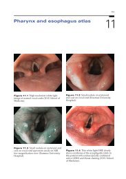
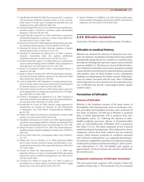
Enzymatic mechanism of bilirubin formation<br />
The microsomal haem oxygenase (HO) enzymes catalyse the<br />
oxidation of haem (Fig. 1). Three molecules of O 2 are consumed
166 2 FUNCTIONS OF THE LIVER<br />
M<br />
b<br />
V<br />
+ 3O 2<br />
5NADPH<br />
Haem<br />
oxygenase<br />
CO<br />
P<br />
M<br />
N<br />
N<br />
Haem<br />
g P<br />
Fe<br />
N<br />
N<br />
a V<br />
Fe 3+<br />
in this reaction and a reducing agent, such as nicotinamide<br />
adenine dinucleotide phosphate hydrogenase (NADPH), is<br />
needed. The α-methene bridge carbon is eliminated as CO<br />
and the iron molecule is released [6]. Of the three forms of HO,<br />
HO-1 is ubiquitous and inducible by haem [8] and stress [7];<br />
HO-2 is a constitutive protein, expressed mainly in the brain<br />
and the testis. The catalytic activity of HO-3 is low, and this<br />
protein may function mainly as a haem binding protein. CO<br />
produced by HO activity has a vasodilatory effect and regulates<br />
the vascular tone in the liver, heart and other organs during<br />
stress. Similarly, biliverdin and its product bilirubin are potent<br />
antioxidants, which may protect tissues under oxidative stress<br />
[7,9] (see below).<br />
Biliverdin is reduced to bilirubin by the action of cytosolic<br />
biliverdin reductases, which require NADH or NADPH for<br />
activity [10]. As discussed later, bilirubin requires energyconsuming<br />
metabolic steps for excretion in bile. Thus, the<br />
physiological advantage of its formation is not clear. The strong<br />
antioxidant activity of bilirubin may be particularly important<br />
during the neonatal period, when other antioxidants are scarce<br />
in body fluids.<br />
Measurement of bilirubin production<br />
M<br />
<strong>Bilirubin</strong> production can be quantified from the turnover of<br />
intravenously administered radioisotopically labelled bilirubin.<br />
Plasma bilirubin clearance is proportional to the reciprocal of<br />
the area under the radiobilirubin disappearance curve [11].<br />
<strong>Bilirubin</strong> removal is calculated as the product of plasma bilirubin<br />
concentration and clearance. At a steady state of plasma<br />
bilirubin concentration, bilirubin removal equals bilirubin<br />
M<br />
d<br />
V<br />
M<br />
P P<br />
M<br />
N<br />
N H<br />
H<br />
N<br />
H<br />
O O<br />
N<br />
Biliverdin<br />
V<br />
M<br />
V<br />
M<br />
M<br />
P<br />
M<br />
<strong>Bilirubin</strong><br />
NN<br />
H<br />
N<br />
H<br />
H 2<br />
C<br />
N<br />
H<br />
O O<br />
P<br />
V<br />
Biliverdin<br />
reductase<br />
NADPH<br />
H<br />
N<br />
M<br />
M<br />
NADP<br />
Fig. 1 Enzyme-catalysed degradation of<br />
haem. Haem degradation begins by haem<br />
oxygenase-catalysed oxidation of the a-bridge<br />
carbon of haem, which is converted to CO,<br />
leading to opening of the tetrapyrrole ring and<br />
release of the iron molecule. The resulting<br />
biliverdin molecule is subsequently reduced to<br />
bilirubin by cytosolic biliverdin reductase.<br />
production. More conveniently, bilirubin formation can be<br />
quantified from CO, which is generated in equimolar amounts<br />
with bilirubin. Following rebreathing in a closed system, CO<br />
production is calculated from the CO concentration in the<br />
rebreathing mask and/or the increment in blood carboxyhaemoglobin<br />
saturation [12]. A small fraction of the CO may be<br />
formed by intestinal bacteria, which can be a significant source<br />
of CO in intestinal bacterial overgrowth syndromes [13].<br />
Inhibition of bilirubin production<br />
Substances, such as tin-protoporphyrin and tin-mesoporphyrin,<br />
that bind irreversibly to HO, but are not broken down, serve<br />
as ‘dead-end’ inhibitors of the enzyme and reduce bilirubin<br />
production [14]. Injection of tin-mesoporphyrin lowers serum<br />
bilirubin levels by 76% in neonates [15].<br />
Chemical characteristics of bilirubin<br />
The tetrapyrrole structure of bilirubin IXα (1,8-dioxo-1,3,6,7tetramethyl-2,8-divinylbiladiene-a,c-dipropionic<br />
acid [17]) was<br />
solved by Fischer and Plieninger [18]. X-ray crystallography has<br />
revealed that the propionic acid side-chains of bilirubin form<br />
hydrogen bonds with the pyrrolic and lactam sites on the opposite<br />
half of the molecule, giving rise to a distorted ‘ridge tile’<br />
structure [19] (Fig. 2). Formation of hydrogen bonds requires<br />
the interpyrrolic bridges at the 5 and 15 position of bilirubin to<br />
be in trans or ‘Z’ configuration, whereby bilirubin is termed<br />
bilirubin IXα-ZZ. Engagement of all polar groups (two propionic<br />
acid carboxyls, four NH groups and two lactam oxygens) of<br />
bilirubin by the hydrogen bonds makes the molecule insoluble
Fig. 2 Internal hydrogen bonding and<br />
photoisomerization of bilirubin. The carboxylic<br />
acid moiety of the propionic acid side-chains of<br />
bilirubin form internal hydrogen bonds with<br />
contralateral NH groups and the lactam<br />
oxygen, thereby engaging all polar groups of<br />
the molecule and making it insoluble in water.<br />
Upon exposure to light, configurational<br />
changes (Z to E) occur at the C4 and C15<br />
interpyrrolic bridges, disrupting the hydrogen<br />
bonds. The bilirubin IXa-4E,15Z configurational<br />
isomer can be cyclized forming the so-called<br />
lumirubin. These configurational and geometric<br />
isomers are more polar than the hydrogenbonded<br />
bilirubin IXa-4Z,15Z and are excreted<br />
in bile without requiring glucuronidation.<br />
in water, necessitating chemical modification for excretion in<br />
bile. Disruption of the hydrogen bonds is accomplished in vivo<br />
by enzyme-catalysed esterification of the propionic acid carboxyl<br />
groups with a glycosyl moiety, mainly glucuronic acid<br />
(vide infra).<br />
The hydrogen bonds ‘bury’ the central methane bridge, so<br />
that the unconjugated bilirubin reacts very slowly with diazo<br />
reagents, whereas bilirubin glucuronides, which lack hydrogen<br />
bonds, react rapidly (‘direct’ van den Bergh reaction). The addition<br />
of ‘accelerators’ such as methanol, ethanol, 6 M urea or<br />
O<br />
O<br />
C<br />
O<br />
4<br />
N<br />
O H<br />
N<br />
H<br />
O<br />
10<br />
C<br />
H<br />
N<br />
H<br />
H<br />
O<br />
<strong>Bilirubin</strong> IXa-4Z,15E<br />
HOOC<br />
COOH<br />
4 10 15<br />
N<br />
H<br />
N<br />
H<br />
N<br />
H<br />
N<br />
H<br />
O<br />
O H<br />
C<br />
O H<br />
N<br />
H<br />
4<br />
N N<br />
15<br />
H 10<br />
N H<br />
O<br />
O H<br />
O C<br />
15<br />
O<br />
N H<br />
N<br />
H<br />
2.3 METABOLISM 167<br />
dimethyl sulphoxide to plasma disrupts the hydrogen bonds of<br />
bilirubin, so that both conjugated and unconjugated bilirubin<br />
react rapidly with diazo reagents (‘total’ van den Bergh reaction).<br />
<strong>Bilirubin</strong> glucuronides in normal bile are 1-O-acyl conjugates<br />
linked to the propionic acid carboxyl of bilirubin in a β-d-ester<br />
linkage, which is hydrolysable by β-glucuronidase. However,<br />
during cholestasis, the migration of the 1-O-acyl bond from the<br />
C1 position to the C2, C3 or C4 position results in the generation<br />
of β-glucuronidase-resistant pigments [20], which are detectable<br />
in serum and bile by chromatographic analysis [21].<br />
4<br />
O<br />
<strong>Bilirubin</strong> IXa-4Z,15Z<br />
O<br />
C O<br />
H O<br />
H N<br />
H<br />
N<br />
10<br />
O<br />
H<br />
H O<br />
C<br />
<strong>Bilirubin</strong> IXa-4E,15Z<br />
N<br />
H O<br />
O<br />
C O H N<br />
O N<br />
4<br />
N<br />
10<br />
O<br />
H<br />
H O<br />
C<br />
Lumirubin<br />
H<br />
N<br />
15<br />
15
168 2 FUNCTIONS OF THE LIVER<br />
In cases of prolonged accumulation of conjugated bilirubin in<br />
plasma, as in cases of cholestasis or Dubin–Johnson syndrome,<br />
the pigment may become covalently bound to albumin [22].<br />
This irreversibly protein-bound form, often termed deltabilirubin,<br />
is included in the ‘direct’ fraction of bilirubin and is<br />
not eliminated in the bile or urine, which results in delayed<br />
clearance even after biliary obstruction or cholestasis is resolved.<br />
Effect of light<br />
The main absorption band of unconjugated bilirubin IXα is at<br />
450–474 nm in most organic solvents. Upon exposure to light,<br />
the ‘Z’ (trans) configuration of the 5 and/or 15 carbon bridges of<br />
bilirubin switches to the ‘E’ (cis) configuration. The resulting<br />
configurational isomers, ZE, EZ or EE, lack internal hydrogen<br />
bonds, are more polar than bilirubin IXα-ZZ and can be<br />
excreted in bile without conjugation [23]. The non-hydrogenbonded<br />
molecule can be stabilized slowly by cyclization of the<br />
vinyl substituent in the endovinyl half of bilirubin IXα-EZ<br />
with the methyl substituent on the internal pyrrole ring, forming<br />
the stable structural isomer, E-cyclobilirubin. Because of its<br />
stability, this molecule is quantitatively important during phototherapy<br />
for neonatal jaundice [24]. Light and oxygen can also<br />
degrade a fraction of the bilirubin molecules into colourless<br />
fragments and biliverdin [25].<br />
Quantification of bilirubin<br />
Bile pigments can be quantified as native or derivatized<br />
tetrapyrroles, or after conversion to azoderivatives. Conversion<br />
to azodipyrroles by reaction with diazo reagents is the most<br />
common method of measuring serum bilirubin levels in<br />
clinical laboratories. Electrophilic attack on the central bridge<br />
splits bilirubin into two diazotized azodipyrrole molecules. As<br />
discussed above, conjugated bilirubin reacts rapidly (‘direct’<br />
fraction), while total bilirubin is determined after adding an<br />
accelerator. Unconjugated bilirubin is calculated by subtracting<br />
the direct fraction from total bilirubin. As 10–15% of unconjugated<br />
bilirubin may give a ‘direct’ diazo reaction, this method<br />
slightly overestimates conjugated bilirubin.<br />
<strong>Bilirubin</strong> and its conjugates in serum or bile can be quantified<br />
more accurately as intact bilirubin tetrapyrroles by high-pressure<br />
liquid chromatography [26–28]. <strong>Bilirubin</strong> mono- and<br />
diconjugates can be converted to methyl esters by alkaline<br />
methanolysis prior to separation [29] but, because the sugar<br />
groups are cleaved off, this method does not permit<br />
identification of specific conjugates.<br />
For repeated bilirubin measurements in jaundiced infants, as<br />
an extension of clinical evaluation of jaundice, bilirubin levels<br />
can be assessed by measurement of the intensity of yellow discoloration<br />
of the skin using a special reflectance photometer<br />
[30]. Two slide tests (Ektachem) are available for determination<br />
of total bilirubin and the unconjugated, conjugated and irreversibly<br />
protein-bound fractions.<br />
<strong>Bilirubin</strong> toxicity<br />
Unconjugated bilirubin is toxic to many cell types, intracellular<br />
organelles and physiological processes. <strong>Bilirubin</strong> inhibits DNA<br />
synthesis [31] and ATPase activity of brain mitochondria [32],<br />
and uncouples oxidative phosphorylation. It has been reported<br />
to inhibit Ca 2+ -activated, phospholipid-dependent protein<br />
kinase C activity and cAMP-dependent protein kinase activity<br />
[33]. Which of these toxic effects is the predominant cause<br />
of bilirubin encephalopathy remains unclear at this time.<br />
Clinically, toxic effects of bilirubin, particularly on the brain, are<br />
seen in neonates and patients with severe inherited deficiency of<br />
bilirubin conjugation. Yellow discoloration of the hippocampus,<br />
basal ganglia and nuclei of the cerebellum and brain stem,<br />
found in infants with acute bilirubin encephalopathy, is termed<br />
kernicterus. Such discoloration is not found in patients with<br />
chronic encephalopathy, in whom focal necrosis of neurons and<br />
glia is seen [34].<br />
As all toxic effects of bilirubin are abrogated by tight binding<br />
to albumin, cerebral toxicity is usually seen when there is a<br />
molar excess of bilirubin over albumin in plasma. At serumunconjugated<br />
bilirubin concentrations over 20 mg/dL, newborn<br />
babies are at risk of kernicterus. However, kernicterus can occur<br />
at lower concentrations in the presence of substances such as<br />
sulphonamides, radiographic contrast dyes and coumarin, which<br />
inhibit albumin–bilirubin binding by competitive or allosteric<br />
displacement [35,36]. Although immaturity of the blood–brain<br />
barrier in neonates has been implicated in the increased susceptibility<br />
of neonates to kernicterus, evidence to support this<br />
concept is insufficient. Normally, bilirubin entering the brain<br />
is cleared rapidly, but the pigment may bind to damaged and<br />
oedematous brain inhibiting its clearance, thereby increasing<br />
the susceptibility to bilirubin encephalopathy [37].<br />
Potential beneficial effects of products<br />
of haem breakdown<br />
Although clinicians are mainly concerned with the importance<br />
of bilirubin levels as a marker of liver disease and with the toxic<br />
effects of the pigment, biliverdin and bilirubin may exert some<br />
beneficial effects by virtue of their strong antioxidant properties.<br />
This may be relevant during the newborn period, when the level<br />
of other natural antioxidants is low. <strong>Bilirubin</strong>, which is toxic to<br />
neuronal cells at high concentrations, has been reported to have<br />
cytoprotective activity at lower concentrations. An inverse relationship<br />
between serum bilirubin levels and risk of ischaemic<br />
coronary artery disease has been observed [38], although<br />
whether such a protective effect extends to subjects with Gilbert<br />
syndrome is questionable [39]. Study of a large number of<br />
subjects in the United States has shown that the odds ratio<br />
for colorectal cancer is reduced to 0.295 in men and 0.186 in<br />
women per 1 mg/dL increment in serum bilirubin levels [40].<br />
Similarly, a previous large study showed an inverse relationship<br />
between serum bilirubin levels and cancer mortality in a Belgian
population [41]. However, such associations do not conclusively<br />
prove a causative role for bilirubin, because possible confounding<br />
variables may exist.<br />
<strong>Bilirubin</strong> in body fluids<br />
About 4% of bilirubin in normal plasma is conjugated, but<br />
the clinical diazo-based methods overexpress this fraction (see<br />
above). In haemolytic jaundice, there is a proportional increase<br />
in plasma-unconjugated and -conjugated bilirubin. In contrast,<br />
in inherited disorders of bilirubin conjugation, the conjugated<br />
bilirubin is absent or reduced in proportion. In biliary obstruction<br />
or hepatocellular diseases, both conjugated and unconjugated<br />
bilirubin accumulate in plasma. <strong>Bilirubin</strong> is present in<br />
exudates and other albumin-containing body fluids and binds<br />
to the elastic tissue of skin and sclera. Haem in subcutaneous<br />
haematomas is sequentially converted to biliverdin and bilirubin,<br />
resulting in a transition from green to yellow discoloration.<br />
Because of tight binding to albumin, unconjugated bilirubin is<br />
not excreted in urine in the absence of albuminuria, but conju-<br />
Fig. 3 Glucuronidation disrupts internal<br />
hydrogen bonding of bilirubin.<br />
Glucuronidation of the propionic acid carboxyl<br />
groups results in disruption of the internal<br />
hydrogen bonds, making the molecule more<br />
polar and secretable in bile. Disruption of<br />
hydrogen bonding exposes the central CHH<br />
bridge to diazo reagents, whereby bilirubin<br />
glucuronides give the direct van den Bergh<br />
reaction.<br />
O<br />
O<br />
HO<br />
H<br />
4<br />
N<br />
N<br />
H H<br />
O<br />
O<br />
A B<br />
N<br />
N<br />
H H<br />
COOH<br />
HO<br />
4<br />
O O<br />
HO<br />
2.3 METABOLISM 169<br />
gated bilirubin, which is less strongly bound to albumin, appears<br />
in urine. <strong>Bilirubin</strong> is present in normal human bile predominantly<br />
as diglucuronide, with bilirubin monoglucuronide and<br />
unconjugated bilirubin accounting for less than 10% and 1–4%<br />
of the pigments respectively. In the presence of reduced bilirubin<br />
glucuronidating capacity of the liver, as in Gilbert syndrome<br />
and Crigler–Najjar syndrome type 2 (see Chapter 16.6), the proportion<br />
of bilirubin monoglucuronide increases to 30% or above.<br />
In addition to the glucuronides, small amounts of glucosyl, xylosyl<br />
and mixed conjugates of bilirubin are found in human bile.<br />
Disposition of bilirubin<br />
Disposition of bilirubin by hepatocytes comprises several<br />
specific steps, including transport of bilirubin to hepatocytes<br />
from sites of production, uptake by and storage within hepatocytes,<br />
enzyme-catalysed conjugation with glucuronic acid,<br />
active transport into the bile canaliculus and degradation in the<br />
intestinal tract. These steps are summarized in Figure 3 and<br />
discussed briefly below.<br />
O<br />
10<br />
<strong>Bilirubin</strong> IXa-4Z,15Z<br />
10<br />
<strong>Bilirubin</strong> IXa-diglucuronide<br />
O<br />
N N<br />
H<br />
H<br />
O<br />
O<br />
15<br />
O<br />
N N<br />
H<br />
H<br />
C D<br />
15<br />
OH<br />
H<br />
OH<br />
O<br />
O<br />
O COOH OH
170 2 FUNCTIONS OF THE LIVER<br />
Transport in plasma<br />
Unconjugated bilirubin circulates in plasma bound tightly but<br />
reversibly to albumin, which prevents its excretion in urine,<br />
except during albuminuria. Albumin binding keeps bilirubin<br />
in solution and abrogates its toxic effects. Conjugated bilirubin<br />
is bound less tightly to albumin, and the unbound fraction is<br />
excreted in the urine. As mentioned above, during prolonged<br />
conjugated hyperbilirubinaemia, a fraction of conjugated bilirubin<br />
becomes irreversibly bound to albumin. This fraction,<br />
termed delta-bilirubin, is not excreted in the bile or urine and<br />
disappears slowly, reflecting the long half-life of albumin [22].<br />
A small unbound fraction of unconjugated bilirubin is thought<br />
to be responsible for its toxicity [42]. Albumin has one highaffinity<br />
primary binding site for bilirubin. Additional sites are<br />
occupied when bilirubin is in molar excess. Normal plasma concentration<br />
of albumin (500–700 µmol/L) exceeds that of bilirubin<br />
(3–17 µmol/L). However, during exaggerated neonatal<br />
jaundice and in patients with Crigler–Najjar syndrome, the<br />
molar concentration of unconjugated bilirubin may exceed that<br />
of albumin. Hypoalbuminaemia resulting from inflammatory<br />
states, chronic malnutrition or liver disease may precipitate<br />
bilirubin toxicity. Sulphonamides, anti-inflammatory drugs,<br />
cholecystographic contrast media, fusidic acid, azapropazone,<br />
sodium caprylate and N-acetyl tryptophan displace bilirubin<br />
from albumin and increase the risk of kernicterus in jaundiced<br />
infants [43]. Binding of short-chain fatty acids to albumin<br />
causes conformational changes, decreasing bilirubin binding.<br />
Because of its pathophysiological importance, various methods<br />
have been devised to measure the unbound fraction of bilirubin<br />
and the reserve albumin binding capacity. These include<br />
ultrafiltration, ultracentrifugation, gel chromatography, affinity<br />
chromatography on albumin agarose polymers, dialysis and<br />
electrophoresis. Rapid degradation of unbound bilirubin by<br />
H 2 O 2 and horseradish peroxidase has been used to distinguish<br />
it from the bound fraction.<br />
Uptake by hepatocytes<br />
At the sinusoidal surface of the hepatocyte (Fig. 4), bilirubin<br />
dissociates from albumin and is taken up by the hepatocyte<br />
by facilitated diffusion that requires inorganic anions, such as<br />
Cl – . The protein(s) involved in sinusoidal bilirubin uptake<br />
have not been identified. A member of the organic anion transport<br />
protein family, termed OATP2 (also termed SLC21A6),<br />
has been proposed as the sinusoidal bilirubin transporter [44],<br />
but its importance in bilirubin transport has been questioned<br />
[45].<br />
Storage within the liver cell<br />
After entering the hepatocyte, bilirubin binds to the major<br />
cytosolic proteins, glutathione-S-transferases (GSTs, formerly<br />
designated ligandin or Y-protein). The GST proteins, which<br />
constitute 5% of the liver cytosol, bind various drugs, hormones,<br />
organic anions [46], a cortisol metabolite [47] and azo-dye carcinogens<br />
[48]. <strong>Bilirubin</strong> is a ligand for GSTs, but not a substrate<br />
for glutathione transfer. Binding to GSTs reduces the efflux of<br />
bilirubin from hepatocytes, thereby increasing its net uptake<br />
(Fig. 4). GST binding inhibits non-specific diffusion of bilirubin<br />
into various subcellular compartments, thereby preventing<br />
specific organellar toxicity, such as inhibition of mitochondrial<br />
respiration by bilirubin that is seen in vitro [49].<br />
Conjugation of bilirubin<br />
Conversion of unconjugated bilirubin to bilirubin diglucuronide<br />
or monoglucuronide by esterification of both or one of<br />
the propionic acid carboxyl groups is critical for efficient biliary<br />
excretion of bilirubin (Fig. 4).<br />
<strong>Bilirubin</strong> uridine diphosphoglucuronate<br />
glucuronosyltransferase<br />
<strong>Bilirubin</strong> is one of the many endogenous and exogenous substrates<br />
whose conjugation with glucuronic acid is mediated<br />
by one or more isoform of uridine diphosphoglucuronate glucuronosyltransferase<br />
(UGTs). UGTs are enzymes concentrated<br />
in the endoplasmic reticulum (ER) and nuclear envelope of<br />
many cell types [50]. They catalyse the transfer of the glucuronic<br />
acid moiety of UDP-glucuronic acid to the aglycone substrates,<br />
forming polar and usually less bioreactive products. <strong>Bilirubin</strong><br />
glucuronidation is catalysed predominantly by a single UGT isoform,<br />
UGT1A1 [51]. The UGT superfamily of genes comprises<br />
two major families, UGT1 and UGT2. Nine isoforms within the<br />
UGT1A subfamily are expressed from a series of exons clustered<br />
in a unique manner on chromosome 2 at the 2q37 region [61].<br />
Four consecutive exons (exons 2–5) located at the 3′ end of the<br />
UGT1A locus are used in nine different mRNAs. These encode<br />
the identical carboxy-terminal domains of these UGT isoforms,<br />
which contain the UDP-glucuronic acid binding site. Upstream<br />
of these four common region exons is a series of unique exons,<br />
each preceded by a separate promoter. Only one of these exons is<br />
utilized in a specific UGT mRNA. The unique exon encodes the<br />
variable N-terminal domain of the nine different UGT isoforms<br />
that impart aglycone specificity to the individual isoforms.<br />
Depending on which promoter is used, transcripts of various<br />
lengths are generated. In all cases, the unique exon, located at the<br />
5′ end of the transcript, is spliced to exon 2, and the intervening<br />
sequence is spliced out. The genes are named according to the<br />
unique first exon. Thus, UGT1A1 utilizes the unique exon 1A1,<br />
UGT1A6 utilizes exon 1A6, etc. [53].<br />
The presence of a separate promoter upstream from each<br />
unique region exon permits differential regulation of individual<br />
UGT isoforms during development and in response to inducing<br />
agents. UGT1A1 is expressed after birth [54] and is induced<br />
by phenobarbital and clofibrate [55]. Delayed expression of<br />
UGT1A1 is a major cause of neonatal hyperbilirubinaemia in<br />
primates. Treatment of rats with triiodothyronine markedly
Fig. 4 <strong>Bilirubin</strong> throughput by hepatocytes.<br />
<strong>Bilirubin</strong> is transported from sites of production<br />
to hepatic sinusoids bound to albumin (1).<br />
At the sinusoidal surface of hepatocytes,<br />
bilirubin dissociates from albumin and enters<br />
hepatocytes by facilitated diffusion (2). Binding<br />
to cytosolic glutathione-S-transferases (GSTs)<br />
increases net uptake of bilirubin by inhibiting its<br />
efflux (3). <strong>Bilirubin</strong> is converted to mono- and<br />
diglucuronide by the action of UGT1A1, which<br />
catalyses the transfer of the glucuronic acid<br />
moiety from UDP-glucuronic acid (UDPGA) to<br />
bilirubin (4). <strong>Bilirubin</strong> glucuronides are actively<br />
transported into bile against a concentration<br />
gradient by the ATP-utilizing pump ABCC2<br />
(also termed MRP2) (5).<br />
ABCC2<br />
(MRP2)<br />
reduces UGT activity towards bilirubin, whereas the activity<br />
towards 4-nitrophenol is increased [56].<br />
In humans, the expression of UGT1A1 is limited to hepatocytes<br />
and, to a lesser extent, the proximal small intestine. UGTs<br />
are integral to ER membranes. In addition to the enzyme<br />
content, UGT1A1 activity is affected by the lipids of the ER<br />
membrane. UGT activity in native microsomal vesicles is latent<br />
[57], probably because the ER membranes pose a barrier to the<br />
polar sugar donor UDP-glucuronic acid or as a result of the<br />
constraint of the enzyme by the membranes. Based on hydrophobicity<br />
analysis, the major portion of mature UGT molecules,<br />
including the UDP-glucuronic acid and the aglycone binding<br />
sites, is thought to be located within the ER cisternae. There<br />
is a single 17-amino-acid membrane-spanning segment and<br />
a 26-amino-acid cytoplasmic tail at the carboxy-terminal<br />
end of the molecule. Full enzyme activity is manifested<br />
in vitro by treatment of the microsomes with membranepermeabilizing<br />
agents, such as digitonin or alamethacin. UDP-<br />
N-acetylglucosamine (UDP-glucNac) stimulates the internalization<br />
of UDP-glucoronic acid into intact microsomal vesicles<br />
and is thought to be the natural activator of UGTs within hepatocytes.<br />
UGT1A1 forms homodimers within the ER membrane,<br />
which may be required for its full catalytic activity [58]. In addition,<br />
it may interact with other UGT isoforms, as well as other<br />
proteins of the ER.<br />
Canalicular excretion of conjugated bilirubin<br />
<strong>Bilirubin</strong> - albumin<br />
1<br />
Conjugated bilirubin undergoes unidirectional transport into<br />
the bile against a concentration gradient, so that bilirubin concentration<br />
in the bile can be as high as 150-fold that in the hepatocyte.<br />
The electrochemical gradient of –35 mV, generated by<br />
the sodium pump, may help in the canalicular transport but, by<br />
itself, is too small to account for this large concentration gradient.<br />
The energy for the uphill transport of bilirubin and many<br />
other non-bile salt organic anions is derived from adenosine<br />
5<br />
4<br />
3<br />
2<br />
GST – <strong>Bilirubin</strong><br />
UGT1A1<br />
<strong>Bilirubin</strong> + albumin<br />
UDPGA<br />
UDP<br />
<strong>Bilirubin</strong> diglucuronide<br />
<strong>Bilirubin</strong> monoglucuronide<br />
2.3 METABOLISM 171<br />
Sinusoidal<br />
surface<br />
Contiguous<br />
surface<br />
Canalicular<br />
(apical)<br />
surface<br />
triphosphate (ATP) hydrolysis by the canalicular ATP-binding<br />
cassette protein, ABCC2 [also termed the MDR-related protein<br />
2 (MRP2) or the multispecific organic anion transporter,<br />
MOAT]. ABCC2 pumps glutathione-, glucuronic acid- or<br />
sulphate-conjugated compounds across the canalicular membrane<br />
[59,60]. Canalicular transport of organic anions is unidirectional<br />
from the cytoplasm of the hepatocyte into the bile.<br />
Canalicular transport may be assisted by the membrane potential,<br />
but the contribution of membrane potential in organic<br />
anion transport has not been quantified. Mutant animals that<br />
lack ATP-dependent canalicular transport of non-bile acid<br />
organic anions retain normal activity with respect to potentialdriven<br />
canalicular transport of non-bile acid organic anions,<br />
including bilirubin glucuronides [60]. The ATP-dependent<br />
canalicular organic anion transport is mediated by a canalicular<br />
membrane protein, termed canalicular multispecific organic<br />
anion transporter (cMOAT) or MRP2 [61].<br />
Maximal bilirubin secretory capacity (Tmax) into the bile<br />
canaliculus depends on bile flow, which has bile salt-dependent<br />
and non-bile salt-dependent components. Bile acids increase<br />
the trafficking of vesicles containing MRP2 and the bile salt<br />
export pump (BSEP) from the Golgi apparatus to the apical<br />
domain of hepatocyte plasma membranes, thereby increasing<br />
the concentration of the transporters in the canalicular membrane<br />
[61].<br />
Fate of bilirubin in the gastrointestinal tract<br />
Although conjugated bilirubin is not substantially absorbed<br />
from the intestines, a fraction of the small amount of unconjugated<br />
bilirubin that is excreted in bile is absorbed and undergoes<br />
enterohepatic circulation. In situations in which increased<br />
amounts of unconjugated bilirubin are excreted in bile, such as<br />
e.g. during phototherapy for neonatal jaundice or Crigler–<br />
Najjar syndrome, absorption of unconjugated bilirubin from<br />
the intestine may be clinically significant [62]. In these cases,
172 2 FUNCTIONS OF THE LIVER<br />
interruption of bilirubin reabsorption by ingestion of various<br />
substances, including calcium salts, can enhance the effect of<br />
phototherapy [63].<br />
Degradation of bilirubin by intestinal bacteria generates<br />
urobilinogen and related products [64]. A major portion of the<br />
urobilinogen reabsorbed from the intestine is excreted in bile,<br />
but a small fraction is excreted in urine. Urobilinogen is colourless;<br />
its oxidation product, urobilin, contributes to the colour of<br />
normal urine and stool. During severe intrahepatic cholestasis<br />
or complete obstruction of the bile duct, urobilinogen and urobilin<br />
are absent in urine and stool, resulting in pale (so-called<br />
clay-coloured) stool. In liver disease and states of increased<br />
bilirubin production, urinary urobilinogen excretion is increased.<br />
Alternative routes of bilirubin elimination<br />
In the absence of bilirubin glucuronidation, a fraction of bilirubin<br />
is excreted as hydroxylated products [65], probably by the<br />
action of microsomal P450s [66] and mitochondrial bilirubin<br />
oxidase in liver [67] and other tissues.<br />
During intrahepatic or extrahepatic cholestasis, conjugated<br />
bilirubin accumulates in plasma. In total biliary obstruction,<br />
renal excretion becomes the major pathway of bilirubin excretion<br />
[68]. Renal excretion of conjugated bilirubin depends on<br />
glomerular filtration of the non-protein-bound fraction of conjugated<br />
bilirubin.<br />
References<br />
1 Chen TS, Chen PS (1984) Understanding the Liver. A History.<br />
Westport, CT: Greenwood Press, p. 99.<br />
2 London IM, West R, Shemin D et al. (1950) On the origin of bile<br />
pigment in normal man. J Biol Chem 184, 351–358.<br />
3 Schwartz S, Johnson JA, Stephenson BD et al. (1971) Erythropoietic<br />
defects in protoporphyria: a study of factors involved in labelling of<br />
porphyrins and bile pigments from ALA- 3 H and glycine- 14 C. J Lab<br />
Clin Med 78, 411–434.<br />
4 Robinson SH (1977) Origins of the early-labeled peak. In: Berk PD,<br />
Berlin NI (eds) Bile Pigments: Chemistry and Physiology. Washington,<br />
DC: US Government Printing Office, pp. 175–188.<br />
5 Come SE, Shohet SB, Robinson SH (1974) Surface remodeling vs.<br />
whole-cell hemolysis of reticulocytes produced with erythroid stimulation<br />
or iron deficiency anemia. Blood 44, 817–830.<br />
6 Tenhunen R, Marver HS, Schmid R (1969) Microsomal heme<br />
oxygenase: characterization of the enzyme. J Biol Chem 244,<br />
6388–6394.<br />
7 Elbirt KK, Bonkovsky HL (1999) Heme oxygenase: recent advances<br />
in understanding its regulation and role. Proc Assoc Am Phys 111,<br />
438–447.<br />
8 Ishizawa S, Yoshida T, Kikuchi G (1983) Induction of heme oxygenase<br />
in rat liver. J Biol Chem 258, 4220–4225.<br />
9 Hayashi S, Takamiya R, Yamaguchi T et al. (1999) Induction of<br />
heme oxygenase-1 suppresses venular leukocyte adhesion elicited by<br />
oxidative stress: role of bilirubin generated by the enzyme. Circ Res 85,<br />
663–671.<br />
10 Tenhunen R, Ross ME, Marver HS et al. (1970) Reduced<br />
nicotinamide-adenine dinucleotide phosphate dependent biliverdin<br />
reductase: partial purification and characterization. Biochemistry 9,<br />
298–303.<br />
11 Jones EA, Bloomer JR, Berk PD et al. (1977) Quantitation of hepatic<br />
bilirubin synthesis in man. In: Berk PD, Berlin NI (eds) Bile Pigments:<br />
Chemistry and Physiology. Washington, DC: US Government Printing<br />
Office, pp. 189–205.<br />
12 Berk PD, Rodkey FL, Blaschke TF et al. (1974) Comparison of plasma<br />
bilirubin turnover and carbon monoxide production in man. J Lab<br />
Clin Med 83, 29–37.<br />
13 Westlake DW, Roxburgh JM, Talbot G (1961) Microbial production<br />
of carbon monoxide from flavinoids. Nature 189, 510–511.<br />
14 Kappas A, Drummond GS, Henschke C et al. (1995) Direct comparison<br />
of tin-mesoporphyrin, an inhibitor of bilirubin production, and<br />
phototherapy in controlling hyperbilirubinemia in term and nearterm<br />
newborns. Pediatrics 95 (4), 468–474.<br />
15 Valaes T, Petmezaki S, Henschke C et al. (1994) Control of jaundice in<br />
preterm newborns by an inhibitor of bilirubin production: studies<br />
with tin-mesoporphyrin. Pediatrics 93 (1), 1–11.<br />
16 Berk PD, Jones EA, Howe RB et al. (1980) Disorders of bilirubin<br />
<strong>metabolism</strong>. In: Bondy PK, Rosenberg LE (eds) Metabolic Control and<br />
Disease, 8th edn. Philadelphia: Saunders, p. 1009.<br />
17 Grandchamp B, Bissel DM, Licko V et al. (1981) Formation and disposition<br />
of newly synthesized heme in adult rat hepatocytes in primary<br />
cultures. J Biol Chem 256, 11677–11683.<br />
18 Fischer H, Plieninger H (1942) Synthese des biliverdins (uteroverdins)<br />
und bilirubins der biliverdine XIII, und III, sowie der Vinulneoxanthosaure.<br />
Hoppe Seyler Z Physiol Chem 274, 231.<br />
19 Bonnett R, Davis E, Hursthouse MB (1976) Structure of bilirubin.<br />
Nature 262 (5566), 327–328.<br />
20 Compernolle F, Van Hees GP, Blanckaert N et al. (1978) Glucuronic<br />
acid conjugates of bilirubin-IXalpha in normal bile compared with<br />
post-obstructive bile. Transformation of the 1-O-acylglucuronide<br />
into 2-, 3-, and 4-O-acylglucuronides. Biochem J 171, 185–201.<br />
21 Jansen PL (1981) β-Glucuronidase-resistant bilirubin glucuronides in<br />
cholestatic liver disease – determination of bilirubin metabolites in<br />
serum by means of high-pressure liquid chromatography. Clin Chim<br />
Acta 110, 309–317.<br />
22 Lauff JJ, Kasper ME, Ambros RT (1983) Quantitative liquid chromatographic<br />
estimation of bilirubin species in pathological serum.<br />
Clin Chem 29, 800–805<br />
23 McDonagh AF, Palma LA, Lightner DA (1982) Phototherapy for<br />
neonatal jaundice. Stereospecific and regiospecific photoisomerization<br />
of bilirubin bound to human serum albumin and NMR characterization<br />
of intramolecularly cyclized photoproducts. J Am Chem Soc<br />
104, 6867.<br />
24 Itho S, Onishi S (1985) Kinetic study of the photochemical changes of<br />
(ZZ)-bilirubin IX bound to human serum albumin. Demonstration<br />
of (EZ)-bilirubin IX as an intermediate in photochemical changes from<br />
(ZZ)-bilirubin IX to (EZ)-cyclobilirubin IX. Biochem J 226, 251–258.<br />
25 McDonagh AF (1975) Thermal and photochemical reactions of<br />
bilirubin IX. Ann NY Acad Sci 244, 553–569.<br />
26 Onishi S, Itho S, Kawade N et al. (1980) An accurate and sensitive<br />
analysis by high pressure liquid chromatography of conjugated and<br />
unconjugated bilirubin IXa and in various biological fluids. Biochem J<br />
185, 281–284.<br />
27 Spivak W, Carey MC (1985) Reverse-phase h.p.l.c. separation,<br />
quantification and preparation of bilirubin and its conjugates from<br />
native bile. Biochem J 225, 787–805.
28 Roy Chowdhury J, Roy Chowdhury N (1982) Quantitation of bilirubin<br />
and its conjugates by high pressure liquid chromatography. Falk<br />
Hepatol 11, 1649–1650.<br />
29 Blanckaert N, Kabra PM, Farina FA et al. (1980) Measurement of<br />
bilirubin and its mono- and diconjugates in human serum by alkaline<br />
methanolysis and high performance liquid chromatography. J Lab<br />
Clin Med 96, 198–212.<br />
30 Schumacher RE, Thornbery JM, Gutcher GR (1985) Transcutaneous<br />
bilirubinometry: a comparison of old and new methods. Pediatrics 76<br />
(1), 10–14.<br />
31 Schiff D, Chan G, Poznasky MJ (1985) <strong>Bilirubin</strong> toxicity in neural cell<br />
lines N115 and NBR10A. Pediatr Res 19 (9), 908–911.<br />
32 Mustafa MG, Cowger ML, King TE (1969) Effects of bilirubin on<br />
mitochondrial reactions. J Biol Chem 244 (23), 6403–6414.<br />
33 Sano K, Nakamura H, Tamotsu M (1985) Mode of inhibitory<br />
action of bilirubin on protein kinase C. Pediatr Res 19 (6), 587–<br />
590.<br />
34 Vaughan VC, Allen FC, Diamond LK (1950) Erythroblastosis fetalis.<br />
IV. Further observations on kernicterus. Pediatrics 6, 706.<br />
35 Gourley GR (1997) <strong>Bilirubin</strong> <strong>metabolism</strong> and kernicterus. Adv Pediatr<br />
44, 173–229.<br />
36 Odell GB (1973) Influence of binding on the toxicity of bilirubin. Ann<br />
NY Acad Sci 226, 225–237.<br />
37 Lee K-S, Gartner LM (1983) Management of unconjugated hyperbilirubinemia<br />
in the newborn. Semin Liver Dis 3 (1), 52–64.<br />
38 Breimer LH, Wannamethee G, Ebrahim S et al. (1995) Serum bilirubin<br />
and risk of ischemic heart disease in middle-aged British men. Clin<br />
Chem 41, 1504–1508.<br />
39 Bosma PJ, van der Meer, IM, Bakker CT et al. (2003) UGT1A1*28<br />
allele and coronary heart disease: the Rotterdam Study. Clin Chem 49,<br />
1180–1181.<br />
40 Zucker SD, Horn PS, Serman KE (2004) Serum bilirubin levels in the<br />
U.S. population: gender effect and inverse correlation with colorectal<br />
cancer. Hepatology 40, 827–835.<br />
41 Temme EHM, Zhang J, Schouten EG et al. (2001) Serum bilirubin and<br />
10-year mortality risk in a Belgian population. Cancer Causes Control<br />
12, 887–894.<br />
42 Bowen WR, Porter E, Waters WF (1959) The protective action of<br />
albumin in bilirubin toxicity in new born puppies. Am J Dis Child 98,<br />
568–573.<br />
43 Brodersen R (1986) Aqueous solubility, albumin binding and tissue<br />
distribution of bilirubin. In: Ostrow JD (ed.) Bile Pigments and<br />
Jaundice. New York: Marcel Dekker, pp. 157–181.<br />
44 Cui Y, Konig J, Leier I et al. (2000) Hepatic uptake of bilirubin and its<br />
conjugates by the human organic anion-transporting polypeptide 2<br />
(symbol SLC21A6). J Biol Chem 276, 9626–9630.<br />
45 Wang P, Kim RB, Roy-Chowdhury J et al. (2003) Organic anion<br />
transport protein SLC21A6 (OATP2) is not sufficient for bilirubin<br />
transport. J Biol Chem 278 (23), 20695–20696<br />
46 Levi AJ, Gatmaitan Z, Arias IM (1969) Two hepatic cytoplasmic protein<br />
fractions, Y and Z, and their possible role in the hepatic uptake of<br />
bilirubin, sulfobromophthalein, and other anions. J Clin Invest 48,<br />
2156–2167.<br />
47 Morey KS, Litwack G (1969) Isolation and properties of cortisol<br />
metabolite binding proteins of rat liver cytosol. Biochemistry 8,<br />
4813–4821.<br />
48 Ketterer B, Ross-Mansell P, Whitehead JK (1967) The isolation of<br />
carcinogen-binding protein from livers of rats given 4-dimethylaminoazobenzene.<br />
Biochem J 103, 316–324.<br />
2.3 METABOLISM 173<br />
49 Kamisaka K, Gatmaitan Z, Moore CL et al. (1975) Ligandin reverses<br />
bilirubin inhibition of liver mitochondrial respiration in vitro. Pediatr<br />
Res 9 (12), 903–905.<br />
50 Roy Chowdhury J, Novikoff PM, Roy Chowdhury N et al. (1985)<br />
Distribution of uridinediphosphoglucuronate glucuronosyl transferase<br />
in rat tissues. Proc Natl Acad Sci USA 82, 2990–2994.<br />
51 Bosma PJ, Seppen J, Goldhoorn B et al. (1994) <strong>Bilirubin</strong> UDPglucuronosyltransferase<br />
1 is the only relevant bilirubin glucuronidating<br />
isoform in man. J Biol Chem 269 (27), 17960–17964<br />
52 Ritter JK, Chen F, Sheen YY et al. (1992) A novel complex locus UGT1<br />
encodes human bilirubin, phenol and other UDP-glucuronosyltransferase<br />
isozymes with identical carboxy termini. J Biol Chem 267 (5),<br />
3257–3261.<br />
53 Mackenzie PI, Owens IS, Burchell B et al. (1997) The UDP glucosyltransferase<br />
gene superfamily: recommended nomenclature update<br />
based on evolutionary divergence. Pharmacogenetics 7 (4), 255–269.<br />
54 Wishart GJ (1978) Functional heterogeneity of UDP-glucuronosyl<br />
transferase as indicated by its differential development and inducibility<br />
by glucocorticoids. Biochem J 174 (2), 485–489.<br />
55 Roy Chowdhury J, Roy Chowdhury N, Moscioni AD et al. (1983)<br />
Differential regulation by triiodothyronine of substrate-specific<br />
uridinediphosphoglucuronate glucuronyl transferases in rat liver.<br />
Biochim Biophys Acta 761 (1), 58–65.<br />
56 Lilienblum W, Walli AK, Bock KW (1982) Differential induction of rat<br />
liver microsomal UDP-glucuronosyltransferase activities by various<br />
inducing agents. Biochem Pharmacol 31 (6), 907–913.<br />
57 Bossuyt X, Blanckaert N (1997) Carrier-mediated transport of uridine<br />
diphosphoglucuronic acid across the endoplasmic reticulum membrane<br />
is a prerequisite for UDP-glucuronosyltransferase activity in rat<br />
liver. Biochem J 323, 645–648.<br />
58 Ghosh SS, Sappal BS, Ganjam VK et al. (2001) Homodimerization of<br />
human bilirubin-uridine-diphosphoglucuronate glucuronosyltransferase-1<br />
(UGT1A1) and its functional implications. J Biol Chem 276,<br />
42108–42115<br />
59 Ishikaowa T, Muller M, Klunemann C et al. (1990) ATP-dependent<br />
primary active transport of cysteinyl leukotrienes transport system for<br />
glutathione S-conjugates. J Biol Chem 265 (31), 19279–19286.<br />
60 Nishida T, Gatmaitan Z, Roy-Chowdhury J et al. (1992) Two distinct<br />
mechanisms for bilirubin glucuronide transport by rat bile canalicular<br />
membrane vesicles. J Clin Invest 90 (5), 2130–2135.<br />
61 Gatmaitan ZC, Nies AT, Arias IM (1997) Regulation and translocation<br />
of ATP-dependent apical membrane proteins in rat liver. Am J Physiol<br />
272, G1041–G1049.<br />
62 Brodersen R, Herman LS (1963) Intestinal reabsorption of unconjugated<br />
bilirubin. A possible contributing factor in neonatal jaundice.<br />
Lancet 1, 1242.<br />
63 Van der Veere CN, Jansen PL, Sinaasappel M et al. (1997) Oral calcium<br />
phosphate: a new therapy for Crigler–Najjar disease? Gastroenterology<br />
112, 455–462.<br />
64 Watson CJ (1977) The urobilinoids: milestones in their history and<br />
some recent developments. In: Berk PD, Berlin NI (eds) Bile Pigments:<br />
Chemistry and Physiology. Washington, DC: US Government Printing<br />
Office, pp. 469–482.<br />
65 Berry CS, Zarembo JE, Ostrow JD (1972) Evidence for conversion of<br />
bilirubin to dihydroxyl derivatives in the Gunn rat. Biochem Biophys<br />
Res Commun 49 (5), 1366–1375.<br />
66 Kapitulnik J, Ostrow JD (1978) Stimulation of bilirubin catabolism in<br />
jaundiced Gunn rats by an inducer of microsomal mixed function<br />
mono oxygenases. Proc Natl Acad Sci USA 75 (2), 682–685.
174 2 FUNCTIONS OF THE LIVER<br />
67 Cardenas-Vazquez R, Yokosuka O et al. (1986) Enzymic oxidation of<br />
unconjugated bilirubin by rat liver. Biochem J 236 (3), 625–633.<br />
68 Cameron JL, Filler RM, Iber FL et al. (1966) Metabolism and excretion<br />
of 1 4 C-labeled bilirubin in children with biliary atresia. N Engl J Med<br />
274 (5), 231–236.<br />
2.3.6 Metabolism of bile acids<br />
Peter L.M. Jansen and Klaas Nico Faber<br />
Introduction<br />
Bile acids are synthesized in the liver from cholesterol; they are<br />
secreted in bile and stored in the gallbladder. After a meal, the<br />
gallbladder contracts, and stored bile is transferred to the duodenum<br />
and via the jejunum to the ileum. This movement is<br />
stimulated by intestinal propulsion. In the ileum, 90–95% of<br />
bile salts are reabsorbed and returned to the liver. The remainder<br />
is lost to the colon, where primary bile salts are transformed by<br />
bacterial <strong>metabolism</strong> into secondary bile salts. Some of the secondary<br />
bile salts are also reabsorbed, and the rest is removed<br />
with the faeces. Primary and secondary bile salts return to the<br />
liver via the portal circulation. In the liver, bile salts are taken up<br />
into hepatocytes, thereby completing the enterohepatic cycle.<br />
Bile acids serve a number of functions: (i) they are the main<br />
solutes in bile and, as such, they are important for the generation<br />
of the so-called bile salt-dependent bile flow; (ii) bile salts are<br />
indispensable for the secretion of cholesterol and phospholipids<br />
from the liver; (iii) in bile, bile salts form mixed micelles that<br />
keep fat-soluble organic compounds in solution, including fatsoluble<br />
vitamins; (iv) in the intestine, bile salts promote the dissolution<br />
and hydrolysis of triglycerides by pancreatic enzymes;<br />
(v) bile salts act as signalling molecules in the regulation of<br />
enzymes and transporters of drug and intermediary <strong>metabolism</strong>.<br />
The adult human liver produces about 500 mg of bile acids<br />
per day [1,2]. About three times this amount represents the total<br />
bile acid pool size that cycles through the enterohepatic circulation<br />
[2]. Bile acids complete an enterohepatic cycle about eight<br />
times per day. Enterohepatic cycling represents an efficient<br />
system for reusage of active components. Enterohepatic cycling<br />
not only serves to reclaim bile acids, but it also enables bile acids<br />
to act as messengers that carry signals from intestine to liver.<br />
Thus, they regulate their own synthesis and transport rates. Bile<br />
acids are also able to repress hepatic fatty acid and triglyceride<br />
synthesis [3,4].<br />
Biosynthesis and metabolic defects<br />
At least 16 different enzymes are involved in the biosynthesis of<br />
bile salts [1,5,6]. Most of these enzymes are active in the neutral<br />
(or classic) and acidic (or alternative) pathways, the two main<br />
routes for the conversion of cholesterol to the primary bile acids<br />
cholic acid (CA) and chenodeoxycholic acid (CDCA) (Fig. 1).<br />
The neutral pathway starts with the hydroxylation of the sterol<br />
nucleus of cholesterol by 7α-hydroxylase (CYP7A1) in the<br />
endoplasmic reticulum. CYP7A1 is regarded as the rate-limiting<br />
enzyme in bile acid biosynthesis, exemplified by the fact that<br />
mice deficient for Cyp7a1 have a 75% reduced bile acid pool<br />
size causing vitamin deficiencies, lipid malabsorption and liver<br />
failure [7–9]. The acidic pathway starts with the hydroxylation<br />
of the cholesterol side-chain by sterol 27-hydroxylase (CYP27).<br />
The CYP27 product, 5-cholesten-3β-27-diol, is not a substrate<br />
for CYP7A1, but is hydroxylated at the C7 position by an alternative<br />
P450 enzyme, CYP7B1. From here on, the neutral and<br />
acidic pathways largely overlap. Double hydroxylated CDCA and<br />
triple hydroxylated CA are the principal bile acids. Their ratio<br />
depends on the activity of sterol 12α-hydroxylase (CYP8B1). Bile<br />
acid synthesis is completed in hepatocyte peroxisomes, where<br />
bile acid coenzyme A:amino acid N-acyltransferase (BAAT)<br />
conjugates either taurine or glycine to CA or CDCA. At least<br />
95% of the bile acid pool is generated through these two pathways.<br />
Extensive intracellular transport of bile acid intermediates<br />
occurs between various organelles. Transport in and out of these<br />
organelles may be mediated by transport proteins, but these<br />
have not been characterized in detail yet.<br />
Bile acid synthesis defects (BASD) are rare genetic disorders<br />
that are the underlying cause of approximately 2% of persistent<br />
cholestasis in infants (see also Chapter 16.10, Genetic cholestatic<br />
diseases). BASDs are recognized by the absence or reduction of<br />
normal primary bile salts in serum and/or urine. Instead, nontypical<br />
bile acids and sterols are often detected in the body fluids<br />
of these patients. These can be identified by fast atom bombardment<br />
ionization–mass spectrometry (FAB-MS) and gas chromatography–mass<br />
spectrometry (GC-MS). Disease-causing<br />
mutations have been identified in 9 out of the 16 bile acid<br />
biosynthesis enzymes (Table 1). Cholestasis is a common clinical<br />
presentation of these diseases. The associated liver diseases<br />
may vary from mild to life-threatening but, in many cases, can<br />
be managed by replacement of deficient primary bile salts. This<br />
not only leads to restoration of normal bile function, but also<br />
induces feedback inhibition on the production of toxic bile acid<br />
intermediates.<br />
Patients with CYP7A1 deficiency have a markedly reduced bile<br />
acid synthesis rate [10]. Symptoms include hyperlipidaemia,<br />
premature vascular disease and gallstones. A mutation in the<br />
CYP7A gene that results in truncation of the enzyme has been<br />
detected in these patients. Only one case of CYP7B1 deficiency<br />
has been reported to date [11]. This child produced no primary<br />
bile acids, and serum concentrations of the toxic 27α-hydroxy<br />
cholesterol were increased. A mutation was identified in the<br />
CYP7B1 gene that truncates and inactivates the enzyme. In<br />
addition, it was found that expression of CYP7A, at both the<br />
mRNA and activity level, was absent. Bile acid treatment was<br />
ineffective, suggesting that the biosynthesis of toxic 27αhydroxy<br />
cholesterol cannot be suppressed.