Chapter 23 The Circulatory, Respiratory, Digestive, and Excretory ...
Chapter 23 The Circulatory, Respiratory, Digestive, and Excretory ...
Chapter 23 The Circulatory, Respiratory, Digestive, and Excretory ...
You also want an ePaper? Increase the reach of your titles
YUMPU automatically turns print PDFs into web optimized ePapers that Google loves.
<strong>Chapter</strong> <strong>23</strong><br />
<strong>The</strong> <strong>Circulatory</strong>, <strong>Respiratory</strong>,<br />
<strong>Digestive</strong>, <strong>and</strong> <strong>Excretory</strong><br />
Systems<br />
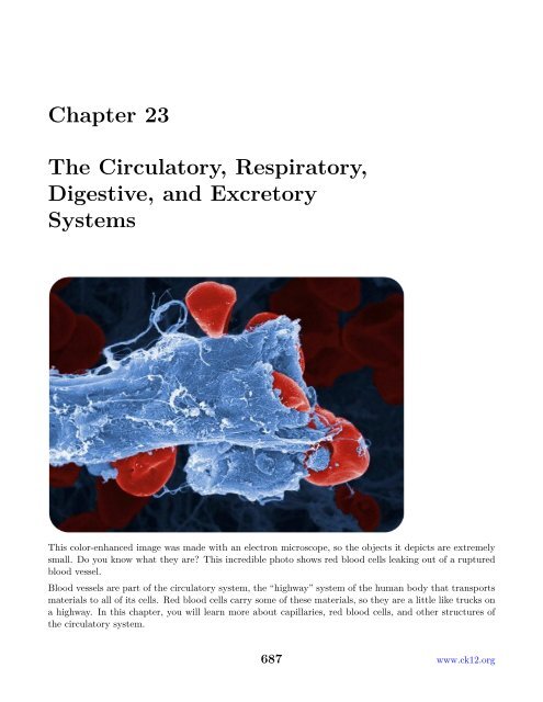
This color-enhanced image was made with an electron microscope, so the objects it depicts are extremely<br />
small. Do you know what they are? This incredible photo shows red blood cells leaking out of a ruptured<br />
blood vessel.<br />
Blood vessels are part of the circulatory system, the “highway” system of the human body that transports<br />
materials to all of its cells. Red blood cells carry some of these materials, so they are a little like trucks on<br />
a highway. In this chapter, you will learn more about capillaries, red blood cells, <strong>and</strong> other structures of<br />
the circulatory system.<br />
687 www.ck12.org
<strong>23</strong>.1 <strong>The</strong> <strong>Circulatory</strong> System<br />
Lesson Objectives<br />
• Explain how the heart pumps blood throughout the body.<br />
• Compare different types of blood vessels <strong>and</strong> their roles.<br />
• Outline pathways of the pulmonary <strong>and</strong> systemic circulations.<br />
• Define cardiovascular disease, <strong>and</strong> list its risk factors.<br />
• Describe blood, blood components, <strong>and</strong> blood pressure.<br />
Vocabulary<br />
• antigen<br />
• artery<br />
• atherosclerosis<br />
• blood<br />
• blood pressure<br />
• blood type<br />
• capillary<br />
• cardiovascular disease (CVD)<br />
• circulatory system<br />
• heart attack<br />
• hypertension<br />
• plasma<br />
• platelet<br />
• pulmonary circulation<br />
• red blood cell<br />
• systemic circulation<br />
• vein<br />
• white blood cell<br />
Introduction<br />
<strong>The</strong> circulatory system can be compared to a system of interconnected, one-way roads that range from<br />
superhighways to back alleys. Like a network of roads, the job of the circulatory system is to allow the<br />
transport of materials from one place to another. As described in Figure <strong>23</strong>.1, the materials carried by the<br />
circulatory system include hormones, oxygen, cellular wastes, <strong>and</strong> nutrients from digested food. Transport<br />
of all these materials is necessary to maintain homeostasis of the body. <strong>The</strong> main components of the<br />
circulatory system are the heart, blood vessels, <strong>and</strong> blood. Each of these components is described in detail<br />
below.<br />
<strong>The</strong> Heart<br />
<strong>The</strong> heart is a muscular organ in the chest. It consists mainly of cardiac muscle tissue <strong>and</strong> pumps blood<br />
through blood vessels by repeated, rhythmic contractions. <strong>The</strong> heart has four chambers, as shown in<br />
Figure <strong>23</strong>.2: two upper atria (singular, atrium) <strong>and</strong> two lower ventricles. Valves between chambers keep<br />
blood flowing through the heart in just one direction. For an animation of the structures of the heart, go<br />
to this link: http://www.byrnehealthcare.com/animations/SutterAnatomy.htm.<br />
www.ck12.org 688
Figure <strong>23</strong>.1: <strong>The</strong> function of the circulatory system is to move materials around the body.<br />
Figure <strong>23</strong>.2: <strong>The</strong> chambers of the heart <strong>and</strong> the valves between them are shown here.<br />
689 www.ck12.org
Blood Flow Through the Heart<br />
Blood flows through the heart in two separate loops, which are indicated by the arrows in Figure <strong>23</strong>.2.<br />
You can also watch an animation of the heart pumping blood at this link: http://www.nhlbi.nih.gov/<br />
health/dci/Diseases/hhw/hhw_pumping.html.<br />
1. Blood from the body enters the right atrium of the heart. <strong>The</strong> right atrium pumps the blood to the<br />
right ventricle, which pumps it to the lungs. This loop is represented by the blue arrows in Figure<br />
<strong>23</strong>.2.<br />
2. Blood from the lungs enters the left atrium of the heart. <strong>The</strong> left atrium pumps the blood to the left<br />
ventricle, which pumps it to the body. This loop is represented by the red arrows in Figure <strong>23</strong>.2.<br />
Heartbeat<br />
Unlike skeletal muscle, cardiac muscle contracts without stimulation by the nervous system. Instead,<br />
specialized cardiac muscle cells send out electrical impulses that stimulate the contractions. As a result,<br />
the atria <strong>and</strong> ventricles normally contract with just the right timing to keep blood pumping efficiently<br />
through the heart. You can watch an animation to see how this happens at this link: http://www.nhlbi.<br />
nih.gov/health/dci/Diseases/hhw/hhw_electrical.html.<br />
Blood Vessels<br />
Blood vessels form a network throughout the body to transport blood to all the body cells. <strong>The</strong>re are<br />
three major types of blood vessels: arteries, veins, <strong>and</strong> capillaries. All three are shown in Figure <strong>23</strong>.3 <strong>and</strong><br />
described below.<br />
Figure <strong>23</strong>.3: Blood vessels include arteries, veins, <strong>and</strong> capillaries.<br />
www.ck12.org 690
• Arteries are muscular blood vessels that carry blood away from the heart. <strong>The</strong>y have thick walls that<br />
can withst<strong>and</strong> the pressure of blood being pumped by the heart. Arteries generally carry oxygen-rich<br />
blood. <strong>The</strong> largest artery is the aorta, which receives blood directly from the heart.<br />
• Veins are blood vessels that carry blood toward the heart. This blood is no longer under much pressure,<br />
so many veins have valves that prevent backflow of blood. Veins generally carry deoxygenated<br />
blood. <strong>The</strong> largest vein is the inferior vena cava, which carries blood from the lower body to the<br />
heart.<br />
• Capillaries are the smallest type of blood vessels. <strong>The</strong>y connect very small arteries <strong>and</strong> veins. <strong>The</strong><br />
exchange of gases <strong>and</strong> other substances between cells <strong>and</strong> the blood takes place across the extremely<br />
thin walls of capillaries.<br />
Blood Vessels <strong>and</strong> Homeostasis<br />
Blood vessels help regulate body processes by either constricting (becoming narrower) or dilating (becoming<br />
wider). <strong>The</strong>se actions occur in response to signals from the autonomic nervous system or the endocrine<br />
system. Constriction occurs when the muscular walls of blood vessels contract. This reduces the amount<br />
of blood that can flow through the vessels (see Figure <strong>23</strong>.4). Dilation occurs when the walls relax. This<br />
increases blood flows through the vessels.<br />
Figure <strong>23</strong>.4: When a blood vessel constricts, less blood can flow through it.<br />
Constriction <strong>and</strong> dilation allow the circulatory system to change the amount of blood flowing to different<br />
organs. For example, during a fight-or-flight response, dilation <strong>and</strong> constriction of blood vessels allow more<br />
blood to flow to skeletal muscles <strong>and</strong> less to flow to digestive organs. Dilation of blood vessels in the skin<br />
allows more blood to flow to the body surface so the body can lose heat. Constriction of these blood vessels<br />
has the opposite effect <strong>and</strong> helps conserve body heat.<br />
691 www.ck12.org
Blood Vessels <strong>and</strong> Blood Pressure<br />
<strong>The</strong> force exerted by circulating blood on the walls of blood vessels is called blood pressure. Blood<br />
pressure is highest in arteries <strong>and</strong> lowest in veins. When you have your blood pressure checked, it is the<br />
blood pressure in arteries that is measured. High blood pressure, or hypertension, is a serious health<br />
risk but can often be controlled with lifestyle changes or medication. You can learn more about hypertension<br />
by watching the animation at this link: http://www.healthcentral.com/high-blood-pressure/<br />
introduction-47-115.html.<br />
Pulmonary <strong>and</strong> Systemic Circulations<br />
<strong>The</strong> circulatory system actually consists of two separate systems: pulmonary circulation <strong>and</strong> systemic<br />
circulation. You can watch animations of both systems at the following link. http://www.pbs.org/wnet/<br />
redgold/journey/phase2_a1.html<br />
Pulmonary Circulation<br />
Pulmonary circulation is the part of the circulatory system that carries blood between the heart <strong>and</strong><br />
lungs (the term pulmonary means “of the lungs”). It is illustrated in Figure <strong>23</strong>.5. Deoxygenated blood<br />
leaves the right ventricle through pulmonary arteries, which transport it to the lungs. In the lungs, the<br />
blood gives up carbon dioxide <strong>and</strong> picks up oxygen. <strong>The</strong> oxygenated blood then returns to the left atrium<br />
of the heart through pulmonary veins.<br />
Figure <strong>23</strong>.5: <strong>The</strong> pulmonary circulation carries blood between the heart <strong>and</strong> lungs.<br />
Systemic Circulation<br />
Systemic circulation is the part of the circulatory system that carries blood between the heart <strong>and</strong> body.<br />
It is illustrated in Figure <strong>23</strong>.6. Oxygenated blood leaves the left ventricle through the aorta. <strong>The</strong> aorta<br />
<strong>and</strong> other arteries transport the blood throughout the body, where it gives up oxygen <strong>and</strong> picks up carbon<br />
dioxide. <strong>The</strong> deoxygenated blood then returns to the right atrium through veins.<br />
www.ck12.org 692
Figure <strong>23</strong>.6: <strong>The</strong> systemic circulation carries blood between the heart <strong>and</strong> body.<br />
693 www.ck12.org
Cardiovascular Disease<br />
Diseases of the heart <strong>and</strong> blood vessels, called cardiovascular diseases (CVD), are very common. <strong>The</strong><br />
leading cause of CVD is atherosclerosis.<br />
Atherosclerosis<br />
Atherosclerosis is the buildup of plaque inside arteries (see Figure <strong>23</strong>.7). Plaque consists of cell debris,<br />
cholesterol, <strong>and</strong> other substances. Factors that contribute to plaque buildup include a high-fat diet <strong>and</strong><br />
smoking. As plaque builds up, it narrows the arteries <strong>and</strong> reduces blood flow. You can watch an animation<br />
about atherosclerosis at these links: http://www.youtube.com/watch?v=fLonh7ZesKs <strong>and</strong> http://www.<br />
youtube.com/watch?v=qRK7-DCDKEA.<br />
Figure <strong>23</strong>.7: <strong>The</strong> fatty material inside the artery on the right is plaque. Notice how much narrower the<br />
artery has become. Less blood can flow through it than the normal artery.<br />
Coronary Heart Disease<br />
Atherosclerosis of arteries that supply the heart muscle is called coronary heart disease. This disease may<br />
or may not have symptoms such as chest pain. As the disease progresses, there is an increased risk of heart<br />
attack. A heart attack occurs when the blood supply to part of the heart muscle is blocked <strong>and</strong> cardiac<br />
muscle fibers die. Coronary heart disease is the leading cause of death of adults in the U.S.<br />
Preventing Cardiovascular Disease<br />
Many factors may increase the risk of developing coronary heart disease <strong>and</strong> other CVDs. <strong>The</strong> risk of<br />
CVDs increases with age <strong>and</strong> is greater in males than females at most ages. Having a close relative with<br />
CVD also increases the risk. <strong>The</strong>se factors cannot be controlled, but other risk factors can, including<br />
smoking, lack of exercise, <strong>and</strong> high-fat diet. By making healthy lifestyle choices, you can reduce your risk<br />
of developing CVD.<br />
www.ck12.org 694
Blood<br />
Blood is a fluid connective tissue. It circulates throughout the body through blood vessels by the pumping<br />
action of the heart. Blood in arteries carries oxygen <strong>and</strong> nutrients to all the body’s cells. Blood in veins<br />
carries carbon dioxide <strong>and</strong> other wastes away from the cells to be excreted. Blood also defends the body<br />
against infection, repairs body tissues, transports hormones, <strong>and</strong> controls the body’s pH.<br />
Composition of Blood<br />
<strong>The</strong> fluid part of blood is called plasma. It is a watery golden-yellow liquid that contains many dissolved<br />
substances <strong>and</strong> blood cells. Types of blood cells in plasma include red blood cells, white blood cells, <strong>and</strong><br />
platelets (see Figure <strong>23</strong>.8). You can learn more about blood <strong>and</strong> its components by watching the animation<br />
What Is Blood? at this link: http://www.apan.net/meetings/busan03/materials/ws/education/<br />
demo-los/blood-rlo/whatisblood.swf.<br />
Figure <strong>23</strong>.8: Cells in blood include red blood cells, white blood cells, <strong>and</strong> platelets.<br />
• <strong>The</strong> trillions of red blood cells in blood plasma carry oxygen. Red blood cells contain hemoglobin,<br />
a protein with iron that binds with oxygen.<br />
• White blood cells are generally larger than red blood cells but far fewer in number. <strong>The</strong>y defend<br />
the body in various ways. For example, white blood cells called phagocytes swallow <strong>and</strong> destroy<br />
microorganisms <strong>and</strong> debris in the blood.<br />
• Platelets are cell fragments involved in blood clotting. <strong>The</strong>y stick to tears in blood vessels <strong>and</strong><br />
to each other, forming a plug at the site of injury. <strong>The</strong>y also release chemicals that are needed for<br />
clotting to occur.<br />
An overview of red blood cells can be viewed at http://www.youtube.com/user/khanacademy#p/c/<br />
7A9646BC5110CF64/36/fLKOBQ6cZHA (16:30).<br />
Hemoglobin is discussed in detail at http://www.youtube.com/user/khanacademy#p/c/7A9646BC5110CF64/<br />
38/LWtXthfG9_M (14:34).<br />
695 www.ck12.org
Blood Type<br />
Figure <strong>23</strong>.9: (Watch Youtube Video)<br />
http://www.ck12.org/flexbook/embed/view/212<br />
Figure <strong>23</strong>.10: (Watch Youtube Video)<br />
http://www.ck12.org/flexbook/embed/view/213<br />
Blood type is a genetic characteristic associated with the presence or absence of certain molecules, called<br />
antigens, on the surface of red blood cells. <strong>The</strong> most commonly known blood types are the ABO <strong>and</strong><br />
Rhesus blood types.<br />
• ABO blood type is determined by two common antigens, often referred to simply as antigens A <strong>and</strong><br />
B. A person may have blood type A (only antigen A), B (only antigen B), AB (both antigens), or O<br />
(no antigens).<br />
• Rhesus blood type is determined by one common antigen. A person may either have the antigen<br />
(Rh + ) or lack the antigen (Rh - ).<br />
Blood type is important for medical reasons. A person who needs a blood transfusion must receive blood<br />
that is the same type as his or her own. Otherwise, the transfused blood may cause a potentially lifethreatening<br />
reaction in the patient’s bloodstream.<br />
Lesson Summary<br />
• <strong>The</strong> heart contracts rhythmically to pump blood to the lungs <strong>and</strong> the rest of the body. Specialized<br />
cardiac muscle cells trigger the contractions.<br />
• Arteries carry blood away from the heart, veins carry blood toward the heart, <strong>and</strong> capillaries connect<br />
arteries <strong>and</strong> veins.<br />
• <strong>The</strong> pulmonary circulation carries blood between the heart <strong>and</strong> lungs. <strong>The</strong> systemic circulation<br />
carries blood between the heart <strong>and</strong> body.<br />
• A disease that affects the heart or blood vessels is called a cardiovascular disease (CVD). <strong>The</strong> leading<br />
cause of CVD is atherosclerosis, or the buildup of plaque inside arteries. Healthy lifestyle choices can<br />
reduce the risk of developing CVD.<br />
www.ck12.org 696
• Blood is a fluid connective tissue that contains a liquid component called plasma. It also contains<br />
dissolved substances <strong>and</strong> blood cells. Red blood cells carry oxygen, white blood cells defend the<br />
body, <strong>and</strong> platelets help blood clot.<br />
A summary of the circulatory system, blood cells <strong>and</strong> hemoglobin is available at http://www.youtube.<br />
com/user/khanacademy#p/c/7A9646BC5110CF64/37/QhiVnFvshZg (14:57).<br />
Lesson Review Questions<br />
Recall<br />
Figure <strong>23</strong>.11: (Watch Youtube Video)<br />
http://www.ck12.org/flexbook/embed/view/215<br />
1. Describe how blood flows through the heart.<br />
2. What controls heartbeat?<br />
3. How do arteries differ from veins?<br />
4. What is blood pressure? What is hypertension?<br />
5. List factors that increase the risk of cardiovascular disease.<br />
6. Identify three types of blood cells <strong>and</strong> their functions.<br />
Apply Concepts<br />
7. To take your pulse, you press your fingers against an artery near the surface of the body. What are you<br />
feeling <strong>and</strong> measuring when you take your pulse? Why can’t you take your pulse by pressing your fingers<br />
against a vein?<br />
8. People with type O blood are called “universal donors” because they can donate blood to anyone else,<br />
regardless of their ABO blood type. Explain why.<br />
Think Critically<br />
9. Compare <strong>and</strong> contrast the pulmonary <strong>and</strong> systemic circulations.<br />
10. Explain the role of blood vessels in homeostasis.<br />
Points to Consider<br />
An important function of the circulatory system is transporting oxygen to cells.<br />
697 www.ck12.org
• Do you know where blood gets the oxygen cells it needs?<br />
• How do you think blood is able to give up its oxygen to cells?<br />
<strong>23</strong>.2 <strong>The</strong> <strong>Respiratory</strong> System<br />
Lesson Objectives<br />
• Define respiration, <strong>and</strong> explain how it differs from cellular respiration.<br />
• Identify the organs of the respiratory system.<br />
• Outline the processes of ventilation, gas exchange, <strong>and</strong> gas transport.<br />
• Describe the role of gas exchange in homeostasis.<br />
• Explain how the rate of breathing is regulated.<br />
• Identify diseases of the respiratory system.<br />
Vocabulary<br />
• asthma<br />
• emphysema<br />
• larynx<br />
• lung<br />
• pharynx<br />
• pneumonia<br />
• respiration<br />
• respiratory system<br />
• trachea<br />
• ventilation<br />
Introduction<br />
Red blood cells are like trucks that transport cargo on a highway system. <strong>The</strong>ir cargo is oxygen, <strong>and</strong> the<br />
highways are blood vessels. Where do red blood cells pick up their cargo of oxygen? <strong>The</strong> answer is the<br />
lungs. <strong>The</strong> lungs are organs of the respiratory system. <strong>The</strong> respiratory system is the body system that<br />
brings air containing oxygen into the body <strong>and</strong> releases carbon dioxide into the atmosphere.<br />
Respiration<br />
<strong>The</strong> job of the respiratory system is the exchange of gases between the body <strong>and</strong> the outside air. This<br />
process, called respiration, actually consists of two parts. In the first part, oxygen in the air is drawn<br />
into the body <strong>and</strong> carbon dioxide is released from the body through the respiratory tract. In the second<br />
part, the circulatory system delivers the oxygen to body cells <strong>and</strong> picks up carbon dioxide from the cells<br />
in return. <strong>The</strong> use of the word respiration in relation to gas exchange is different from its use in the term<br />
cellular respiration. Recall that cellular respiration is the metabolic process by which cells obtain energy<br />
by “burning” glucose. Cellular respiration uses oxygen <strong>and</strong> releases carbon dioxide. Respiration by the<br />
respiratory system supplies the oxygen <strong>and</strong> takes away the carbon dioxide.<br />
An overview of breathing is shown at http://www.youtube.com/user/khanacademy#p/c/7A9646BC5110CF64/<br />
35/SPGRkexI_cs (20:33).<br />
www.ck12.org 698
Figure <strong>23</strong>.12: (Watch Youtube Video)<br />
http://www.ck12.org/flexbook/embed/view/216<br />
Organs of the <strong>Respiratory</strong> System<br />
<strong>The</strong> organs of the respiratory system that bring air into the body are shown in Figure <strong>23</strong>.13. Refer to<br />
the figure as you read below about the passage of air through these organs. You can also watch a detailed<br />
animation of the respiratory system at this link: http://www.youtube.com/watch?v=HiT621PrrO0.<br />
Figure <strong>23</strong>.13: <strong>The</strong> organs of the respiratory system move air into <strong>and</strong> out of the body.<br />
Journey of a Breath of Air<br />
Take in a big breath of air through your nose. As you inhale, you may feel the air pass down your throat <strong>and</strong><br />
notice your chest exp<strong>and</strong>. Now exhale <strong>and</strong> observe the opposite events occurring. Inhaling <strong>and</strong> exhaling<br />
may seem like simple actions, but they are just part of the complex process of respiration, which includes<br />
these four steps:<br />
699 www.ck12.org
1. Ventilation<br />
2. Pulmonary gas exchange<br />
3. Gas transport<br />
4. Peripheral gas exchange<br />
Ventilation<br />
Respiration begins with ventilation. This is the process of moving air in <strong>and</strong> out of the lungs. <strong>The</strong> lungs<br />
are the organs in which gas exchange takes place between blood <strong>and</strong> air.<br />
• Air enters the respiratory system through the nose. As the air passes through the nasal cavity, mucus<br />
<strong>and</strong> hairs trap any particles in the air. <strong>The</strong> air is also warmed <strong>and</strong> moistened so it won’t harm delicate<br />
tissues of the lungs.<br />
• Next, the air passes through the pharynx, a long tube that is shared with the digestive system. A<br />
flap of connective tissue called the epiglottis closes when food is swallowed to prevent choking.<br />
• From the pharynx, air next passes through the larynx, or voice box. <strong>The</strong> larynx contains vocal<br />
cords, which allow us to produce vocal sounds<br />
• After the larynx, air moves into the trachea, or wind pipe. This is a long tube that leads down to<br />
the chest.<br />
• In the chest, the trachea divides as it enters the lungs to form the right <strong>and</strong> left bronchi. <strong>The</strong> bronchi<br />
contain cartilage, which prevents them from collapsing. Mucus in the bronchi traps any remaining<br />
particles in air. Tiny hairs called cilia line the bronchi <strong>and</strong> sweep the particles <strong>and</strong> mucus toward the<br />
throat so they can be expelled from the body.<br />
• Finally, air passes from the bronchi into smaller passages called bronchioles. <strong>The</strong> bronchioles end in<br />
tiny air sacs called alveoli.<br />
Pulmonary Gas Exchange<br />
Pulmonary gas exchange is the exchange of gases between inhaled air <strong>and</strong> the blood. It occurs in the<br />
alveoli of the lungs. Alveoli (singular, alveolus) are grape-like clusters surrounded by networks of thinwalled<br />
pulmonary capillaries. After you inhale, there is a greater concentration of oxygen in the alveoli<br />
than in the blood of the pulmonary capillaries, so oxygen diffuses from the alveoli into the blood across<br />
the capillaries (see Figure <strong>23</strong>.14). Carbon dioxide, in contrast, is more concentrated in the blood of the<br />
pulmonary capillaries than in the alveoli, so it diffuses in the opposite direction. This link has an animation<br />
of pulmonary gas exchange: http://www.youtube.com/watch?v=Z1h29R82mVc&NR=1.<br />
Gas Transport<br />
After the blood in the pulmonary capillaries becomes saturated with oxygen, it leaves the lungs <strong>and</strong><br />
travels to the heart. <strong>The</strong> heart pumps the oxygen-rich blood into arteries, which carry it throughout the<br />
body. Eventually, the blood travels into capillaries that supply body tissues. <strong>The</strong>se capillaries are called<br />
peripheral capillaries.<br />
Peripheral Gas Exchange<br />
<strong>The</strong> cells of the body have a much lower concentration of oxygen than does the oxygenated blood in the<br />
peripheral capillaries. <strong>The</strong>refore, oxygen diffuses from the peripheral capillaries into body cells. Carbon<br />
www.ck12.org 700
Figure <strong>23</strong>.14: Alveoli are tiny sacs in the lungs where gas exchange takes place.<br />
dioxide is produced by cells as a byproduct of cellular respiration, so it is more concentrated in the cells<br />
than in the blood of the peripheral capillaries. As a result, carbon dioxide diffuses in the opposite direction.<br />
Back to the Lungs<br />
<strong>The</strong> carbon dioxide from body cells travels in the blood from the peripheral capillaries to veins <strong>and</strong> then<br />
to the heart. <strong>The</strong> heart pumps the blood to the lungs, where the carbon dioxide diffuses into the alveoli.<br />
<strong>The</strong>n, the carbon dioxide passes out of the body through the other structures of the respiratory system,<br />
bringing the process of respiration full circle.<br />
Gas Exchange <strong>and</strong> Homeostasis<br />
Gas exchange is needed to provide cells with the oxygen they need for cellular respiration. Cells cannot<br />
survive for long without oxygen. Gas exchange is also needed to carry away carbon dioxide waste. Some<br />
of the carbon dioxide in the blood dissolves to form carbonic acid, which keeps blood pH within a normal<br />
range.<br />
Blood pH may become unbalanced if the rate of breathing is too fast or too slow. When breathing is too<br />
fast, blood contains too little carbon dioxide <strong>and</strong> becomes too basic. When breathing is too slow, blood<br />
contains too much carbon dioxide <strong>and</strong> becomes too acidic. Clearly, to maintain proper blood pH, the rate<br />
of breathing must be regulated.<br />
Regulation of Breathing<br />
To underst<strong>and</strong> how breathing is regulated, you first need to underst<strong>and</strong> how breathing occurs.<br />
701 www.ck12.org
How Breathing Occurs<br />
Inhaling is an active movement that results from the contraction of a muscle called the diaphragm. <strong>The</strong><br />
diaphragm is large, sheet-like muscle below the lungs (see Figure <strong>23</strong>.15). When the diaphragm contracts,<br />
the ribcage exp<strong>and</strong>s <strong>and</strong> the contents of the abdomen move downward. This results in a larger chest<br />
volume, which decreases air pressure inside the lungs. With lower air pressure inside than outside the<br />
lungs, air rushes into the lungs. When the diaphragm relaxes, the opposite events occur. <strong>The</strong> volume<br />
of the chest cavity decreases, air pressure inside the lungs increases, <strong>and</strong> air flows out of the lungs, like<br />
air rushing out of a balloon. You can watch an animation showing how breathing occurs at this link:<br />
http://www.youtube.com/watch?v=hp-gCvW8PRY&feature=related.<br />
Control of Breathing<br />
Figure <strong>23</strong>.15: Breathing depends on contractions of the diaphragm.<br />
<strong>The</strong> regular, rhythmic contractions of the diaphragm are controlled by the brain stem. It sends nerve<br />
impulses to the diaphragm through the autonomic nervous system. <strong>The</strong> brain stem monitors the level of<br />
carbon dioxide in the blood. If the level becomes too high, it “tells” the diaphragm to contract more often.<br />
Breathing speeds up, <strong>and</strong> the excess carbon dioxide is released into the air. <strong>The</strong> opposite events occur<br />
when the level of carbon dioxide in the blood becomes too low. In this way, breathing keeps blood pH<br />
within a narrow range.<br />
Diseases of the <strong>Respiratory</strong> System<br />
When you have a cold, your nasal passages may become so congested that it’s hard to breathe through<br />
your nose. Many other diseases also affect the respiratory system, most of them more serious than the<br />
common cold. <strong>The</strong> following list includes just a sample of respiratory system diseases.<br />
• Asthma is a disease in which the air passages of the lungs periodically become too narrow, often<br />
with excessive mucus production. This causes difficulty breathing, coughing, <strong>and</strong> chest tightness. An<br />
asthma attack may be triggered by allergens, strenuous exercise, stress, or other factors. You can<br />
learn more about asthma by watching the animation at this link: http://www.youtube.com/watch?<br />
v=S04dci7NTPk&feature=reated.<br />
www.ck12.org 702
• Pneumonia is a disease in which some of the alveoli of the lungs fill with fluid so gas exchange<br />
cannot occur. Symptoms usually include coughing, chest pain, <strong>and</strong> difficulty breathing. Pneumonia<br />
may be caused by an infection or injury of the lungs.<br />
• Emphysema is a lung disease in which walls of the alveoli break down so less gas can be exchanged<br />
in the lungs (see Figure <strong>23</strong>.16). This causes shortness of breath. <strong>The</strong> damage to the alveoli is usually<br />
caused by smoking <strong>and</strong> is irreversible.<br />
Figure <strong>23</strong>.16: Pneumonia <strong>and</strong> emphysema are caused by damage to the alveoli of the lungs.<br />
Cigarette Health Warnings<br />
Beginning in September 2012, the U.S. Food <strong>and</strong> Drug Administration will require larger, more prominent<br />
cigarette health warnings on all cigarette packaging <strong>and</strong> advertisements in the United States. <strong>The</strong>se<br />
warnings are a significant advancement in communicating the dangers of smoking. <strong>The</strong>se new cigarette<br />
health warnings contains nine different warnings that will increase awareness of the specific health risks<br />
associated with smoking, such as death, addiction, lung disease, cancer, stroke <strong>and</strong> heart disease. <strong>The</strong>se<br />
warnings include:<br />
1. cigarettes are addictive<br />
2. tobacco smoke can harm your children<br />
3. cigarettes cause fatal lung disease<br />
4. cigarettes cause cancer<br />
5. cigarettes cause strokes <strong>and</strong> heart disease<br />
6. smoking during pregnancy can harm your baby<br />
7. smoking can kill you<br />
8. tobacco smoke causes fatal lung disease in nonsmokers<br />
9. quitting smoking now greatly reduces serious risks to your health.<br />
See http://www.fda.gov/TobaccoProducts/Labeling/CigaretteWarningLabels/default.htm for additional<br />
information.<br />
703 www.ck12.org
Figure <strong>23</strong>.17: Cigarette warning labels unveiled on 6/21/2011 by the U.S. Food <strong>and</strong> Drug Administration.<br />
Lesson Summary<br />
• Respiration is the process in which gases are exchanged between the body <strong>and</strong> the outside air. <strong>The</strong><br />
lungs <strong>and</strong> other organs of the respiratory system bring oxygen into the body <strong>and</strong> release carbon<br />
dioxide into the atmosphere.<br />
• Respiration begins with ventilation, the process of moving air into <strong>and</strong> out of the lungs. Gas exchange<br />
in the lungs takes place in across the thin walls of pulmonary arteries in tiny air sacs called alveoli.<br />
Oxygenated blood is transported by the circulatory system from lungs to tissues throughout the<br />
body. Gas exchange between blood <strong>and</strong> body cells occurs across the walls of peripheral capillaries.<br />
• Gas exchange helps maintain homeostasis by supplying cells with oxygen, carrying away carbon<br />
dioxide waste, <strong>and</strong> maintaining proper pH of the blood.<br />
• Breathing occurs due to repeated contractions of a large muscle called the diaphragm. <strong>The</strong> rate of<br />
breathing is regulated by the brain stem. It monitors the level of carbon dioxide in the blood <strong>and</strong><br />
triggers faster or slower breathing as needed to keep the level within a narrow range.<br />
• Diseases of the respiratory system include asthma, pneumonia, <strong>and</strong> emphysema.<br />
Lesson Review Questions<br />
Recall<br />
1. What is respiration? What is ventilation?<br />
2. How is respiration different from cellular respiration?<br />
3. Outline the pathway of a breath of air from the nose to the alveoli.<br />
4. Describe how pulmonary gas exchange occurs.<br />
5. Identify three diseases of the respiratory system, <strong>and</strong> state what triggers or causes each disease.<br />
www.ck12.org 704
Apply Concepts<br />
6. Sometimes people who are feeling anxious breathe too fast <strong>and</strong> become lightheaded. This is called<br />
hyperventilation. Hyperventilation can upset the pH balance of the blood, resulting in blood that is too<br />
basic. Explain why.<br />
Think Critically<br />
7. Compare <strong>and</strong> contrast pulmonary <strong>and</strong> peripheral gas exchange.<br />
8. Explain why contraction of the diaphragm causes the lungs to fill with air.<br />
9. Explain how the rate of breathing is controlled.<br />
Points to Consider<br />
Oxygen is just one substance transported by the blood. <strong>The</strong> blood also transports nutrients such as glucose.<br />
• What are nutrients? What other substances do you think might be nutrients?<br />
• Where do you think nutrients enter the bloodstream? How might this occur?<br />
<strong>23</strong>.3 <strong>The</strong> <strong>Digestive</strong> System<br />
Lesson Objectives<br />
• Identify the organs <strong>and</strong> functions of the digestive system.<br />
• Outline the roles of the mouth, esophagus, <strong>and</strong> stomach in digestion.<br />
• Explain how digestion <strong>and</strong> absorption occur in the small intestine.<br />
• List functions of the large intestine.<br />
• Describe common diseases of the digestive system.<br />
• Identify classes of nutrients <strong>and</strong> their functions in the human body.<br />
• Explain how to use MyPyramid <strong>and</strong> food labels as tools for balanced eating.<br />
Vocabulary<br />
• absorption<br />
• bile<br />
• body mass index (BMI)<br />
• chemical digestion<br />
• digestion<br />
• digestive system<br />
• eating disorder<br />
• elimination<br />
• esophagus<br />
• feces<br />
• gall bladder<br />
• gastrointestinal (GI) tract<br />
• large intestine<br />
• liver<br />
705 www.ck12.org
• macronutrient<br />
• mechanical digestion<br />
• micronutrient<br />
• mineral<br />
• MyPlate<br />
• MyPyramid<br />
• nutrient<br />
• obesity<br />
• peristalsis<br />
• small intestine<br />
• stomach<br />
• villi<br />
• vitamin<br />
Introduction<br />
<strong>The</strong> respiratory <strong>and</strong> circulatory systems work together to provide cells with the oxygen they need for<br />
cellular respiration. Cells also need glucose for cellular respiration. Glucose is a simple sugar that comes<br />
from the food we eat. To get glucose from food, digestion must occur. This process is carried out by the<br />
digestive system.<br />
Overview of the <strong>Digestive</strong> System<br />
<strong>The</strong> digestive system consists of organs that break down food <strong>and</strong> absorb nutrients such as glucose.<br />
Organs of the digestive system are shown in Figure <strong>23</strong>.18. Most of the organs make up the gastrointestinal<br />
tract. <strong>The</strong> rest of the organs are called accessory organs.<br />
(GI) system.<br />
<strong>The</strong> following interactive animation demonstrates the flow of food through the gastrointestinal<br />
<strong>The</strong> Gastrointestinal Tract<br />
<strong>The</strong> gastrointestinal (GI) tract is a long tube that connects the mouth with the anus. It is more than 9<br />
meters (30 feet) long in adults <strong>and</strong> includes the esophagus, stomach, <strong>and</strong> small <strong>and</strong> large intestines. Food<br />
enters the mouth, passes through the other organs of the GI tract, <strong>and</strong> then leaves the body through the<br />
anus. At the following link, you can watch an animation that shows what happens to food as it passes<br />
through the GI tract. http://www.youtube.com/watch?v=QtDgQjOGPJM.<br />
<strong>The</strong> organs of the GI tract are lined with mucous membranes that secrete digestive enzymes <strong>and</strong> absorb<br />
nutrients. <strong>The</strong> organs are also covered by layers of muscle that enable peristalsis. Peristalsis is an<br />
involuntary muscle contraction that moves rapidly along an organ like a wave (see Figure <strong>23</strong>.20). You can<br />
watch an animation of peristalsis at this link: http://en.wikipedia.org/wiki/File:Peristalsis.gif.<br />
www.ck12.org 706
Figure <strong>23</strong>.18: <strong>The</strong> digestive system includes organs from the mouth to the anus.<br />
Figure <strong>23</strong>.19: (Watch Remote Swf Video)<br />
http://www.ck12.org/flexbook/embed/view/221<br />
707 www.ck12.org
Accessory Organs of Digestion<br />
Figure <strong>23</strong>.20: Peristalsis pushes food through the GI tract.<br />
Other organs involved in digestion include the liver, gall bladder, <strong>and</strong> pancreas. <strong>The</strong>y are called accessory<br />
organs because food does not pass through them. Instead, they secrete or store substances needed for<br />
digestion.<br />
Functions of the <strong>Digestive</strong> System<br />
<strong>The</strong> digestive system has three main functions: digestion of food, absorption of nutrients, <strong>and</strong> elimination<br />
of solid food waste. Digestion is the process of breaking down food into components the body can absorb.<br />
It consists of two types of processes: mechanical digestion <strong>and</strong> chemical digestion.<br />
• Mechanical digestion is the physical breakdown of chunks of food into smaller pieces. This type<br />
of digestion takes place mainly in the mouth <strong>and</strong> stomach.<br />
• Chemical digestion is the chemical breakdown of large, complex food molecules into smaller,<br />
simpler nutrient molecules that can be absorbed by the blood. This type of digestion begins in the<br />
mouth <strong>and</strong> stomach but occurs mainly in the small intestine.<br />
After food is digested, the resulting nutrients are absorbed. Absorption is the process in which substances<br />
pass into the bloodstream, where they can circulate throughout the body. Absorption of nutrients occurs<br />
mainly in the small intestine. Any remaining matter from food that cannot be digested <strong>and</strong> absorbed<br />
passes into the large intestine as waste. <strong>The</strong> waste later passes out of the body through the anus in the<br />
process of elimination.<br />
<strong>The</strong> Start of Digestion: Mouth to Stomach<br />
Does the sight or aroma of your favorite food make your mouth water? When this happens, you are getting<br />
ready for digestion.<br />
Mouth<br />
<strong>The</strong> mouth is the first digestive organ that food enters. <strong>The</strong> sight, smell, or taste of food stimulates the<br />
release of digestive enzymes by salivary gl<strong>and</strong>s inside the mouth. <strong>The</strong> major salivary enzyme is amylase.<br />
It begins the chemical digestion of carbohydrates by breaking down starch into sugar.<br />
www.ck12.org 708
<strong>The</strong> following interactive animation demonstrates the chewing <strong>and</strong> swallowing process.<br />
Figure <strong>23</strong>.21: (Watch Remote Swf Video)<br />
http://www.ck12.org/flexbook/embed/view/222<br />
<strong>The</strong> mouth also begins the process of mechanical digestion. Sharp teeth in the front of the mouth cut or<br />
tear food when you bite into it (see Figure <strong>23</strong>.22). Broad teeth in the back of the mouth grind food when<br />
you chew. Food is easier to chew because it is moistened by saliva from the salivary gl<strong>and</strong>s. <strong>The</strong> tongue<br />
helps mix the food with saliva <strong>and</strong> also helps you swallow. After you swallow, the chewed food passes into<br />
the pharynx.<br />
Esophagus<br />
Figure <strong>23</strong>.22: Teeth are important for mechanical digestion.<br />
From the pharynx, the food moves into the esophagus. <strong>The</strong> esophagus is a long, narrow tube that passes<br />
food from the pharynx to the stomach by peristalsis. <strong>The</strong> esophagus has no other digestive functions. At<br />
the end of the esophagus, a muscle called a sphincter controls the entrance to the stomach. <strong>The</strong> sphincter<br />
opens to let food into the stomach <strong>and</strong> then closes again to prevent food from passing back into the<br />
esophagus.<br />
709 www.ck12.org
Stomach<br />
<strong>The</strong> stomach is a sac-like organ in which food is further digested both mechanically <strong>and</strong> chemically. (To<br />
see an animation of how the stomach digests food, go to the link below.) Churning movements of the<br />
stomach’s thick, muscular walls complete the mechanical breakdown of food. <strong>The</strong> churning movements<br />
also mix food with digestive fluids secreted by the stomach. One of these fluids is hydrochloric acid. It kills<br />
bacteria in food <strong>and</strong> gives the stomach the low pH needed by digestive enzymes that work in the stomach.<br />
<strong>The</strong> main enzyme is pepsin, which chemically digests protein. See http://www.youtube.com/watch?v=<br />
URHBBE3RKEs&feature=related for additional information.<br />
<strong>The</strong> stomach stores the partly digested food until the small intestine is ready to receive it. When the small<br />
intestine is empty, a sphincter opens to allow the partially digested food to enter the small intestine.<br />
<strong>The</strong> following interactive animation demonstrates the processes that occur in the stomach.<br />
Figure <strong>23</strong>.<strong>23</strong>: (Watch Remote Swf Video)<br />
http://www.ck12.org/flexbook/embed/view/2<strong>23</strong><br />
Digestion <strong>and</strong> Absorption: <strong>The</strong> Small Intestine<br />
<strong>The</strong> small intestine is a narrow tube about 7 meters (<strong>23</strong> feet) long in adults. It is the site of most<br />
chemical digestion <strong>and</strong> virtually all absorption. <strong>The</strong> small intestine consists of three parts: the duodenum,<br />
jejunum, <strong>and</strong> ileum (see Figure <strong>23</strong>.18).<br />
Digestion in the Small Intestine<br />
<strong>The</strong> duodenum is the first <strong>and</strong> shortest part of the small intestine. Most chemical digestion takes place<br />
here, <strong>and</strong> many digestive enzymes are active in the duodenum (see Table <strong>23</strong>.1). Some are produced by<br />
the duodenum itself. Others are produced by the pancreas <strong>and</strong> secreted into the duodenum. To see animations<br />
about digestive enzymes in the duodenum, use these links: http://www.youtube.com/watch?v=<br />
bNMsNHqxszc&feature=related (0:40) <strong>and</strong> http://www.youtube.com/watch?v=IxNpXO8gGFM(2:45).<br />
Table <strong>23</strong>.1: <strong>Digestive</strong> Enzymes Active in the Duodenum<br />
Enzyme What It Digests Where It Is Made<br />
Amylase carbohydrates pancreas<br />
Trypsin proteins pancreas<br />
Lipase lipids pancreas, duodenum<br />
Maltase carbohydrates duodenum<br />
www.ck12.org 710
Table <strong>23</strong>.1: (continued)<br />
Enzyme What It Digests Where It Is Made<br />
Peptidase proteins duodenum<br />
<strong>The</strong> liver is an organ of both digestion <strong>and</strong> excretion. It produces a fluid called bile, which is secreted<br />
into the duodenum. Some bile also goes to the gall bladder, a sac-like organ that stores <strong>and</strong> concentrates<br />
bile <strong>and</strong> then secretes it into the small intestine. In the duodenum, bile breaks up large globules of lipids<br />
into smaller globules that are easier for enzymes to break down. Bile also reduces the acidity of food<br />
entering from the highly acidic stomach. This is important because digestive enzymes that work in the<br />
duodenum need a neutral environment. <strong>The</strong> pancreas contributes to the neutral environment by secreting<br />
bicarbonate, a basic substance that neutralizes acid.<br />
Absorption in the Small Intestine<br />
<strong>The</strong> jejunum is the second part of the small intestine, where most nutrients are absorbed into the blood. As<br />
shown in Figure <strong>23</strong>.24, the mucous membrane lining the jejunum is covered with millions of microscopic,<br />
fingerlike projections called villi (singular, villus). Villi contain many capillaries, <strong>and</strong> nutrients pass from<br />
the villi into the bloodstream through the capillaries. Because there are so many villi, they greatly increase<br />
the surface area for absorption. In fact, they make the inner surface of the small intestine as large as<br />
a tennis court! You can watch an animation of absorption across intestinal villi at this link: http:<br />
//www.youtube.com/watch?v=P1sDOJM65Bc&feature=related.<br />
Figure <strong>23</strong>.24: This image shows intestinal villi greatly magnified. <strong>The</strong>y are actually microscopic.<br />
<strong>The</strong> ileum is the third part of the small intestine. A few remaining nutrients are absorbed here. Like the<br />
jejunum, the inner surface of the ileum is covered with villi that increase the surface area for absorption.<br />
<strong>The</strong> Large Intestine <strong>and</strong> Its Functions<br />
From the small intestine, any remaining food wastes pass into the large intestine. <strong>The</strong> large intestine is<br />
a relatively wide tube that connects the small intestine with the anus. Like the small intestine, the large<br />
711 www.ck12.org
intestine also consists of three parts: the cecum (or caecum), colon, <strong>and</strong> rectum. Follow food as it moves<br />
through the digestive system at http://www.youtube.com/watch?v=Uzl6M1YlU3w&feature=related<br />
(1:37).<br />
<strong>The</strong> digestive system song Where Will I Go can be heard at http://www.youtube.com/watch?v=OYWVbt6t2mw&#<br />
38;feature=related (3:27).<br />
waste.<br />
<strong>The</strong> following interactive animation demonstrates how the gastrointestinal (GI) system eliminates<br />
Figure <strong>23</strong>.25: (Watch Remote Swf Video)<br />
http://www.ck12.org/flexbook/embed/view/224<br />
Absorption of Water <strong>and</strong> Elimination of Wastes<br />
<strong>The</strong> cecum is the first part of the large intestine, where wastes enter from the small intestine. <strong>The</strong> wastes<br />
are in a liquid state. As they passes through the colon, which is the second part of the large intestine,<br />
excess water is absorbed. <strong>The</strong> remaining solid wastes are called feces. Feces accumulate in the rectum,<br />
which is the third part of the large intestine. As the rectum fills, the feces become compacted. After a<br />
certain amount of feces accumulate, they are eliminated from the body. A sphincter controls the anus <strong>and</strong><br />
opens to let feces pass through.<br />
Bacteria in the Large Intestine<br />
Trillions of bacteria normally live in the large intestine. Most of them are helpful. In fact, we wouldn’t<br />
be able to survive without them. Some of the bacteria produce vitamins, which are absorbed by the large<br />
intestine. Other functions of intestinal bacteria include:<br />
• Controlling the growth of harmful bacteria.<br />
• Breaking down indigestible food components.<br />
• Producing substances that help prevent colon cancer.<br />
• Breaking down toxins before they can poison the body.<br />
Diseases of the <strong>Digestive</strong> System<br />
Many diseases can affect the digestive system. Three of the most common are food allergies, ulcers, <strong>and</strong><br />
heartburn.<br />
www.ck12.org 712
• Food allergies occur when the immune system reacts to substances in food as though they were<br />
harmful “foreign invaders.” Foods that are most likely to cause allergies are pictured in Figure<br />
<strong>23</strong>.26. Symptoms of food allergies often include vomiting <strong>and</strong> diarrhea.<br />
• Ulcers are sores in the lining of the stomach or duodenum that are usually caused by bacterial<br />
infections. Symptoms typically include abdominal pain <strong>and</strong> bleeding. You can see how stomach<br />
ulcers develop at this link: http://www.youtube.com/watch?v=4bXZRgJ-1fk.<br />
• Heartburn is a painful burning sensation in the chest caused by stomach acid backing up into the<br />
esophagus. <strong>The</strong> stomach acid may eventually cause serious damage to the esophagus unless the<br />
problem is corrected.<br />
Figure <strong>23</strong>.26: <strong>The</strong>se foods are the most common causes of food allergies.<br />
KQED: Hepatitis C: <strong>The</strong> Silent Epidemic<br />
Hepatitis C is a virus that causes cirrhosis of the liver <strong>and</strong> liver cancer. It’s the leading cause for liver<br />
transplants in the U.S., <strong>and</strong> an estimated 4 million Americans have the disease. Current treatments are<br />
difficult to tolerate <strong>and</strong> are often ineffective, but recent breakthroughs from scientists may soon produce a<br />
cure for the disease that claims more than 10,000 American lives each year. See http://www.kqed.org/<br />
quest/television/hepatitis-c-the-silent-epidemic for additional information.<br />
Food <strong>and</strong> Nutrients<br />
Did you ever hear the saying, “You are what you eat”? It’s not just a saying. It’s actually true. What<br />
you eat plays an important role in your health. Eating a variety of the right types of foods promotes good<br />
health <strong>and</strong> provides energy for growth <strong>and</strong> activity. This is because healthful foods are rich in nutrients.<br />
Nutrients are substances the body needs for energy, building materials, <strong>and</strong> control of body processes.<br />
713 www.ck12.org
Figure <strong>23</strong>.27: (Watch Youtube Video)<br />
http://www.ck12.org/flexbook/embed/view/1541<br />
<strong>The</strong>re are six main classes of nutrients: carbohydrates, proteins, lipids, water, vitamins, <strong>and</strong> minerals.<br />
<strong>The</strong>se six classes are categorized as macronutrients or micronutrients depending on how much of them the<br />
body needs.<br />
Macronutrients<br />
Nutrients the body needs in relatively large amounts are called macronutrients. <strong>The</strong>y include carbohydrates,<br />
proteins, lipids, <strong>and</strong> water. All macronutrients except water can be used by the body for energy.<br />
(<strong>The</strong> energy in food is measured in a unit called a Calorie.) <strong>The</strong> exact amount of each macronutrient that<br />
an individual needs depends on many factors, including gender <strong>and</strong> age. Recommended daily intakes by<br />
teens of three macronutrients are shown in Table <strong>23</strong>.2. Based on your gender <strong>and</strong> age, how many grams<br />
of proteins should you eat each day?<br />
Table <strong>23</strong>.2: Recommended Intakes of Macronutrients<br />
Gender/Age Carbohydrates (g/day) Proteins (g/day) Water (L/day) (includes<br />
water in food)<br />
Males 9–13 years 130 34 2.4<br />
Males 14-18 years 130 52 3.3<br />
Females 9-13 years 130 34 2.1<br />
Females 14-18 years 130 46 2.3<br />
• Carbohydrates include sugars, starches, <strong>and</strong> fiber. Sugars <strong>and</strong> starches are used by the body for<br />
energy. One gram of carbohydrates provides 4 Calories of energy. Fiber, which is found in plant<br />
foods, cannot be digested but is needed for good health.<br />
• Dietary proteins are broken down during digestion to provide the amino acids needed for protein<br />
synthesis. Any extra proteins in the diet not needed for this purpose are used for energy or stored<br />
as fat. One gram of proteins provides 4 Calories of energy.<br />
• Lipids provide the body with energy <strong>and</strong> serve other vital functions. One gram of lipids provides 9<br />
Calories of energy. You need to eat small amounts of lipids for good health. However, large amounts<br />
can be harmful, especially if they contain saturated fatty acids from animal foods.<br />
• Water is essential to life because biochemical reactions take place in water. Most people can survive<br />
only a few days without water.<br />
Micronutrients<br />
Nutrients the body needs in relatively small amounts are called micronutrients. <strong>The</strong>y include vitamins<br />
<strong>and</strong> minerals. Vitamins are organic compounds that are needed by the body to function properly. Several<br />
vitamins are described in Table ref table|table:vitamins|below}. Vitamins play many roles in good health,<br />
www.ck12.org 714<br />
ranging from maintaining good vision to helping blood clot. Vitamin B12 is produced by bacteria in the<br />
large intestine. Vitamin D is synthesized by the skin when it is exposed to UV light. Most other vitamins<br />
must be obtained from foods like those listed in Table <strong>23</strong>.3.
Table <strong>23</strong>.3: (continued)<br />
Vitamin Function Good Food Sources<br />
C making connective tissue oranges, red peppers<br />
D healthy bones <strong>and</strong> teeth salmon, eggs<br />
E normal cell membranes vegetable oils, nuts<br />
K blood clotting spinach, soybeans<br />
Minerals are chemical elements that are essential for body processes. <strong>The</strong>y include calcium, which helps<br />
form strong bones <strong>and</strong> teeth, <strong>and</strong> potassium, which is needed for normal nerve <strong>and</strong> muscle function. Good<br />
sources of minerals include green leafy vegetables, whole grains, milk, <strong>and</strong> meats. Vitamins <strong>and</strong> minerals<br />
do not provide energy, but they are still essential for good health. <strong>The</strong> needed amounts generally can be<br />
met with balanced eating. However, people who do not eat enough of the right foods may need vitamin<br />
or mineral supplements.<br />
Balanced Eating<br />
Balanced eating is a way of eating that promotes good health. It means eating the right balance of different<br />
foods to provide the body with all the nutrients it needs. Fortunately, you don’t need to measure <strong>and</strong><br />
record the amounts of different nutrients you each day in order to balance your eating. Instead, you can<br />
use MyPlate <strong>and</strong> food labels.<br />
MyPyramid <strong>and</strong> MyPlate<br />
MyPyramid shows the relative amounts of foods in different food groups you should eat each day (see<br />
Figure <strong>23</strong>.28). You can visit the MyPyramid Web site (http://www.mypyramid.gov) to learn more about<br />
MyPyramid <strong>and</strong> customize it for your own gender, age, <strong>and</strong> activity level.<br />
Each food group represented by a colored b<strong>and</strong> in MyPyramid is a good source of nutrients. <strong>The</strong> key in<br />
Figure <strong>23</strong>.28 shows the food group each b<strong>and</strong> represents. <strong>The</strong> wider the b<strong>and</strong>, the more you should eat<br />
from that food group. <strong>The</strong> white tip of MyPyramid represents foods that should be eaten only once in<br />
awhile, such as ice cream <strong>and</strong> potato chips. <strong>The</strong>y contain few nutrients <strong>and</strong> may contribute excess Calories<br />
to the diet. <strong>The</strong> figure “walking” up the side of MyPyramid represents the role of physical activity in<br />
balanced eating. Regular exercise helps you burn any extra energy that you consume in foods <strong>and</strong> provides<br />
many other health benefits. You should be active for about an hour a day most days of the week. <strong>The</strong><br />
more active you are, the more energy you will use.<br />
In June 2011, the United States Department of Agriculture replaced My Pyramid with MyPlate. MyPlate<br />
depicts the relative daily portions of various food groups. See http://www.choosemyplate.gov/ for further<br />
information.<br />
<strong>The</strong> following guidelines accompany MyPlate:<br />
1. Balancing Calories<br />
• Enjoy your food, but eat less.<br />
• Avoid oversized portions.<br />
2. Foods to Increase<br />
• Make half your plate fruits <strong>and</strong> vegetables.<br />
715 www.ck12.org
Figure <strong>23</strong>.28: MyPyramid is a visual guideline for balanced eating.<br />
www.ck12.org 716
Figure <strong>23</strong>.30: Nutrition facts labels like this one can help you make good food choices.<br />
estimate of the fat content of the body. It is calculated by dividing a person’s weight (in kilograms) by<br />
the square of the person’s height (in meters). Obesity increases the risk of health problems such as type 2<br />
diabetes <strong>and</strong> hypertension.<br />
Eating Disorders<br />
Some people who are obese have an eating disorder, called binge eating disorder, in which they compulsively<br />
overeat. An eating disorder is a mental illness in which people feel compelled to eat in a way that causes<br />
physical, mental, <strong>and</strong> emotional health problems. Other eating disorders include anorexia nervosa <strong>and</strong><br />
bulimia nervosa. Treatments for eating disorders include counseling <strong>and</strong> medication.<br />
Lesson Summary<br />
• <strong>The</strong> digestive system consists of organs that break down food, absorb nutrients, <strong>and</strong> eliminate waste.<br />
<strong>The</strong> breakdown of food occurs in the process of digestion.<br />
• Digestion consists of mechanical <strong>and</strong> chemical digestion. Mechanical digestion occurs in the mouth<br />
<strong>and</strong> stomach. Chemical digestion occurs mainly in the small intestine. <strong>The</strong> pancreas <strong>and</strong> liver secrete<br />
fluids that aid in digestion.<br />
• Virtually all absorption of nutrients takes place in the small intestine, which has a very large inner<br />
surface area because it is covered with millions of microscopic villi.<br />
• <strong>The</strong> absorption of water from digestive wastes <strong>and</strong> the elimination of the remaining solid wastes occur<br />
in the large intestine. <strong>The</strong> large intestine also contains helpful bacteria.<br />
• <strong>Digestive</strong> system diseases include food allergies, ulcers, <strong>and</strong> heartburn.<br />
• Nutrients are substances that the body needs for energy, building materials, <strong>and</strong> control of body<br />
processes. Carbohydrates, proteins, lipids, <strong>and</strong> water are nutrients needed in relatively large amounts.<br />
Vitamins <strong>and</strong> minerals are nutrients needed in much smaller amounts.<br />
• Balanced eating promotes good health. MyPyramid <strong>and</strong> food labels are two tools that can help you<br />
choose the right foods for balanced eating. Eating too much <strong>and</strong> exercising too little can lead to<br />
weight gain <strong>and</strong> obesity. Some people who are obese have an eating disorder. Eating disorders are<br />
mental illnesses that require treatment by health professionals.<br />
www.ck12.org 718
Interactive Puzzles<br />
Figure <strong>23</strong>.31: (Watch Remote Swf Video)<br />
http://www.ck12.org/flexbook/embed/view/225<br />
Figure <strong>23</strong>.32: (Watch Remote Swf Video)<br />
http://www.ck12.org/flexbook/embed/view/226<br />
Figure <strong>23</strong>.33: (Watch Remote Swf Video)<br />
http://www.ck12.org/flexbook/embed/view/227<br />
719 www.ck12.org
Lesson Review Questions<br />
Recall<br />
1. What organs make up the gastrointestinal tract? What are the accessory organs of digestion?<br />
2. Describe peristalsis <strong>and</strong> its role in digestion.<br />
3. Define mechanical <strong>and</strong> chemical digestion.<br />
4. Describe functions of the stomach.<br />
5. Where are most nutrients absorbed?<br />
6. What role do villi play in absorption?<br />
7. How do bacteria in the large intestine help keep us healthy?<br />
8. Describe two diseases of the digestive system.<br />
9. What is an eating disorder? Give an example.<br />
Apply Concepts<br />
10. Assume that a person has a disease that prevents the pancreas from secreting digestive enzymes.<br />
Explain how digestion might be affected.<br />
11. Aleesha weighs 80 kg <strong>and</strong> is 1.6 m tall. What is her body mass index? Is she obese?<br />
Think Critically<br />
12. Compare <strong>and</strong> contrast macronutrients <strong>and</strong> micronutrients.<br />
13. Explain how to use MyPyramid <strong>and</strong> food labels to choose foods for balanced eating.<br />
Points to Consider<br />
In this lesson, you learned that the large intestine eliminates solid wastes that are left after digestion occurs.<br />
• Wastes are also produced when cells break down nutrients for energy <strong>and</strong> building materials. How do<br />
you think these wastes are removed from the body? Do you think they are eliminated by the large<br />
intestine as well?<br />
• Might there be other ways to remove wastes from the body? What about liquid wastes <strong>and</strong> excess<br />
water?<br />
<strong>23</strong>.4 <strong>The</strong> <strong>Excretory</strong> System<br />
Lesson Objectives<br />
• Define excretion, <strong>and</strong> identify organs of the excretory system.<br />
• Explain how the urinary system filters blood <strong>and</strong> excretes wastes.<br />
• Describe the roles of the kidneys in homeostasis.<br />
• Identify kidney diseases, <strong>and</strong> describe dialysis.<br />
www.ck12.org 720
Vocabulary<br />
• bladder<br />
• dialysis<br />
• excretion<br />
• excretory system<br />
• kidney failure<br />
• nephron<br />
• ureter<br />
• urethra<br />
• urinary system<br />
• urination<br />
• urine<br />
Introduction<br />
If you exercise on a hot day, you are likely to lose a lot of water in sweat. <strong>The</strong>n, for the next several hours,<br />
you may notice that you do not pass urine as often as normal <strong>and</strong> that your urine is darker than usual.<br />
Do you know why this happens? Your body is low on water <strong>and</strong> trying to reduce the amount of water lost<br />
in urine. <strong>The</strong> amount of water lost in urine is controlled by the kidneys, the main organs of the excretory<br />
system.<br />
Excretion<br />
Excretion is the process of removing wastes <strong>and</strong> excess water from the body. It is one of the major ways<br />
the body maintains homeostasis. Although the kidneys are the main organs of excretion, several other<br />
organs also excrete wastes. <strong>The</strong>y include the large intestine, liver, skin, <strong>and</strong> lungs. All of these organs of<br />
excretion, along with the kidneys, make up the excretory system. This lesson focuses on the role of the<br />
kidneys in excretion. <strong>The</strong> roles of the other excretory organs are summarized below:<br />
• <strong>The</strong> large intestine eliminates solid wastes that remain after the digestion of food.<br />
• <strong>The</strong> liver breaks down excess amino acids <strong>and</strong> toxins in the blood.<br />
• <strong>The</strong> skin eliminates excess water <strong>and</strong> salts in sweat.<br />
• <strong>The</strong> lungs exhale water vapor <strong>and</strong> carbon dioxide.<br />
Urinary System<br />
<strong>The</strong> kidneys are part of the urinary system, which is shown in Figure <strong>23</strong>.34. <strong>The</strong> main function of<br />
the urinary system is to filter waste products <strong>and</strong> excess water from the blood <strong>and</strong> excrete them from the<br />
body.<br />
Kidneys <strong>and</strong> Nephrons<br />
<strong>The</strong> kidneys are a pair of bean-shaped organs just above the waist. A cross-section of a kidney is shown<br />
in Figure <strong>23</strong>.35. <strong>The</strong> function of the kidney is to filter blood <strong>and</strong> form urine. Urine is the liquid waste<br />
product of the body that is excreted by the urinary system. Nephrons are the structural <strong>and</strong> functional<br />
units of the kidneys. A single kidney may have more than a million nephrons!<br />
721 www.ck12.org
Figure <strong>23</strong>.34: <strong>The</strong> kidneys are the chief organs of the urinary system.<br />
Figure <strong>23</strong>.35: Each kidney is supplied by a renal artery <strong>and</strong> renal vein.<br />
www.ck12.org 722
<strong>The</strong> kidney <strong>and</strong> nephron are discussed at http://www.youtube.com/user/khanacademy#p/c/7A9646BC5110CF64/<br />
57/cc8sUv2SuaY (18:38).<br />
Figure <strong>23</strong>.36: (Watch Youtube Video)<br />
http://www.ck12.org/flexbook/embed/view/228<br />
Additional information about the nephron is shown at http://www.youtube.com/user/khanacademy#p/<br />
c/7A9646BC5110CF64/58/czY5nyvZ7cU.<br />
Figure <strong>23</strong>.37: (Watch Youtube Video)<br />
http://www.ck12.org/flexbook/embed/view/229<br />
Filtering Blood <strong>and</strong> Forming Urine<br />
As shown in Figure <strong>23</strong>.38, each nephron is like a tiny filtering plant. It filters blood <strong>and</strong> forms urine in<br />
the following steps:<br />
1. Blood enters the kidney through the renal artery, which branches into capillaries. When blood<br />
passes through capillaries of the glomerulus of a nephron, blood pressure forces some of the water<br />
<strong>and</strong> dissolved substances in the blood to cross the capillary walls into Bowman’s capsule.<br />
2. <strong>The</strong> filtered substances pass to the renal tubule of the nephron. In the renal tubule, some of the<br />
filtered substances are reabsorbed <strong>and</strong> returned to the bloodstream. Other substances are secreted<br />
into the fluid.<br />
3. <strong>The</strong> fluid passes to a collecting duct, which reabsorbs some of the water <strong>and</strong> returns it to the<br />
bloodstream. <strong>The</strong> fluid that remains in the collecting duct is urine.<br />
Excretion of Urine<br />
From the collecting ducts of the kidneys, urine enters the ureters, two muscular tubes that move the<br />
urine by peristalsis to the bladder (see Figure <strong>23</strong>.34). <strong>The</strong> bladder is a hollow, sac-like organ that stores<br />
urine. When the bladder is about half full, it sends a nerve impulse to a sphincter to relax <strong>and</strong> let urine<br />
flow out of the bladder <strong>and</strong> into the urethra. <strong>The</strong> urethra is a muscular tube that carries urine out of the<br />
7<strong>23</strong> www.ck12.org
Figure <strong>23</strong>.38: <strong>The</strong> parts of a nephron <strong>and</strong> their functions are shown in this diagram.<br />
body. Urine leaves the body through another sphincter in the process of urination. This sphincter <strong>and</strong><br />
the process of urination are normally under conscious control.<br />
Kidneys <strong>and</strong> Homeostasis<br />
<strong>The</strong> kidneys play many vital roles in homeostasis. <strong>The</strong>y filter all the blood in the body many times each<br />
day <strong>and</strong> produce a total of about 1.5 liters of urine. <strong>The</strong> kidneys control the amount of water, ions, <strong>and</strong><br />
other substances in the blood by excreting more or less of them in urine. <strong>The</strong> kidneys also secrete hormones<br />
that help maintain homeostasis. Erythropoietin, for example, is a kidney hormone that stimulates bone<br />
marrow to produce red blood cells when more are needed.<br />
<strong>The</strong> kidneys themselves are also regulated by hormones. For example, antidiuretic hormone from the<br />
hypothalamus stimulates the kidneys to produce more concentrated urine when the body is low on water.<br />
Homeostasis <strong>and</strong> feedback mechanisms are discussed in the following two videos: http://www.youtube.<br />
com/watch?v=FTkwJuazIwA&p=5D2D4<strong>23</strong>94C49020A&playnext=1&index=3 (8:24) <strong>and</strong> http:<br />
//www.youtube.com/watch?v=kIVix1O6qEI&p=5D2D4<strong>23</strong>94C49020A&index=5 (8:40).<br />
Kidney Disease <strong>and</strong> Dialysis<br />
A person can live a normal, healthy life with just one kidney. However, at least one kidney must function<br />
properly to maintain life. Diseases that threaten the health <strong>and</strong> functioning of the kidneys include kidney<br />
stones, infections, <strong>and</strong> diabetes.<br />
• Kidney stones are mineral crystals that form in urine inside the kidney. <strong>The</strong>y may be extremely<br />
painful. If they block a ureter, they must be removed so urine can leave the kidney <strong>and</strong> be excreted.<br />
• Bacterial infections of the urinary tract, especially the bladder, are very common. Bladder infections<br />
can be treated with antibiotics prescribed by a doctor. If untreated, they may lead to kidney damage.<br />
• Uncontrolled diabetes may damage capillaries of nephrons. As a result, the kidneys lose much of their<br />
ability to filter blood. This is called kidney failure. <strong>The</strong> only cure for kidney failure is a kidney<br />
www.ck12.org 724
transplant, but it can be treated with dialysis. Dialysis is a medical procedure in which blood is<br />
filtered through a machine (see Figure <strong>23</strong>.39).<br />
Lesson Summary<br />
Figure <strong>23</strong>.39: A dialysis machine filters a patient’s blood.<br />
• Excretion is the process of removing wastes <strong>and</strong> excess water from the body. It is one of the major<br />
ways the body maintains homeostasis. Organs of excretion make up the excretory system. <strong>The</strong>y<br />
include the kidneys, large intestine, liver, skin, <strong>and</strong> lungs.<br />
• <strong>The</strong> kidneys filter blood <strong>and</strong> form urine. <strong>The</strong>y are part of the urinary system, which also includes<br />
the ureters, bladder, <strong>and</strong> urethra.<br />
• Each kidney has more than a million nephrons, which are the structural <strong>and</strong> functional units of the<br />
kidney. Each nephron is like a tiny filtering plant.<br />
• <strong>The</strong> kidneys maintain homeostasis by controlling the amount of water, ions, <strong>and</strong> other substances in<br />
the blood. <strong>The</strong>y also secrete hormones that have other homeostatic functions.<br />
• Kidney diseases include kidney stones, infections, <strong>and</strong> kidney failure due to diabetes. Kidney failure<br />
may be treated with dialysis.<br />
Lesson Review Questions<br />
Recall<br />
1. What is excretion?<br />
725 www.ck12.org
2. List organs of the excretory system <strong>and</strong> their functions.<br />
3. Describe how nephrons filter blood <strong>and</strong> form urine.<br />
4. State the functions of the ureters, bladder, <strong>and</strong> urethra.<br />
Apply Concepts<br />
5. Tom was seriously injured in a car crash. As a result, he had to have one of his kidneys removed. Does<br />
Tom need dialysis? Why or why not?<br />
Think Critically<br />
6. Explain how the kidneys maintain homeostasis.<br />
Points to Consider<br />
Infections caused by microorganisms may affect any of the organ systems described in this chapter. For<br />
example, you have just read that bacterial infections of the bladder are common.<br />
• What defenses do you think the body has to keep out microorganisms?<br />
• Do you know if there are other defenses against microorganisms if they manage to get inside the<br />
body?<br />
Image Sources<br />
(1) CK-12 Foundation. . CC-BY-NC-SA 3.0.<br />
(2) (Milk) Janine Chedid; (Shellfish) Frank C. Müller; (Nuts) Courtesy of Alice Welch/US Department<br />
of Agriculture; (Grains) Courtesy of Peggy Greb/Agricultural Research Service; (Egg) David<br />
Benbennick. [(Milk) http://en.wikipedia.org/wiki/File:Milk.jpg; (Shellfish)<br />
http://commons.wikimedia.org/wiki/File:Garnelen_im_Verkauf_fcm.jpg; (Nuts)<br />
http://commons.wikimedia.org/wiki/File:Peanuts.jpg; Grains)<br />
http://commons.wikimedia.org/wiki/File:Various_grains.jpg; (Egg)<br />
http://commons.wikimedia.org/wiki/File:Fried_egg,_sunny_side_up.jpg ]. (Milk)<br />
CC-BY-SA 3.0; (Shellfish) Public Domain; (Nuts) Public Domain; (Grains) Public Domain; (Egg)<br />
Public Domain.<br />
(3) delldot, modified by CK-12 Foundation.<br />
http://commons.wikimedia.org/wiki/File:Fluid-filled_alveolus1.svg. CC-BY-SA 3.0.<br />
(4) Sansculotte, modified by CK-12 Foundation.<br />
http://commons.wikimedia.org/wiki/Image:Grafik_blutkreislauf.jpg. CC-BY-SA-2.5.<br />
(5) Image copyright Alila Sao Mai, 2011. http://www.shutterstock.com. Used under license from<br />
Shutterstock.com.<br />
(6) Brbbl, modified by CK-12 Foundation.<br />
http://commons.wikimedia.org/wiki/File:Diafragma_ademhaling.gif. CC-BY-SA 3.0.<br />
www.ck12.org 726
(7) Image Copyright Zoltan Pataki, 2010. http://www.shutterstock.com. Used under license from<br />
Shutterstock.com.<br />
(8) CK-12 Foundation. . CC-BY-NC-SA 3.0.<br />
(9) Mariana Ruiz Villarreal.<br />
http://commons.wikimedia.org/wiki/File:<strong>Digestive</strong>_system_simplified.svg. Public<br />
Domain.<br />
(10) Boumphreyfr, modified by CK-12 Foundation.<br />
http://commons.wikimedia.org/wiki/File:Peristalsis.png. CC-BY-SA 3.0.<br />
(11) Image Copyright Oguz Ara, 2010. http://www.shutterstock.com. Used under license from<br />
Shutterstock.com.<br />
(12) CK-12 Foundation. . CC-BY-NC-SA 3.0.<br />
(13) Courtesy of National Cancer Institute/SEER Training Modules.<br />
http://en.wikipedia.org/wiki/File:Illu_conducting_passages.jpg. Public Domain.<br />
(14) Piotr Michał Jaworski. http://commons.wikimedia.org/wiki/File:Kidney_PioM.png.<br />
CC-BY-SA 3.0.<br />
(15) Image copyright Sebastian Kaulitzki, 2011. http://www.shutterstock.com. Used under license<br />
from Shutterstock.com.<br />
(16) http://commons.wikimedia.org/wiki/File:Illu_systemic_circuit.svg. CC-BY-SA 3.0.<br />
(17) Image Copyright Sebastian Kaulitzki, 2010. http://www.shutterstock.com. Used under license<br />
from Shutterstock.com.<br />
(18) http://www.cnn.com/2011/HEALTH/06/21/cigarette.labels.gallery/index.html.<br />
(19) CK-12 Foundation. . CC-BY-NC-SA 3.0.<br />
(20) Wapcaplet, modified by Yaddah. <strong>The</strong> direction of blood flow through the heart. GNU-FDL 1.2.<br />
(21) CK-12 Foundation. . CC-BY-NC-SA 3.0.<br />
(22) Courtesy of National Cancer Institute/SEER Training Modules.<br />
http://commons.wikimedia.org/wiki/File:Illu_urinary_system.svg. Public Domain.<br />
(<strong>23</strong>) Courtesy of National Kidney <strong>and</strong> Urologic Diseases Information Clearinghouse.<br />
http://kidney.niddk.nih.gov/kudiseases/pubs/yourkidneys/images/hemo.gif. Public<br />
Domain.<br />
(24) Courtesy of MyPyramid.gov. http://www.mypyramid.gov/. Public Domain.<br />
(25) http://www.choosemyplate.gov/. Public Domian.<br />
Opening image copyright by Anne Weston (http://io9.com/#!373166/when-microscopic-blood-vessels-explode<br />
<strong>and</strong> is under the Creative Commons license CC-BY-NC-ND.<br />
727 www.ck12.org


