the functional design of the insect excretory system - The Journal of ...
the functional design of the insect excretory system - The Journal of ...
the functional design of the insect excretory system - The Journal of ...

Create successful ePaper yourself
Turn your PDF publications into a flip-book with our unique Google optimized e-Paper software.
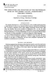
F. exp. Biol. (1981), 90, 1-15<br />
|Pi
Mid-gut<br />
Rectum<br />
S. H. P. MADDRELL<br />
Anterior<br />
hind-gut<br />
Malpighian<br />
tubule<br />
Fig. i. Fluid movements in <strong>the</strong> <strong>insect</strong> <strong>excretory</strong> <strong>system</strong>. Fluid is transported into <strong>the</strong> Malpighian<br />
tubules where many haemolymph solutes can diffuse across <strong>the</strong> walls into <strong>the</strong> lumen.<br />
Much <strong>of</strong> <strong>the</strong> water is <strong>the</strong>n recovered in <strong>the</strong> anterior hindgut and rectum toge<strong>the</strong>r with o<strong>the</strong>r<br />
useful substances.<br />
resulting fluid is <strong>the</strong>n passed out <strong>of</strong> <strong>the</strong> body. Such a <strong>system</strong> is more energy consuming<br />
than might seem necessary. Because <strong>of</strong> <strong>the</strong> relatively unselective nature <strong>of</strong> <strong>the</strong> filtration<br />
process, large quantities <strong>of</strong> useful substances (water, ions, sugars, amino acids, etc.)<br />
have continually to be reabsorbed. It would seem that if <strong>the</strong> filter were more selective,<br />
savings <strong>of</strong> energy might be made and evolution would act to favour animals with such<br />
a selective filter. However, this would expose <strong>the</strong>m to <strong>the</strong> danger <strong>of</strong> toxic material not<br />
able to pass through <strong>the</strong> filter. Although evolution has led to <strong>the</strong> development <strong>of</strong><br />
active transport <strong>system</strong>s able to eliminate toxic materials that <strong>of</strong>ten appear in circulation,<br />
<strong>the</strong> animal would still be vulnerable to toxins not encountered before (Ramsay,<br />
1958). Harmful proteins and/or cells held back by <strong>the</strong> filter are dealt with by separate<br />
mechanisms, e.g. antibodies and phagocytosis.<br />
In accord with <strong>the</strong>se ideas, <strong>the</strong> accepted view <strong>of</strong> <strong>the</strong> <strong>insect</strong> <strong>excretory</strong> <strong>system</strong> is that<br />
<strong>the</strong> haemolymph is 'filtered' through permeable Malpighian tubules, with reabsorption<br />
<strong>of</strong> useful substances from <strong>the</strong> filtrate, in <strong>the</strong> anterior hindgut and rectum (Fig. 1).<br />
One <strong>of</strong> <strong>the</strong> main objects <strong>of</strong> <strong>the</strong> present paper is to draw attention to <strong>the</strong> hc0<br />
surprising at first sight, that <strong>the</strong> effective permeability <strong>of</strong> <strong>insect</strong> Malpighian tubules is~
<strong>The</strong> <strong>functional</strong> <strong>design</strong> <strong>of</strong> <strong>the</strong> <strong>insect</strong> <strong>excretory</strong> <strong>system</strong> 3<br />
very considerably less than <strong>the</strong> analogous filtration sites in most o<strong>the</strong>r animals, both<br />
invertebrates and vertebrates, and to consider <strong>the</strong> implications <strong>of</strong> this for <strong>the</strong> operation<br />
<strong>of</strong> <strong>the</strong> <strong>insect</strong>'s <strong>excretory</strong> <strong>system</strong>.<br />
In <strong>the</strong> fluid filtered by pressure through <strong>the</strong> glomeruli <strong>of</strong> vertebrates, even substances<br />
as large as inulin occur at concentrations negligibly different from that in <strong>the</strong><br />
blood on <strong>the</strong> o<strong>the</strong>r side <strong>of</strong> <strong>the</strong> filter (Smith, 1951). Similarly, Kirschner & Wagner<br />
(1965) showed that <strong>the</strong> filtration site <strong>of</strong> <strong>the</strong> crayfish antennal gland, <strong>the</strong> coelomosac,<br />
allows free penetration <strong>of</strong> compounds up to 50000 in molecular weight. <strong>The</strong>y concluded<br />
that '<strong>the</strong> evidence indicates that <strong>the</strong> permeability <strong>of</strong> <strong>the</strong> antennal gland exceeds<br />
that <strong>of</strong> <strong>the</strong> vertebrate glomerulus'. Braun, Kummel & Mangos (1966), working on<br />
<strong>the</strong> protonephridium <strong>of</strong> <strong>the</strong> rotifer, Asplanchna priodonta, provided evidence that, as<br />
in <strong>the</strong> vertebrate nephron, [ 14 C] inulin passes freely through <strong>the</strong> walls <strong>of</strong> <strong>the</strong> filtering<br />
terminal organs (<strong>the</strong> flame bulbs). Reabsorption <strong>of</strong> water and electrolytes in <strong>the</strong><br />
succeeding protonephridial tubules produced a urine in which <strong>the</strong> inulin concentration<br />
always very significantly exceeded that <strong>of</strong> <strong>the</strong> body fluid. In sharp contrast, <strong>the</strong> fluid<br />
secreted by Malpighian tubules <strong>of</strong> <strong>insect</strong>s contains even such small molecules as<br />
sugars and amino acids at concentrations markedly less than in <strong>the</strong> haemolymph<br />
(Ramsay, 1958; Maddrell & Gardiner, 1974). Rapidly secreting Malpighian tubules<br />
<strong>of</strong> Rhodnius, for instance, produce fluid containing glycine and inulin at concentrations<br />
which are only about 1 % <strong>of</strong> those in <strong>the</strong> bathing fluid (Maddrell & Gardiner,<br />
1974; Maddrell & Gardiner, 1980). It might be argued that <strong>the</strong>se results are merely<br />
consequences <strong>of</strong> <strong>the</strong> fact that <strong>the</strong> force driving such solutes into <strong>the</strong> primary <strong>excretory</strong><br />
fluid <strong>of</strong> <strong>insect</strong>s is not pressure as in most o<strong>the</strong>r animals but only diffusion acting under<br />
<strong>the</strong> influence <strong>of</strong> <strong>the</strong> concentration gradient between <strong>the</strong> secreted fluid and <strong>the</strong> bathing<br />
extracellular fluid. This can be discounted, however, as <strong>the</strong> Malpighian tubules <strong>of</strong><br />
<strong>the</strong> pill millipede, Glomeris marginata, which is not an <strong>insect</strong>, can rapidly secrete fluid<br />
extremely similar in composition to <strong>the</strong> bathing fluid (Farquharson, 19746). Here, as<br />
with <strong>insect</strong> Malpighian tubules, <strong>the</strong> secreted fluid is not filtered by pressure into <strong>the</strong><br />
lumina <strong>of</strong> <strong>the</strong> tubules and presumably most haemolymph solutes find <strong>the</strong>ir way into<br />
<strong>the</strong> lumen by diffusion. <strong>The</strong> secreted fluid none<strong>the</strong>less has virtually <strong>the</strong> same ionic<br />
composition as <strong>the</strong> bathing fluid even when this is varied over a wide range and even<br />
such large solutes as inulin and dextrans <strong>of</strong> molecular weight up to 16000 appear in<br />
<strong>the</strong> secreted fluid at concentrations only a little depressed from those <strong>of</strong> <strong>the</strong> bathing<br />
fluid (Farquharson, 19746).<br />
Ano<strong>the</strong>r possible explanation <strong>of</strong> <strong>the</strong> under-representation <strong>of</strong> haemolymph solutes<br />
in <strong>the</strong> fluid secreted by <strong>insect</strong> Malpighian tubules could be that such solutes are<br />
actively reabsorbed from <strong>the</strong> lumen. Indeed, such reabsorption is known to occur -<br />
<strong>the</strong> Malpighian tubules <strong>of</strong> CalUphora voimtoria limit <strong>the</strong> loss <strong>of</strong> D-glucose from <strong>the</strong><br />
haemolymph by a reabsorptive process which can be blocked by treatment with<br />
phlorizin (Knowles, 1975). However, nei<strong>the</strong>r L-glucose which is not actively transported,<br />
nor D-glucose in phlorizin-treated tubules appear in <strong>the</strong> luminal fluid at<br />
concentrations any higher than 50% <strong>of</strong> those occurring in <strong>the</strong> bathing fluid (Knowles,<br />
1975)-<br />
To emphasize <strong>the</strong> low permeability <strong>of</strong> <strong>insect</strong> Malpighian tubules and, incidentally,<br />
display one <strong>of</strong> <strong>the</strong> advantages <strong>of</strong> such low permeability, it is worth recounting <strong>the</strong><br />
Results <strong>of</strong> some recent work on <strong>the</strong> blood sucking <strong>insect</strong>, Rhodnius. Amino acids
4 S. H. P. MADDRELL<br />
passively cross <strong>the</strong> walls <strong>of</strong> this <strong>insect</strong>s's Malpighian tubules so slowly that whefl,<br />
after a blood meal, <strong>the</strong> tubules rapidly eliminate a volume <strong>of</strong> fluid equivalent to 5-10<br />
times that <strong>of</strong> <strong>the</strong> haemolymph, <strong>the</strong>re is a loss <strong>of</strong> only about 2 % <strong>of</strong> <strong>the</strong> haemolymph<br />
content <strong>of</strong> amino acids (Maddrell & Gardiner, 1980). No reabsorption <strong>of</strong> amino acids<br />
is required during this rapid excretion <strong>of</strong> fluid.<br />
Insect Malpighian tubules thus have only limited permeability to haemolymph<br />
solutes. Three questions now arise, (i) How can such low permeability be reconciled<br />
with <strong>the</strong> overriding need, emphasized above, for an <strong>excretory</strong> <strong>system</strong> to contain a<br />
relatively non-selective and permeable nitration site ? (ii) How are <strong>the</strong> walls <strong>of</strong><br />
Malpighian tubules structurally organized to provide a low effective permeability ?<br />
(iii) What are <strong>the</strong> advantages to be derived from having an <strong>excretory</strong> <strong>system</strong> <strong>of</strong> only<br />
limited permeability to haemolymph solutes ?<br />
It will be <strong>the</strong> main object <strong>of</strong> <strong>the</strong> remainder <strong>of</strong> this paper to answer <strong>the</strong>se questions.<br />
OPERATION OF AN EXCRETORY SYSTEM OF LOW PERMEABILITY<br />
To answer <strong>the</strong> first question, it has to be pointed out that although such small<br />
solutes as amino acids can only penetrate <strong>the</strong> walls <strong>of</strong> <strong>insect</strong> Malpighian tubules<br />
slowly, <strong>the</strong> tubules yet have measurable permeabilities to substances as large as inulin<br />
(Ramsay & Riegel, 1961; Maddrell & Gardiner, 1974). This is a clear indication that<br />
<strong>the</strong> apparently low overall permeability <strong>of</strong> <strong>the</strong> tubule wall might be due more to a<br />
limitation in area <strong>of</strong> <strong>the</strong> permeable sites than to any restriction at <strong>the</strong> sites <strong>of</strong> permeation.<br />
This is very important, as it means that toxic molecules, even those <strong>of</strong> moderate<br />
size, will still be removed passively, albeit slowly, from <strong>the</strong> <strong>insect</strong>'s haemolymph.<br />
This in turn suggests that <strong>insect</strong>s might in fact use <strong>the</strong> same basis for <strong>the</strong> operation<br />
<strong>of</strong> <strong>the</strong>ir <strong>excretory</strong> <strong>system</strong>s as do o<strong>the</strong>r animals, but that <strong>the</strong> rate <strong>of</strong> 'filtration' <strong>of</strong> <strong>the</strong><br />
extracellular fluid is very much reduced. But how are <strong>insect</strong>s able to survive with<br />
such a slowly operating <strong>system</strong> and gain <strong>the</strong> advantages described below, when o<strong>the</strong>r<br />
animals seem not to be able to ?<br />
A possible explanation is along <strong>the</strong> following lines. Insects as a group are<br />
characterized by <strong>the</strong>ir small bodily size, which gives <strong>the</strong>m a high surface-area/volume<br />
ratio and makes <strong>the</strong>ir body fluids more liable to be affected by changes in <strong>the</strong> environment.<br />
In spite <strong>of</strong> this, <strong>insect</strong>s seem only to be found in habitats which impose some<br />
form <strong>of</strong> osmotic and/or ionic stress (thus <strong>the</strong>y appear on land, and in fresh, brackish,<br />
and hypersaline bodies <strong>of</strong> water, but not in such a stable environment as <strong>the</strong> sea). Of<br />
course, a crucial element in <strong>the</strong>ir ability to survive in such environments is <strong>the</strong><br />
possession <strong>of</strong> a wax-covered integument which greatly restricts ion and water fluxes<br />
between <strong>the</strong>ir internal and external environments (Beament, 1961). It is <strong>of</strong>ten thought<br />
that <strong>the</strong> haemolymph <strong>of</strong> <strong>insect</strong>s, because <strong>of</strong> its supposedly large volume, might also<br />
help in that it could act as a water store and as a buffer against changes in composition.<br />
While this could be true for some <strong>insect</strong>s, such as caterpillars, which have particularly<br />
large volumes <strong>of</strong> haemolymph, recent measurements <strong>of</strong> haemolymph volume in o<strong>the</strong>r<br />
<strong>insect</strong>s make this less likely. <strong>The</strong>y show that many <strong>insect</strong>s, particularly flying <strong>insect</strong>s,<br />
have haemolymph volumes which are, if anything, smaller in relation to total body<br />
weight than, for example, <strong>the</strong> extracellular fluid <strong>of</strong> vertebrates. Table 1 shows <strong>the</strong><br />
results <strong>of</strong> determinations <strong>of</strong> haemolymph volume in a series <strong>of</strong> <strong>insect</strong>s. <strong>The</strong>se resul<br />
are to be compared with determinations <strong>of</strong> extracellular fluid volume in vertebrate^.
Order<br />
Diptera<br />
Lepidoptera<br />
Coleoptera<br />
Hemiptera<br />
Orthoptera<br />
<strong>The</strong> <strong>functional</strong> <strong>design</strong> <strong>of</strong> <strong>the</strong> <strong>insect</strong> <strong>excretory</strong> <strong>system</strong><br />
Table i. Volumes <strong>of</strong> haemolymph in <strong>insect</strong>s<br />
Species<br />
Musca domestica<br />
Musca domestica<br />
Sarcophaga barbata<br />
Pierit brasticae<br />
Pierit brasticae<br />
Tenebrio molitor<br />
Rhodnius prolixus<br />
Carausius morosus<br />
Carausius morosus<br />
Periplaneta americana<br />
Periplaneta americana<br />
Arenivaga »p.<br />
Schistocerca gregaria<br />
Locusta vngratoria<br />
Locusta migratoria<br />
Stage<br />
Adult S<br />
Adult 6*<br />
Adult (30 h after<br />
emergence)<br />
Larva<br />
Adult<br />
Larva<br />
Young 5th instar<br />
Adult ?<br />
Adult §<br />
Larva<br />
Adult 6"<br />
Larva<br />
Adult 6*<br />
5th instar<br />
Adult<br />
Haemolymph<br />
volume (as %<br />
<strong>of</strong> body weight) References<br />
18.6 Bondaryk & Morrison (1966)<br />
206 Bondaryk & Morrison (1966)<br />
9 Cottrell (1962)<br />
34'S<br />
7-2<br />
10<br />
309<br />
23<br />
15<br />
164<br />
178<br />
175<br />
137<br />
171<br />
182<br />
Nicolson(i975)<br />
Nicolson(i97s)<br />
Jones (1957)<br />
Maddrell & Gardiner (1976)<br />
Nicolson et 0/(1974)<br />
Ramsay (1955)<br />
Edney(i968)<br />
Wall (1970)<br />
Edney(i968)<br />
Samaranayaka (1975)<br />
Loughton & Tobij (1969)<br />
Loughton & Tobe (1969)<br />
As far as blood volume goes <strong>the</strong>re seems to be little variation between vertebrate<br />
species; determinations on eight different vertebrates gave closely similar results with<br />
an average value <strong>of</strong> 7 % <strong>of</strong> <strong>the</strong> body weight (Sjostrand, 1962). To this figure must be<br />
added <strong>the</strong> volume <strong>of</strong> <strong>the</strong> lymph, about 2 % <strong>of</strong> <strong>the</strong> body weight, and <strong>the</strong> interstitial<br />
fluid, about 16% <strong>of</strong> <strong>the</strong> body weight (Pitts, 1968), giving a total extracellular fluid<br />
volume <strong>of</strong> about 25 % <strong>of</strong> <strong>the</strong> body weight for vertebrates. (Direct measurements using<br />
a range <strong>of</strong> markers (Swan, Madisso & Pitts, 1954) gave extracellular fluid volumes <strong>of</strong><br />
between 22 and 23 % for dogs.) <strong>The</strong>se figures suggest that, in general, <strong>insect</strong>s are in<br />
no better a position than vertebrates to withstand water loss. Indeed, Edney (1977)<br />
has found that, even in desert-living <strong>insect</strong>s, it is not so much tolerance <strong>of</strong> water loss<br />
but ra<strong>the</strong>r its prevention that is <strong>the</strong> important factor in <strong>the</strong>ir survival. <strong>The</strong>y can<br />
withstand losses <strong>of</strong> 30% or so <strong>of</strong> <strong>the</strong>ir body weight, but this is not greatly different<br />
from <strong>the</strong> abilities <strong>of</strong> desert-living cattle, camels, and donkeys. <strong>The</strong>se desert vertebrates<br />
are all well able to survive water losses <strong>of</strong> 20% <strong>of</strong> <strong>the</strong>ir body weight (Siebert &<br />
MacFarlane, 1975), and in extreme cases can tolerate losses up to 27 % <strong>of</strong> body weight<br />
• in <strong>the</strong> camel (Schmidt-Nielsen, 1964) and up to 30% in <strong>the</strong> donkey (Maloij, 1970).<br />
In general, <strong>insect</strong>s are very much smaller than vertebrates living in <strong>the</strong> same<br />
environment. For example, <strong>the</strong> smallest terrestrial adult <strong>insect</strong>s, species <strong>of</strong> Thrips,<br />
weigh only a few micrograms (C. P. Ellington, personal communication) as against<br />
<strong>the</strong> smallest terrestrial vertebrates such as <strong>the</strong> Etruscan shrew which, as an adult,<br />
weighs about 2 g (Miller, 1964). At <strong>the</strong> o<strong>the</strong>r end <strong>of</strong> <strong>the</strong> scale, <strong>the</strong> largest terrestrial<br />
<strong>insect</strong>s weigh less than 100 g, while a bull elephant can weigh more than 10 000 000 g.<br />
<strong>The</strong>se differences imply a surface-area/volume ratio for terrestrial <strong>insect</strong>s which is<br />
close to 100 times larger than for terrestrial vertebrates. So although <strong>insect</strong>s are well<br />
protected by <strong>the</strong>ir wax-covered integument, <strong>the</strong>y are so very much smaller than<br />
vertebrates and have a so much higher surface-area/volume ratio that it is hard to<br />
avoid <strong>the</strong> conclusion that <strong>insect</strong> tissues must have evolved a greater tolerance to varied<br />
conditions in <strong>the</strong> haemolymph. Perhaps this may explain <strong>the</strong> relative ease with which<br />
fei vitro preparations <strong>of</strong> many <strong>insect</strong> tissues can be made. However, some <strong>insect</strong><br />
"tissues are less tolerant. In this respect, it is noteworthy that <strong>insect</strong>s, alone among <strong>the</strong>
6 S. H. P. MADDRELL<br />
invertebrates, have developed a series <strong>of</strong> epi<strong>the</strong>lia, characterized by <strong>the</strong> presence oF<br />
simple tight junctions, to protect such potentially sensitive tissues as <strong>the</strong> central<br />
nervous <strong>system</strong>, <strong>the</strong> eyes, and <strong>the</strong> testis (Lane & Skaer, 1980). <strong>The</strong> regulatory properties<br />
<strong>of</strong> one <strong>of</strong> <strong>the</strong>se epi<strong>the</strong>lia, <strong>the</strong> perineurium, which envelops much <strong>of</strong> <strong>the</strong> nervous<br />
<strong>system</strong>, has been particularly well worked out (Treherne, 1974; Treherne & Sch<strong>of</strong>ield,<br />
1978). Oxygen supplies for <strong>the</strong>se tissues reaches <strong>the</strong>m directly by <strong>the</strong> tracheal <strong>system</strong><br />
so that <strong>the</strong>ir protective epi<strong>the</strong>lia are no liability in this respect. In addition, as <strong>the</strong><br />
haemolymph is not responsible for circulating oxygen, it contains no respiratory<br />
pigment which could be adversely affected by changes in <strong>the</strong> haemolymph. (<strong>The</strong><br />
single exception to this is <strong>the</strong> occurrence <strong>of</strong> haemoglobin in <strong>the</strong> haemolymph <strong>of</strong> <strong>the</strong><br />
aquatic larvae <strong>of</strong> certain Chironomidae. However, this pigment only releases oxygen<br />
under conditions <strong>of</strong> extreme oxygen paucity (Wigglesworth, 1974).) It may well,<br />
<strong>the</strong>refore, be possible for <strong>insect</strong>s to survive haemolymph conditions which vary in<br />
composition within wider limits than would be tolerable in o<strong>the</strong>r animals.<br />
If, <strong>the</strong>n, <strong>insect</strong>s can tolerate changes in <strong>the</strong>ir internal milieu, one can argue that <strong>the</strong><br />
<strong>excretory</strong> <strong>system</strong> need not operate so rapidly as it o<strong>the</strong>rwise would have to, to control<br />
<strong>the</strong> composition <strong>of</strong> <strong>the</strong> haemolymph. Perhaps <strong>insect</strong> tissues can tolerate or are protected<br />
from potentially deleterious changes in <strong>the</strong>ir environment for long enough to<br />
allow <strong>the</strong> <strong>excretory</strong> <strong>system</strong> to correct <strong>the</strong> situation at a relatively leisurely rate. It may<br />
not matter so much how quickly novel toxic materials are removed, so long as <strong>the</strong>y<br />
are eventually removed. A <strong>system</strong> which provides for slow passive removal <strong>of</strong> substances,<br />
even those as large as inulin, would, <strong>the</strong>refore, suit <strong>insect</strong>s well, and as we<br />
have seen, that is exactly what <strong>the</strong>y have. To back up this passive <strong>system</strong>, evolution<br />
has led to <strong>the</strong> appearance in Malpighian tubules <strong>of</strong> active transport mechanisms that<br />
can rapidly eliminate a very wide range <strong>of</strong> substances that <strong>insect</strong>s repeatedly need to<br />
excrete (Maddrell, 1977). Peculiarly, <strong>the</strong>se mechanisms appear not to involve nitrogenous<br />
wastes, usually thought to be a very important class <strong>of</strong> substances for <strong>excretory</strong><br />
<strong>system</strong>s to eliminate. Although <strong>insect</strong>s excrete many different nitrogenous compounds,<br />
<strong>the</strong> most widely encountered is uric acid (Bursell, 1967). Surprisingly, <strong>the</strong> speed at<br />
which uric acid is eliminated from <strong>the</strong> body seems not to have been <strong>the</strong> subject <strong>of</strong><br />
strong selection pressure in <strong>the</strong> evolution <strong>of</strong> <strong>insect</strong>s. Indeed, deposits <strong>of</strong> uric acid<br />
occur in <strong>the</strong> fat bodies <strong>of</strong> parasitic <strong>insect</strong>s (Corbet & Ro<strong>the</strong>ram, 1965), and in <strong>the</strong> fat<br />
bodies <strong>of</strong> normally hydrated and fed cockroaches, where it is even suggested that <strong>the</strong>y<br />
act as stores <strong>of</strong> nitrogen (Tucker, 1977). Uric acid also appears in great quantities in<br />
<strong>the</strong> accessory glands <strong>of</strong> male cockroaches (Roth & Dateo, 1965) and it is actually used<br />
as a pigment in <strong>the</strong> bodies and wings <strong>of</strong> o<strong>the</strong>r <strong>insect</strong>s (Berridge, 1965; Harmsen,<br />
1966). Even in Rhodnius, where <strong>the</strong> diet is very rich in protein, <strong>the</strong> indications are<br />
that uric acid crosses <strong>the</strong> wall <strong>of</strong> <strong>the</strong> upper tubule entirely passively (Maddrell &<br />
Gardiner, 1974). <strong>The</strong>se findings all support <strong>the</strong> view that <strong>the</strong> slow elimination <strong>of</strong><br />
nitrogenous waste in <strong>insect</strong>s is readily tolerated and that selection pressure for an<br />
<strong>excretory</strong> <strong>system</strong> that operates more rapidly does not arise from this source.<br />
THE STRUCTURE OF INSECT MALPIGHIAN TUBULES<br />
<strong>The</strong> <strong>insect</strong> <strong>excretory</strong> <strong>system</strong> operates as if it has permeable areas limited in <strong>the</strong>ir<br />
extent. Does <strong>the</strong> structure <strong>of</strong> <strong>insect</strong> Malpighian tubules bear this out ? Fig. 2 showi<br />
that it does. Compared with <strong>the</strong> situation in, say, <strong>the</strong> podocytes <strong>of</strong> <strong>the</strong> Bowman's
(Facing p. 6)
<strong>Journal</strong> <strong>of</strong> Experimental Biology, Vol. 90 Fig.<br />
Fig. 2. Electron micrographs <strong>of</strong> sections through <strong>the</strong> walls <strong>of</strong> <strong>the</strong> Malpighian tubules <strong>of</strong> (a) a<br />
millipede, Glomeris marginata ( x 20000; micrograph courtesy <strong>of</strong> Dr J. A. Riegel), and (b) an<br />
<strong>insect</strong>, Rhodnius proluxus (X3000; micrograph courtesy <strong>of</strong> Dr B. S. Hill). In <strong>the</strong> wall <strong>of</strong> <strong>the</strong><br />
tubule <strong>of</strong> FViodnius, intercellular clefts that could act as pathways for materials to cross <strong>the</strong> wall<br />
passively are not <strong>of</strong>ten encountered even in low-power micrographs such as that shown. In<br />
contrast, <strong>the</strong> wall <strong>of</strong> <strong>the</strong> tubule <strong>of</strong> Glomeris shows many such clefts, even in a higher-power<br />
micrograph <strong>of</strong> a section taken from a part <strong>of</strong> <strong>the</strong> cell near <strong>the</strong> nucleus.<br />
S. H. P. MADDRELL
journal <strong>of</strong> Experimental Biology, Vol. 90<br />
{ b y . .•. •.,<br />
S. H. P. MADDRELL<br />
Fig. zb
<strong>The</strong> <strong>functional</strong> <strong>design</strong> <strong>of</strong> <strong>the</strong> <strong>insect</strong> <strong>excretory</strong> <strong>system</strong> 7<br />
capsule <strong>of</strong> <strong>the</strong> vertebrate kidney or <strong>the</strong> podocytes <strong>of</strong> <strong>the</strong> antennal glands <strong>of</strong> Crustacea<br />
(or, indeed, <strong>the</strong> filtration sites <strong>of</strong> <strong>the</strong> <strong>excretory</strong> <strong>system</strong>s in virtually all o<strong>the</strong>r animals<br />
studied - see Kummel (1973) for an excellent account <strong>of</strong> such filtration structures),<br />
<strong>the</strong> intercellular clefts <strong>of</strong> <strong>insect</strong> Malpighian tubules occupy very much less <strong>of</strong> <strong>the</strong><br />
frontal area <strong>of</strong> <strong>the</strong> epi<strong>the</strong>lium. In Rhodnius, for example, it has been calculated that<br />
<strong>the</strong> area <strong>of</strong> <strong>the</strong> clefts occupies only 0-03 % <strong>of</strong> <strong>the</strong> epi<strong>the</strong>lial surface (Maddrell, 1980a).<br />
<strong>The</strong> contrast with <strong>the</strong> multiply perforated wall <strong>of</strong> <strong>the</strong> very permeable Malpighian<br />
tubules <strong>of</strong> <strong>the</strong> millipede, Glomeris marginata (Fig. 2 and see Farquharson, 19746)<br />
could scarcely be more marked. It is relevant to add that <strong>the</strong> overall dimensions <strong>of</strong> <strong>the</strong><br />
tubules are ra<strong>the</strong>r similar. Rhodnius has four Malpighian tubules, each with a fluid<br />
secreting region 30 mm long and about 100 fim in diameter, while Glomeris has only<br />
two tubules but each has a fluid secreting portion about 60 mm long and around 80 fim.<br />
in diameter (Farquharson, 1974a).<br />
<strong>The</strong> restricted area <strong>of</strong> <strong>the</strong> intercellular clefts in <strong>insect</strong> Malpighian tubules raises<br />
ano<strong>the</strong>r question. What is <strong>the</strong> function <strong>of</strong> <strong>the</strong> extensive area <strong>of</strong> <strong>the</strong> cells <strong>of</strong> <strong>the</strong> tubules'<br />
walls ? Could not a low rate <strong>of</strong> passive filtration <strong>of</strong> <strong>the</strong> haemolymph simply be achieved<br />
by much less extensive tubules with intercellular clefts less widely separated ? Part <strong>of</strong><br />
<strong>the</strong> explanation could be that <strong>insect</strong>s may need occasionally to be able to excrete large<br />
quantities <strong>of</strong> fluid or to filter <strong>the</strong>ir haemolymph much more rapidly, and this requires<br />
an accelerated rate <strong>of</strong> fluid secretion provided in turn by many tubule cells (Maddrell,<br />
1980a).<br />
Blood-sucking <strong>insect</strong>s, sap-feeding <strong>insect</strong>s, and <strong>insect</strong>s that live in fresh waters are<br />
obvious cases where rapid fluid excretion is important. <strong>The</strong> need for <strong>the</strong> tubules <strong>of</strong><br />
o<strong>the</strong>r <strong>insect</strong>s to cater for varying rates <strong>of</strong> fluid production has a more subtle explanation.<br />
As will be argued below, an advantage <strong>of</strong> slow filtration <strong>of</strong> <strong>the</strong> haemolymph is<br />
that <strong>the</strong> energy-consuming process <strong>of</strong> reabsorption can <strong>the</strong>n also be slow. <strong>The</strong> price<br />
<strong>of</strong> this, as we have seen, is that toxins are more slowly removed. Now, if passive<br />
filtration <strong>of</strong> haemolymph solutes is by diffusion, its rate will depend on <strong>the</strong> difference<br />
in concentration <strong>of</strong> <strong>the</strong>se solutes between <strong>the</strong> haemolymph and lumen. If fluid<br />
secretion is slow, <strong>the</strong> luminal concentration will be high and solutes will enter <strong>the</strong><br />
tubule only slowly; reabsorption need also only be slow. If, however, fluid secretion<br />
is accelerated, this will continuously carry away solutes diffusing across <strong>the</strong> walls and<br />
so lower <strong>the</strong> concentration <strong>of</strong> <strong>the</strong>se solutes in <strong>the</strong> lumen. <strong>The</strong>y will thu8 enter <strong>the</strong><br />
tubule much faster. A similar argument applies to substances which are actively<br />
transported into <strong>the</strong> tubule lumen. <strong>The</strong>se will tend to diffuse back out <strong>of</strong> <strong>the</strong> tubule<br />
through <strong>the</strong> permeable intercellular clefts at a rate dependent on <strong>the</strong>ir concentration<br />
in <strong>the</strong> lumen. So <strong>the</strong> rate at which <strong>the</strong>y can be removed from <strong>the</strong> haemolymph will<br />
also depend on <strong>the</strong> rate <strong>of</strong> fluid secretion, as this strongly affects <strong>the</strong>ir concentration<br />
in <strong>the</strong> lumen. Fig. 3 shows, for one particularly well studied case, <strong>the</strong> Malpighian<br />
tubules <strong>of</strong> Calliphora, how <strong>the</strong> net rates <strong>of</strong> removal <strong>of</strong> substances from <strong>the</strong> haemolymph<br />
are affected by changes in <strong>the</strong> rate <strong>of</strong> fluid secretion. Clearly, <strong>the</strong> rate <strong>of</strong> fluid secretion<br />
can have very dramatic effects on <strong>the</strong> rates at which substances are removed from <strong>the</strong><br />
haemolymph, regardless <strong>of</strong> whe<strong>the</strong>r <strong>the</strong>y cross <strong>the</strong> tubule walls actively or passively.<br />
<strong>The</strong> shapes <strong>of</strong> <strong>the</strong> curves show that <strong>the</strong> rate <strong>of</strong> elimination <strong>of</strong> substances from <strong>the</strong><br />
haemolymph is more affected by changes in rate <strong>of</strong> fluid secretion at low rates than at<br />
higher rates. So, if <strong>the</strong> <strong>insect</strong> is to enjoy <strong>the</strong> advantages <strong>of</strong> being able to control <strong>the</strong>
S. H. P. MADDRELL<br />
15 20 25 30 35 40 45<br />
Rate <strong>of</strong> fluid secretion (nl min" 1 )<br />
Removal aided by<br />
active transport<br />
50 55 60<br />
Fig. 3. Calculated rates at which a substance, present at 1 mM in <strong>the</strong> haemolymph, is removed<br />
from circulation by a Malpighian tubule secreting fluid at different rates. <strong>The</strong> calculations are<br />
based on <strong>the</strong> properties <strong>of</strong> Malpighian tubules from Calliphora (for details see Berridge, 1965;<br />
Maddrell & Gardiner, 1974; Schwartz & Reynolds, 1979). For <strong>the</strong> lower line it was assumed<br />
that loss was by passive diffusion across <strong>the</strong> tubule wall with <strong>the</strong> wall having a permeability to<br />
<strong>the</strong> substance <strong>of</strong> 10" 5 cm s~'. For <strong>the</strong> upper line it was fur<strong>the</strong>r assumed that <strong>the</strong> substance was<br />
subjected to active transport towards <strong>the</strong> lumen at a rate <strong>of</strong> 75 pmol min" 1 . For an explanation<br />
<strong>of</strong> <strong>the</strong> ma<strong>the</strong>matical basis <strong>of</strong> <strong>the</strong> calculation see Maddrell & Gardiner (1980). <strong>The</strong> rates <strong>of</strong> fluid<br />
secretion normally shown by unstimulated and by maximally hormonally stimulated Malpighian<br />
tubules are shown by <strong>the</strong> vertical lines so that <strong>the</strong> stippled area between <strong>the</strong>m encompasses<br />
<strong>the</strong> normal range <strong>of</strong> rates that <strong>the</strong> tubules can achieve.<br />
rate <strong>of</strong> elimination <strong>of</strong> <strong>the</strong>se substances, it would make more sense for it to control <strong>the</strong><br />
rate <strong>of</strong> fluid secretion over a range where this has large effects on <strong>the</strong> rate <strong>of</strong> removal<br />
<strong>of</strong> substances from <strong>the</strong> haemolymph. Fig. 3 shows that for Calliphora this expectation<br />
is realized; <strong>the</strong> range <strong>of</strong> rates <strong>of</strong> secretion that can be achieved does not extend up to<br />
values where <strong>the</strong> rate <strong>of</strong> solute removal is becoming insensitive to changes in rate <strong>of</strong><br />
fluid secretion.<br />
If <strong>the</strong> above arguments have any force, <strong>the</strong>n an ability to control <strong>the</strong> rate <strong>of</strong> fluid<br />
secretion by <strong>the</strong>ir Malpighian tubules should be widespread among <strong>insect</strong>s. Table 2<br />
lists those <strong>insect</strong>s for which <strong>the</strong>re is evidence that <strong>the</strong>y can alter <strong>the</strong> rate <strong>of</strong> secretion<br />
by <strong>the</strong>ir tubules; <strong>the</strong> list covers virtually all those <strong>insect</strong>s where fluid secretion by<br />
<strong>the</strong>ir Malpighian tubules has been studied. Control <strong>of</strong> fluid secretion by Malpighian<br />
tubules is known in most cases to be effected by hormones (<strong>the</strong>se hormones are<br />
usually called diuretic hormones, but since tubule secretion can be accelerated greatly<br />
without <strong>the</strong>re necessarily being a loss <strong>of</strong> water from <strong>the</strong> <strong>insect</strong>, <strong>the</strong> water <strong>of</strong>ten being<br />
reabsorbed in <strong>the</strong> hindgut, <strong>the</strong> term diuretic is not always appropriate). An excellent<br />
example <strong>of</strong> an <strong>insect</strong> deriving an advantage from accelerating fluid secretion by its<br />
Malpighian tubules is <strong>the</strong> locust. Locusts in flight are, <strong>of</strong> course, extremely active<br />
metabolically, and since metabolism is likely to produce useless, if not harmful!<br />
by-products, <strong>the</strong>re will, during flight, be a need to filter <strong>the</strong> haemolymph more
<strong>The</strong> <strong>functional</strong> <strong>design</strong> <strong>of</strong> <strong>the</strong> <strong>insect</strong> <strong>excretory</strong> <strong>system</strong><br />
Table 2. Insects for which <strong>the</strong>re is evidence that <strong>the</strong> rate <strong>of</strong> fluid transport<br />
by <strong>the</strong>ir Malpighian tubules is regulated by hormones<br />
Order Species References<br />
Hymenoptera Apis mellifera Altmann (1956)<br />
Diptera<br />
Lepidoptera<br />
Coleoptera<br />
Hemiptera<br />
Orthoptera<br />
Calliphora vomitoria<br />
CaUiphora erythrocephala<br />
Glossina morsitans<br />
Aedes taemorhynchus<br />
Anopheles freeborni<br />
Pieris brassicae<br />
Calpodes ethlius<br />
Danaus plexippus<br />
Anisotarsus cupripennis<br />
Rhodnius prolixus<br />
Triatoma ivjestans<br />
Triatoma phyllosoma<br />
Dipetalogaster maxima<br />
Dysdercus fasciatus<br />
Periplaneta americana<br />
Locus ta rmgratoria<br />
Schxstocerca gregaria<br />
Carausius morosus<br />
Acheta domes tua<br />
Knowles(i976)<br />
Schwartz & Reynolds (1979)<br />
Gee (197s)<br />
Maddrell & Phillips (1978)<br />
Nijhout & Carrow (1978)<br />
Nicolson(i976)<br />
Ryerse(i978)<br />
Dores, Dallman & Nerman (1979)<br />
Nufiez(i9s6)<br />
Maddrell (1962)<br />
Maddrell (unpublished results)<br />
Maddrell (unpublished results)<br />
Maddrell (unpublished results)<br />
Berridge(i966)<br />
Mills (1967)<br />
Cazal&Girardie(i968)<br />
Mordue(i969)<br />
Pilcher(i97o)<br />
Geldiay & Edwards (1976)<br />
rapidly. Appropriately, flying locusts release hormone(8) to accelerate both fluid secretion<br />
by <strong>the</strong>ir Malpighian tubules and fluid reabsorption in <strong>the</strong> hindgut (Mordue, 1969).<br />
If we accept that <strong>insect</strong>s need to be able to secrete fluid at a range <strong>of</strong> rates, <strong>the</strong>n it<br />
now becomes clear why <strong>the</strong> Malpighian tubules have to have a considerable area <strong>of</strong><br />
fluid secreting epi<strong>the</strong>lium, and this <strong>the</strong> tubule cells provide.<br />
ADVANTAGES OF AN EXCRETORY SYSTEM WITH RESTRICTED •<br />
PERMEABLE SITES FOR FILTRATION<br />
What advantages do <strong>insect</strong>s derive from <strong>the</strong>ir somewhat unusual <strong>excretory</strong> <strong>system</strong> ?<br />
As will be clear from what has already been said, an obvious advantage <strong>of</strong> a <strong>system</strong><br />
which operates slowly is that reabsorption <strong>of</strong> useful substances from <strong>the</strong> filtered fluid<br />
need only be slow. Presumably <strong>the</strong> fluid flow in <strong>the</strong> <strong>system</strong> need also only be slow, at<br />
least when no hormonal stimulation <strong>of</strong> secretion goes on. It is possible to check this<br />
point by reference to <strong>the</strong> known rates <strong>of</strong> Malpighian tubule fluid production. Table 3<br />
compares <strong>the</strong> rates at which <strong>the</strong> extracellular fluid is passed through <strong>the</strong> <strong>excretory</strong><br />
<strong>system</strong> <strong>of</strong> <strong>insect</strong>s with similar figures for vertebrates. Clearly, most vertebrates filter<br />
<strong>the</strong>ir extracellular fluid 10-20 times more rapidly than do most <strong>insect</strong>s. Taking into<br />
account <strong>the</strong> fact that, in <strong>insect</strong>s, many haemolymph solutes <strong>of</strong> even quite small<br />
molecular size only appear in <strong>the</strong> filtered fluid at concentrations in <strong>the</strong> range 10-50%<br />
<strong>of</strong> those in <strong>the</strong> haemolymph, it is clear that reabsorption <strong>of</strong> such solutes need only<br />
occur in <strong>insect</strong>s at about 1-2% <strong>of</strong> <strong>the</strong> rate required in many vertebrates.<br />
<strong>The</strong> figures in Table 3 for a snake and a frog suggest that filtration <strong>of</strong> extracellular<br />
fluid may be slower in ecto<strong>the</strong>rmic animals, perhaps reflecting <strong>the</strong>ir lower rates <strong>of</strong><br />
metabolism. If so, <strong>the</strong>n <strong>the</strong> slow rate at which <strong>insect</strong>s pass fluid through <strong>the</strong>ir Malpighian<br />
tubules might be attributable more to <strong>the</strong> fact <strong>the</strong>y are ecto<strong>the</strong>rms than to
10 S. H. P. MADDRELL<br />
Table 3. Times taken for volumes <strong>of</strong> fluid equivalent to <strong>the</strong> total extracellular flutS<br />
(a) to be filtered through <strong>the</strong> glomeruli <strong>of</strong> some higher vertebrates or (b) to be passed through<br />
<strong>the</strong> Malpighian tubules <strong>of</strong> <strong>insect</strong>s<br />
(a) Vertebrates*<br />
Frog<br />
Snake<br />
Chicken<br />
Mouse<br />
Sand Rat<br />
Rat<br />
Rabbit<br />
Cat<br />
Dog<br />
Pig<br />
Goat<br />
Sheep<br />
Camel<br />
Ox<br />
Horse<br />
Man<br />
(ft) Insects<br />
Calliphora erythrocephala<br />
Dysdercut faiciattu<br />
SchUtocerca gregaria<br />
Carauriiu morosus<br />
Rhodnius prolixui<br />
Aedes taemorhynchut<br />
Times (min)<br />
285<br />
S2S<br />
138<br />
38<br />
19<br />
42<br />
83<br />
127<br />
57<br />
178<br />
185<br />
158<br />
218<br />
82<br />
322<br />
135<br />
625<br />
1 500<br />
1 700<br />
2750<br />
10000<br />
20 (during diuresis)<br />
2000<br />
References<br />
Osbaldiston (1974)<br />
Osbaldiston (1974)<br />
Renkin & Gilmore (197;<br />
Siebert & MacFarlane (1975!<br />
Siebert & MacFarlane (1975]<br />
Renkin & Gilmore (1973)<br />
Renkin & Gilmore (1973)<br />
Berridge(i968)<br />
Berridge(i966)<br />
Phillips (1964)<br />
Nicolson, et al. (1974)<br />
Ramsay (1954)<br />
Maddrell (1964)<br />
Maddrell & Gardiner (1976)<br />
Maddrell & Phillips (1978)<br />
•<strong>The</strong> extracellular fluid <strong>of</strong> <strong>the</strong>se vertebrates is assumed to be 25% <strong>of</strong> <strong>the</strong> body weight (Pitts, 1968).<br />
any difference special to <strong>insect</strong>s. Very likely this is part <strong>of</strong> <strong>the</strong> explanation but <strong>the</strong>re<br />
is ano<strong>the</strong>r factor to be taken into account. <strong>The</strong> <strong>insect</strong>s used as examples in Table 3<br />
are all considerably smaller than frogs and snakes and so, <strong>of</strong> course, would be expected<br />
(Schmidt-Nielsen, 1979) to have higher specific metabolic rates (metabolic rate per<br />
unit <strong>of</strong> body weight). This would suggest that <strong>the</strong>y should need to filter <strong>the</strong> haemolymph<br />
faster than do larger ecto<strong>the</strong>rms. <strong>The</strong> few figures so far available (Table 3)<br />
indicate that, if anything, <strong>the</strong> opposite is <strong>the</strong> case and that <strong>insect</strong>s filter <strong>the</strong> haemolymph<br />
considerably more slowly than would be predicted.<br />
Direct evidence for <strong>the</strong> slow passive removal <strong>of</strong> substances from <strong>insect</strong> haemolymph<br />
comes from experiments on <strong>the</strong> rate <strong>of</strong> elimination <strong>of</strong> inulin injected into locusts.<br />
Although locust Malpighian tubules are permeable to inulin, its elimination is at a<br />
rate with a half-time <strong>of</strong> about 8 days (Maddrell & Gardiner, 1974). For comparison, it<br />
can be calculated from <strong>the</strong> known rate <strong>of</strong> glomerular filtration in man (Pitts, 1968)<br />
that inulin would be eliminated from <strong>the</strong> extracellular fluid with a half-time <strong>of</strong> about<br />
100 min.<br />
A major advantage logically follows from <strong>the</strong> fact that substances are only slowly<br />
removed passively from <strong>insect</strong> haemolymph. It becomes feasible for <strong>insect</strong>s to<br />
maintain in <strong>the</strong> haemolymph high concentrations <strong>of</strong> useful substances <strong>of</strong> low molecular<br />
weight. Flying <strong>insect</strong>s can, as fuels for flight, have 100 mM <strong>of</strong> <strong>the</strong> disaccharide<br />
trehalose and/or diglyceride lipid in <strong>the</strong> haemolymph (Weis-Fogh, 1967). Many
<strong>The</strong> <strong>functional</strong> <strong>design</strong> <strong>of</strong> <strong>the</strong> <strong>insect</strong> <strong>excretory</strong> <strong>system</strong> 11<br />
Trisects in <strong>the</strong> orders Lepidoptera, Hymenoptera, and Coleoptera contain 100-200 mM<br />
<strong>of</strong> amino acids in <strong>the</strong> haemolymph (Maddrell, 1971). Finally, hormones that are<br />
released into circulation will only slowly be removed by <strong>the</strong> <strong>excretory</strong> <strong>system</strong> and so<br />
can more easily function for useful lengths <strong>of</strong> time. A potential disadvantage is that<br />
<strong>insect</strong>icidal substances once <strong>the</strong>y reach <strong>the</strong> haemolymph have longer to produce<br />
damaging effects than if <strong>the</strong>y were more rapidly eliminated (Maddrell, 19806).<br />
Insects have evolved a series <strong>of</strong> active transport mechanisms that move substances<br />
into or out <strong>of</strong> <strong>the</strong> tubule lumen. This would not be possible in a more permeable<br />
tubule as <strong>the</strong> resulting gradients would rapidly be cancelled by diffusion. Examples<br />
<strong>of</strong> <strong>the</strong>se <strong>system</strong>s include active transport into <strong>the</strong> lumen <strong>of</strong> phosphate, magnesium,<br />
and sulphate ions, sulphonates, acylamides, alkaloids, and glycosides (Maddrell, 1977)<br />
and <strong>the</strong> active reabsorption <strong>of</strong> glucose (Knowles, 1975). Nearly all Malpighian tubules<br />
secrete fluid much richer in potassium than is <strong>the</strong> haemolymph. This has to be<br />
reabsorbed in some <strong>insect</strong>s but in herbivorous <strong>insect</strong>s that face <strong>the</strong> problem <strong>of</strong> excess<br />
potassium in <strong>the</strong> diet, <strong>the</strong> use <strong>of</strong> potassium transport as <strong>the</strong> driving force for fluid<br />
formation in <strong>the</strong> Malpighian tubules allows <strong>the</strong>m both to rid <strong>the</strong>mselves <strong>of</strong> potassium<br />
and to produce <strong>the</strong> essential primary <strong>excretory</strong> fluid with a single transport process.<br />
With a more permeable tubule, potassium excretion would require additional<br />
potassium transport in a less permeable part <strong>of</strong> <strong>the</strong> <strong>excretory</strong> <strong>system</strong>.<br />
We have seen that acceleration <strong>of</strong> fluid secretion in <strong>the</strong> Malpighian tubules<br />
accelerates <strong>the</strong> 'filtering' <strong>of</strong> <strong>the</strong> haemolymph in that haemolymph solutes pass more<br />
rapidly into <strong>the</strong> tubule lumen (Fig. 3). This is closely similar to <strong>the</strong> effect <strong>of</strong> an<br />
increase in glomerular filtration rate in vertebrates. However, if <strong>the</strong> rate <strong>of</strong> fluid<br />
secretion is increased still more, <strong>the</strong> relation breaks down and <strong>the</strong> rate <strong>of</strong> passage <strong>of</strong><br />
solutes into <strong>the</strong> lumen is scarcely any fur<strong>the</strong>r affected (Fig. 3). Essentially this is<br />
because <strong>the</strong> acceleration <strong>of</strong> solute movement from <strong>the</strong> haemolymph follows from<br />
increasing dilution <strong>of</strong> solute in <strong>the</strong> tubule lumen at higher rates <strong>of</strong> fluid secretion;<br />
diffusion back into <strong>the</strong> haemolymph falls to low levels. Once <strong>the</strong> concentration <strong>of</strong> a<br />
solute in <strong>the</strong> lumen reaches a very low level, however, fur<strong>the</strong>r increases in <strong>the</strong> rate <strong>of</strong><br />
fluid secretion have virtually no more effect on solute entry. This allows <strong>the</strong> evolution<br />
in <strong>insect</strong>s that need to eliminate fluid from <strong>the</strong> haemolymph <strong>of</strong> a <strong>system</strong> whereby <strong>the</strong><br />
tubules secrete fluid at very high rates. Since <strong>the</strong> excess fluid is <strong>the</strong>n quickly removed,<br />
and contains only traces <strong>of</strong> haemolymph solutes, no reabsorption <strong>of</strong> <strong>the</strong> solutes is<br />
needed. Such an adaptation is, <strong>of</strong> course, particularly suited to blood-sucking <strong>insect</strong>s<br />
such as Rhodnius, which take very large blood meals that necessitate <strong>the</strong> elimination<br />
<strong>of</strong> much excess fluid. <strong>The</strong>se ideas may well be important in explaining why many<br />
blood-sucking <strong>insect</strong>s can excrete fluid at what seem unnecessarily high rates;<br />
Rhodnius after feeding can eliminate fluid equivalent in volume to its total haemolymph<br />
in as short a time as 30 min.<br />
INSECTS IN THE TERRESTRIAL ENVIRONMENT<br />
As pointed out earlier, <strong>insect</strong>s seem only to be found in habitats characterized by<br />
osmotic and/or ionic stress. In particular, <strong>the</strong>y are most numerous in <strong>the</strong> terrestrial<br />
•environment. Here <strong>the</strong> probleVn is one <strong>of</strong> resisting desiccation and it is worth con-<br />
Kidering what role <strong>the</strong> <strong>insect</strong> <strong>excretory</strong> <strong>system</strong> plays in this. Obviously, its main
12 S. H. P. MADDRELL<br />
contribution must be that <strong>of</strong> limiting water loss. It seems that <strong>the</strong>re are two distinctive<br />
features <strong>of</strong> <strong>insect</strong>s which are important in this.<br />
<strong>The</strong> first is that <strong>the</strong> flow <strong>of</strong> water through <strong>the</strong> <strong>excretory</strong> <strong>system</strong> is comparatively<br />
slow. Selection pressure for effective water reabsorption, strong in all terrestrial<br />
animals, is likely to be still more severe in <strong>insect</strong>s because <strong>of</strong> <strong>the</strong>ir small size. It is<br />
scarcely surprising that many terrestrial <strong>insect</strong>s have evolved with highly developed<br />
abilities for reabsorbing water from <strong>the</strong> primary <strong>excretory</strong> fluid. Given <strong>the</strong> low rate<br />
<strong>of</strong> production <strong>of</strong> this fluid, water reabsorption is very effective, recovering, in many<br />
cases, virtually all <strong>the</strong> water from <strong>the</strong> <strong>excretory</strong> material. So effective are <strong>the</strong>se water<br />
recovery <strong>system</strong>s that in Tenebrto (Ramsay, 1971) and in <strong>The</strong>rmobia (Noble-Nesbitt,<br />
1970), for example, water vapour can be taken up from moist air drawn into <strong>the</strong><br />
hindgut.<br />
A second characteristic <strong>of</strong> <strong>insect</strong>s may have been <strong>of</strong> crucial importance in <strong>the</strong><br />
evolution <strong>of</strong> such effective water reabsorption. This is that <strong>the</strong> hindgut, which is<br />
responsible for water recovery, is lined with cuticle. <strong>The</strong> significance <strong>of</strong> <strong>the</strong> restricted<br />
permeability <strong>of</strong> this cuticular lining was first recognized by Phillips & Dockrill (1968).<br />
<strong>The</strong>y pointed out that <strong>the</strong> very effective water reabsorption carried out by <strong>the</strong> rectal<br />
cells might well only be possible because <strong>the</strong>y are protected by <strong>the</strong> cuticle from<br />
mounting concentrations <strong>of</strong> potentially interfering compounds in <strong>the</strong> luminal fluid.<br />
As would be expected, <strong>the</strong>n, evolution has favoured <strong>the</strong> hindgut as <strong>the</strong> site <strong>of</strong><br />
mechanisms concerned with <strong>the</strong> final stages <strong>of</strong> water recovery in <strong>insect</strong>s. <strong>The</strong> o<strong>the</strong>r<br />
very successful terrestrial animals, <strong>the</strong> vertebrates, do not have <strong>the</strong> advantage <strong>of</strong> such<br />
a cuticular intima and could not have developed such a <strong>system</strong>. Indeed, water recovery<br />
in vertebrates is achieved by epi<strong>the</strong>lia that are in direct contact with <strong>the</strong> urine. It is<br />
possible that this difference was one <strong>of</strong> <strong>the</strong> most important factors in preventing <strong>the</strong><br />
evolution <strong>of</strong> terrestrial vertebrates small enough to compete effectively with <strong>insect</strong>s<br />
for <strong>the</strong> niches available for animals in this size range.<br />
SUMMARIZING REMARKS<br />
Insects have a slowly operating <strong>excretory</strong> <strong>system</strong> in which <strong>the</strong> rate <strong>of</strong> passive<br />
movement <strong>of</strong> haemolymph solutes into a slowly secreted primary <strong>excretory</strong> fluid is<br />
restricted by a reduction in <strong>the</strong> area available for passive transfer. <strong>The</strong>y may have<br />
come to possess such an energy-saving <strong>system</strong> as a result <strong>of</strong> <strong>the</strong>ir evolution as small<br />
animals in osmotically and ionically stressful environments. Although <strong>the</strong> possession<br />
<strong>of</strong> a waxy cuticle is a major element in <strong>the</strong>ir ability to live in such environments,<br />
<strong>insect</strong>s have a very high surface-area/volume ratio and this is likely to have conferred<br />
a selective advantage on individuals able to withstand unusually variable extracellular<br />
conditions. Among <strong>the</strong>ir major adaptations evolved to allow <strong>the</strong>m to tolerate such<br />
conditions are <strong>the</strong> lack <strong>of</strong> a blood-borne respiratory pigment to be affected and <strong>the</strong><br />
development <strong>of</strong> a <strong>system</strong> whereby <strong>the</strong>ir most sensitive tissues are protected by <strong>the</strong><br />
regulatory activities <strong>of</strong> special covering epi<strong>the</strong>lia. Because <strong>of</strong> <strong>the</strong>se features it follows<br />
that <strong>the</strong>re has been less evolutionary pressure for rapid <strong>excretory</strong> control <strong>of</strong> <strong>the</strong><br />
haemolymph composition. With an <strong>excretory</strong> <strong>system</strong> that only slowly filters <strong>the</strong><br />
haemolymph, less energy expenditure is involved in <strong>the</strong> production <strong>of</strong> <strong>the</strong> primary<br />
<strong>excretory</strong> fluid and in reabsorption <strong>of</strong> useful substances from it. In addition, <strong>insect</strong>s
<strong>The</strong> <strong>functional</strong> <strong>design</strong> <strong>of</strong> <strong>the</strong> <strong>insect</strong> <strong>excretory</strong> <strong>system</strong> 13<br />
^re able to maintain in circulation high concentrations <strong>of</strong> substances 9uch as amino<br />
acids, trehalose, and lipids. <strong>The</strong>y can also eliminate excess fluid at very high rates<br />
with <strong>the</strong> loss <strong>of</strong> only trace amounts <strong>of</strong> haemolymph solutes. In <strong>the</strong> terrestrial environment,<br />
where <strong>insect</strong>s are particularly successful, <strong>the</strong> slow production <strong>of</strong> primary<br />
<strong>excretory</strong> fluid may have been important in allowing <strong>the</strong> evolution <strong>of</strong> very effective<br />
water recovery mechanisms. That <strong>the</strong>se mechanisms have been developed in <strong>the</strong><br />
hindgut is attributable to <strong>the</strong> advantage conferred by <strong>the</strong> cuticular lining <strong>of</strong> allowing<br />
water reabsorption by <strong>the</strong> hindgut cells to go on protected from mounting concentrations<br />
<strong>of</strong> potentially interfering compounds.<br />
I am indebted to many <strong>of</strong> my colleagues and students who have debated with me<br />
many <strong>of</strong> <strong>the</strong> points that are discussed above. I particularly wish to thank Dr G. E.<br />
Pratt for some characteristically pertinent and helpful comments.<br />
REFERENCES<br />
ALTMANN, G. (1956). Die Regulation des Wasserhaushaltea der Honigbiene. Insectes soc. 3, 33-40.<br />
BEAMENT, J. W. L. (1961). <strong>The</strong> water relations <strong>of</strong> <strong>the</strong> <strong>insect</strong> cuticle. Biol. Rev. 36, 281-320.<br />
BERRIDGH, M. J. (1965). <strong>The</strong> physiology <strong>of</strong> excretion in <strong>the</strong> cotton stainer, Dysdercui fatciatus Signoret.<br />
III. Nitrogen excretion and <strong>excretory</strong> metabolism. J. exp. Biol. 43, 535-552.<br />
BHRRIDGB, M. J. (1966). <strong>The</strong> physiology <strong>of</strong> excretion in <strong>the</strong> cotton stainer, Dysdercus fatciatut Signoret.<br />
IV. Hormonal control <strong>of</strong> excretion. J. exp. Biol. 44, 553—566.<br />
BERRIDGE, M. J. (1968). Urine formation by <strong>the</strong> Malpighian tubules <strong>of</strong> Calliphora erythrocephala.<br />
I. Cations. J. exp. Biol. 48, 159-174.<br />
BONDARYK, R. P. & MORRISON, P. E. (1966). <strong>The</strong> relationship between nutrition, haemolymph proteins<br />
and ovarian development in Musca domestica L. J. Insect Pkysiol. 13, 963-976.<br />
BRAUN, G., KOMMEL, G. & MANGOS, J. A. (1966). Studies on <strong>the</strong> ultrastructure and function <strong>of</strong> a<br />
primitive <strong>excretory</strong> organ, <strong>the</strong> protonephridium <strong>of</strong> <strong>the</strong> rotifer Asplanchna priodonia. PftUgers Arch.<br />
ges. Physiol. 289, 141—154.<br />
BURSELL, E. (1967). Excretion <strong>of</strong> nitrogen in <strong>insect</strong>s. Adv. Insect Physiol. 4, 33—67.<br />
•CAZAL, M. & GIRARDIE, A. (1968). Controle humoral de l'equilibre hydrique chez Locusta nrigratoria<br />
migratorioides. J. Insect Physiol. 14, 655-668.<br />
CORBET, S. A. & ROTHBRAM, S. (1965). <strong>The</strong> life history <strong>of</strong> <strong>the</strong> ichneumonid Nemeritis (Devorgilla)<br />
canexcens (Gravenhorst) as a parasite <strong>of</strong> <strong>the</strong> Mediterranean flour moth, Ephestia (Anagasta) kuehniclla<br />
Zeller, under laboratory conditions. Proc. R. ent. Soc. hand. A 40, 67—79.<br />
COTTRELL, C. B. (1962). <strong>The</strong> imaginal ecdysis <strong>of</strong> blowflies. Observations on <strong>the</strong> hydrostatic mechanisms<br />
involved in digging and expansion. J. exp. Biol. 39, 431-448.<br />
DORES, R. M., DALLMANN, S. & H., HERMAN, W. S. (1979). <strong>The</strong> regulation <strong>of</strong> post-eclosion and<br />
post-feeding diuresis in <strong>the</strong> monarch butterfly, Danaus plexippus. J. Insect Physiol. 25, 895-901.<br />
EDNEY, E. B. (1968). <strong>The</strong> effect <strong>of</strong> water loss on <strong>the</strong> haemolymph <strong>of</strong> Arenivaga sp. and Periplaneta<br />
americana. Comp. Biochem. Physiol. 25, 149—158.<br />
EDNEY, E. B. (1977). Water Balance in Land Arthropods. Berlin, Heidelberg and New York: Springer-<br />
Verlag.<br />
FARQUHARSON, P. A. (1974a). A study <strong>of</strong> <strong>the</strong> Malpighian tubules <strong>of</strong> <strong>the</strong> pill millipede, Glomeris marginata<br />
(Villers). I. <strong>The</strong> isolation <strong>of</strong> <strong>the</strong> tubules in a Ringer solution. J. exp. Biol. 60, 13-28.<br />
FARQUHARSON, P. A. (19746). A study <strong>of</strong> <strong>the</strong> Malpighian tubules <strong>of</strong> <strong>the</strong> pill millipede, Glomeris marginata<br />
(Viller3). III. <strong>The</strong> permeability characteristics <strong>of</strong> <strong>the</strong> tubule. J. exp. Biol. 60, 41-51.<br />
GEE, J. D. (1975). <strong>The</strong> control <strong>of</strong> diuresis in <strong>the</strong> tsetse fly Glossina autteni: a preliminary investigation<br />
<strong>of</strong> <strong>the</strong> diuretic hormone. J. exp. Biol. 63, 391-401.<br />
GELDIAY, L. S. & EDWARDS, J. S. (1976). Neurosecretion and water balance in <strong>the</strong> house cricket Acheta<br />
domesticus L. Gen. comp. Endocrinol. 28, 163—170.<br />
HARMSBN, R. (1966). <strong>The</strong> <strong>excretory</strong> role <strong>of</strong> pteridines in <strong>insect</strong>s. J. exp. Biol. 45, 1-13.<br />
JONES, J. C. (1957). DDT and <strong>the</strong> hemocyte picture <strong>of</strong> <strong>the</strong> mealworm, Tenebrio molitor L. J. cell comp.<br />
Pltysiol. 50, 423-428.<br />
KIRSCHNER, L. B. & WAGNER, S. (1965). <strong>The</strong> site and permeability <strong>of</strong> <strong>the</strong> filtration locus in <strong>the</strong> crayfish<br />
antennal gland. J. exp. Biol. 43, 385-395.<br />
fcNOWLES, G. (1975). <strong>The</strong> reduced glucose permeability <strong>of</strong> <strong>the</strong> isolated Malpighian tubules <strong>of</strong> <strong>the</strong><br />
'blowfly Calliphora vomitoria. J. exp. Biol. 6a, 327-340.
14 S. H. P. MADDRELL<br />
KNOWLES, G. (1976). <strong>The</strong> action <strong>of</strong> <strong>the</strong> <strong>excretory</strong> apparatus <strong>of</strong> Calliphora vormtoria in handling injected 1<br />
sugar solution. J. exp. Biol. 64, 131-140.<br />
KOMMEL, G. (1973). Filtration structures in <strong>excretory</strong> <strong>system</strong>s - A comparison. In Comparative<br />
Physiology: Locomotion, Respiration, Transport and Blood (ed. L. Bolia, K. Schmidt-Nielsen and<br />
S. H. P. Maddrell), pp. 221-240. Amsterdam: North Holland.<br />
LANE, N. J. & SKAER, H. B. (1980). Intercellular junctions in <strong>insect</strong>s. Adv. Insect Pkysiol. 15, 35-213.<br />
LOUGHTON, B. G. & TOBE, S. S. (1969). Blood volume in <strong>the</strong> African migratory locust. Can. J. Zool.<br />
47. 1333-1338.<br />
MADDRELL, S. H. P. (1962). A diuretic hormone in Rhodnius prolixus Stal. Nature, Lond. 194, 605—606.<br />
MADDRELL, S. H. P. (1064). Excretion in <strong>the</strong> blood-sucking bug, Rhodnius prolixus Stal. II. <strong>The</strong> normal<br />
course <strong>of</strong> diuresis and <strong>the</strong> effect <strong>of</strong> temperature. J. exp. Biol. 41, 163-176.<br />
MADDRELL, S. H. P. (1971). Inorganic ions and amino acids in haemolymph: <strong>insect</strong>s. In Respiration<br />
and Circulation (ed. P. L. Altman and D. S. Dittman), pp. 1917-1918. Federation <strong>of</strong> American<br />
Societies for Experimental Biology.<br />
MADDRELL, S. H. P. (1977). Insect Malpighian tubules. In Transport <strong>of</strong> Ions and Water in Animal<br />
Tissues (ed. B. L. Gupta, R. B. Moreton, J. L. Oschman and B. J. Wall), pp. 541-569. London:<br />
Academic Press.<br />
MADDRELL, S. H. P. (1980a). Characteristics <strong>of</strong> epi<strong>the</strong>lial transport in <strong>insect</strong> Malpighian tubules.<br />
Current Topics in Membranes and Transport (in <strong>the</strong> Press).<br />
MADDRELL, S. H. P. (19806). <strong>The</strong> <strong>insect</strong> neuroendocrine <strong>system</strong> as a target for <strong>insect</strong>icides. Insect<br />
Neurobiology and Pesticide Action, pp. 329-334. Society for Chemical Industry, London.<br />
MADDRELL, S. H. P. & GARDINER, B. O. C. (1974). <strong>The</strong> passive permeability <strong>of</strong> <strong>insect</strong> Malpighian<br />
tubules to organic solutes. J. exp. Biol. 60, 641-652.<br />
MADDRELL, S. H. P. & GARDINER, B. O. C. (1976). Diuretic hormone in adult Rhodnius prolixus; total<br />
store and speed <strong>of</strong> release. Physiol. Entomol. 1, 265-269.<br />
MADDRELL, S. H. P. & GARDINER, B. O. C. (1980). <strong>The</strong> retention <strong>of</strong> amino acids in <strong>the</strong> haemolymph<br />
during diuresis in Rhodnius prolixus. J. exp. Biol. 87, 315-329.<br />
MADDRELL, S. H. P. & PHILLIPS, J. E. (1978). Induction <strong>of</strong> sulphate transport and hormonal control<br />
<strong>of</strong> fluid secretion by Malpighian tubules <strong>of</strong> larvae <strong>of</strong> <strong>the</strong> mosquito Aedes taeniorhynchus. J. exp. Biol.<br />
75. 133-145-<br />
MALOIJ, G. M. O. (1970). Water economy <strong>of</strong> <strong>the</strong> Somali donkey. Am. J. Physiol. 319, 1522-1527.<br />
MILLS, R. R. (1967). Hormonal control <strong>of</strong> excretion in <strong>the</strong> American cockroach. I. Release <strong>of</strong> a diuretic<br />
hormone from <strong>the</strong> terminal abdominal ganglion. J. exp. Biol. 46, 35-41.<br />
MORDUE, W. (1069). Hormonal control <strong>of</strong> Malpighian tubules and rectal function in <strong>the</strong> desert locust<br />
Schistocerca gregaria. J. Insect Physiol. 15, 273-285.<br />
NICOLSON, S. W. (1975). Osmoregulation, metamorphosis and <strong>the</strong> diuretic hormone <strong>of</strong> <strong>the</strong> cabbage .<br />
white butterfly, Pieris brassicae. Ph.D. <strong>The</strong>sis, University <strong>of</strong> Cambridge.<br />
NICOLSON, S. W. (1976). <strong>The</strong> hormonal control <strong>of</strong> diuresis in <strong>the</strong> cabbage white butterfly, Pieris brassicae.<br />
J. exp. Biol. 65, 565-575.<br />
NICOLSON, S. W., HORSFIELD, P. M., GARDINER, B. O. C. & MADDRELL, S. H. P. (1974). Effects <strong>of</strong><br />
starvation and dehydration on osmotic and ionic balance in Carausius morosus. J. Insect Physiol. 30,<br />
2061-3069.<br />
NIJHOUT, H. F. & CARROW, G. M. (1978). Diuresis after a blood meal in female Anopheles freehand.<br />
J. Insect Physiol. 24, 293-208.<br />
NOBLE-NESBITT, J. (1970). Water balance in <strong>the</strong> Firebrat, <strong>The</strong>rmobia domestica (Packard). <strong>The</strong> site <strong>of</strong><br />
uptake <strong>of</strong> water from <strong>the</strong> atmosphere. J. exp. Biol. 5a, 193-200.<br />
NUNEZ, J. A. (1956). Untersuchungen liber die Regelung des Wasserhaushaltes bei Anisotarsus cupripenms<br />
Germ. Z. vergl. Physiol. 38, 341-354.<br />
OSBALDISTON, G. W. (1974). Renal function tests: vertebrates. Part III. Poikilo<strong>the</strong>rmic animals, pp.<br />
2008-9 m Biology Data Book (2nd edition), volume III. Federation <strong>of</strong> American Societie* for<br />
Experimental Biology, Be<strong>the</strong>sda, Maryland.<br />
PHILLIPS, J. E. (1964). Rectal absorption in <strong>the</strong> desert locust, Schistocerca gregaria Forskal. III. <strong>The</strong><br />
nature <strong>of</strong> <strong>the</strong> <strong>excretory</strong> process. J. exp. Biol. 41, 69-80.<br />
PHILLIPS, J. E. & DOCKRILL, A. A. (1968). Molecular sieving <strong>of</strong> hydrophilic molecules by <strong>the</strong> rectal<br />
intima <strong>of</strong> <strong>the</strong> desert locust (Schistocerca gregaria). J. exp. Biol. 48, 521-532.<br />
PILCHER, D. E. M. (1970). <strong>The</strong> influence <strong>of</strong> <strong>the</strong> diuretic hormone on <strong>the</strong> process <strong>of</strong> urine secretion by<br />
<strong>the</strong> Malpighian tubules <strong>of</strong> Carausius morosus. J. exp. Biol. 53, 653—665.<br />
PILCHER, D. E. M. (1070). Hormonal control <strong>of</strong> <strong>the</strong> Malpighian tubules <strong>of</strong> <strong>the</strong> stick <strong>insect</strong> Carausius<br />
morosus. J. exp. Biol. 5a, 653-665.<br />
PITTS, R. F. (1968). Physiology <strong>of</strong> <strong>the</strong> Kidney and Body Fluids. Chicago: Year Book Medical Publishers.<br />
RAMSAY, J. A. (1954). Active transport <strong>of</strong> water by <strong>the</strong> Malpighian tubules <strong>of</strong> <strong>the</strong> stick <strong>insect</strong>, Dixippuu<br />
morosus (Orthoptera, Phasmidae). J. exp. Biol. 31, 104-113.
<strong>The</strong> <strong>functional</strong> <strong>design</strong> <strong>of</strong> <strong>the</strong> <strong>insect</strong> <strong>excretory</strong> <strong>system</strong> 15<br />
RAMSAY, J. A. (1955). <strong>The</strong> excretion <strong>of</strong> 8odium, potassium and water by <strong>the</strong> Malpighian tubules <strong>of</strong> <strong>the</strong><br />
stick <strong>insect</strong>, Dixippus moronu (Orthoptera, Phasmidae). J. exp. Biol. 33, 200-216.<br />
RAMSAY, J. A. (1958). Excretion by <strong>the</strong> Malpighian tubules <strong>of</strong> <strong>the</strong> stick <strong>insect</strong> Dixippus morosui (Orthoptera,<br />
Phasmidae): amino acids, sugars and urea. J. exp. Biol. 35, 871-891.<br />
RAMSAY, J. A. (1971). Insect rectum. Phil. Trans. R. Soc. Lond. B. 36a, 251-260.<br />
RAMSAY, J. A. & RIEOBL, J. A. (1961). Excretion <strong>of</strong> inulin by Malpighian tubules. Nature, Lond. 191,<br />
RKNKIN, E. M. & GILMORE, J. P. (i973). Glomendar Filtration. In Handbook <strong>of</strong> Physiology. Section 8.<br />
Renal Physiology, pp. 185-248. American Physiological Society, Washington, D.C.<br />
ROTH, I. M. & DATEO, G. P. (1965). Uric acid storage and excretion by accessory sex glands <strong>of</strong> male<br />
cockroaches. J. Insect Physiol. 11, 1023-1029.<br />
RYBRSE, J. S. (1978). Developmental changes in Malpighian tubule fluid transport. J. Insect Physiol.<br />
M, 3I5-3I9-<br />
SAMARANAYAKA, A. U. M. D. (1975). Studies on <strong>the</strong> effects <strong>of</strong> <strong>insect</strong>icides on Sckistocerca gregaria. Ph.D.<br />
<strong>The</strong>sis, University <strong>of</strong> Cambridge.<br />
SCHMIDT-NIELSEN, K. (1979). Animal Physiology: Adaptation and Environment (2nd edition). Cambridge:<br />
Cambridge University Press.<br />
SCHWARTZ, L. M. & REYNOLDS, S. E. (1979). Fluid transport in CaUiphora Malpighian tubules: a<br />
diuretic hormone from <strong>the</strong> thoracic ganglion and abdominal nerves. J. Insect Physiol. a$, 847-854.<br />
SIBBEHT, B. D. & MACFARLANB, W. V. (1975). Dehydration in desert cattle and camels. Physiol. Zool.<br />
48, 36-48.<br />
SJOSTRAND, T. (1962). Blood volume. Chapter 4 (pp. 51-62) in Handbook <strong>of</strong> Physiology. Circulation.<br />
Vol. 1. American Physiological Society, Washington, D.C.<br />
SMITH, H. W. (195 I). <strong>The</strong> Kidney. New York: Oxford University Press.<br />
SWAN, R. C, MADISSO, H. & PITTS, R. F. (1954). Measurement <strong>of</strong> extracellular fluid volume in nephrectomized<br />
dogs. J. Clin. Invest. 33, 1447.<br />
TREHERNE, J. E. (1974). <strong>The</strong> environment and function <strong>of</strong> <strong>insect</strong> nerve cells. In Insect Neurophysiology<br />
(ed. J. E. Treherne), pp. 187—244. Amsterdam: North Holland.<br />
TREHERNE, J. E. & SCHOFIELD, P. K. (1978). A model for extracellular sodium regulation in <strong>the</strong> central<br />
nervous <strong>system</strong> <strong>of</strong> an <strong>insect</strong> (Periplanetaamericana).J. exp. Biol. 77,251-254.<br />
TUCKER, L. E. (1977). <strong>The</strong> influence <strong>of</strong> diet, age and state <strong>of</strong> hydration on Na, K and urate balance in<br />
<strong>the</strong> fat body <strong>of</strong> <strong>the</strong> cockroach Periplaneta americana.J. exp. Biol. 71, 67-79.<br />
WALKER, E. P. (1964). Mammals <strong>of</strong> <strong>the</strong> World. Baltimore: Johns Hopkins.<br />
WALL, B. J. (1970). Effects <strong>of</strong> dehydration and rehydration on Periplaneta americana. J. Insect Physiol.<br />
16, 1027-1042.<br />
WEIS-FOOH, T. (1967). Metabolism and weight economy in migrating animals, particularly birds and<br />
<strong>insect</strong>s. In Insects and Physiology (ed. J. W. L. Beament and J. E. Treherne), pp. 143-159. Edinburgh<br />
and London: Oliver and Boyd.<br />
WiCGLESWORTH, V. B. (1974). Insect Physiology. London: Chapman & Hall.