Slipped Capital Femoral Epiphysis: Diagnosis and Management
Slipped Capital Femoral Epiphysis: Diagnosis and Management
Slipped Capital Femoral Epiphysis: Diagnosis and Management
Create successful ePaper yourself
Turn your PDF publications into a flip-book with our unique Google optimized e-Paper software.
<strong>Slipped</strong> <strong>Capital</strong> <strong>Femoral</strong> <strong>Epiphysis</strong>:<br />
<strong>Diagnosis</strong> <strong>and</strong> <strong>Management</strong><br />
DAVID PECK, MD, Providence Athletic Medicine, Novi, Michigan<br />
<strong>Slipped</strong> capital femoral epiphysis is the most common hip disorder in adolescents, <strong>and</strong> it has a<br />
prevalence of 10.8 cases per 100,000 children. It usually occurs in children eight to 15 years of<br />
age, <strong>and</strong> it is one of the most commonly missed diagnoses in children. <strong>Slipped</strong> capital femoral<br />
epiphysis is classified as stable or unstable based on the stability of the physis. The condition<br />
is associated with obesity <strong>and</strong> growth surges, <strong>and</strong> it is occasionally associated with endocrine<br />
disorders such as hypothyroidism, growth hormone supplementation, hypogonadism, <strong>and</strong><br />
panhypopituitarism. Patients usually present with limping <strong>and</strong> poorly localized pain in the<br />
hip, groin, thigh, or knee. <strong>Diagnosis</strong> is confirmed by bilateral hip radiography, which needs to<br />
include anteroposterior <strong>and</strong> frog-leg lateral views in patients with stable slipped capital femoral<br />
epiphysis, <strong>and</strong> anteroposterior <strong>and</strong> cross-table lateral views in patients with the unstable form.<br />
The goals of treatment are to prevent slip progression <strong>and</strong> avoid complications such as avascular<br />
necrosis <strong>and</strong> chondrolysis. Stable slipped capital femoral epiphysis is usually treated using<br />
in situ screw fixation. Treatment of unstable slipped capital femoral epiphysis usually involves<br />
in situ fixation, but there is controversy about the timing of surgery, value of reduction, <strong>and</strong><br />
whether traction should be used. (Am Fam Physician. 2010;82(3):258-262. Copyright © 2010<br />
American Academy of Family Physicians.)<br />
Patient information:<br />
A patient education h<strong>and</strong>out<br />
on this topic is available<br />
at http://familydoctor.<br />
org/282.xml.<br />
▲<br />
<strong>Slipped</strong> capital femoral epiphysis<br />
(SCFE) is the most common hip<br />
disorder in adolescents, usually occurring<br />
between eight <strong>and</strong> 15 years of<br />
age. 1,2 The condition is defined as the posterior<br />
<strong>and</strong> inferior slippage of the proximal femoral<br />
epiphysis on the metaphysis (femoral neck),<br />
which occurs through the epiphyseal plate<br />
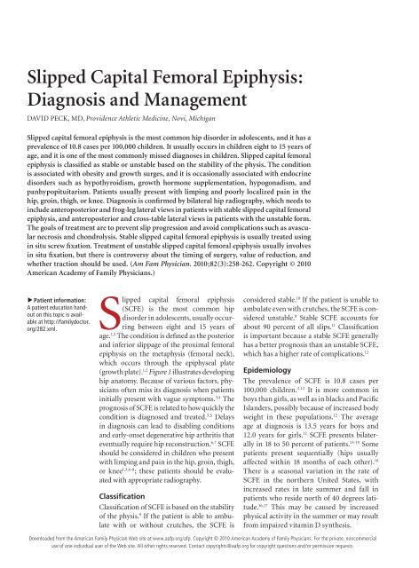
(growth plate). 1,2 Figure 1 illustrates developing<br />
hip anatomy. Because of various factors, physicians<br />
often miss its diagnosis when patients<br />
initially present with vague symptoms. 3,4 The<br />
prognosis of SCFE is related to how quickly the<br />
condition is diagnosed <strong>and</strong> treated. 3,5 Delays<br />
in diagnosis can lead to disabling conditions<br />
<strong>and</strong> early-onset degenerative hip arthritis that<br />
eventually require hip reconstruction. 6,7 SCFE<br />
should be considered in children who present<br />
with limping <strong>and</strong> pain in the hip, groin, thigh,<br />
or knee 1,3,6-8 ; these patients should be evaluated<br />
with appropriate radiography.<br />
Classification<br />
Classification of SCFE is based on the stability<br />
of the physis. 9 If the patient is able to ambulate<br />
with or without crutches, the SCFE is<br />
considered stable. 10 If the patient is unable to<br />
ambulate even with crutches, the SCFE is considered<br />
unstable. 9 Stable SCFE accounts for<br />
about 90 percent of all slips. 11 Classification<br />
is important because a stable SCFE generally<br />
has a better prognosis than an unstable SCFE,<br />
which has a higher rate of complications. 12<br />
Epidemiology<br />
The prevalence of SCFE is 10.8 cases per<br />
100,000 children. 2,12 It is more common in<br />
boys than girls, as well as in blacks <strong>and</strong> Pacific<br />
Isl<strong>and</strong>ers, possibly because of increased body<br />
weight in these populations. 12 The average<br />
age at diagnosis is 13.5 years for boys <strong>and</strong><br />
12.0 years for girls. 12 SCFE presents bilaterally<br />
in 18 to 50 percent of patients. 13-15 Some<br />
patients present sequentially (hips usually<br />
affected within 18 months of each other). 10<br />
There is a seasonal variation in the rate of<br />
SCFE in the northern United States, with<br />
increased rates in late summer <strong>and</strong> fall in<br />
patients who reside north of 40 degrees latitude.<br />
16,17 This may be caused by increased<br />
physical activity in the summer or may result<br />
from impaired vitamin D synthesis.<br />
Downloaded from the American Family Physician Web site at www.aafp.org/afp. Copyright © 2010 American Academy of Family Physicians. For the private, noncommercial<br />
use of one individual user of the Web site. All other rights reserved. Contact copyrights@aafp.org for copyright questions <strong>and</strong>/or permission requests.<br />
258 American Family Physician www.aafp.org/afp Volume 82, Number 3 ◆ August 1, 2010
ILLUSTRATION BY dAvId kLemm<br />
Proximal femoral<br />
capital epiphysis<br />
Physeal plate<br />
Etiology<br />
The etiology of SCFE is thought to be multifactorial<br />
<strong>and</strong> may include obesity, growth surges, <strong>and</strong>, less commonly,<br />
endocrine disorders. 18-21 Of children diagnosed<br />
with SCFE, 63 percent have a weight in the 90th percentile<br />
or higher. 22,23 Related endocrine disorders include<br />
hypothyroidism, growth hormone supplementation,<br />
hypogonadism, <strong>and</strong> panhypopituitarism. 2 An endocrine<br />
disorder should be considered in SCFE with unusual<br />
presentations, including patients who are younger than<br />
eight years, older than 15 years, or underweight. 20<br />
Table 1. Differential <strong>Diagnosis</strong> of Hip Pain in the Young Patient<br />
<strong>Slipped</strong> <strong>Capital</strong> <strong>Femoral</strong> <strong>Epiphysis</strong><br />
History <strong>and</strong> Physical Examination<br />
Condition Age (years) Clinical features Frequency <strong>Diagnosis</strong><br />
Apophyseal<br />
avulsion fracture<br />
12 to 25 Pain after sudden forceful<br />
movement<br />
Table 1 outlines the differential diagnosis of a young<br />
patient presenting with hip pain. The most common<br />
symptoms of SCFE are limping <strong>and</strong> pain that is poorly<br />
localized to the hip, groin, thigh, or knee. 8 Knee or distal<br />
thigh pain is the presenting symptom in 15 percent of<br />
patients with the condition. 24 History of trauma to the<br />
area is rare. 9,25<br />
On physical examination, the patient may be unable to<br />
bear weight with a severe slip. Limited internal rotation of<br />
the hip is the most telling sign in the diagnosis of SCFE. 8<br />
Obligatory external rotation is noted in the involved hip<br />
of patients with SCFE when the hip is passively flexed to<br />
90 degrees 1,4,8 (Figure 2). Unless the patient has bilateral<br />
SCFE, it is helpful to compare range of motion with the<br />
uninvolved hip.<br />
Radiography<br />
Radiography is needed for patients eight to 15 years of age<br />
with new-onset limping <strong>and</strong> pain in the hip, groin, thigh,<br />
or knee. It is important to inform the radiologist of the<br />
Often History of trauma; radiography<br />
Hip apophysitis 12 to 25 Activity-related hip pain Often History of overuse; radiography to rule out fractures<br />
Transient synovitis < 10 Limping or hip pain Often Radiography; laboratory testing; ultrasonography<br />
Fracture All ages Pain after traumatic event Occasionally History of trauma; radiography<br />
<strong>Slipped</strong> capital<br />
femoral epiphysis<br />
Legg-Calvé-<br />
Perthes disease<br />
Greater trochanter<br />
Lesser trochanter<br />
Figure 1. Anatomy of the developing hip.<br />
10 to 15 Hip, groin, thigh, or knee<br />
pain; limping<br />
4 to 9 Vague hip pain, decreased<br />
internal rotation of hip<br />
Occasionally Bilateral hip radiography (anteroposterior <strong>and</strong><br />
lateral)<br />
Infrequently Hip radiography or magnetic resonance imaging<br />
Septic arthritis All ages Fever, limping, hip pain Infrequently Radiography; laboratory testing; joint aspiration<br />
nOTe: Table is sorted by frequency of conditions.<br />
Metaphysis<br />
(femoral neck)<br />
Figure 2. Obligatory external rotation of the hip.<br />
August 1, 2010 ◆ Volume 82, Number 3 www.aafp.org/afp American Family Physician 259
<strong>Slipped</strong> <strong>Capital</strong> <strong>Femoral</strong> <strong>Epiphysis</strong><br />
Figure 3. Frog-leg lateral radiography of mild stable<br />
slipped capital femoral epiphysis.<br />
Figure 4. Anteroposterior radiography of left-sided<br />
slipped capital femoral epiphysis. Radiologic signs include:<br />
(A) Steel sign—on anteroposterior radiography, a double<br />
density is found at the metaphysis (caused by the posterior<br />
lip of the epiphysis being superimposed on the metaphysis);<br />
(B) widening of the growth plate (physis) compared<br />
with the uninvolved side; (C) decreased epiphyseal height<br />
compared with the uninvolved side; (D) klein’s line—on<br />
anteroposterior radiography, a line drawn along the superior<br />
edge of the femoral neck should normally cross the<br />
epiphysis; the epiphysis will fall below this line in slipped<br />
capital femoral epiphysis; <strong>and</strong> (E) lesser trochanter prominence,<br />
which is caused by external rotation of the femur.<br />
Information from reference 26.<br />
clinical context <strong>and</strong> that SCFE is suspected so that the<br />
diagnosis can be ruled out. Radiography needs to include<br />
anteroposterior <strong>and</strong> frog-leg lateral views of both hips to<br />
diagnose stable SCFE (Figure 3). In unstable SCFE, anteroposterior<br />
<strong>and</strong> cross-table lateral views of the involved side<br />
should be compared with the uninvolved side because of<br />
the decreased range of motion of the hip. 1,9 The radiologic<br />
signs of SCFE are shown in Figures 4 26 <strong>and</strong> 5.<br />
Radiography is used to grade the severity of the slip<br />
in SCFE. The Wilson method measures the relative displacement<br />
of the epiphysis on the metaphysis in a frog-leg<br />
lateral radiograph. A mild slip involves epiphysis displace-<br />
A<br />
e<br />
C<br />
B<br />
D<br />
Steel sign<br />
Widening of physis<br />
Relative decreased<br />
height of epiphysis<br />
Loss of intersection of the<br />
epiphysis by a lateral<br />
cortical line along<br />
femoral neck<br />
(Klein’s line)<br />
Klein’s line<br />
Figure 5. diagrams of radiographic signs of slipped capital<br />
femoral epiphysis.<br />
ment less than one third of the width of the metaphysis; a<br />
moderate slip involves displacement between one third <strong>and</strong><br />
one half of the width; <strong>and</strong> a severe slip involves displacement<br />
greater than one half of the width. 9 The Southwick<br />
method measures the epiphyseal shaft angle on the frog-leg<br />
lateral radiograph 27 (Figure 6). The angle is calculated by<br />
subtracting the epiphyseal shaft angle on the uninvolved<br />
side from that on the side with SCFE. A mild slip is less than<br />
30 degrees, a moderate slip is between 30 <strong>and</strong> 50 degrees,<br />
<strong>and</strong> a severe slip is greater than 50 degrees. 28<br />
Treatment<br />
Once the diagnosis of SCFE is made, the patient should<br />
be placed on non–weight-bearing crutches or in a wheelchair<br />
<strong>and</strong> quickly referred to an orthopedic surgeon<br />
familiar with the treatment of SCFE. 1 The initial goals<br />
of treatment are to prevent slip progression <strong>and</strong> avoid<br />
complications. 9,29<br />
260 American Family Physician www.aafp.org/afp Volume 82, Number 3 ◆ August 1, 2010<br />
ILLUSTRATIONS BY dAvId kLemm
ILLUSTRATION BY dAvId kLemm<br />
Normal SCFE<br />
12°<br />
b<br />
c<br />
Prophylactic treatment of the contralateral hip in<br />
patients with SCFE is controversial, but it is not recommended<br />
in most patients. Prophylactic pinning may be<br />
indicated in patients at high risk of subsequent slips, such<br />
as patients with obesity or an endocrine disorder, or those<br />
who have a low likelihood of follow-up. 11,15,30<br />
STABLE SCFE<br />
a<br />
Figure 6. Southwick method for determining slipped capital<br />
femoral epiphysis (SCFe) severity using a frog-leg lateral<br />
radiograph. The first line (a) is drawn from the anterior to<br />
the posterior epiphyseal edges. Next, a line (b) is drawn<br />
perpendicular to the first line. A third line (c) is drawn down<br />
the middle of the femoral diaphysis. The angle formed by<br />
lines b <strong>and</strong> c is the lateral epiphyseal-shaft angle (LeSA).<br />
The actual slip angle is the difference between the LeSA of<br />
the SCFe hip <strong>and</strong> that of the uninvolved hip.<br />
The st<strong>and</strong>ard treatment of stable SCFE is in situ fixation<br />
with a single screw 1,4,9,31 (Figure 7). Case series <strong>and</strong> animal<br />
model studies have shown this to be a simple technique<br />
with low rates of recurrence <strong>and</strong> complications. 4,9,31 After<br />
SORT: KEY RECOMMENDATIONS FOR PRACTICE<br />
Clinical recommendation<br />
Family physicians should consider SCFe when<br />
a child presents with limping <strong>and</strong> groin, hip,<br />
thigh, or knee pain.<br />
Physical examination of patients with SCFe<br />
usually shows decreased internal rotation of<br />
the hip <strong>and</strong> obligatory external rotation.<br />
Radiography to rule out SCFe should include<br />
anteroposterior <strong>and</strong> lateral views of the hips<br />
(frog-leg lateral views for stable SCFe; crosstable<br />
lateral views for unstable SCFe).<br />
The st<strong>and</strong>ard treatment of stable SCFe is in<br />
situ fixation with a single screw.<br />
SCFE = slipped capital femoral epiphysis.<br />
a<br />
c<br />
b<br />
40°<br />
Evidence<br />
rating References<br />
C 1, 3, 6-8<br />
C 1, 4, 8<br />
C 1, 9<br />
C 1, 4, 9, 31<br />
A = consistent, good-quality patient-oriented evidence; B = inconsistent or limitedquality<br />
patient-oriented evidence; C = consensus, disease-oriented evidence, usual<br />
practice, expert opinion, or case series. For information about the SORT evidence<br />
rating system, go to http://www.aafp.org/afpsort.xml.<br />
<strong>Slipped</strong> <strong>Capital</strong> <strong>Femoral</strong> <strong>Epiphysis</strong><br />
Figure 7. Anteroposterior radiography of bilateral stable<br />
slipped capital femoral epiphysis treated with in situ fixation<br />
with a single screw.<br />
closure of the growth plate, progression of athletic activities<br />
may be allowed, including running <strong>and</strong>, eventually,<br />
participating in contact sports. 1 Most patients with mild<br />
to moderate SCFE who are treated with in situ fixation<br />
have good to excellent long-term outcomes. 11<br />
UNSTABLE SCFE<br />
Unstable SCFE is a much more severe injury than stable<br />
SCFE. The rate of osteonecrosis is as high as 20 to<br />
50 percent in patients with the unstable form. 9,32 Treatment<br />
goals are similar to those of stable SCFE with in<br />
situ fixation, but there is controversy as to the specifics<br />
of treatment, including timing of surgery,<br />
value of reduction, <strong>and</strong> whether traction<br />
should be used. 1,31<br />
Complications<br />
AVASCULAR NECROSIS<br />
Avascular necrosis occurs in up to 60 percent<br />
of patients with unstable SCFE. 31 It<br />
is a serious complication associated with<br />
severe displacement <strong>and</strong> fixation with more<br />
than one screw. 8,29 Avascular necrosis often<br />
leads to advanced <strong>and</strong> early degenerative<br />
osteoarthritis. 33<br />
CHONDROLYSIS<br />
Chondrolysis is the acute loss of articular<br />
cartilage, which causes joint stiffness <strong>and</strong><br />
pain. 9 It is usually reported as a complication<br />
of surgical treatment of SCFE, but it<br />
can occur with the use of a hip spica cast<br />
<strong>and</strong> in untreated advanced SCFE. With the<br />
improvement of surgical techniques, the<br />
August 1, 2010 ◆ Volume 82, Number 3 www.aafp.org/afp American Family Physician 261
<strong>Slipped</strong> <strong>Capital</strong> <strong>Femoral</strong> <strong>Epiphysis</strong><br />
incidence of chondrolysis has decreased from a rate of<br />
5 to 7 percent to a rate of 1 to 2 percent in patients treated<br />
for SCFE. 8,29<br />
The Author<br />
DAVID PECK, MD, FACSM, CAQSM, is the research <strong>and</strong> educational director<br />
for the Providence Athletic Medicine Fellowship Program in Novi, Mich.<br />
Address correspondence to David Peck, MD, Providence Athletic<br />
Medicine, 26750 Providence Pkwy., Ste. 210, Novi, MI 48374 (e-mail:<br />
dmpeck99@aol.com). Reprints are not available from the author.<br />
Author disclosure: Nothing to disclose.<br />
REFERENCES<br />
1. Loder RT. <strong>Slipped</strong> capital femoral epiphysis [published correction appears<br />
in Am Fam Physician. 1998;58(1):52]. Am Fam Physician. 1998;57(9):<br />
2135-2142, 2148-2150.<br />
2. Gholve PA, Cameron DB, Millis MB. <strong>Slipped</strong> capital femoral epiphysis<br />
update. Curr Opin Pediatr. 2009;21(1):39-45.<br />
3. Rahme D, Comley A, Foster B, Cundy P. Consequences of diagnostic<br />
delays in slipped capital femoral epiphysis. J Pediatr Orthop B. 2006;<br />
15(2):93-97.<br />
4. Katz DA. <strong>Slipped</strong> capital femoral epiphysis: the importance of early<br />
diagnosis. Pediatr Ann. 2006;35(2):102-111.<br />
5. Loder RT. Correlation of radiographic changes with disease severity <strong>and</strong><br />
demographic variables in children with stable slipped capital femoral<br />
epiphysis. J Pediatr Orthop. 2008;28(3):284-290.<br />
6. Kocher MS, Bishop JA, Weed B, et al. Delay in diagnosis of slipped capital<br />
femoral epiphysis. Pediatrics. 2004;113(4):e322-e325.<br />
7. Green DW, Reynolds RA, Khan Sn, Tolo V. The delay in diagnosis of<br />
slipped capital femoral epiphysis: a review of 102 patients. HSS J. 2005;<br />
1(1):103-106.<br />
8. Reynolds RA. <strong>Diagnosis</strong> <strong>and</strong> treatment of slipped capital femoral epiphysis.<br />
Curr Opin Pediatr. 1999;11(1):80-83.<br />
9. Loder RT, Richards BS, Shapiro PS, Reznick LR, Aronson DD. Acute<br />
slipped capital femoral epiphysis: the importance of physeal stability.<br />
J Bone Joint Surg Am. 1993;75(8):1134-1140.<br />
10. Loder RT. <strong>Slipped</strong> capital femoral epiphysis in children. Curr Opin Pediatr.<br />
1995;7(1):95-97.<br />
11. Loder RT, Starnes T, Dikos G, Aronsson DD. Demographic predictors of<br />
severity of stable slipped capital femoral epiphyses. J Bone Joint Surg<br />
Am. 2006;88(1):97-105.<br />
12. Lehmann CL, Arons RR, Loder RT, Vitale MG. The epidemiology of<br />
slipped capital femoral epiphysis: an update. J Pediatr Orthop. 2006;<br />
26(3):286-290.<br />
13. Loder RT. The demographics of slipped capital femoral epiphysis. An international<br />
multicenter study. Clin Orthop Relat Res. 1996;(322):8-27.<br />
14. Koenig KM, Thomson JD, Anderson KL, Carney BT. Does skeletal maturity<br />
predict sequential contralateral involvement after fixation of slipped<br />
capital femoral epiphysis? J Pediatr Orthop. 2007;27(7):796-800.<br />
15. Riad J, Bajelidze G, Gabos PG. Bilateral slipped capital femoral epiphysis:<br />
predictive factors for contralateral slip. J Pediatr Orthop. 2007;<br />
27(4):411-414.<br />
16. Loder RT. A worldwide study on the seasonal variation of slipped capital<br />
femoral epiphysis. Clin Orthop Relat Res. 1996;(322):28-36.<br />
17. Brown D. Seasonal variation of slipped capital femoral epiphysis in the<br />
United States. J Pediatr Orthop. 2004;24(2):139-143.<br />
18. Murray AW, Wilson nI. Changing incidence of slipped capital femoral epiphysis:<br />
a relationship with obesity? J Bone Joint Surg Br. 2008;90(1):92-94.<br />
19. Bhatia nn, Pirpiris M, Otsuka nY. Body mass index in patients with slipped<br />
capital femoral epiphysis. J Pediatr Orthop. 2006;26(2):197-199.<br />
20. Papavasiliou KA, Kirkos JM, Kapetanos GA, Pournaras J. Potential influence<br />
of hormones in the development of slipped capital femoral epiphysis:<br />
a preliminary study. J Pediatr Orthop B. 2007;16(1):1-5.<br />
21. nourbakhsh A, Ahmed HA, McAuliffe TB, Garges KJ. Case report: bilateral<br />
slipped capital femoral epiphyses <strong>and</strong> hormone replacement. Clin<br />
Orthop Relat Res. 2008;466(3):743-748.<br />
22. Houghton KM. Review for the generalist: evaluation of pediatric hip<br />
pain. Pediatr Rheumatol Online J. 2009;7:10.<br />
23. Manoff eM, Banffy MB, Winell JJ. Relationship between body mass<br />
index <strong>and</strong> slipped capital femoral epiphysis. J Pediatr Orthop. 2005;<br />
25(6):744-746.<br />
24. Matava MJ, Patton CM, Luhmann S, Gordon Je, Schoenecker PL. Knee pain<br />
as the initial symptom of slipped capital femoral epiphysis: an analysis of initial<br />
presentation <strong>and</strong> treatment. J Pediatr Orthop. 1999;19(4):455-460.<br />
25. Kasper JC, Gerhardt MB, M<strong>and</strong>elbaum BR. Stress injury leading to<br />
slipped capital femoral epiphysis in a competitive adolescent tennis<br />
player: a case report. Clin J Sport Med. 2007;17(1):72-74.<br />
26. Mitchell SR, Tennent TD, Brown RR, Monsell F. <strong>Slipped</strong> capital femoral<br />
epiphysis. Hip Int. 2007;17(4):185-193.<br />
27. Jacobs B. <strong>Diagnosis</strong> <strong>and</strong> natural history of slipped capital femoral epiphysis.<br />
Instr Course Lect. 1972;21:167-173.<br />
28. Southwick WO. Compression fixation after biplane intertrochanteric<br />
osteotomy for slipped capital femoral epiphysis. A technical improvement.<br />
J Bone Joint Surg Am. 1973;55(6):1218-1224.<br />
29. Aronsson DD, Loder RT. Treatment of the unstable (acute) slipped capital<br />
femoral epiphysis. Clin Orthop Relat Res. 1996;(322):99-110.<br />
30. Lim YJ, Lam KS, Lee eH. Review of the management outcome of slipped<br />
capital femoral epiphysis <strong>and</strong> the role of prophylactic contra-lateral pinning<br />
re-examined. Ann Acad Med Singapore. 2008;37(3):184-187.<br />
31. Kalogrianitis S, Tan CK, Kemp GJ, Bass A, Bruce C. Does unstable slipped<br />
capital femoral epiphysis require urgent stabilization? J Pediatr Orthop<br />
B. 2007;16(1):6-9.<br />
32. Loder RT. Unstable slipped capital femoral epiphysis. J Pediatr Orthop.<br />
2001;21(5):694-699.<br />
33. Boero S, Brunenghi GM, Carbone M, Stella G, Calevo MG. Pinning in<br />
slipped capital femoral epiphysis: long-term follow-up study. J Pediatr<br />
Orthop B. 2003;12(6):372-379.<br />
262 American Family Physician www.aafp.org/afp Volume 82, Number 3 ◆ August 1, 2010


