unusual x-ray characteristics of vermiculite from wiry - Clay Minerals ...
unusual x-ray characteristics of vermiculite from wiry - Clay Minerals ...
unusual x-ray characteristics of vermiculite from wiry - Clay Minerals ...

You also want an ePaper? Increase the reach of your titles
YUMPU automatically turns print PDFs into web optimized ePapers that Google loves.
<strong>Clay</strong>s and <strong>Clay</strong> <strong>Minerals</strong>, Vol. 49, No. 3, 197-203, 2001.<br />
UNUSUAL X-RAY CHARACTERISTICS OF VERMICULITE FROM WIRY,<br />
LOWER SILESIA, POLAND<br />
BORIS A. SAKHAROV 1, ELZBIETA DUBINSKA 2, PAWEL BYLINA 3 AND GRZEGORZ KAPROlq 2<br />
1 Geological Institute, Russian Academy <strong>of</strong> Sciences, Pyzhevsky Street 7, 109017 Moscow, Russia<br />
2 Institute <strong>of</strong> Geochemistry,.Mineralogy and Petrology, Faculty <strong>of</strong> Geology, Warsaw University,<br />
al. Zwirki i Wigury 93, 02~089 Warsaw, Poland<br />
3 Institute <strong>of</strong> Geological Sciences, Polish Academy <strong>of</strong> Sciences, ul. Twarda 51/55, 00-818 Warsaw, Poland<br />
Abstract Coarse-grained <strong>vermiculite</strong> <strong>from</strong> a serpentinite-pegmafite thermal zone displays a rational<br />
series <strong>of</strong> narrow 14.4 A basal reflections and an <strong>unusual</strong> broad 28 A peak. X-<strong>ray</strong> diffraction simulations<br />
and fitting techniques show that the 28 ,~ peak is related to 28 .~ domains consisting <strong>of</strong> elongated 2:1<br />
layers <strong>of</strong> different lengths. The domains are located at the crystal edges <strong>of</strong> the <strong>vermiculite</strong>.<br />
Key Words----Crystal Edge, Interstratified Mineral, Vermiculite, XRD Characteristics.<br />
INTRODUCTION<br />
The X-<strong>ray</strong> identification <strong>of</strong> <strong>vermiculite</strong> would ap-<br />
pear to be simple: it is distinguishable <strong>from</strong> other clay<br />
minerals after undergoing various treatments (e.g. eth-<br />
ylene glycol, Mg 2+ + glycerol, heating, and saturation<br />
with several exchangeable cations). However, sponta-<br />
neous rehydration after heating and ambiguous results<br />
<strong>of</strong> swelling tests, primarily related to slow adsorption<br />
<strong>of</strong> ethylene glycol and glycerol, make unambiguous<br />
identification <strong>of</strong> <strong>vermiculite</strong> time consuming and dif-<br />
ficult (MacEwan and Wilson, 1980; de la Calle and<br />
Suquet, 1988; Pons et al., 1989; Reichenbach and Be-<br />
yer, 1994, 1995; Reichenbach and Schfitte, 1995;<br />
Moore and Reynolds, 1997). The diffraction patterns<br />
<strong>of</strong> regularly interstratified (Reichweite, R = 1) min-<br />
erals with <strong>vermiculite</strong> layers, e.g. high-charge corren-<br />
site and hydrobiotite, display strong low-angle super-<br />
structure peaks both in air-dried and ethylene glycol-<br />
treated samples. Nevertheless, the correct identifica-<br />
tion <strong>of</strong> expandable layers in the trioctahedral<br />
interstratified minerals may be problematic.<br />
This study concerns a detailed analysis <strong>of</strong> <strong>unusual</strong><br />
diffraction <strong>characteristics</strong> <strong>of</strong> <strong>vermiculite</strong> <strong>from</strong> Wiry,<br />
Lower Silesia, Poland. These features occur mainly on<br />
the low-angle diffraction peak. The <strong>vermiculite</strong> <strong>from</strong><br />
Wiry is a product <strong>of</strong> phlogopite alteration, found in<br />
the contact zone between serpentinite and granite-type<br />
apophysis. Details <strong>of</strong> the <strong>vermiculite</strong> occurrence are<br />
given in Sachanbifiski (1993), Jelitto et al. (1993), Du-<br />
bifiska et al. (1995), and Janeczek and Sachanbifiski<br />
(1995).<br />
MATERIAL AND METHODS<br />
Grain fractions were obtained by soaking the sample<br />
in double-distilled water until it disaggregated. Coarse<br />
flakes were separated using a nylon sieve (0.1 ram)<br />
and purified by hand, using a binocular microscope.<br />
Fine-grained fractions <strong>of</strong> the same sample were sepa-<br />
Copyright 9 2001, The <strong>Clay</strong> <strong>Minerals</strong> Society 197<br />
rated using repeated centrifugal sedimentation in double-distilled<br />
water.<br />
All X-<strong>ray</strong> diffraction (XRD) patterns were obtained<br />
on a DRON-1 diffractometer, using CoKct radiation<br />
(Fe-filtered), 1.0 and 0.5 mm divergence slits, 0.25 mm<br />
receiving slit, 0.04~ steps, a counting time <strong>of</strong> 5 s per<br />
step, and a 1.5-45~ angular range. Oriented clay aggregates<br />
were prepared on 7 cm length glass slides by<br />
the pipette method using 2.5 mg/cm 2 <strong>of</strong> specimen.<br />
Samples were treated overnight with liquid ethylene<br />
glycol. The specimens were heated for 2 h on a homemade<br />
thermal stage and XRD patterns were recorded<br />
at temperatures <strong>of</strong> 75, 150 and 250~ The XRD patterns<br />
<strong>of</strong> specimens heated at 500~ were recorded after<br />
cooling the specimen. The patterns were collected using<br />
the DRONEK s<strong>of</strong>tware (W. Musial, unpublished<br />
manuscript, 1992).<br />
Simulated diffraction patterns <strong>of</strong> various structural<br />
models were calculated using the ASN program (Drits<br />
and Sakharov, 1976). The z atomic coordinates for different<br />
types <strong>of</strong> layers for simulations were obtained<br />
<strong>from</strong> Walker (1975) and Moore and Reynolds (1997).<br />
The instrumental factors recommended by Drits and<br />
Tchoubar (1990), i.e. sizes <strong>of</strong> the divergent and receiving<br />
slits, distances between X-<strong>ray</strong> source, sample<br />
and detector, and specimen size and thickness were<br />
used. The particle orientation factor, tr*, <strong>of</strong> 12 ~ (Reynolds,<br />
1986) was also included in the calculations. The<br />
lognormal distribution <strong>of</strong> the thickness <strong>of</strong> the coherent<br />
scattering domain (CSD) was adopted (Drits et al.,<br />
1998) to obtain the best fit between calculated and<br />
experimental curves. The authors changed the independent<br />
parameters for each calculated X-<strong>ray</strong> curve to<br />
accommodate d-values, intensity ratios, and pr<strong>of</strong>iles on<br />
experimental and simulated patterns.<br />
The chemical composition <strong>of</strong> the sample studied<br />
(calculated on the basis <strong>of</strong> O10(OH) 2 per formula unit)<br />
is: Mgo.a9Cao.ol (Mg2.73Fe3 +0.27)( Si2.93A11.05Fe3 +0.02)O lo(On)2 ,<br />
where Fe 3+ is equal to total Fe on the basis <strong>of</strong> wet chem-
198 Sakharov, Dubfnska, Bylina and Kapr<strong>of</strong>i <strong>Clay</strong>s and <strong>Clay</strong> <strong>Minerals</strong><br />
i<br />
!<br />
A<br />
14.3<br />
--i 28.4 3.59 2.88<br />
ik-'~"~-J ~. 7.17 4.79 ,,t ]L j~ natural<br />
_=!<br />
B 14.34<br />
14.32-,<br />
~ .~5.g3 ~ 250~<br />
~<br />
10.04<br />
(1.28 ~ 2.88<br />
A288<br />
~.~,2 4"82 3"59 3"22 "~<br />
320 f/<br />
-~--- - after 2 weeks<br />
1'0 1'5 :~0 2~5 30 35<br />
~<br />
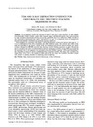
Figure 1. Experimental (observed) XRD patterns <strong>of</strong> a vermiculitic sample <strong>from</strong> Wiry, using oriented aggregate specimens.<br />
(A) Natural--air-dried specimen, eth. glycol---ethylene glycol-solvated specimen, 250~ recorded using a heating<br />
stage. The upper line represents an enlarged pattern. The sample was not completely anhydrous and it port<strong>ray</strong>s an irregularly<br />
interstratified structure (R = 0) composed <strong>of</strong> 10.2 A layers (anhydrous <strong>vermiculite</strong>, 85 vol.%), 11.6 A layers (one-layer H20<br />
complex <strong>of</strong> <strong>vermiculite</strong>, 7.5 vol.%) and 14.4 ,~ layers (two-layer H20 complex <strong>of</strong> <strong>vermiculite</strong>, 7.5 vol.%). (B) XRD patterns<br />
<strong>of</strong> the specimen heated to 500~ in an oven, recorded immediately after cooling and two weeks later. The FWHM values are<br />
given in parentheses; the intensity <strong>of</strong> the --14 A peak on the XRD pattern <strong>of</strong> heated specimen in (B) is --5 smaller than<br />
the corresponding peak <strong>of</strong> the untreated sample in (A); CoKa radiation, grain fraction >0.1 mm, flakes selected under binocular<br />
microscope.<br />
ical analysis, i.e. using atomic absorption spectroscopy<br />
(AAS) for Si, A1, Ti, Fe, Mn, Ni, Cr, Zn and Cu and<br />
flame atomic emission spectroscopy (FAES) for Na,<br />
K, Ca, Li, Rb and Cs. For the analyses we used HF-<br />
or HCl-digested samples. We assumed that Fe 3+ can<br />
be placed in tetrahedral sites to complete a total <strong>of</strong> 4.0<br />
tetrahedral cations. Simulated XRD patterns were cal-<br />
culated using fixed element ratios.<br />
RESULTS AND DISCUSSION<br />
The XRD pattern <strong>of</strong> the natural and glycolated<br />
coarse flakes shows a series <strong>of</strong> sharp 00l reflections <strong>of</strong><br />
typical <strong>vermiculite</strong>, and a broad low-angle band at<br />
--28 A (Figure 1A). The reflection positions, intensi-<br />
ties, and pr<strong>of</strong>iles are almost the same in both the nat-<br />
ural and glycolated specimens. The 28 ,~ diffraction<br />
maximum was obtained at high relative humidity (r.h.<br />
> 80%). The maximum disappeared at low r.h. (10%)<br />
and only a rational series <strong>of</strong> 14.3 ,~ basal reflections<br />
<strong>of</strong> <strong>vermiculite</strong> were visible. This effect is reversible,<br />
i.e. the maximum appears again with increasing r.h.<br />
The XRD pattem <strong>of</strong> the sample heated at 500~ and<br />
recorded after co<strong>of</strong>ing the specimen (Figure 1B) reveals<br />
two series <strong>of</strong> peaks: (1) --14.3 ,~ (full width at half-<br />
maximum peak height, FWHM, 0.4-0.6~ and (2) two<br />
broad bands: --10 ,~ (FWHM at 1.28~ and 3.2 ,~<br />
(FWHM at 5.35~ are presumably several broad and<br />
poorly resolved diffraction maxima). Both series are re-<br />
lated to the unstable irregularly interstratified structures<br />
composed <strong>of</strong> common <strong>vermiculite</strong> and partly and/or fully<br />
dehydrated <strong>vermiculite</strong>. Rehydration occurred upon (or<br />
immediately after) cooling <strong>of</strong> the specimen. Our efforts<br />
to avoid spontaneous rehydration <strong>of</strong> the <strong>vermiculite</strong> by<br />
using conventional heating procedures failed.
Vol. 49, No. 3, 2001 Unusual XRD <strong>characteristics</strong> <strong>of</strong> <strong>vermiculite</strong> 199<br />
14.3<br />
..~. 11.7<br />
28.4 11 2.88<br />
~ ~,~.6 7.17 4~9 3~ L. 2000<br />
383 75~<br />
10 20 30 40<br />
~<br />
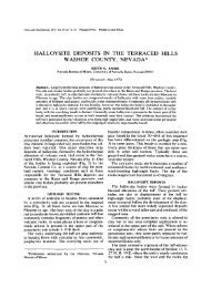
Figure 2. Experimental (observed) XRD patterns <strong>of</strong> a vermiculitic sample <strong>from</strong> Wiry (grain-size <strong>of</strong> >0.1 mm, flakes selected<br />
under microscope) using oriented aggregate specimens. The traces were recorded after 1 h heating on a heating stage at each<br />
temperature. CoKa radiation.<br />
r<br />
m<br />
14.3<br />
/l~ 14"6<br />
Bi - biotite<br />
T - talc<br />
14.5 flakes<br />
~'~ ~~- 5-10pm<br />
~0.1 mm, 5-10 p.m, 2-5 p.m,<br />
200 Sakharov, Dubfnska, Bylina and Kapr<strong>of</strong>i <strong>Clay</strong>s and <strong>Clay</strong> <strong>Minerals</strong><br />
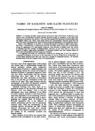
Figure 4. (A) Experimental (observed) XRD pattern <strong>of</strong> the coarse flake-fraction treated with ethylene glycol and a calculated<br />
regularly-interstratified <strong>vermiculite</strong>-saponite pattern, with R = 1. The <strong>vermiculite</strong> layers contain a single layer <strong>of</strong> glycol<br />
molecules, the saponite layers contain two layers <strong>of</strong> ethylene glycol, with the total Fe content located in saponite layers.<br />
(]3) Experimental XRD pattern <strong>of</strong> the coarse flake-fraction heated to 250~ (heating stage) and a calculated pattern <strong>of</strong> the<br />
mixture composed <strong>of</strong> regularly interstratified chlorite-<strong>vermiculite</strong> (90 vol.%, <strong>vermiculite</strong> layers contracted) and regular ver-<br />
miculite (10 vol.%, contracted layers). The g<strong>ray</strong> bars represent reflections on calculated traces, absent <strong>from</strong> experimental<br />
patterns. CoKcx radiation.<br />
_.g,<br />
r<br />
c"<br />
14.3<br />
1'0<br />
4.79<br />
3.59<br />
2.88<br />
A ~ ~ experimental<br />
~ ~ calculated<br />
10020 ~-- :30 ~ ,~0<br />
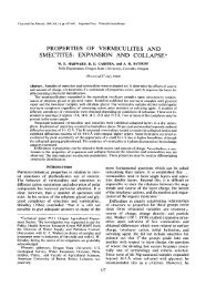
Figure 5. Calculated X-<strong>ray</strong> traces <strong>of</strong> <strong>vermiculite</strong> and <strong>unusual</strong> 28 A domain. The calculated pattern represents a mixture <strong>of</strong><br />
regular <strong>vermiculite</strong> (93 vol.%) and 28 ,~ edge layers (7 vol.%). CoKa radiation.
Vol. 49, No. 3, 2001 Unusual XRD <strong>characteristics</strong> <strong>of</strong> <strong>vermiculite</strong> 201<br />
Figure 6. Model <strong>of</strong> <strong>vermiculite</strong> structure with 28 ~, domains stable in high r.h. conditions.<br />
showed that the 28 ,~ peak disappeared immediately<br />
when layer ratios were not equal to 1:1, or when the<br />
regularity <strong>of</strong> interstratification decreased.<br />
lnterstratified <strong>vermiculite</strong>-smectite, R = 1. Because<br />
observed X-<strong>ray</strong> tracings <strong>of</strong> the glycolated sample do<br />
not show swelling effects, there is no evidence sup-<br />
Figure 7. Model <strong>of</strong> 'ski-like' terminal edges <strong>of</strong> dehydrated<br />
domain.<br />
porting an interstratified <strong>vermiculite</strong>-smectite model<br />
(Figure 4A). We considered several models <strong>of</strong> vermic-<br />
ulite-smectite interstratification, including different co-<br />
herently scattering domain sizes and distributions and<br />
different locations <strong>of</strong> heavy cation and different num-<br />
bers <strong>of</strong> octahedral vacancies. The results <strong>of</strong> these cal-<br />
culations were inconsistent with the observed patterns.<br />
Interstratified <strong>vermiculite</strong>-mica, R = 1. Interstratified<br />
<strong>vermiculite</strong>-mica models must be rejected because the<br />
sample does not contain K, Na, or other elements that<br />
can be placed into a mica-like interlayer.<br />
Vermiculite-type mineral comprising layers with an<br />
inhomogeneous distribution <strong>of</strong> 2:1 layers and~or<br />
interlayers<br />
Inhomogeneity in <strong>vermiculite</strong> can result <strong>from</strong>: (1)<br />
different octahedral Fe content in adjacent layers; (2)<br />
polar 2:1 layers owing to different A1-Si substitution<br />
in the tetrahedral sheets and thus, different layer<br />
charge and interlayer cation and water contents; and<br />
(3) various interlayer cations and water contents with<br />
different positions.<br />
These models were examined in detail. However,<br />
the resulting calculated patterns did not compare well<br />
with the observed XRD patterns. Although some cal-<br />
culated patterns show a 28 .& peak, these patterns al-<br />
ways showed a relatively intense 9.6 .& peak; more-<br />
over, the observed 28 A peak was broader than the<br />
calculated peak, whereas other calculated parts <strong>of</strong> the<br />
pattern fit well with the observed patterns. Thus, mod-<br />
els involving inhomogeneity <strong>of</strong> <strong>vermiculite</strong> are reject-<br />
ed.
202 Sakharov, Dubfnska, Bylina and Kapr<strong>of</strong>i <strong>Clay</strong>s and <strong>Clay</strong> <strong>Minerals</strong><br />
A mixture <strong>of</strong> <strong>vermiculite</strong> and interstratified mineral<br />
The data observed can be interpreted tentatively as<br />
a mixture <strong>of</strong> <strong>vermiculite</strong> as the dominant phase with<br />
minor interstratified chlorite-<strong>vermiculite</strong> or inhomo-<br />
geneous <strong>vermiculite</strong>. We examined these mixtures us-<br />
ing fitting procedures for simulated and observed XRD<br />
patterns. All trials were unsuccessful. Figure 4B shows<br />
an example <strong>of</strong> a mixture composed <strong>of</strong> interstratified<br />
chlorite-<strong>vermiculite</strong> and <strong>vermiculite</strong>. The X-<strong>ray</strong> pat-<br />
terns <strong>of</strong> both untreated and glycolated samples can be<br />
accepted provisionally. However, heated corrensite<br />
produces an XRD pattern which differs <strong>from</strong> the ob-<br />
served pattern (Figure 4B).<br />
A model <strong>of</strong> a mixture composed <strong>of</strong> Mg-exchanged<br />
<strong>vermiculite</strong> with a large CSD thickness and inhomo-<br />
geneous <strong>vermiculite</strong> with relatively small CSD thick-<br />
ness produced both 28 ]~ and 9.6 A peaks, but the<br />
latter peak was absent <strong>from</strong> the observed patterns.<br />
A <strong>vermiculite</strong> structure with <strong>unusual</strong> crystal domains<br />
The models described above cannot explain the ob-<br />
served diffraction effects. The 28 ]k reflection is also<br />
more prominent in the coarse-grained <strong>vermiculite</strong><br />
(Figure 3). Therefore, we suggest that the observed<br />
XRD effects arise <strong>from</strong> different domains consisting<br />
<strong>of</strong> 2:1 layers with a periodicity <strong>of</strong> 14 and 28 .~. These<br />
domains scatter X-<strong>ray</strong>s independently. The 14 A do-<br />
mains represent normal <strong>vermiculite</strong>, whereas the 28 ,~<br />
domains are composed <strong>of</strong> 2:1 layers with interlayers<br />
containing unidentified material which scatters X-<strong>ray</strong>s<br />
incoherently. Figure 5 shows the agreement <strong>of</strong> X-<strong>ray</strong><br />
patterns calculated on the basis <strong>of</strong> the proposed model<br />
and observed curves. The reflection positions, inten-<br />
sifies and pr<strong>of</strong>iles fit well.<br />
A reasonable explanation <strong>of</strong> the 28 A domains is<br />
the following: <strong>vermiculite</strong> crystals have ordered alter-<br />
nating and variable elongated 2:1 layers with differ-<br />
ences in length at the crystal edges. Thus, 28 A do-<br />
mains can occur along with 14 ,~ layer stacking (Fig-<br />
ure 6). The average thickness <strong>of</strong> the 14 ,~ and 28 A<br />
domains is equal to 20 and 3 layers, respectively. An<br />
approximate content <strong>of</strong> the 28 A edge layers is --7%<br />
<strong>of</strong> the total <strong>vermiculite</strong> volume. The 28 A domains<br />
appear to lose their periodicity in low humidity and<br />
during heating to produce a 'ski-like' pattern (Figure 7)<br />
similar to edges observed by Reichenbach et al. (1988)<br />
on high-resolution transmission electron microscope<br />
images.<br />
CONCLUSIONS<br />
The X-<strong>ray</strong> pattern <strong>of</strong> Wiry <strong>vermiculite</strong> is <strong>unusual</strong>.<br />
The diffraction maximum at 28 ,~ is definitely not an<br />
artifact <strong>of</strong> instrumental parameters and was obtained<br />
only at exceptionally high relative humidity.<br />
The <strong>vermiculite</strong> <strong>from</strong> Wiry is a transitional mineral<br />
in the sequence phlogopite --~ interstratified vermicu-<br />
lite-phlogopite --~ <strong>vermiculite</strong> --+ saponite, where hon-<br />
eycomb saponite aggregates unequivocally form ow-<br />
ing to dissolution-precipitation (Jelitto et aL, 1993;<br />
Dubifiska et al., 1995). The 28 ,~ domains may be<br />
where initial dissolution <strong>of</strong> the <strong>vermiculite</strong> occurs.<br />
ACKNOWLEDGMENTS<br />
We thank Z. J<strong>of</strong>ica for performing the wet chemical anal-<br />
yses. We are grateful to J. Mogcicki for flake separation. We<br />
thank W. Musia/for the DRONEK s<strong>of</strong>tware. We also thank<br />
S. Tsipursky for useful discussions, and D. McCarty, J. Walk-<br />
er, H. Graf yon Reichenbach and S. Guggenheim for their<br />
helpful suggestions to improve this manuscript. This inves-<br />
tigation was supported by State Committee for Scientific Re-<br />
search (KBN 6P04D 014 09) and the Russian Science Foun-<br />
dation.<br />
REFERENCES<br />
Collins, D.R., Fitch, A.N. and Catlow, R.A. (1992) Dehydra-<br />
tion <strong>of</strong> <strong>vermiculite</strong>s and montmorillonites: a time-resolved<br />
powder neutron diffraction study. Journal <strong>of</strong> Materials<br />
Chemistry, 2, 865-873.<br />
de la Calle, C. and Suquet, H. (1988) Vermiculite. Pp. 455-<br />
496 in: Hydrous Phyllosilicates (exclusive <strong>of</strong> micas) (S.W.<br />
Bailey, editor). Reviews in Mineralogy, 19, Mineralogical<br />
Society <strong>of</strong> America, Washington, D.C.<br />
Drits, V.A. and Sakharov, B.A. (1976) X-<strong>ray</strong> Structural Anal-<br />
ysis <strong>of</strong> Mixed-layer <strong>Minerals</strong>. Transactions <strong>of</strong> Academy <strong>of</strong><br />
Sciences U.S.S.R., 295, Moscow, 252 pp. (in Russian).<br />
Drits, V.A. and Tchoubar, C. (1990) X-Ray Diffraction by La-<br />
mellar Structures. Theory and Application to Microdivided<br />
Silicates and Carbons. Springer-Verlag, Berlin-Heidelberg-<br />
New York-London-Paris-Tokyo-Hong Kong-Barcelona,<br />
371 pp.<br />
Drits, V.A., Eberl, D.D. and Srod<strong>of</strong>i, J. (1998) XRD mea-<br />
surement <strong>of</strong> mean thickness, thickness distribution and<br />
strain for illite and illite/smectite crystallites by the Bertant-<br />
Warren-Awerbach technique. <strong>Clay</strong>s and <strong>Clay</strong> <strong>Minerals</strong>, 46,<br />
38-50.<br />
Dubifiska, E., Jelitto, J. and Kozlowski, A. (1995) Origin and<br />
evolution <strong>of</strong> granite-serpentinite reaction zones at Wiry,<br />
Lower Silesia. Acta Geologica Polonica, 45, 41-82.<br />
Janeczek, J. and Sachanbifiski, M. (1995) New data on hy-<br />
bridal pegmatite in serpentinite <strong>from</strong> magnesite mine in<br />
Wiry (Lower Silesia). Przeglqd Geologiczny, 53, 777-782<br />
(in Polish).<br />
Jelitto, J., Dubifiska, E., Wiewi6ra A. and Bylina, P. (1993)<br />
Layer silicates <strong>from</strong> serpentinite-pegmatite contact (Wiry,<br />
Lower Silesia, Poland). <strong>Clay</strong>s and <strong>Clay</strong> <strong>Minerals</strong>, 41,693-<br />
701.<br />
MacEwan, D.M.C. and Wilson, M.J. (1980) Interlayer and<br />
intercalation complexes <strong>of</strong> clay minerals. Pp. 197-248 in:<br />
Crystal Structures <strong>of</strong> <strong>Clay</strong> <strong>Minerals</strong> and their X-<strong>ray</strong> Iden-<br />
tification (G.W. Brindley and G. Brown, editors). Mono-<br />
graph, 5. Mineralogical Society, London.<br />
Moore, D.M. and Reynolds, R.C. (1997) X-<strong>ray</strong> Diffraction<br />
and the Identification and Analysis <strong>of</strong> <strong>Clay</strong> <strong>Minerals</strong>. Ox-<br />
ford University Press, Oxford-New York, 378 pp.<br />
Pons, C.H., Pozzuoli, A., Rausell-Colon, J.A. and de la Calle,<br />
C. (1989) Mechanisme de passage de l'rtat hydrat6 ~ une<br />
couche ~t l'rtat "zero couche" d'une <strong>vermiculite</strong>-Li de San-<br />
ta-Olalla. <strong>Clay</strong> <strong>Minerals</strong>, 24, 479-493.<br />
Reichenbach, H. Graf v., Wachsmuth, H. and Marcks C.<br />
(1988) Observations at the mica-<strong>vermiculite</strong> interface with<br />
HRTEM. Colloid & Polymer Science, 266, 652-656.
Vol. 49, No. 3, 2001 Unusual XRD <strong>characteristics</strong> <strong>of</strong> <strong>vermiculite</strong> 203<br />
Reichenbach, H. Graf v. and Beyer, J. (1994) Dehydration<br />
and rehydration <strong>of</strong> <strong>vermiculite</strong>s: I. Phlogopitic Mg-vermic-<br />
ulite. <strong>Clay</strong> <strong>Minerals</strong>, 29, 327-340.<br />
Reichenbach, H. Graf v. and Beyer, J. (1995) Dehydration<br />
and rehydration <strong>of</strong> <strong>vermiculite</strong>s: II. Phlogopitic Ca-vermic-<br />
ulite. <strong>Clay</strong> <strong>Minerals</strong>, 30, 273-286.<br />
Reichenbach, H. Graf v. and Schiitte, R. (1995) PH2o-T sta-<br />
bility diagrams <strong>of</strong> hydrated <strong>vermiculite</strong>s, <strong>Clay</strong>s: Controlling<br />
the Environment. Pp. 235-239 in: Proceedings <strong>of</strong> the lOth<br />
International <strong>Clay</strong> Conference Adelaide, Australia 1993,<br />
(G.J. Churchman, R.W. Fitzpatrick and R. A. Eggleton,<br />
editors). CSIRO Publishing Melbourne, Australia.<br />
Reynolds, R.C. (1986) The Lorentz-polarization factor and<br />
preferred orientation in oriented clay aggregates. <strong>Clay</strong>s and<br />
<strong>Clay</strong> <strong>Minerals</strong>, 34, 359-367.<br />
Ruiz-Conde, A., Ruiz-Amil, A., Prrez-Rodriguez, J.L. and<br />
S~inchez-Soto, EJ. (1996) Dehydration-rehydration in mag-<br />
nesium <strong>vermiculite</strong>: Conversion <strong>from</strong> two-one and one-two<br />
water layer hydration states through the formation <strong>of</strong> inter-<br />
stratified phases. Journal <strong>of</strong> Materials Chemistry, 6, 1557-<br />
1566.<br />
Sachanbifiski, M. (1993) Talc and <strong>vermiculite</strong> occurrence in<br />
ultrarnafic rocks <strong>of</strong> the Sl~a ophiolite (Lower Silesia). Acta<br />
Universitatis Wraclaviensis, 1412, 63-117 (in Polish).<br />
Walker, G.W. (1975) Vermiculites. Pp. 155-189 in: Soil Com-<br />
ponents, Vol. 2: Inorganic Components (J.E. Gieseking, ed-<br />
itor). Springer, New York-Berlin-Heidelberg.<br />
E-mail <strong>of</strong> corresponding author: dubinska@geo.uw.edu.pl<br />
(Received 21 June 2000; accepted 12 November 2000; Ms.<br />
462, A.E. Douglas K. McCarty)