Aromatase and gynecomastia - Endocrine-Related Cancer
Aromatase and gynecomastia - Endocrine-Related Cancer
Aromatase and gynecomastia - Endocrine-Related Cancer

You also want an ePaper? Increase the reach of your titles
YUMPU automatically turns print PDFs into web optimized ePapers that Google loves.
<strong>Aromatase</strong> <strong>and</strong> <strong>gynecomastia</strong><br />
G D Braunstein<br />
Department of Medicine, Cedars-Sinai Medical Center, UCLA School of Medicine, Los Angeles, California, USA<br />
(Requests for offprints should be addressed to G D Braunstein, Room B-118, Cedars-Sinai Medical Center,<br />
8700 Beverly Blvd, Los Angeles, California 90048, USA)<br />
<strong>Endocrine</strong>-<strong>Related</strong> <strong>Cancer</strong> (1999) 6 315-324<br />
Abstract<br />
An imbalance between estrogen action relative to <strong>and</strong>rogen action at the breast tissue level results in<br />
<strong>gynecomastia</strong>. Enhancement of aromatization of <strong>and</strong>rogens to estrogens is important in the<br />
pathogenesis of <strong>gynecomastia</strong> associated with obesity, aging, puberty, liver disease, thyrotoxicosis,<br />
17-oxosteroid reductase deficiency. Klinefelter’s syndrome, <strong>and</strong> neoplasms of the testes, adrenals <strong>and</strong><br />
liver. A primary aromatase excess syndrome with exuberant <strong>gynecomastia</strong> had been found both<br />
sporadically <strong>and</strong> in a familial setting. Although aromatase inhibition would appear to be an important<br />
class of drugs to treat <strong>gynecomastia</strong>, relatively little published data with these drugs exist <strong>and</strong> most<br />
concern the use of Δ 1 -testolactone, which reduces the size of the breast gl<strong>and</strong>ular tissue, but does<br />
not completely ameliorate the problem. Studies with the newer generation of more potent aromatase<br />
inhibitors need to be carried out.<br />
<strong>Endocrine</strong>-<strong>Related</strong> <strong>Cancer</strong> (1999) 6 315-324<br />
Introduction<br />
Gynecomastia, which represents a benign proliferation of<br />
the breast gl<strong>and</strong>ular tissue, can be detected in up to 70%<br />
of boys during puberty <strong>and</strong> between one-third <strong>and</strong> twothirds<br />
of adults (Braunstein 1993). This common clinical<br />
condition results from an imbalance in estrogen action<br />
relative to <strong>and</strong>rogen action at the breast tissue level.<br />
The estrogen/<strong>and</strong>rogen imbalance may result from an<br />
increase in free estrogens through direct secretion from the<br />
testes or adrenal gl<strong>and</strong>s, extragl<strong>and</strong>ular aromatization of<br />
estrogen precursors, displacement of more estrogen than<br />
<strong>and</strong>rogen from the blood transport protein, sex hormonebinding<br />
globulin (SHBG), by certain drugs such as<br />
spironolactone or ketoconazole, decreased or altered<br />
metabolism of estrogens, or through the administration or<br />
exposure to exogenous estrogen or estrogen-like drugs.<br />
The imbalance may also occur from a decrease in free<br />
<strong>and</strong>rogens through decreased secretion from the testes,<br />
altered metabolism of <strong>and</strong>rogens or increased binding of<br />
<strong>and</strong>rogens relative to estrogens by SHBG. Androgen<br />
receptor defects, either due to mutations in the receptor<br />
that reduce its function or to competitive displacement of<br />
<strong>and</strong>rogens from the receptors by drugs such as<br />
spironolactone, flutamide, or cimetidine also reduce<br />
<strong>and</strong>rogen action <strong>and</strong>, hence, the <strong>and</strong>rogen antagonism of<br />
estrogen effect on the breast. Finally, in some individuals,<br />
<strong>gynecomastia</strong> may result from an enhanced sensitivity of<br />
breast tissue to normal concentrations of free estrogens<br />
<strong>and</strong> <strong>and</strong>rogens (Braunstein 1993).<br />
Table 1 lists the various causes of <strong>gynecomastia</strong> under<br />
their primary pathophysiological mechanism (Mathur &<br />
Braunstein 1997). However, it should be noted that, in<br />
many patients, multiple pathophysiological mechanisms<br />
account for the estrogen-to-<strong>and</strong>rogen imbalance. The<br />
<strong>gynecomastia</strong> found with aging serves as an example of<br />
the many factors that may account for the breast<br />
stimulation. Aging is associated with an increase in body<br />
fat which results in enhanced aromatization of<br />
<strong>and</strong>rostenedione to estrone <strong>and</strong> testosterone to estradiol<br />
(Siiteri & MacDonald 1973). Testosterone production also<br />
decreases with aging <strong>and</strong> this, together with the elevation<br />
in serum concentrations of SHBG seen with aging, leads<br />
to a reduction in the free testosterone levels. Finally, older<br />
individuals use a multitude of medications, many of which<br />
may be associated with <strong>gynecomastia</strong> (Table 2) (Mathur &<br />
Braunstein 1997).<br />
Role of aromatase in <strong>gynecomastia</strong><br />
<strong>Aromatase</strong> or estrogen synthetase plays a pivotal role in<br />
the production of estrogens in men. The adult testes<br />
normally directly secrete almost 15% <strong>and</strong>
Braunstein: <strong>Aromatase</strong> <strong>and</strong> <strong>gynecomastia</strong><br />
316<br />
Table 1 Causes of <strong>gynecomastia</strong> 1<br />
Physiological<br />
Neonatal<br />
Pubertal<br />
Aging<br />
Pathological<br />
Idiopathic<br />
Drug induced<br />
Increased serum estrogen<br />
Increased aromatization (peripherally or gl<strong>and</strong>ular)<br />
Sertoli cell tumors<br />
Sex cord tumors<br />
Testicular germ cell tumors<br />
Leydig cell tumors<br />
Adrenocortical tumors<br />
Hermaphroditism<br />
Obesity<br />
Hyperthyroidism<br />
Liver disease<br />
Testicular feminization<br />
Refeeding after starvation<br />
Primary aromatase excess<br />
Displacement of estrogen from SHBG<br />
Spironolactone<br />
Ketoconazole<br />
Decreased estrogen metabolism<br />
Cirrhosis (?)<br />
Exogenous sources<br />
Topical estrogen creams <strong>and</strong> lotions<br />
Ingestion of estrogen<br />
Embalming fluid<br />
Eutopic hCG production<br />
Choriocarcinoma<br />
Ectopic hCG production<br />
Lung carcinoma<br />
Liver carcinoma<br />
Kidney carcinoma<br />
Gastric carcinoma<br />
Decreased testosterone synthesis<br />
Primary gonadal failure, congenital<br />
Anorchia<br />
Klinefelter’s syndrome<br />
Hermaphroditism<br />
Hereditary defects in testosterone synthesis<br />
Primary gonadal failure, acquired<br />
Viral orchitis<br />
Castration<br />
Granulomatous disease (including leprosy)<br />
Testicular failure due to hypothalamic <strong>and</strong>/or pituitary disease<br />
Androgen resistance due to <strong>and</strong>rogen receptor defects<br />
Other<br />
Chronic renal failure<br />
Chronic illness<br />
HIV<br />
Enhanced breast tissue sensitivity<br />
1The various disorders are listed under their primary pathophysiological<br />
mechanism. Modified from Mathur & Braunstein (1997).
Table 2 Drugs associated with <strong>gynecomastia</strong><br />
Hormones<br />
Androgens<br />
Anabolic steroids<br />
Chorionic gonadotropin<br />
Estrogens<br />
Growth hormone<br />
Anti<strong>and</strong>rogens/inhibitors of <strong>and</strong>rogen synthesis<br />
Cyproterone acetate<br />
Flutamide<br />
Finasteride<br />
Antibiotics<br />
Ethionamide<br />
Isoniazid<br />
Ketoconazole<br />
Metronidazole<br />
Antiulcer drugs<br />
Cimetidine<br />
Ranitidine<br />
Omeprazole<br />
<strong>Cancer</strong> chemotherapeutic drugs<br />
Alkylating agents<br />
Methotrexate<br />
Vinca alkaloids<br />
Combination chemotherapy<br />
Cardiovascular drugs<br />
Amiodarone<br />
Captopril<br />
Digitoxin<br />
Diltiazem<br />
Enalapril<br />
Methyldopa<br />
Nifedipine<br />
Reserpine<br />
Spironolactone<br />
Verapamil<br />
Psychoactive drugs<br />
Diazepam<br />
Haloperidol<br />
Phenothiazines<br />
Tricyclic antidepressants<br />
Drugs of abuse<br />
Alcohol<br />
Amphetamines<br />
Heroin<br />
Marijuana (phytoestrogens)<br />
Methadone<br />
Other<br />
Auranofin<br />
Diethylpropion<br />
Domperidone<br />
Etretinate<br />
Metoclopramide<br />
Phenytoin<br />
Penicillamine<br />
Sulindac<br />
Theophylline<br />
The association between many of the drugs listed <strong>and</strong><br />
<strong>gynecomastia</strong> is based on case reports <strong>and</strong> therefore may<br />
not represent a true cause-<strong>and</strong>-effect relationship. From<br />
Mathur & Braunstein (1997).<br />
<strong>Endocrine</strong>-<strong>Related</strong> <strong>Cancer</strong> (1999) 6 315-324<br />
testosterone through the action of 17-oxosteroid<br />
reductase, which is distributed widely in various tissues<br />
(Wilson et al. 1980, Braunstein 1993) (Fig. 1).<br />
<strong>Aromatase</strong> activity is an enzymatic complex<br />
composed of the product of the CYP19 gene, aromatase<br />
cytochrome P450 (P450 arom), which binds C 19 steroid<br />
substrates <strong>and</strong> converts their A rings to a phenolic ring,<br />
<strong>and</strong> its associated flavoprotein, NADPH-cytochrome<br />
P450 reductase, present in the endoplasmic reticulum <strong>and</strong><br />
which transfers reducing equivalents from NADPH to<br />
P450 (Simpson et al. 1994). The CYP19 is located on<br />
chromosome 15q21 <strong>and</strong> contains 10 exons (Chen et al.<br />
1988, Toda et al. 1990). Regulation of P450 arom mRNA is<br />
tissue specific <strong>and</strong> involves alternate splicing of exon I <strong>and</strong><br />
exon II whose expression is directed by five or more<br />
promoters (Bulun et al. 1993, Simpson et al. 1994).<br />
<strong>Aromatase</strong> activity has been demonstrated in the<br />
placenta, ovary, testes, brain, skin fibroblasts, adipocytes,<br />
normal breast stromal cells, <strong>and</strong> fetal tissues (Simpson<br />
et al. 1994, Sasano et al. 1996, Santner et al. 1997).<br />
Promoter I.1 directs aromatase expression in the placenta,<br />
promoters I.3, I.4 <strong>and</strong> II in adipose tissue fibroblasts in a<br />
hormone- or cytokine-dependent fashion, promoter II in<br />
the ovary <strong>and</strong> testes, <strong>and</strong> promoter I.4 in fetal liver,<br />
intestine, brain <strong>and</strong> skin fibroblasts (Simpson et al. 1994).<br />
As noted in Table 1, a number of conditions have been<br />
found to be associated with increased aromatization.<br />
Based upon steroid production rate <strong>and</strong> precursor-product<br />
studies in various conditions as well as the more recent<br />
molecular biological investigations which have defined<br />
the transcripts of P450 arom present in certain disease<br />
states, a functional classification of aromatase-associated<br />
<strong>gynecomastia</strong> can be developed (Table 3).<br />
Relative or absolute increased circulating concentrations<br />
of <strong>and</strong>rostenedione have been found in several<br />
conditions. Pubertal <strong>gynecomastia</strong> has been extensively<br />
studied <strong>and</strong> various pathophysiological causes have been<br />
implicated, including enhanced sensitivity of breast tissue,<br />
a transient elevation of estradiol levels at the onset of<br />
puberty, <strong>and</strong> relatively higher levels of estradiol in<br />
comparison to testosterone during the pubertal transition,<br />
since estradiol rises 3-fold from the prepubertal to adult<br />
state, while testosterone rises 30-fold, so adult or nearadult<br />
male estrogen levels may be reached before adult<br />
<strong>and</strong>rogen concentrations are achieved. In addition, there is<br />
increased estrone production from <strong>and</strong>rostenedione,<br />
which may be related to enhanced <strong>and</strong>rostenedione<br />
production which, in turn, is related to body surface area,<br />
both of which increase during adrenarche (Hemsell et al.<br />
1977, Wilson et al. 1980, Braunstein 1995).<br />
Elevated production rates of <strong>and</strong>rostenedione have<br />
been found in patients with feminizing adrenocortical<br />
neoplasms which would also lead to enhanced estrogen<br />
production from extragl<strong>and</strong>ular aromatization (Zayed<br />
317
Braunstein: <strong>Aromatase</strong> <strong>and</strong> <strong>gynecomastia</strong><br />
et al. 1994). However, this is not the only mechanism by<br />
which adrenal tumors lead to <strong>gynecomastia</strong>. Young et al.<br />
(1996) described a 29-year-old male with progressive<br />
bilateral <strong>gynecomastia</strong> who had plasma estrone <strong>and</strong><br />
estradiol levels that were 10 to 20 times higher than in<br />
normal men, but whose peripheral <strong>and</strong> tumor vein plasma<br />
<strong>and</strong>rostenedione was within the normal range. The adrenal<br />
tumor venous estrone level was 8-fold higher than the<br />
peripheral venous plasma level <strong>and</strong>, thus, was due to the<br />
direct secretion of estrone by the tumor. In contrast to the<br />
normal adrenal in which P450 arom activity is not detected,<br />
a high level was found in the tumor tissue <strong>and</strong> only the<br />
gonadal type promoter II-specific transcripts were found<br />
(Young et al. 1996). Therefore, feminizing adrenocortical<br />
carcinoma may be associated with <strong>gynecomastia</strong> by either<br />
excessive precursor production <strong>and</strong>/or excessive aromatase<br />
activity.<br />
Gynecomastia is a prominent feature of 17-oxosteroid<br />
reductase deficiency. Since this enzyme catalyzes the<br />
interconversion of <strong>and</strong>rostenedione to testosterone <strong>and</strong><br />
estrone to estradiol, patients have high concentrations of<br />
<strong>and</strong>rostenedione, <strong>and</strong> hence, estrone, with low levels of<br />
testosterone <strong>and</strong> estradiol (Castro-Magana et al. 1993).<br />
In patients with cirrhosis, the production rate <strong>and</strong><br />
plasma concentrations of <strong>and</strong>rostenedione are increased<br />
318<br />
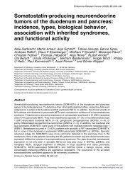
Figure 1 Gl<strong>and</strong>ular <strong>and</strong> peripheral origins <strong>and</strong> interrelations of testosterone, <strong>and</strong>rostenedione, estrone <strong>and</strong> estradiol in<br />
males. Circled numbers denote the following enzymes: (1) cytochrome P450scc (cholesterol side-chain cleavage enzyme),<br />
(2) 3β−hydroxysteroid dehydrogenase <strong>and</strong> Δ 5 , Δ 4 -isomerase, (3) cytochrome P450c17 (mediating 17α-hydroxylase<br />
activity), (4) cytochrome P450c17 (mediating 17,20-lyase activity), (5) 17-oxosteroid reductase, <strong>and</strong> (6) aromatase. The<br />
thick arrows denote the major source of the hormone. From Braunstein (1993).<br />
without an alteration in the metabolic clearance rate of the<br />
hormone. The conversion rate of <strong>and</strong>rostenedione to<br />
estrone <strong>and</strong> testosterone <strong>and</strong> the conversion rate of<br />
testosterone to estrone <strong>and</strong> <strong>and</strong>rostenedione are also<br />
increased, leading to elevated estradiol levels, while the<br />
plasma testosterone concentrations are decreased (Gordon<br />
et al. 1975, Olivo et al. 1975). Increased <strong>and</strong>rostenedione<br />
production has also been found in thyrotoxicosis<br />
(Southren 1974).<br />
Increased aromatase activity in which normal amounts<br />
of precursors are produced but are converted into<br />
estrogens at an enhanced rate can be due to increased<br />
activity in normal tissues, dysregulation of P450 arom or<br />
they may be due to mechanisms that have yet to be defined<br />
(Table 3). Epidemiological studies have clearly shown<br />
that the prevalence of <strong>gynecomastia</strong> is related to body<br />
weight, in particular the fat compartment. Niewoehner <strong>and</strong><br />
Nuttall (1984) found a close correlation between the<br />
percentage of patients with <strong>gynecomastia</strong> <strong>and</strong> the body<br />
mass index (BMI) (Fig. 2). In addition, they noted a<br />
significant correlation (r=0.52, P
Table 3 <strong>Aromatase</strong>-associated causes of <strong>gynecomastia</strong><br />
I. Increased precursors<br />
Puberty<br />
Adrenal tumors<br />
17-oxosteroid reductase deficiency<br />
Liver disease<br />
Thyrotoxicosis<br />
II. Increased aromatase activity<br />
Increased activity in normal tissue<br />
Obesity<br />
Aging<br />
<strong>Aromatase</strong> dysregulation<br />
Familial aromatase excess syndrome<br />
Neoplasms<br />
Eutopic production<br />
Sertoli cell tumors<br />
Isolated<br />
Peutz-Jegher’s Syndrome<br />
Carney complex<br />
Trophoblastic tumors<br />
Ectopic production<br />
Feminizing adrenocortical neoplasms<br />
Hepatocellular carcinoma<br />
?Melanoma<br />
Mechanism unknown<br />
Klinefelter’s syndrome<br />
Idiopathic <strong>gynecomastia</strong><br />
Thyrotoxicosis<br />
Spironolactone<br />
those without. Studies carried out in postmenopausal<br />
women have shown a close correlation (r=0.74) between<br />
body weight <strong>and</strong> extent of conversion of <strong>and</strong>rostenedione<br />
to estrone (Siiteri & MacDonald 1973) (Fig. 4), <strong>and</strong><br />
similar results were reported by Schneider et al. (1979) in<br />
<strong>Endocrine</strong>-<strong>Related</strong> <strong>Cancer</strong> (1999) 6 315-324<br />
obese men. These latter investigators also noted a<br />
progressive increase in urinary estrogen production rate<br />
with increasing obesity (Fig. 5). Since aromatase activity<br />
has been localized to adipose tissue, both histochemically<br />
(Sasano et al. 1996) <strong>and</strong> through adipose tissue cDNA<br />
analysis (Simpson et al. 1994), it is reasonable to conclude<br />
that enhanced conversion of <strong>and</strong>rostenedione to estrone<br />
<strong>and</strong> testosterone to estradiol in obesity is due to the<br />
quantitative elevation of P450 arom activity present in the<br />
exp<strong>and</strong>ed fat mass.<br />
Aging has also been associated with an increase in<br />
conversion of <strong>and</strong>rostenedione to estrone (r=0.62,<br />
P
Braunstein: <strong>Aromatase</strong> <strong>and</strong> <strong>gynecomastia</strong><br />
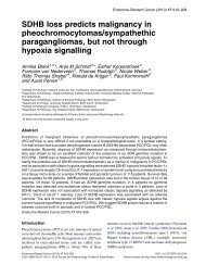
Figure 3 Correlation of breast tissue diameter with body<br />
mass index in 214 subjects. r=0.52, P< 0.001. From<br />
Niewoehner & Nuttall (1984).<br />
estrone sulfate. The authors hypothesized that the<br />
syndrome was the result of a failure in the normal decline<br />
in expression of both P450 arom <strong>and</strong> sulfokinase enzyme<br />
activities after birth (Hemsell et al. 1977). Subsequently,<br />
several families with this syndrome have been described<br />
with apparent autosomal dominant <strong>and</strong> X-linked recessive<br />
or sex-linked autosomal dominant modes of inheritance<br />
described (Berkovitz et al. 1985, Leiberman & Zachmann<br />
320<br />
1992, Stratakis et al. 1998). Males exhibit heterosexual<br />
precocity with <strong>gynecomastia</strong>, accelerated height <strong>and</strong> bone<br />
age in childhood <strong>and</strong> adolescence <strong>and</strong> shortened final<br />
adult height, while females have isosexual precocity <strong>and</strong><br />
macromastia. Studies by Stratakis <strong>and</strong> colleagues (1998)<br />
in one family have shown that the disorder cosegregates<br />
with a polymorphism of the P450 arom gene <strong>and</strong> appears to<br />
be associated with the utilization of a novel exon I of the<br />
P450 arom cDNA. Bulun <strong>and</strong> co-workers (1997) studied a<br />
17-year-old male with this syndrome <strong>and</strong> showed that the<br />
P450 arom mRNA levels in buttock <strong>and</strong> thigh adipose tissue<br />
were 14 to 21 times higher than in a normal adolescent<br />
boy. The aromatase expression was regulated by<br />
promoters I.3 <strong>and</strong> II, similar to those found in normal<br />
adipose tissue. Recently, Bulun (1998) described a male<br />
with the syndrome apparently occurring sporadically who<br />
had an inversion mutation in chromosome 15q21.1 which<br />
gave rise to a direction reversal of the promoter of an<br />
unrelated gene which caused the aberrant transcription of<br />
the P450 arom gene.<br />
Dysregulation of P450 arom is also found in patients<br />
with large cell calcifying Sertoli cell (sex-cord) tumors of<br />
the testicle, which can occur as an isolated abnormality or<br />
in association with the autosomal dominant Peutz-<br />
Jegher’s syndrome (gastrointestinal polyposis <strong>and</strong> oval,<br />
irregularly pigmented lip macules) or with the autosomal<br />
dominant Carney complex (cardiac myxomas, spotty<br />
cutaneous pigmentation, primary pigmented nodular<br />
adrenocortial disease with hypercortisolism) (Coen et al.<br />
1991, Young et al. 1995, Berensztein et al. 1995,<br />
Diamond et al. 1996). The gonadal promoter II directs the<br />
P450 arom gene expression in the tumors associated with<br />
the Peutz-Jegher’s syndrome (Bulun et al. 1993, 1997).<br />
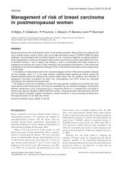
Figure 4 Correlation of extent of conversion of <strong>and</strong>rostenedione to estrone<br />
with body weight in postmenopausal women (r=0.74). From Siiteri &<br />
MacDonald (1973).
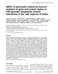
Figure 5 Urinary estradiol <strong>and</strong> estrone production<br />
rates, expressed as micrograms per day <strong>and</strong> plotted<br />
against the percentage above ideal body weight for 10<br />
obese men. (●), estradiol (E 2 ) production; (■), estrone<br />
(E 1 ) production; (shaded area), normal range. From<br />
Schneider et al. (1979).<br />
Testicular trophoblastic tumors are also capable of<br />
converting estrogen precursors to estrogen (MacDonald &<br />
Siiteri 1966), although the tissue-specific P450 arom<br />
promoter type has not been identified.<br />
Ectopic production of aromatase refers to aromatase<br />
expression by neoplasms that use promoters that are not<br />
normally expressed by the tissue from which the tumor<br />
arose. As noted above, the gonadal promoter II has been<br />
found in association with an aromatase-expressing<br />
adrenocortical carcinoma (Young et al. 1996), <strong>and</strong><br />
recently Agarwal et al. (1998) described a 17½-year-old<br />
male with severe <strong>gynecomastia</strong> <strong>and</strong> a large fibrolamellar<br />
hepatocellular carcinoma that exhibited high levels of<br />
P450 arom that was not present in the adjacent normal liver<br />
or adult liver samples. Promoters I.3 <strong>and</strong> II, rather than the<br />
normal fetal liver I.4 promoter, were used by the tumor to<br />
direct the P450 arom expression. <strong>Aromatase</strong> activity has<br />
also been found in some malignant melanomas (Santen<br />
et al. 1988). Whether the aromatase activity is responsible<br />
for the occasional patient found with <strong>gynecomastia</strong> in<br />
association with the tumor or due to the ectopic production<br />
of human chorionic gonadotropin is not known at this time<br />
(Braunstein 1991).<br />
Finally, several conditions have been shown to be<br />
associated with increased conversion of <strong>and</strong>rostenedione<br />
to estrone <strong>and</strong>/or testosterone to estradiol without the<br />
mechanism being defined. These include Klinefelter’s<br />
syndrome (Wang et al. 1975), thyrotoxicosis (Southren<br />
et al. 1974, Olivo et al. 1975), <strong>and</strong> the use of<br />
spironolactone (Huffman & Azarnoff 1975). Bulard <strong>and</strong><br />
<strong>Endocrine</strong>-<strong>Related</strong> <strong>Cancer</strong> (1999) 6 315-324<br />
Figure 6 Correlation of the extent of conversion of<br />
<strong>and</strong>rostenedione to estrone with age in males (r=0.62).<br />
From Siiteri & MacDonald (1973).<br />
co-workers (1987) found increased aromatase activity in<br />
pubic skin fibroblasts from six patients with persistent<br />
pubertal <strong>gynecomastia</strong> <strong>and</strong> two with idiopathic<br />
<strong>gynecomastia</strong>, raising the possibility that local dysregulation<br />
of breast tissue aromatase may lead to a local<br />
estrogen to <strong>and</strong>rogen imbalance.<br />
Use of aromatase inhibitors<br />
for <strong>gynecomastia</strong><br />
Considering the important role that aromatase plays in the<br />
production of elevated quantities of estrogen in males with<br />
<strong>gynecomastia</strong>, it would be anticipated that aromatase<br />
inhibitors would have been a mainstay in the therapy of the<br />
disorder. However, there is relatively little information<br />
available <strong>and</strong> what published data exist concern only the<br />
early generation aromatase inhibitor, Δ 1 -testolactone.<br />
Coen <strong>and</strong> colleagues (1991) treated a 5½-year-old boy<br />
with Peutz-Jehger’s syndrome <strong>and</strong> prepubertal <strong>gynecomastia</strong><br />
from an aromatase-producing sex-cord tumor<br />
with 450 mg Δ1 -testolactone given orally daily for 11<br />
months. This led to a slight decrease in height velocity, but<br />
did not affect the advancing bone age. On the medication<br />
there was an increase in serum levels of testosterone <strong>and</strong><br />
<strong>and</strong>rostenedione, but no alteration in serum estradiol or<br />
estrone levels. Lieberman & Zachmann (1992) described<br />
a family in whom 5 of 10 members had <strong>gynecomastia</strong>,<br />
early growth, advanced bone age, <strong>and</strong> short final stature,<br />
presumably due to excessive aromatization of adrenal<br />
precursors. A 13-year-old male member of that family<br />
321
Braunstein: <strong>Aromatase</strong> <strong>and</strong> <strong>gynecomastia</strong><br />
with severe <strong>gynecomastia</strong> was treated with 450 mg/day<br />
Δ 1 -testolactone for 6 months with ‘moderate regression of<br />
the gynaecomastia’. However, after 6 months he escaped<br />
from the effects of the drug both clinically <strong>and</strong><br />
biochemically. During the first three months on Δ 1 -<br />
testolactone, the serum levels of testosterone increased<br />
twofold, the <strong>and</strong>rostenedione levels by tenfold, <strong>and</strong><br />
estradiol was decreased by 50% without a change in<br />
estrone levels, but the levels returned to baseline by 6<br />
months. Stratakis et al. (1998) treated a brother <strong>and</strong> sister,<br />
aged 10 <strong>and</strong> 7½ years respectively, who suffered from the<br />
excessive peripheral aromatase syndrome, with a<br />
combination of gonadotropin releasing hormone analog<br />
<strong>and</strong> Δ 1 -testolactone (40 mg/kg/day orally). The<br />
combination decreased pubertal progression, skeletal age,<br />
estrone <strong>and</strong> estradiol levels, but the authors did not<br />
comment on the effects on breast development, other than<br />
to state that the boy was treated with bilateral reductive<br />
mammoplasties.<br />
Zachmann <strong>and</strong> colleagues (1986) treated 22 boys with<br />
pubertal <strong>gynecomastia</strong> with 450 mg Δ 1 -testolactone by<br />
mouth daily for two to six months without side effects.<br />
Before therapy, the mean breast diameter was 4.4 cm<br />
(median=3.8, n=22). After 2 months of therapy, the mean<br />
diameter had decreased to 3.3 cm (median=3.0, n=22),<br />
after 4 months to a mean of 3.2 cm (median=2.8, n=14),<br />
<strong>and</strong> after 6 months to a mean of 1.7 cm (median=1.5,<br />
n=4). They noted that several weeks before the reduction<br />
in the breast size there was a softening of the gl<strong>and</strong>ular<br />
tissue. During therapy, pubic hair <strong>and</strong> testicular volume<br />
increased normally. While on therapy, there were<br />
significant increases in serum testosterone (to a maximum<br />
of 1.5 times baseline), <strong>and</strong>rostenedione (13.5 times<br />
baseline), dehydroepi<strong>and</strong>rosterone (1.2 times baseline),<br />
estrone (1.6 times baseline) <strong>and</strong> follicle-stimulating<br />
hormone (1.3 times baseline), but no significant change in<br />
estradiol, luteinizing hormone, or prolactin concentrations.<br />
These authors did not note how many of their<br />
patients had complete disappearance of the <strong>gynecomastia</strong><br />
or how many were satisfied with the results.<br />
In an unpublished, prospective study of 4 patients with<br />
idiopathic <strong>gynecomastia</strong> of long st<strong>and</strong>ing duration, we<br />
gave escalating doses of Δ 1 -testolactone at doses of<br />
150 mg for 2 months, then 300 mg for 2 months, <strong>and</strong><br />
finally 750 mg for 2 months. On this regimen, the mean<br />
breast size decreased 28% at 2 months, 32% at 4 months,<br />
<strong>and</strong> 47% at 6 months (P
<strong>Aromatase</strong> <strong>and</strong> its Inhibitors. New Biology <strong>and</strong> Clinical<br />
Perspectives, Prague, September 3-6 1998.<br />
Bulun SE, Rosenthal IM, Brodie AMH, Inkster SE, Zeller WP,<br />
DiGeorge AM, Frasher SD, Kilgore MW & Simpson ER 1993<br />
Use of tissue-specific promoters in the regulation of aromatase<br />
cytochrome P450 gene expression in human testicular <strong>and</strong><br />
ovarian sex cord tumors, as well as in normal fetal <strong>and</strong> adult<br />
gonads. Journal of Clinical Endocrinology <strong>and</strong> Metabolism<br />
77 1616-1621.<br />
Bulun SE, Noble LS, Takayama K, Michael MD, Agarwal V,<br />
Fisher C, Zhao Y, Hinshelwood MM, Ito Y & Simpsom ER<br />
1997 <strong>Endocrine</strong> disorders associated with inappropriately<br />
high aromatase expression. Journal of Steroid Biochemistry<br />
<strong>and</strong> Molecular Biology 61 133-139.<br />
Castro-Magana M, Angulo M & Uy J 1993 Male hypogonadism<br />
with <strong>gynecomastia</strong> caused by late-onset deficiency of<br />
testicular 17-ketosteroid reductase. New Engl<strong>and</strong> Journal of<br />
Medicine 328 1297-1301.<br />
Chen S, Besman MJ, Sparkes RS, Zollman S, Klisak I, Moh<strong>and</strong>as<br />
T, Hall PF & Shively JE 1988 Human aromatase: cDNA<br />
cloning, Southern blot analysis, <strong>and</strong> assignment of the gene to<br />
chromosome 15. DNA 7 27-38.<br />
Clel<strong>and</strong> WH, Mendelson CR & Simpson ER 1985 Effects of<br />
aging <strong>and</strong> obesity on aromatase activity of human adipose<br />
cells. Journal of Clinical Endocrinology <strong>and</strong> Metabolism 60<br />
174-177.<br />
Coen P, Kulin H, Ballantine T, Zaino R, Frauenhoffer E, Boal D,<br />
Inkster S, Brodie A & Santen R 1991 An aromatase-producing<br />
sex-cord tumor resulting in prepubertal <strong>gynecomastia</strong>. New<br />
Engl<strong>and</strong> Journal of Medicine 324 317-322.<br />
Diamond FB Jr, Root AW, Hoover DL & Monteforte H 1996<br />
Hetero- <strong>and</strong> isosexual pseudoprecocity associated with<br />
testicular sex-cord tumors in an 8-year-old male. Journal of<br />
Pediatric Endocrinology <strong>and</strong> Metabolism 9 407-414.<br />
Forbes GB & Reina JC 1970 Adult lean body mass declines with<br />
age: some longitudinal observations. Metabolism 19 653-663.<br />
Georgiadis E, Pap<strong>and</strong>reou L, Evangelopoulou C, Aliferis C,<br />
Lymberis C, Panitsa C & Batrinos M 1994 Incidence of<br />
gynaecomastia in 954 young males <strong>and</strong> its relationship to<br />
somatometric parameters. Annals of Human Biology 21 579-<br />
587.<br />
Gordon GG, Olivo J, Rafii F & Southren AL 1975 Conversion of<br />
<strong>and</strong>rogens to estrogens in cirrhosis of the liver. Journal of<br />
Clinical Endocrinology <strong>and</strong> Metabolism 40 1018-1026.<br />
Hemsell DL, Grodin JM, Brenner PF, Siiteri PK & MacDonald<br />
PC 1974 Plasma precursors of estrogen. II. Correlation of the<br />
extent of conversion of plasma <strong>and</strong>rostenedione to estrone<br />
with age. Journal of Clinical Endocrinology <strong>and</strong> Metabolism<br />
38 476-479.<br />
Hemsell DL, Edman CD, Marks JF, Siiteri P & MacDonald PC<br />
1977 Massive extragl<strong>and</strong>ular aromatization of plasma<br />
<strong>and</strong>rostenedione resulting in feminization of a prepubertal<br />
boy. Journal of Clinical Investigation 60 455-464.<br />
Huffman DH & Azarnoff DL 1975 Effect of spironolactone on<br />
metabolic conversion of <strong>and</strong>rogens to estrogens: a preliminary<br />
report. Clinical Research 23 476A.<br />
<strong>Endocrine</strong>-<strong>Related</strong> <strong>Cancer</strong> (1999) 6 315-324<br />
Leiberman E & Zachmann M 1992 Familial adrenal feminization<br />
probably due to increased steroid aromatization. Hormone<br />
Research 37 96-102.<br />
MacDonald PC & Siiteri PK 1966 The in vivo mechanism of<br />
origin of estrogen in subjects with trophoblastic tumors.<br />
Steroids 8 589-603.<br />
Mathur R & Braunstein GD 1997 Gynecomastia:<br />
pathomechanisms <strong>and</strong> treatment strategies. Hormone<br />
Research 48 95-102.<br />
Nicolis GL, Modlinger RS & Gabrilove JL 1971 A study of the<br />
histopathology of human <strong>gynecomastia</strong>. Journal of Clinical<br />
Endocrinology <strong>and</strong> Metabolism 32 173-178.<br />
Niewoehner CB & Nuttall FQ 1984 Gynecomastia in a<br />
hospitalized male population. American Journal of Medicine<br />
77 633-638.<br />
Novak LP 1972 Aging, total body potassium, fat-free mass, <strong>and</strong><br />
cell mass in males <strong>and</strong> females between ages 18 <strong>and</strong> 85 years.<br />
Journal of Gerontology 27 438-443.<br />
Olivo J, Gordon GG, Rafii F & Southren AL 1975 Estrogen<br />
metabolism in hyperthyroidism <strong>and</strong> in cirrhosis of the liver.<br />
Steroids 26 47-56.<br />
Santen RJ, Santner SJ, Harvey HA, Lipton A, Simmonds M, Feil<br />
PD, M<strong>and</strong>ers E & Davis TS 1988 Marked heterogeneity of<br />
aromatase activity in human malignant melanoma tissue.<br />
European Journal of <strong>Cancer</strong> <strong>and</strong> Clinical Oncology 24 1811-<br />
1816.<br />
Santner SJ, Pauley RJ, Tait L, Kaseta J & Santen R 1997<br />
<strong>Aromatase</strong> activity <strong>and</strong> expression in breast cancer <strong>and</strong> benign<br />
breast tissue stromal cells. Journal of Clinical Endocrinology<br />
<strong>and</strong> Metabolism 82 200-208.<br />
Sasano H, Kimura M, Shizawa S, Kimura N & Nagura H 1996<br />
<strong>Aromatase</strong> <strong>and</strong> steroid receptors in <strong>gynecomastia</strong> <strong>and</strong> male<br />
breast carcinoma: an immunohistochemical study. Journal of<br />
Clinical Endocrinology <strong>and</strong> Metabolism 81 3063-3067.<br />
Schneider G, Kirschner MA, Berkowitz R & Ertel NH 1979<br />
Increased estrogen production in obese men. Journal of<br />
Clinical Endocrinology <strong>and</strong> Metabolism 48 633-638.<br />
Siiteri PK & MacDonald PC 1973 Role of extragl<strong>and</strong>ular<br />
estrogen in human endocrinology. In H<strong>and</strong>book of Physiology,<br />
pp 615-629. Eds RO Greep & EB Astwood EB. Washington<br />
DC: American Physiology Society.<br />
Simpson ER, Mahendroo MS, Means GD, Kilgore MW,<br />
Hinshelwood MM, Graham-Lorence S, Amarneh B, Ito Y,<br />
Fisher CR, Michael MD, Mendelson CR & Bulun SE 1994<br />
<strong>Aromatase</strong> cytochrome P450, the enzyme responsible for<br />
estrogen biosynthesis. <strong>Endocrine</strong> Reviews 15 342-355.<br />
Southren AL, Olivo J, Gordon GG, Vittek J, Brener J & Rafii F<br />
1974 The conversion of <strong>and</strong>rogens to estrogens in<br />
hyperthyroidism. Journal of Clinical Endocrinology <strong>and</strong><br />
Metabolism 38 207-214.<br />
Stratakis CA, Vottero A, Brodie A, Kirschner LS, DeAtkine D,<br />
Lu, Q, Yue W, Mitsiades CS, Flor AW & Chrousos GP 1998<br />
The aromatase excess syndrome is associated with<br />
feminization of both sexes <strong>and</strong> autosomal dominant<br />
transmission of aberrant P450 aromatase gene transcription.<br />
Journal of Clinical Endocrinology <strong>and</strong> Metabolism 83 1348-<br />
1357.<br />
323
Braunstein: <strong>Aromatase</strong> <strong>and</strong> <strong>gynecomastia</strong><br />
Toda K, Terashima M, Kawamoto M, Sumimoto H, Yokoyama<br />
Y, Kuribayashi I, Mitsuuchi Y, Maeda T, Yamamoto Y, Sagara<br />
Y, Ikeda H & Shizuta Y 1990 Structural <strong>and</strong> functional<br />
characterization of human aromatase P450 gene. European<br />
Journal of Biochemistry 193 559-565.<br />
Wang C, Baker HWG, Burger HG, DeKretser DM & Hudson B<br />
1975 Hormonal studies in Klinefelter’s syndrome. Clinical<br />
Endocrinology 4 399-411.<br />
Wilson JD, Aiman J & Macdonald PC 1980 The pathogenesis of<br />
<strong>gynecomastia</strong>. Advances in Internal Medicine 25 1-32.<br />
Young S, Gooneratne S, Straus FH, Zeller WP, Bulun SE &<br />
Rosenthal IM 1995 Feminizing Sertoli cell tumors in boys<br />
with Peutz-Jegher’s syndrome. American Journal of Surgical<br />
Pathology 19 50-58.<br />
324<br />
Young J, Bulun SE, Agarwal V, Couzinet B, Mendelson CR,<br />
Simpson ER & Schaison G 1996 <strong>Aromatase</strong> expression in a<br />
feminizing adrenocortical tumor. Journal of Clinical<br />
Endocrinology <strong>and</strong> Metabolism 81 3173-3176.<br />
Zachmann M, Eiholzer U, Muritano M, Werder EA & Manella B<br />
1986 Treatment of pubertal gynaecomastia with testolactone.<br />
Acta Endocrinologica (Suppl) 279 218-226.<br />
Zayed A, Stock JL, Liepman MK, Wollin M & Longcope C 1994<br />
Feminization as a result of both peripheral conversion of<br />
<strong>and</strong>rogens <strong>and</strong> direct estrogen production from an<br />
adrenocortical carcinoma. Journal of Endocrinological<br />
Investigation 17 275-278.