Uveal Coloboma and True Klinefelter Syndrome - Journal of Medical ...
Uveal Coloboma and True Klinefelter Syndrome - Journal of Medical ...
Uveal Coloboma and True Klinefelter Syndrome - Journal of Medical ...

You also want an ePaper? Increase the reach of your titles
YUMPU automatically turns print PDFs into web optimized ePapers that Google loves.
Downloaded from<br />
jmg.bmj.com on April 5, 2013 - Published by group.bmj.com<br />
<strong>Journal</strong> <strong>of</strong> <strong>Medical</strong> Genetics (1970). 7, 213.<br />
<strong>Uveal</strong> <strong>Coloboma</strong> <strong>and</strong> <strong>True</strong> <strong>Klinefelter</strong> <strong>Syndrome</strong><br />
J. FRAN(;OIS,"* M. Th. MATTON-VAN LEUVEN, <strong>and</strong> Ph. GOMBAULT<br />
The clinical features <strong>of</strong> '<strong>Klinefelter</strong>' syndrome<br />
were first described by <strong>Klinefelter</strong>, Reifenstein, <strong>and</strong><br />
Albright (1942). The true <strong>Klinefelter</strong> syndrome<br />
is chromatin-positive <strong>and</strong> is due to X chromosome<br />
polysomy, most frequently 47,XXY (Jacobs <strong>and</strong><br />
Strong, 1959), but karyotypes with one or more<br />
X's or Y's additional to the XXY formula, such as<br />
48,XXXY, 49,XXXXY, <strong>and</strong> mosaicisms <strong>of</strong> XXY<br />
with other stem-lines, are variants <strong>of</strong> the syndrome<br />
(Barr et al., 1959; Ford et al., 1959; Fraccaro <strong>and</strong><br />
Lindsten, 1960; Muldal <strong>and</strong> Ockey, 1960; Ellis<br />
et al., 1961; Harnden <strong>and</strong> Jacobs, 1961). The somatic<br />
abnormalities <strong>of</strong> the syndrome are relatively<br />
minor, especially at an early age <strong>and</strong> mostly only observed<br />
at or after puberty. They are: hypogonadism,<br />
testicular underdevelopment <strong>and</strong> atrophy<br />
(hyalinization <strong>of</strong> seminiferous tubules), inconstant<br />
gynaecomastia, <strong>and</strong> hormonal deviations, such as<br />
increased excretion <strong>of</strong> gonadotrophins. The following<br />
extragenital manifestations are frequent: poorly<br />
developed musculature, osteoporosis, skeletal anomalies,<br />
cutaneous angiomata, <strong>and</strong> mental retardation.<br />
The clinical picture <strong>of</strong> XXXY <strong>and</strong> other variants<br />
is similar to that <strong>of</strong> XXY subjects, though there<br />
seems to be a higher frequency <strong>of</strong> additional congenital<br />
abnormalities.<br />
The purpose <strong>of</strong> this paper is to report the presence<br />
<strong>of</strong> bilateral uveal coloboma in a 46,XX/47,XXY<br />
chromatin-positive boy.<br />
Case History<br />
This boy (H. Franky) was referred to us at the age <strong>of</strong> 4<br />
months; he was the son <strong>of</strong> healthy but consanguineous<br />
parents (Fig. 1), both aged 23 years, after a full-term<br />
uneventful pregnancy. The mother had had one x-ray<br />
<strong>of</strong> the abdominal region at the age <strong>of</strong> 15 years after an<br />
injury. The child had seizures on the 5th day, which<br />
lasted 20 minutes. The temperature <strong>and</strong> blood calcium<br />
levels were normal.<br />
Intramuscular administration <strong>of</strong> phenobarbitone<br />
(25 mg.) was helpful. Three days later, there was sudden<br />
high fever <strong>and</strong> his general condition became poor, due to<br />
* Address: Cytogenetics Laboratory, Ophthalmological Clinic <strong>of</strong><br />
the University <strong>of</strong> Ghent, De Pintelaan 115, Ghent, Belgium.<br />
213<br />
a urinary infection (Esch. coli) which was treated with<br />
'pentrexyl'. The EEG was disturbed <strong>and</strong> showed<br />
paroxysmal bisynchronous spike waves. After a week<br />
the patient was free <strong>of</strong> fever <strong>and</strong> urography was normal.<br />
The EEG returned to normal.<br />
The patient's physical <strong>and</strong> psychological development<br />
continued satisfactorily <strong>and</strong> he could walk without<br />
help at 12 months. The genitalia were normally developed<br />
for his age: both testes had descended since<br />
birth (Fig. 2).<br />
Ocular examination. This revealed bilateral coloboma<br />
<strong>of</strong> the iris (Fig. 3) <strong>and</strong> choroid, more pronounced<br />
in the right eye, where it included the optic nerve. In<br />
the left eye, the macular region <strong>and</strong> optic nerve have<br />
been spared. Both fundi had an albinoid aspect, normal<br />
for his age.<br />
The child was last seen at the age <strong>of</strong> 16 months. He<br />
was alert <strong>and</strong> his mental status seemed entirely normal.<br />
Careful questioning <strong>of</strong> the parents <strong>and</strong> personal examination<br />
<strong>of</strong> several members <strong>of</strong> the prob<strong>and</strong>'s <strong>and</strong> <strong>of</strong> the<br />
parents' generation did not reveal the occurrence <strong>of</strong> a<br />
colobomatous lesion or equivalent.<br />
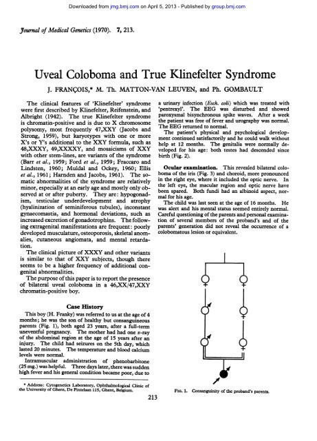
FIG. 1. Consanguinity <strong>of</strong> the prob<strong>and</strong>'s parents.
Downloaded from<br />
jmg.bmj.com on April 5, 2013 - Published by group.bmj.com<br />
214 Frangois, Matton-Van Leuven, <strong>and</strong> Gombault<br />
Cytogenetic examination. This was performed in<br />
leucocytes, cultured following a micromethod seen in<br />
Pr<strong>of</strong>essor Lejeune's laboratory. A total <strong>of</strong> 75 cells was<br />
analysed numerically. In 5 cells, 2 or more chromosomes<br />
had been lost during the techmcal in vitro<br />
manipulations. In the remaining 70 cells, 2n was distributed<br />
as follows: 45: 8%/', 46: 37%, 47: 490o, <strong>and</strong><br />
48: 6%. In each <strong>of</strong> these groups, karyotypes were<br />
\ Ww analysed (28 in total). Of the cells with 2n=47,<br />
11 karyotypes were prepared. They constantly were<br />
47,XXY (Fig. 4). Of the cells with 2n=46, 12 karyotypes<br />
x a _ s:were analysed: 10 <strong>of</strong> them were 46,XX (Fig. 5) <strong>and</strong> 2<br />
<strong>of</strong> them were 46,XY. Of the cells with 2n = 45, 3 karyotypes<br />
were analysed: 2 <strong>of</strong> them had lost an autosome<br />
each in a different group <strong>and</strong> could be interpreted as<br />
45,XX; the remaining one had an autosome missing in<br />
.........;. f the C group <strong>and</strong> could be interpreted as 45,X. Of the<br />
FIG. 2. Outer genitalia. cells with 2n = 48, 2 karyotypes were prepared: one could<br />
be interpreted as 48,XXXY <strong>and</strong> the other possibly as<br />
48,XXYY.<br />
The sex chromosomal configuration is thus distributed<br />
in the 28 karyotypes as follows: XX in 12 karyotypes<br />
(43%O); XXY in 11 (39%); XY in 2 (7%); XXXY in 1<br />
.... (35%); XXYY in 1 (3,5%); XO in 1 (in vitro loss?)<br />
(3-5%).<br />
Structurally, the chromosomes were normal. In<br />
many karyotypes, however, the Y chromosome had long<br />
long arms. The mean Y/F index, i.e. the total length <strong>of</strong><br />
the Y expressed as a ratio <strong>of</strong> the mean total length <strong>of</strong> the F<br />
chromosomes, was 0-96. The karyotypes were the result<br />
<strong>of</strong> three different blood cultures: the first time only<br />
XXY karyotypes were obtained. As the quality <strong>of</strong> the<br />
metaphases was not very good, a second blood culture<br />
revealed a high majority <strong>of</strong> cells with 46,XX. A third<br />
culture revealed a majority <strong>of</strong> cells with 47,XXY.<br />
The sex chromatin was evaluated in buccal mucosa on<br />
three occasions by the same observer: the first time Barr<br />
FIG. 3. Bilateral iris coloboma. bodies were seen in 12o% <strong>of</strong> the cells (orcein stain), the<br />
:. .. :: ... s ;: . . .s; - , : .:........<br />
...... ...-RRSo "-'';......................<br />
A.. > &.: ,:'<br />
............................................ b':<br />
.~~I...... ...... .... ... ...<br />
-: '.-;.. ....... '- -. .A<br />
* ...... .. .-eE y E - . ' ........ . f X -; . y; ;-X~~~~~~~~~~~~~~~~~~~~~~~~~~~~~~~~~~ ....... .. .. .. ...<br />
119 ........... X i _ ... ......<br />
.......... ... .. a._ ........ ...:t_:.-<br />
_._?... :........ ...:..... ......<br />
. --::--.<br />
.. * W0JU,<br />
' ';'X' '........::. y''<br />
1 ... . : .~~~~~~~~~.......<br />
:.: :;<br />
* Z :<br />
,,,.:.::;:. .<br />
4Ut:Q :<br />
FIG. 4. Karyotype with 2n = 47,XXY (propositus).<br />
;:::. ::.............<br />
'M
Downloaded from<br />
jmg.bmj.com on April 5, 2013 - Published by group.bmj.com<br />
<strong>Uveal</strong> <strong>Coloboma</strong> <strong>and</strong> <strong>True</strong> <strong>Klinefelter</strong> <strong>Syndrome</strong><br />
,,,., %....A<br />
second time in 160o (orcein stain), the third time in 10%<br />
(Shorr stain). These results indicated the presence <strong>of</strong><br />
two X chromosomes.<br />
The drumstick evaluation revealed 0-4% polymorphs<br />
with a drumstick appendage. The lobulation <strong>of</strong> the<br />
polymorphs was extremely low.<br />
Cytogenetic evaluation was also performed in the<br />
patient's parents. The karyotype <strong>of</strong> the mother was<br />
normal (46,XX), both numerically <strong>and</strong> structurally.<br />
The karyotype <strong>of</strong> the father was numerically normal<br />
(46,XY). Structurally, most karyotypes showed a long<br />
Y chromosome, the relative length <strong>of</strong> which was generally<br />
even longer than in the prob<strong>and</strong> (Fig. 6). The mean<br />
Y/F index was 1-13.<br />
Dermatoglyphic examination (Table I). This<br />
was carried out on the prob<strong>and</strong> <strong>and</strong> his parents.<br />
Total finger ridge count. Prob<strong>and</strong>, not determined;<br />
ulnar loops on all fingers, except on right thumb on<br />
which a whorl was present. Father, 68; mother, 146.<br />
Axial triradius <strong>and</strong> atd angle. Prob<strong>and</strong>, distal displacement<br />
to position t", more pronounced on the left<br />
FIG. 6. G group chromosomes in karyotypes <strong>of</strong> the patient <strong>and</strong> his<br />
father.<br />
FIG. 5. Karyotype with 2n=46,XX (propositus).<br />
h<strong>and</strong>; atd right, 570; atd left, 68°. Father, triradius in<br />
position t; atd right, 430; atd left, 40°. Mother, distal<br />
displacement <strong>of</strong> axial triradius to position t'; atd right,<br />
48°; atd left, 540.<br />
Digital triradii. Prob<strong>and</strong>, left h<strong>and</strong>; triradius c absent.<br />
Father, all present; mother, all present.<br />
a-b ridge count. Prob<strong>and</strong>, right 32, left 40, summed<br />
72. Father, right, not counted because <strong>of</strong> burn scars;<br />
left 40. Mother, right 37, left 37, summed 74.<br />
.............<br />
TABLE I<br />
FORMULAE OF DERMATOGLYPHS<br />
Main Line Index<br />
Prob<strong>and</strong> r fRight Left 11.9.0.5'.13.t'.0.0.0.0.0 11.9.7.5'.13.tr.O.O.O.L.0 16<br />
Father fRight Not RLeft 11.7.7.3.13.t.O.O.O.O.L determined<br />
14<br />
Mother fRight 9.9.57.5'.13t'°.O.O.O.L.D 14<br />
Left 11.9.7.5.'.13.t'.O.O.O.L.0 16<br />
215<br />
Discussion<br />
A: Sex Chromosomal Anomalies<br />
The sex chromatin values suggested the presence <strong>of</strong><br />
two X chromosomes. The extremely low nuclear<br />
lobulation <strong>of</strong> the polymorphonuclear leucocytes,<br />
observed in our prob<strong>and</strong>, has been reported as<br />
typical for <strong>Klinefelter</strong> syndrome (Mittwoch, 1964;<br />
Fr0l<strong>and</strong>, 1969).<br />
The blood cultures revealed the presence <strong>of</strong> two<br />
main cell lines <strong>of</strong> about equal importance: 46,XX/<br />
47,XXY. The XX/XXY type <strong>of</strong> mosaicism has<br />
been reported a few times in the <strong>Klinefelter</strong><br />
syndrome (Ford et al., 1959; Hayward, 1960;<br />
Rohde, 1963; Court Brown et al., 1964).
216<br />
Downloaded from<br />
jmg.bmj.com on April 5, 2013 - Published by group.bmj.com<br />
Three questions have to be answered:<br />
(1) Do these two cell lines exist in vivo ?<br />
(2) If they exist in vivo, how did they arise <strong>and</strong><br />
how are they distributed in the patient ?<br />
(3) Have the minor cell lines 45,X, 46,XY,<br />
48,XXXY, <strong>and</strong> 48,XXYY originated in vitro<br />
through erroneous mitoses, or do they also exist in<br />
vivo ?<br />
Though the different ratios obtained in the three<br />
cultures might indicate the origin or the preferential<br />
growth <strong>of</strong> one <strong>of</strong> the cell lines in vitro, we feel that<br />
the two main cell lines exist in vivo <strong>and</strong> that the<br />
patient's karyotype is 46,XX/47,XXY. Indeed,<br />
our results were obtained from short-term blood<br />
cultures in which only one or two mitoses take<br />
place, <strong>and</strong> in which it is less likely than in long-term<br />
fibroblast cultures that selective forces for preferential<br />
growth for one or other cell line play an important<br />
role, or that a cell line with a karyotype other<br />
than the original one arises.<br />
There are two ways in which the two main cell<br />
lines may have arisen in the patient.<br />
(1) From an XY zygote. This hypothesis requires<br />
the occurrence <strong>of</strong> at least 2 successive mitotic<br />
errors: (a) mitotic nondisjunction <strong>of</strong> the X chromosome<br />
at the first division <strong>of</strong> the XY zygote, with<br />
production <strong>of</strong> XXY <strong>and</strong> YO cells <strong>and</strong> loss <strong>of</strong> the<br />
non-viable YO cell; (b) further early post-zygotic<br />
mitotic error, i.e. the loss <strong>of</strong> a Y chromosome at<br />
division <strong>of</strong> an XXY cell with production <strong>of</strong> XXY<br />
<strong>and</strong> XX cells.<br />
(2) From an XXY zygote. This requires the<br />
subsequent occurrence <strong>of</strong> two different pathological<br />
conditions: an imbalanced gamete <strong>and</strong> erroneous<br />
somatic mitoses at or shortly after the first cleavage<br />
division.<br />
(a) Imbalanced gamete. As the parents' karyotypes<br />
were normal, the possibility <strong>of</strong> a parental<br />
numerical chromosomal abnormality can be ruled<br />
out. The possibility <strong>of</strong> a gametogenetic error<br />
can neither be proved nor rejected. The hypothesis<br />
<strong>of</strong> meiotic nondisjunction could not be substantiated<br />
by data on the parents' ages which were<br />
normal (i.e. both 23 years), or by other cases <strong>of</strong><br />
aneuploidy in the family, or by a history <strong>of</strong> a virus<br />
infection <strong>of</strong> the mother. The x-ray examination <strong>of</strong><br />
the abdominal region at the age <strong>of</strong> 15 years can be<br />
rejected as responsible for an erroneous cell division<br />
taking place years later at ovulation.<br />
The father had, however, an unusually long Y<br />
chromosome. The question whether such a Y can<br />
determine abnormal behaviour <strong>of</strong> the XY bivalent<br />
at meiosis with consecutive nondisjunction is not<br />
Frangois, Matton-Van Leuven, <strong>and</strong> Gombault<br />
yet solved. More reports on the presence <strong>of</strong> long Y<br />
chromosomes in the families <strong>of</strong> aneuploid patients<br />
are needed to evaluate a possible causal relation.<br />
In order to have an idea <strong>of</strong> the variation <strong>of</strong> the<br />
length <strong>of</strong> the Y chromosome in the Belgian population,<br />
we determined the Y/F index in the karyotypes<br />
<strong>of</strong> 120 males who were referred to our laboratory,<br />
either as patients or as normal relatives <strong>of</strong><br />
affected subjects. This control series had a Y/F<br />
index= 087 + 0-16.<br />
Data from published reports on the Y/F value are<br />
as follows: 088±0-11 in Asiatic Indians, 1-00+<br />
0-15 in Japanese, 0-92 ± 0-09 in American Negroes,<br />
0 94 + 0 10 in Jews <strong>of</strong> eastern European extraction,<br />
<strong>and</strong> 0-86 ± 0 09 in non-Jews <strong>of</strong> Anglo Saxon origin<br />
(Cohen, Shaw, <strong>and</strong> MacCluer, 1966). In Japan,<br />
the Y/F was reported to vary between 0-78-0-93 in<br />
control patients with a normal Y, <strong>and</strong> between<br />
1-07-1-56 in patients with a long Y (Makino et al.,<br />
1963; Tonomura <strong>and</strong> Ono, 1963; Makino <strong>and</strong><br />
Takagi, 1965).<br />
Though it is mostly accepted that a long Y<br />
chromosome is a variation <strong>of</strong> the normal karyotype,<br />
its presence in association with somatic <strong>and</strong> genital<br />
malformations, <strong>and</strong> also in association with other<br />
karyotypic anomalies, might indicate a possible adverse<br />
effect. Association <strong>of</strong> a long Y chromosome<br />
with sexual anomalies was reported by Van Wijck,<br />
Tijdink, <strong>and</strong> Stolte (1962), de la Chapelle <strong>and</strong><br />
Hortling (1963), Gropp et al. (1963), Gustavson et<br />
al. (1964b), Makino et al. (1964), Makino <strong>and</strong> Takagi<br />
(1965), Kjessler (1966), Fraccaro et al. (1966),<br />
Nusso et al. (1967), Beer, Pfeiffer, <strong>and</strong> Dammer<br />
(1 967), <strong>and</strong> Dumars <strong>and</strong> Fisher (1968). Association<br />
<strong>of</strong> a long Y chromosome with other cytogenetic<br />
abnormalities was observed in trisomy 21 (de la<br />
Chapelle <strong>and</strong> Hortling, 1963; Jacobs <strong>and</strong> Harnden,<br />
cited by Penrose, 1961; Bishop, Blank, <strong>and</strong> Hunter,<br />
1962; Gripenberg, 1964; Makino <strong>and</strong> Takagi,<br />
1965). Familial occurrence <strong>of</strong> a long Y chromosome<br />
in families with a mongoloid prob<strong>and</strong> was<br />
noted by Bishop et al. (1962), Dekaban, Bender, <strong>and</strong><br />
Economos (1963), <strong>and</strong> Makino et al. (1963).<br />
A mosaic karyotype similar to the one found in<br />
our patient, 46,XX/47,XXY, where the Y chromosome<br />
was long, was reported by Burgio, Severi, <strong>and</strong><br />
Biscatti (1965) in a patient with a right endoabdominal<br />
testis <strong>and</strong> left gonadic agenesis.<br />
Comparison <strong>of</strong> the Y/F index <strong>of</strong> our prob<strong>and</strong><br />
(0 96) <strong>and</strong> <strong>of</strong> his father (1 -13) with, on the one h<strong>and</strong><br />
the data from published reports, <strong>and</strong> on the other<br />
h<strong>and</strong> the values in the Belgian controls, shows that<br />
the father certainly has a longer Y chromosome than<br />
normal, but that the prob<strong>and</strong>, though at the upper<br />
limit, is still within the normal variation. This is a
Downloaded from<br />
jmg.bmj.com on April 5, 2013 - Published by group.bmj.com<br />
puzzling fact. However, in the karyotypes which<br />
showed 5 chromosomes in the G group, the 5th<br />
chromosome was interpreted as a Y. Indeed, if<br />
there were an autosomal anomaly, the child would<br />
be expected to be mentally retarded, which is not<br />
the case in our prob<strong>and</strong>. The child is well developed<br />
both physiologically <strong>and</strong> psychologically<br />
<strong>and</strong> is by no means mentally retarded.<br />
(b) Mitotic error in the 47,XXY foetus, early<br />
post-zygotic after the first cleavage division, such as<br />
the following:<br />
(i) Somatic non-disjunction <strong>of</strong> 47,XXY cells with<br />
production <strong>of</strong> either 48,XXXY <strong>and</strong> 46,XY cells or<br />
46,XX <strong>and</strong> 48,XXYY cells. This is not very likely<br />
in view <strong>of</strong> the percentages obtained for the different<br />
cell lines.<br />
(ii) Direct loss <strong>of</strong> a Y chromosome giving rise to<br />
47,XXY <strong>and</strong> 46,XX cells, which is more likely to<br />
have occurred.<br />
The percentages suggest that whether the karyotype<br />
<strong>of</strong> the zygote was 46,XY or 47,XXY, a divisional<br />
error (including loss <strong>of</strong> a Y chromosome)<br />
must have occurred very early, resulting in the production<br />
<strong>of</strong> 46,XX <strong>and</strong> 47,XXY cell lines in about<br />
the same quantity.<br />
Mitotic errors may have occurred occasionally in<br />
the XXY cells, with production <strong>of</strong> the minor cell<br />
lines, 45,X, 46,XY, 48,XXXY, <strong>and</strong> 48,XXYY,<br />
either very late in embryonic life or in vitro during<br />
culture. In view <strong>of</strong> the extremely low frequency<br />
<strong>of</strong> these cell lines, it was impossible to judge<br />
whether they represented true mosaicism or accidental<br />
deviations. The 46,XY configuration was<br />
indeed found only in two <strong>and</strong> the 48,XXXY,<br />
48,XXYY, <strong>and</strong> 45,X configurations, respectively,<br />
only in one karyotype, whereas the 47,XXY <strong>and</strong><br />
46,XX cells represented the great majority <strong>of</strong> cells.<br />
It was impossible to perform a skin or fascia<br />
biopsy so that the frequency <strong>of</strong> the different cell<br />
lines in tissue other than blood could not be studied.<br />
In any case, blood mosaicism indicates that the<br />
error must have occurred at or before the gastrula<br />
stage, i.e. around or shortly after the 14th day.<br />
The clinical picture <strong>of</strong> mosaic patients is probably<br />
less dependent on the number <strong>of</strong> cell lines<br />
than on the relative frequency <strong>of</strong> the cells in each<br />
cell line. This frequency depends on the stage <strong>of</strong><br />
embryonic development at which the non-disjunction<br />
which causes the mosaicism takes place. The<br />
earlier mosaicism originates in embryonic life, the<br />
more severe probably is the influence on the phenotype.<br />
The relative influence <strong>of</strong> the different cell<br />
lines will only become evident at puberty. Repeated<br />
cultures are sometimes necessary in order to<br />
reveal the existence <strong>of</strong> mosaicism. A mosaic<br />
<strong>Uveal</strong> <strong>Coloboma</strong> <strong>and</strong> <strong>True</strong> <strong>Klinefelter</strong> <strong>Syndrome</strong><br />
217<br />
pattern can indeed be overlooked if an inadequate<br />
number <strong>of</strong> cells is counted. We can compare in<br />
this respect our patient with the patient reported by<br />
Hecht et al. (1966) who was originally described as a<br />
46,XX patient without Y chromosomes, in whom,<br />
when further investigations were carried out, a<br />
mosaic cell line (1 %) was detected.<br />
B: <strong>Coloboma</strong><br />
<strong>Coloboma</strong> results from abnormal closure <strong>of</strong> the<br />
foetal cup cleft soon after gastrulation at the end <strong>of</strong><br />
the first or the beginning <strong>of</strong> the second foetal<br />
month, when the optic vesicle is in an active state <strong>of</strong><br />
differentiation (Duke-Elder, 1940; Mann, 1957).<br />
This faulty development <strong>of</strong> the ectodermal eye<br />
structures can be caused by chromosomal, Mendelian,<br />
<strong>and</strong> environmental intrauterine teratogenous<br />
factors.<br />
(I) Chromosomal anomalies. <strong>Coloboma</strong> <strong>and</strong><br />
other eye defects have been found in anomalies <strong>of</strong><br />
all the autosomal groups <strong>and</strong> are not specific for any<br />
particular chromosomal abnormality: A group<br />
(Lele, Dent, <strong>and</strong> Delhanty, 1965; El Massri <strong>and</strong><br />
Riad, 1965), B group (Hirschhorn <strong>and</strong> Cooper,<br />
1961; Hirschhorn, Cooper, <strong>and</strong> Firschein, 1965;<br />
Gustavson et al., 1964b; Shaw, Cohen, <strong>and</strong><br />
Hildebr<strong>and</strong>t, 1965; Wolf et al., 1965; D. Jesberg,<br />
personal communication, 1966), C group (Salonius<br />
<strong>and</strong> Opitz, 1964), D group (Patau et al., 1960),<br />
<strong>and</strong> E group (Francois et al., 1965; D. Jesberg,<br />
personal communication, 1966).<br />
<strong>Coloboma</strong>tous lesions <strong>of</strong> the eye ball have not<br />
been found in trisomy 21.<br />
A t(B/G) translocation trisomy was found in a<br />
child with multiple anomalies among which were<br />
blepharophimosis <strong>and</strong> colobomata <strong>of</strong> the irides<br />
(Gustavson et al., 1964a). <strong>Uveal</strong> colobomata are<br />
part <strong>of</strong> the oculo-anal syndrome, which is caused by<br />
an extra small submedian chromosome in girls<br />
with mongoloid slant <strong>of</strong> the eyelids, hypertelorism,<br />
microphthalmia, anal atresia, recto-vaginal fistulae,<br />
pre-auricular fistulae, renal malformations, <strong>and</strong><br />
mental retardation (Schachenmann et al., 1965;<br />
Cagianut, 1968).<br />
<strong>Coloboma</strong>ta were also found associated with<br />
oligophrenia, various congenital abnormalities,<br />
exotropia, myopia, <strong>and</strong> the presence <strong>of</strong> a small<br />
extrametacentric chromosome in a sex chromatinnegative<br />
boy (Heimann, Jaeger, <strong>and</strong> Dollmann,<br />
1968).<br />
The importance <strong>of</strong> a chromosome aberration as a<br />
causal factor should not be overstressed.<br />
(i) In the case <strong>of</strong> autosomal aberrations, the coloboma<br />
is only part <strong>of</strong> the general pathological<br />
condition.
218<br />
Downloaded from<br />
jmg.bmj.com on April 5, 2013 - Published by group.bmj.com<br />
(ii) Chromosomal aberrations are usually absent<br />
in cases <strong>of</strong> isolated coloboma with an otherwise<br />
normal phenotype.<br />
(iii) When a coloboma is found in association with<br />
a chromosomal peculiarity, but when the latter is<br />
also present in one <strong>of</strong> the phenotypically normal<br />
parents (Lele et al., 1965), the causal relation between<br />
the coloboma <strong>and</strong> the chromosomal peculiarity<br />
becomes doubtful.<br />
(iv) There exist several reports <strong>of</strong> coloboma<br />
patients with associated multiple congenital malformations,<br />
who had a normal karyotype (Edwards,<br />
Young, <strong>and</strong> Finley, 1961; Angelman, 1961).<br />
In sex chromosomal aberrations, ocular anomalies<br />
are infrequent <strong>and</strong> uveal coloboma is exceptional.<br />
In the <strong>Klinefelter</strong> syndrome the eyes are usually<br />
normal. Other severe somatic malformations are<br />
also extremely infrequent. A possible explanation<br />
is that the major part <strong>of</strong> the extra X chromosome is<br />
genetically inactivated. Out <strong>of</strong> the impressive<br />
bibliography on <strong>Klinefelter</strong> syndrome, including<br />
review studies on large series (Court Brown et al.,<br />
1964; Frol<strong>and</strong>, 1969), on smaller series, <strong>and</strong> the<br />
numerous individual case reports (Nowakowski,<br />
1961; de la Chapelle <strong>and</strong> Hortling, 1963;<br />
Prader, Murset, <strong>and</strong> Hauschteck, 1964, etc.), totalling<br />
400-500 karyotyped or sex chromatin typed<br />
<strong>Klinefelter</strong> patients, the following ocular morphological<br />
features were noted.<br />
47,XXY karyotype: Myopic astigmatism (Nowakowski,<br />
1961, case 6), high myopia (Nowakowski, 1961, case 7),<br />
bilateral coloboma <strong>of</strong> iris <strong>and</strong> choroid (Appelmans,<br />
Michiels, <strong>and</strong> Guzik, 1965), Marfan syndrome (Bessiere<br />
et al., 1962), pigmentary retinopathy (Nagy, Leowei, <strong>and</strong><br />
Kakuk, 1965), associated achondroplasia, <strong>and</strong> posterior<br />
corneal dysgenesis <strong>and</strong> atrophy <strong>of</strong> iris root, condensations<br />
<strong>of</strong> the Schwalbe ring, <strong>and</strong> abnormal insertion <strong>of</strong> iris root,<br />
forming radial arcades which masked the ciliary b<strong>and</strong><br />
(Amalric et al., 1966), hypertelorism in a premature child<br />
with multiple malformations <strong>and</strong> triploidy (2n = 69:<br />
66A + XXY) (Bernard et al., 1967).<br />
48,XXXY karyotype. Severe myopia (Ferguson-<br />
Smith, Johnston, <strong>and</strong> H<strong>and</strong>maker, 1960).<br />
49, XXXXY karyotype. Esotropia <strong>and</strong> myopia (-18D)<br />
(Anders et al., 1960), hypertelorism (Fraccaro, Klinger,<br />
<strong>and</strong> Schutt, 1962; Barr et al., 1962), hypertelorism,<br />
epicanthus, strabismus internus concomitans, myopia<br />
(- 3D) (Pfeiffer, 1962), alternating esotropia <strong>and</strong> hypertropia<br />
caused by obvious overaction <strong>of</strong> the inferior<br />
obliques (Atkins <strong>and</strong> Connelly, 1963), conspicuous<br />
epicanthus (Farquhar <strong>and</strong> Walker, 1964), hypertelorism,<br />
palpebral fissures, slanting downwards <strong>and</strong> medially<br />
(Joseph, Anders, <strong>and</strong> Taylor, 1964), strabismus (Fraser<br />
et al., 1961), divergent strabismus <strong>and</strong> hypertelorism<br />
(Barr et al., 1962), strabismus, bilateral coloboma <strong>of</strong><br />
iris <strong>and</strong> optic nerve (Joannides <strong>and</strong> Tsenki, 1968),<br />
pronounced epicanthus (Frol<strong>and</strong>, 1969, case 47),<br />
Frangois, Matton-Van Leuven, <strong>and</strong> Gombault<br />
pronounced epicanthus, slight hypertelorism, alternating<br />
external strabismus, high myopia (-20D), <strong>and</strong> myopic<br />
fundus degeneration (Frol<strong>and</strong>, 1969, case 48).<br />
49,XXXYY karyotype. Blepharophimosis <strong>and</strong> prominent<br />
supra-orbital ridges (Bray <strong>and</strong> Ann Josephine,<br />
1963).<br />
48,XXYY karyotype. Cataract, strabismus, high myopia,<br />
<strong>and</strong> choroidal atrophy (Laurence, Ishmael, <strong>and</strong><br />
Davies, 1963), epicanthus (Mirouze et al., 1964),<br />
myopia (Herbeuval et al., 1965), iris aplasia (Warburg,<br />
1968), prominent supra-orbital ridges (Ellis et al.,<br />
1961). In a case <strong>of</strong> Amalric et al. (1966) there was a<br />
uveal dystrophy: iris atrophy <strong>and</strong> atrophic insertion <strong>of</strong><br />
this iris root. The iris atrophy was more pronounced in<br />
the periphery. The sphincter zone was yellow. The<br />
zone <strong>of</strong> the dilator muscle fibres was grey. This bicolour<br />
iris atrophy reminded one <strong>of</strong> the atrophies found<br />
in genetic mesodermal syndromes. Atrophy in the<br />
chamber angle was especially pronounced between 4 <strong>and</strong><br />
6 o'clock. The iris root continued there only in a thin<br />
grey-yellow strip, attaching itself very high up. This<br />
atrophic iris insertion masked all the underlying structures.<br />
Mosaic karyotypes<br />
48,XXYY/49,XXXYY: epicanthus, right iris coloboma,<br />
<strong>and</strong> bilateral choroidal colobomata adjacent to the<br />
papilla in a 48, XXYY/71, XXXYY infant with peculiar<br />
facies, syndactyly, <strong>and</strong> multiple minor malformations<br />
(Schmid <strong>and</strong> Vischer, 1967).<br />
47,XXY/46,XX/46,XY/45,X: severe myopia <strong>and</strong> pigmentary<br />
retinopathy (Nowakowski, 1961; Bergman,<br />
Nowakowski, <strong>and</strong> Reitalu, 1969).<br />
47,XYY/48,XXYY: Prominent supra-orbital ridges<br />
<strong>and</strong> myopia (Spencer, Eyles, <strong>and</strong> Mason, 1969), prominent<br />
supra-orbital ridges <strong>and</strong> optic atrophy (Spencer<br />
et al., 1969).<br />
48,XXXY/49,XXXXY/50,XXXXXY: high myopia<br />
(- 18D) <strong>and</strong> convergent strabism (Anders et al., 1960).<br />
Long Y: coloboma, developmental errors <strong>of</strong> the brain<br />
<strong>and</strong> the heart, cleft scrotum, small penis, in a child in<br />
whom 'preliminary investigations pointed to some<br />
chromosome abnormality' (Angelman, 1961): the boy<br />
had 46 chromosomes, but had a larger Y than normal<br />
(H. Angleman, personal communication, 1969).<br />
47,XXY+ chromosome resembling a no. 15: myopic<br />
astigmatism (Nowakowski, 1961: Case 9), myopia<br />
(Nowakowski, 1961: Case 11).<br />
This review shows that the association <strong>of</strong> coloboma<br />
with <strong>Klinefelter</strong> syndrome has been reported<br />
only three times (Appelmans et al., 1965; Schmid<br />
<strong>and</strong> Vischer, 1967; Joannides <strong>and</strong> Tsenki, 1968).<br />
To our knowledge, our prob<strong>and</strong> is the fourth case.<br />
In this connexion it is interesting to note that the<br />
peculiar iris observed by Amalric et al. (1966) in the<br />
48,XXYY <strong>Klinefelter</strong> patient <strong>and</strong> in the 47,XXY<br />
<strong>Klinefelter</strong>/achondroplasic patient, was most pronounced<br />
at the lower half <strong>of</strong> the angle, i.e. at the<br />
localization <strong>of</strong> the foetal cleft. The lesions found
Downloaded from<br />
jmg.bmj.com on April 5, 2013 - Published by group.bmj.com<br />
in the anterior chamber were classified by these<br />
authors as belonging to the malformative group <strong>of</strong><br />
cleavage <strong>of</strong> the anterior chamber together with other<br />
pathological conditions, such as persistence <strong>of</strong><br />
mesenchymatous tissue, posterior embryotoxon,<br />
hyaline corneal membrane, posterior marginal dysplasia<br />
<strong>of</strong> Streiff, <strong>and</strong> mesodermic dysgenesis <strong>of</strong><br />
Rieger (Amalric et al., 1966).<br />
The small number <strong>of</strong> reported ocular anomalies<br />
in the <strong>Klinefelter</strong> syndrome might be due to incomplete<br />
examination <strong>of</strong> the patient, omitting an<br />
ophthalmological examination, or to the fact that<br />
the ocular functions are not always disturbed, so<br />
that the patient does not complain <strong>and</strong> is not referred<br />
to an ophthalmological department. The<br />
frequent associated mental retardation also makes<br />
complete ophthalmic examination difficult. <strong>Coloboma</strong><br />
<strong>of</strong> the iris, being an easily seen congenital<br />
anomaly, is however not likely to be overlooked, or to<br />
be omitted in a report when it is present in a patient.<br />
We examined extensively the eyes <strong>of</strong> 3 adult<br />
<strong>Klinefelter</strong> patients, including evaluation <strong>of</strong> the<br />
refraction, mobility, fundus, <strong>and</strong> anterior chamber<br />
angle: these patients were karyotyped in our<br />
laboratory <strong>and</strong> all were 47,XXY, <strong>and</strong> we did not<br />
notice any ocular abnormality (Table II).<br />
The results <strong>of</strong> different sex chromatin surveys in<br />
newborn males, normal adult males, males with<br />
mental retardation, <strong>and</strong> other institutionalized<br />
patients, indicated that an additional X chromosome<br />
was present in some or all <strong>of</strong> the cells <strong>of</strong> 2-07 per<br />
1000 newborn male babies <strong>and</strong> <strong>of</strong> 1 96-2-18 per<br />
1000 adult males (Prader et al., 1958; Moore, 1959;<br />
Berlemann, 1961; Maclean, Harnden, <strong>and</strong> Court<br />
Brown, 1961; Maclean et al., 1964; Paulsen et al.,<br />
1964; Maclean, 1966; Taylor <strong>and</strong> Moores, 1967;<br />
Maclean et al., 1968; Fr0l<strong>and</strong>, 1969).<br />
<strong>Coloboma</strong> <strong>of</strong> the iris is judged to have an incidence<br />
<strong>of</strong> about 1:6000 (Duke-Elder, 1940), i.e.<br />
1:12,000 males. Knowing that the frequency <strong>of</strong><br />
<strong>Klinefelter</strong> syndrome is 2:1000 males, it can be<br />
calculated that there is a probability <strong>of</strong> about 1 in 6<br />
million that the two conditions would occur in the<br />
same individual by chance alone.<br />
The fact that in the published reports <strong>of</strong> 400-500<br />
karyotyped or sex chromatin-typed <strong>Klinefelter</strong><br />
patients, coloboma was found 4 times ( ± 1%), can<br />
indicate that coloboma may possibly be part <strong>of</strong> the<br />
phenotypic expression <strong>of</strong> <strong>Klinefelter</strong> syndrome,<br />
though the causal mechanism remains entirely obscure.<br />
The idea has gained ground that the human sex<br />
chromosomes act in two different ways (Commoner,<br />
1964).<br />
(a) The euchromatic X chromosome exerts its<br />
<strong>Uveal</strong> <strong>Coloboma</strong> <strong>and</strong> <strong>True</strong> <strong>Klinefelter</strong> <strong>Syndrome</strong><br />
219<br />
influence via sex-linked Mendelian genes <strong>and</strong> control<br />
biochemical specificity. (b) The second heterochromatic<br />
X chromosome would act as an agent <strong>of</strong><br />
nucleotide sequestration <strong>and</strong> regulate metabolic<br />
processes, which result in quantitative differences.<br />
The difference between the sexes would be <strong>of</strong> a<br />
quantitative rather than <strong>of</strong> a qualitative nature, <strong>and</strong><br />
would not be correlated with any biochemical<br />
specificity. In this respect, <strong>Klinefelter</strong> patients can<br />
be considered as males having a quantitatively altered<br />
production <strong>of</strong> sex hormones. According to Ohno<br />
(1967) <strong>and</strong> Mittwoch (1967), the metabolic pathway<br />
<strong>of</strong> steroid sex hormone synthesis is such that 'the<br />
functional difference between <strong>and</strong>rogen-producing<br />
interstitial cells <strong>and</strong> estrogen-producing follicular cells<br />
is not qualitative, but quantitative. The former inhibits<br />
further conversion <strong>of</strong> <strong>and</strong>rostenedione, while<br />
the latter does not permit the accumulation <strong>of</strong> <strong>and</strong>rostenedione,<br />
but converts it further to estrone.'<br />
(II) Mendelian heredity. Though uveal coloboma<br />
occurs most frequently as a sporadic condition,<br />
it can be familial <strong>and</strong> is then usually transmitted as<br />
an irregular dominant trait. On rare occasions,<br />
however, recessive transmission has been reported<br />
(Waardenburg, 1934; Gesell <strong>and</strong> Blake, 1936;<br />
McLain <strong>and</strong> Dekaban, 1963). Consanguinity <strong>of</strong><br />
the parents was reported by Waardenburg (1932).<br />
We have observed an occurrence <strong>of</strong> coloboma <strong>of</strong><br />
the uvea, which can best be explained by recessive<br />
transmission rather than by dominant transmission<br />
(Franqois, 1966): a father <strong>and</strong> a daughter are<br />
affected, but the mother also has a nephew who<br />
suffers from it. Therefore, a marriage between a<br />
homozygote <strong>and</strong> a heterozygote giving rise to the<br />
manifestation in the daughter (pseudo-dominance)<br />
could be postulated.<br />
Isolated cases <strong>of</strong> <strong>Klinefelter</strong> syndrome have been<br />
associated with other Mendelian diseases, such as<br />
Ehlers-Danlos syndrome (Hsu, Geller, <strong>and</strong> Nemhauser,<br />
1966), achondroplasia (Sendrail et al.,<br />
1967),* familial spino-cerebellar degeneration,t<br />
(Indemini <strong>and</strong> Ammann, 1961), <strong>and</strong> thyroid hyp<strong>of</strong>unction<br />
(Herbeuval et al., 1968). In the present<br />
case family data were entirely negative. So, though<br />
the parents were consanguineous, the chance exists<br />
that the coloboma in our prob<strong>and</strong> is not due to<br />
Mendelian heredity.<br />
(Ill) Disturbed embryogenesis. As possible<br />
adverse influences on the closing <strong>of</strong> the optic cup the<br />
following have been suggested: inflammation<br />
* The frequency <strong>of</strong> achondroplasia is estimated to be around 0.1<br />
per 1000; it is inherited either in a dominant or a recessive mode.<br />
t In a sibship <strong>of</strong> 5, <strong>Klinefelter</strong> syndrome was associated with a<br />
neurological syndrome including ocular manifestations; 3 sibs had a<br />
paresis <strong>of</strong> an external ocular muscle; 2 had Fuch's heterochromia.
220<br />
Downloaded from<br />
jmg.bmj.com on April 5, 2013 - Published by group.bmj.com<br />
(Mann, 1957), vascular defect (Stroeva, 1961),<br />
germinal defect in the epiblast (Von Szily, 1924),<br />
<strong>and</strong> defect in the mesoblastic tissue surrounding the<br />
optic cup (Von Hippel, 1903).<br />
C: Dermatoglyphs<br />
Developmental disturbances taking place during<br />
the third <strong>and</strong> fourth months <strong>of</strong> foetal life affect the<br />
arrangements <strong>of</strong> the dermal ridges. Contrary to the<br />
specific dermatoglyphs, discernible on first view,<br />
which are found in autosomal trisomies, the peculiarities<br />
in <strong>Klinefelter</strong> syndrome are mainly quantitative,<br />
<strong>and</strong> consist <strong>of</strong> a tendency to fingertip patterns<br />
with a low ridge count. Many loops have so few<br />
ridges that they constitute a transition to arches.<br />
The frequency <strong>of</strong> true arches is also increased. The<br />
mean total finger ridge count (TFRC) <strong>of</strong> <strong>Klinefelter</strong>s<br />
(1178) was found to be about 30 ridges<br />
lower than the mean <strong>of</strong> control males, <strong>and</strong> about<br />
9 ridges lower than the mean <strong>of</strong> control females.<br />
As in all races, normal females have more arches,<br />
fewer whorls, <strong>and</strong> lower ridge counts; this finding<br />
has been interpreted as suggestive for sex reversal<br />
(Forbes, 1964).<br />
Our prob<strong>and</strong> could not be tested for fingertip<br />
counting. His father had a very low TFRC (68),<br />
whereas his mother had a high TFRC (146).<br />
The axial triradius tends to be situated low in the<br />
palm in <strong>Klinefelter</strong> patients (Holt, 1968), corresponding<br />
with a normal maximum atd angle. Surprisingly,<br />
our prob<strong>and</strong>'s axial triradius is displaced<br />
distally, a phenomenon that is not typical <strong>of</strong><br />
<strong>Klinefelter</strong> syndrome.<br />
According to Holt (1968), genetic as well as environmental<br />
factors must play a role in the determination<br />
<strong>of</strong> the position <strong>of</strong> the axial triradius. In<br />
this respect it must be mentioned that the atd<br />
angles <strong>of</strong> the father were normal, i.e. less than 450<br />
(corresponding with position t <strong>of</strong> the axial triradius),<br />
but that the atd angles <strong>of</strong> the mother were intermediate<br />
(490 <strong>and</strong> 540) corresponding with position<br />
t' <strong>of</strong> the axial triradius. This makes it difficult to<br />
judge whether the distal displacement <strong>of</strong> the axial<br />
triradius in the prob<strong>and</strong> is inherited from the mother<br />
or whether it reveals an intrauterine disturbance<br />
during the third or fourth month.<br />
The fact that the triradius c was absent needs to<br />
be stressed. An absent triradius c on both palms,<br />
d being sited nearer to the radial border <strong>of</strong> each<br />
palm than normal, was reported by Jancar (1964)<br />
in a 49,XXXXY patient. Vague et al. (1968) reported<br />
<strong>Klinefelter</strong> syndrome in monozygotic twins,<br />
in one <strong>of</strong> whom triradius c was absent on the left<br />
h<strong>and</strong>. In our own series, the triradius c was absent<br />
in the prob<strong>and</strong> <strong>and</strong> in another patient on the left<br />
Frangois, Matton-Van Leuven, <strong>and</strong> Gombault<br />
TABLE II<br />
OCULAR AND DERMATOGLYPHIC<br />
EVALUATION OF KLINEFELTER PATIENTS<br />
Patient Karyotype Dermatoglyphs Eye<br />
V. d. V. Willy 47,XXY<br />
TFRC low (42);<br />
bilateral, very<br />
proximal<br />
Normal<br />
V. E. Gerard 47,XXY Normal Normal<br />
V. C. Eric 47,XXY<br />
Left h<strong>and</strong>,<br />
absent<br />
triradius c<br />
Slight<br />
astigmatism,<br />
otherwise<br />
normal<br />
H. Franky 46,XX/47,XXY<br />
Bilateral distal<br />
displacementBiael<br />
to tr <strong>of</strong> axialBiael<br />
triradius; left coloboma <strong>of</strong><br />
h<strong>and</strong>, irss <strong>and</strong><br />
triradtius c choroid<br />
absent<br />
h<strong>and</strong> (Table II, V. C. Eric). It has to be remembered,<br />
however, that even in a normal population in<br />
some palms one <strong>of</strong> the digital triradii, most commonly<br />
c, is absent. More data on dermatoglyphs<br />
in <strong>Klinefelter</strong> patients are needed in order to interpret<br />
this finding.<br />
In <strong>Klinefelter</strong> syndrome the distance between<br />
triradii a <strong>and</strong> b tends to be shortened as compared<br />
with the distance in the general population, where<br />
the range <strong>of</strong> ab counts summed for both h<strong>and</strong>s lies<br />
between 47 <strong>and</strong> 122 ridges. The ab ridge count is<br />
genetically controlled, though environmental factors<br />
exert a greater effect on the ab ridge count in early<br />
prenatal life than they do on the finger ridge count<br />
(Holt, 1968). The ab ridge count <strong>of</strong> our family<br />
did not allow any conclusion regarding a possible<br />
diminution <strong>of</strong> the a-b distance in the prob<strong>and</strong>.<br />
As can be seen from the palmar formulae, the<br />
direction <strong>of</strong> the main lines is normal, <strong>and</strong> no special<br />
patterns were present in the thenar or hypothenar<br />
areas. The main line indices were also within<br />
normal range, as compared with the mean main<br />
line index <strong>of</strong> the population: 16 38 (Holt, 1968).<br />
Conclusions<br />
The prob<strong>and</strong> presents the association <strong>of</strong> three<br />
pathological conditions which may have arisen at<br />
different embryonic stages.<br />
(1) Sex chromosomal aneuploidy <strong>and</strong> mosaicism<br />
47,XXY/46,XX around or shortly after the 14th day.<br />
(2) <strong>Uveal</strong> colobomata at the end <strong>of</strong> the first or the<br />
beginning <strong>of</strong> the second foetal month.<br />
(3) Abnormal dermatoglyphs during the third or<br />
fourth months.<br />
Whether the three conditions are causally linked<br />
can neither be proved nor rejected. Published data<br />
indicate however that coloboma may be part <strong>of</strong> the
Downloaded from<br />
jmg.bmj.com on April 5, 2013 - Published by group.bmj.com<br />
phenotypic expression <strong>of</strong> the <strong>Klinefelter</strong> syndrome.<br />
Except for the consanguinity <strong>of</strong> the parents, no<br />
familial data indicate a possible Mendelian origin <strong>of</strong><br />
the coloboma, so that it can also be the result <strong>of</strong> disturbed<br />
embryogenesis.<br />
The major cell lines, 46,XX <strong>and</strong> 47,XXY, are<br />
assumed to be present in the prob<strong>and</strong> <strong>and</strong> not to<br />
have arisen in vitro. The minor cell lines,<br />
48,XXXY <strong>and</strong> 48,XXYY, could be present in<br />
the patient, but might well have arisen in vitro.<br />
The mosaicism can as well have arisen from an XY<br />
zygote as from an XXY zygote. In the latter<br />
case the father's long Y chromosome may have<br />
played a causal role in nondisjunction. In both<br />
cases a divisional error including the loss <strong>of</strong> a Y<br />
chromosome must have occurred early in the postzygotic<br />
stage.<br />
The effect <strong>of</strong> the mosaicism on the clinical (sexual)<br />
phenotype <strong>of</strong> the patient will only become evident at<br />
puberty. The abnormal dermatoglyphs are not<br />
typical for <strong>Klinefelter</strong> syndrome, <strong>and</strong> can be the<br />
result <strong>of</strong> a disturbed embryogenesis. The displaced<br />
axial triradius may however also be inherited<br />
from the mother.<br />
REFERENCES<br />
Amalric, P., Ruffie, J., Colombies, P., <strong>and</strong> Bessou, P. (1966).<br />
Dysgenesie camerulaire chez deux malades atteints de syndrome de<br />
<strong>Klinefelter</strong>. Bulletins et Memoires de la Societe Francaise d'Ophtalmologie,<br />
79, 238-250.<br />
Anders, G., Prader, A., Hauschteck, E., Scharer, K., Siebenmann,<br />
R. E., <strong>and</strong> Heller, R. (1960). Multiples Sex-Chromatin und<br />
Komplexes chromosomales Mosaik bei einem Knaben mit<br />
Idiotie und multiplen Missbildungen. Helvetica Paediatrica<br />
Acta, 15, 515-532.<br />
Angelman, H. (1961). <strong>Syndrome</strong> <strong>of</strong> coloboma with multiple congenital<br />
abnormalities in infancy. British <strong>Medical</strong> <strong>Journal</strong>, 1,<br />
1212-1214.<br />
Appelmans, M., Michiels, J., <strong>and</strong> Guzik, A. (1965). Colobome<br />
bilateral complet associe au syndrome de <strong>Klinefelter</strong>-Albright-<br />
Reifenstein par erreur chromosomiale XXY. Bulletin de la<br />
Societe Belge d'Ophtalmologie, 140, 456-468.<br />
Atkins, L., <strong>and</strong> Connelly, J. P. (1963). XXXXY sex chromosome<br />
abnormality. American <strong>Journal</strong> <strong>of</strong> Diseases <strong>of</strong> Children, 106, 514-<br />
519.<br />
Barr, M. L., Carr, D. H., Pozonyi, J., Wilson, R. A., Dunn, H. G.<br />
Jacobson, T. S., <strong>and</strong> Miller, I. R. (1962). The XXXXY sexchromosome<br />
abnormality. Canadian <strong>Medical</strong> Association <strong>Journal</strong><br />
87, 891-901.<br />
-, Shaver, E. L., Carr, D. H., <strong>and</strong> Plunkett, E. R. (1959).<br />
unusual sex chromatin pattern in three mentally deficient subjects.<br />
<strong>Journal</strong> <strong>of</strong> Mental Deficiency Research, 3, 78-87.<br />
Beer, H., Pfeiffer, R. A., <strong>and</strong> Dammer, H. (1967). The so-called<br />
long Y chromosome in man. Fortschritte der Medizin, 85, 467-<br />
470.<br />
Bergman, S., Nowakowski, H., <strong>and</strong> Reitalu, J. (1969). Cytological<br />
<strong>and</strong> clinical observations in XXY/XX/XY/XO mosaicism.<br />
Hereditas, 61, 276-278.<br />
Berlemann, E. (1961). Geschlechtschromatinbestimmungen am<br />
Neugeborenen. Schweizerische medizinische Wochenschrift, 91,<br />
292-294.<br />
Bernard, R., Stahl, A., Coigquet, Y. B., <strong>and</strong> Passeron, P. (1967). Triploidie<br />
chomosomique chez un nouveau-ne poly-malforme.<br />
Annales de Gdnitique, 10, 70-74.<br />
Bessiere, E., Riviere, J., Leuret, J. Ph., <strong>and</strong> Mme. Le rebeller (1962).<br />
Sur une association de maladie de <strong>Klinefelter</strong> et d'anomalies congenitales<br />
(camptodactylie, macrophakie). Bulletin des Societis<br />
d'Ophtalmologie de France, 62, 197-200.<br />
<strong>Uveal</strong> <strong>Coloboma</strong> <strong>and</strong> <strong>True</strong> <strong>Klinefelter</strong> <strong>Syndrome</strong><br />
An<br />
221<br />
Bishop, A., Blank, C. E., <strong>and</strong> Hunter, H. (1962). Heritable variation<br />
in the length <strong>of</strong> the Y chromosome. Lancet, 2, 18-20.<br />
Bray, P., <strong>and</strong> Sister Ann Josephine (1963). An XXXYY sexchromosome<br />
anomaly. Report <strong>of</strong> a mentally deficient male. J7ournal<br />
<strong>of</strong> the American <strong>Medical</strong> Association, 184, 179-182.<br />
Burgio, R. G., Severi, F., <strong>and</strong> Biscatti, G. (1965). Dysgenesie<br />
gonadique mixte i caryotype XY/XX/XXY. Pediatrie, 21,687-697.<br />
Cagianut, B. (1968). Augenbefunde bei Chromosomenkrankheiten.<br />
Ophthalmologica, 155, 148-166.<br />
Cohen, M. M., Shaw, M. W., <strong>and</strong> MacCluer, J. W. (1966). Racial<br />
differences in the length <strong>of</strong> the human Y chromosome. Cytogenetics,<br />
5, 34-52.<br />
Commoner, B. (1964). Roles <strong>of</strong> deoxyribonucleic acid in inheritance.<br />
Nature (London), 202, 960-968.<br />
Court Brown, W. M., Harnden, D. G., Jacobs, P. A., Maclean, N.,<br />
<strong>and</strong> Mantle, D. J. (1964). Abnormalities <strong>of</strong> the sex chromosome<br />
complement in man. <strong>Medical</strong> Research Council. Special Report<br />
Series, No. 305.<br />
Dekaban, A. S., Bender, M. A., <strong>and</strong> Economos, G. E. (1963).<br />
Chromosome studies in mongoloids <strong>and</strong> their families. Cytogetics,<br />
2, 61-75.<br />
de la Chapelle, A., <strong>and</strong> Hortling, H. (1963). Cytogenetical <strong>and</strong><br />
clinical observations in male hypogonadism. Acta Endocrinologica,<br />
44, 165-182.<br />
Duke-Elder, W. S. (1940). Textbook <strong>of</strong> Ophthalmology, Vol. II.<br />
Mosby, St. Louis.<br />
Dumars, K. W., Jr., <strong>and</strong> Fisher, C. E. (1968). Gynecomastia, hypospermatogenesis<br />
<strong>and</strong> a large Y chromosome in a man. California<br />
Medicine, 108, 52-54.<br />
Edwards, J. H., Young, R. B., <strong>and</strong> Finley, H. V. L. (1961). <strong>Coloboma</strong><br />
with multiple congenital anomalies. British <strong>Medical</strong><br />
<strong>Journal</strong>, 2, 586-587.<br />
El Massri, A., <strong>and</strong> Riad, Z. (1965). Abnormal chromcsomal<br />
pattern in familial iridochoroidal coloboma.-Orient. Archives<br />
<strong>of</strong> Ophthalmology (Bombay), 3, 228-237.<br />
Ellis, J. R., Miller, 0. J., Penrose, L. S., <strong>and</strong> Scott, G. E. B. (1961).<br />
A male with XXYY chromosomes. Annals <strong>of</strong> Human Genetics,<br />
25, 145-151.<br />
Farquhar, H. G., <strong>and</strong> Walker, S. (1964). An XXXXY chromosome<br />
abnormality. Annals <strong>of</strong> Human Genetics, 28, 11-19.<br />
Ferguson-Smith, M. A., Johnston, A. W., <strong>and</strong> H<strong>and</strong>maker, S. D.<br />
(1960). Primary amentia <strong>and</strong> micro-orchidism associated with<br />
<strong>and</strong> XXXY sex-chromosome constitution. Lancet, 2, 184-187.<br />
Forbes, A. P. (1964). Fingerprints <strong>and</strong> palmprints (dermatoglyphics)<br />
<strong>and</strong> palmar-flexion creases in gonadal dysgenesis, pseudohypoparathyroidism<br />
<strong>and</strong> <strong>Klinefelter</strong>'s syndrome. New Engl<strong>and</strong><br />
<strong>Journal</strong> <strong>of</strong> Medicine, 270, 1268-1277.<br />
Ford, C. E., Polani, P. E., Briggs, J. H., <strong>and</strong> Bishop, P. M. F. (1959).<br />
A presumptive human XXY/XX mosaic. Nature (London), 183,<br />
1030-1032.<br />
Fraccaro, M., Klinger, H. P., <strong>and</strong> Schutt, W. (1962). A male with<br />
XXXXY sex chromosomes. Cytogenetics, 1, 52-64.<br />
-, <strong>and</strong> Lindsten, J. (1960). A child with 49 chromosomes.<br />
Lancet, 2, 1303.<br />
-, -, Klinger, H. P., Tiepolo, L., Bergstr<strong>and</strong>, C. G., Herrlin,<br />
K. M., Livaditus, A., Pehrson, M., <strong>and</strong> Tillinger, K. G. (1966).<br />
Cytogenetical <strong>and</strong> clinical investigations in four subjects with<br />
anomalies <strong>of</strong> sexual development. Annals <strong>of</strong> Human Genetics, 29,<br />
281-304.<br />
Fran4ois, J. (1966). Heredite des affections colobomateuses du<br />
globe oculaire. Annales d'Oculistique, 199, 545-573.<br />
--, Matton-Van Leuven, M. Th., Cieters, P., <strong>and</strong> Coppieters, R.<br />
(1965). Malformations oculaires dans la trisomie 18. Bulletin de<br />
la Societe Belge d'Ophtalmologie, 140, 444-455.<br />
Fraser, J. H., Boyd, E., Lennox, B., <strong>and</strong> Dennison, W. M. (1961). A<br />
case <strong>of</strong> XXXXY <strong>Klinefelter</strong>'s syndrome. Lancet, 2, 1064-1067.<br />
Frol<strong>and</strong>, A. (1969). <strong>Klinefelter</strong>'s syndrome, clinical, endocrinological<br />
<strong>and</strong> cytogenetical studies. Danish <strong>Medical</strong> Bulletin, 16,<br />
Suppl. VI, 1-108.<br />
Gesell, A., <strong>and</strong> Blake, E. M. (1936). Twinning <strong>and</strong> ocular pathology<br />
with a report <strong>of</strong> bilateral macular coloboma in monozygotic<br />
twins. Archives <strong>of</strong> Ophthalmology, 15, 1050-1071.<br />
Gripenberg, U. (1964). Size variation <strong>and</strong> orientation <strong>of</strong> the human<br />
Y chromosome. Chromosoma, 15, 618-629.<br />
Gropp, A., Brodehl, J., Schumacher, H., <strong>and</strong> Homstein, 0. (1963).<br />
Testiculare Feminisierung bei einem 5 Monate alten Kind,<br />
kombiniert mit familiirem, abnorm grossem Y-Chromosom.<br />
Klinische Wochenschrift, 41, 690-694.
222<br />
Downloaded from<br />
jmg.bmj.com on April 5, 2013 - Published by group.bmj.com<br />
Frangois, Matton-Van Leuven, <strong>and</strong> Gombault<br />
Gustavson, K.-H., Finley, S. C., Finley, W. H., <strong>and</strong> Jalling, B.<br />
(1964a). A 4-5/21-22 chromosomal translocation associated with<br />
multiple congenital anomalies. Acta Paediatrica, 53, 172-181.<br />
-, Hagberg, B., Knutson, H., <strong>and</strong> Sjolin, S. (1964b). The<br />
pterygium-colli syndrome in the male. Acta Paediatrica, 53,<br />
454-464.<br />
Harnden, D. G., <strong>and</strong> Jacobs, P. A. (1961). Cytogenetics <strong>of</strong> abnormal<br />
sexual development in man. British <strong>Medical</strong> Bulletin, 17,<br />
206-212.<br />
Hayward, M. D. (1960). Sex chromosome mosaicism in man.<br />
Heredity, 15, 235-240.<br />
Hecht, F., Antonius, J. I., McGuire, P., <strong>and</strong> Hale, C. G. (1966).<br />
XXY cells in predominantly XX human male: evidence for cell<br />
selection. Pediatrics, 38, 982-985.<br />
Heimann, K., Jaeger, W., <strong>and</strong> Dollmann, A. (1968). Augenmissbildungen<br />
bei einem Kind mit uberzahligem Chromosomenfragment.<br />
Ophthalmologica, 155, 390-398.<br />
Herbeuval, R., Gilgenkrantz, S., Guerci, O., <strong>and</strong> Thibaut, G.<br />
(1965). <strong>Syndrome</strong> de <strong>Klinefelter</strong> a formule XXYY. Presse<br />
<strong>Medical</strong>e, 73, 2987-2990.<br />
-, Guerci, O., Gilgenkrantz, S., Macinot, C., <strong>and</strong> Neimann, J. L.<br />
(1968). <strong>Syndrome</strong> de myxoedeme congenital et de <strong>Klinefelter</strong><br />
Revue Francaise associes.<br />
d'Endocrinologie Clinique, Nutrition et<br />
Metabolisme, 9, 394-406.<br />
Hirschhorn, K., <strong>and</strong> Cooper, H. L. (1961). Apparent deletion <strong>of</strong><br />
short arms <strong>of</strong> one chromosome (4 or 5) in a child with defects <strong>of</strong><br />
midline fusion. Human Chromosome Newsletter, 4, 14.<br />
--, -, <strong>and</strong> Firschein, I. L. (1965). Deletion <strong>of</strong> short arms <strong>of</strong><br />
chromosome 4-5 in a child with defects <strong>of</strong> midline fusion.<br />
Humangenetik, 1, 479-482.<br />
Holt, S. B. (1968). The Genetics <strong>of</strong> Dermal Ridges. Thomas,<br />
Springfield, Illinois.<br />
Hsu, L. Y. F., Geller, J., <strong>and</strong> Nemhauser, I. (1966). Triple chromosomal<br />
mosaicism XY/XXY/XXXY in <strong>Klinefelter</strong>'s syndrome.<br />
<strong>Journal</strong> <strong>of</strong> Clinical Endocrinology, 26, 104-110.<br />
Indemini, M., <strong>and</strong> Ammann, F. (1961). Heredo-degenerescence<br />
spino-cerebelleuse (HDSC) associee au syndrome de <strong>Klinefelter</strong>.<br />
<strong>Journal</strong> de Genetique Humaine, 10, 297-325.<br />
Jacobs, P. A., <strong>and</strong> Harnden, D. G., cited by Penrose, L. S. (1961).<br />
British <strong>Medical</strong> Bulletin, 17, 188.<br />
-, <strong>and</strong> Strong, J. A. (1959). A case <strong>of</strong> human intersexuality<br />
having a possible XXY sex-determining mechanism. Nature<br />
(London), 183, 302-303.<br />
Jancar, J. (1964). Mentally defective males with XXXXY chromosomes.<br />
In Proceedings <strong>of</strong> the International Copenhagen<br />
Congress on Scientific Study <strong>of</strong> Mental Retardation, Vol. 1, p. 179.<br />
Joannides, T., <strong>and</strong> Tsenki, C. (1968). Ueber ein Kind mit 49<br />
Chromosomen (XXXXY). Deutsche Ophthalmologische Gesellschaft.<br />
Bericht, 69, 419-420.<br />
Joseph, M. C., Anders, J. M., <strong>and</strong> Taylor, A. I. (1964). A boy with<br />
XXXXY sex chromosomes. <strong>Journal</strong><strong>of</strong> <strong>Medical</strong> Genetics, 1,95-101.<br />
Kjessler, B. (1966). Karyotype meiosis <strong>and</strong> spermatogenesis in a<br />
sample <strong>of</strong> men attending an infertility clinic. Monographs in<br />
Human Genetics, 2, 1-74.<br />
<strong>Klinefelter</strong>, H. F., Jr., Reifenstein, E. C., Jr., <strong>and</strong> Albright, F.<br />
(1942). <strong>Syndrome</strong> characterised by gynecomastia, aspermatogenesis<br />
with A-Leydigism <strong>and</strong> increased secretion <strong>of</strong> follicle<br />
stimulating hormone. <strong>Journal</strong> <strong>of</strong> Clinical Endocrinology <strong>and</strong> Metabolism,<br />
2, 615-627.<br />
Laurence, K. M., Ishmael, J., <strong>and</strong> Davies, T. S. (1963). A case <strong>of</strong> a<br />
mentally defective male with XXYY sex chromosome constitution.<br />
Cytogenetics, 2, 50-54.<br />
Lele, K. P., Dent, T., <strong>and</strong> Delhanty, J. D. A. (1965). Chromosome<br />
studies in five cases <strong>of</strong> coloboma <strong>of</strong> the iris. Lancet, 1, 576-578.<br />
McLain, W., <strong>and</strong> Dekaban, A. (1963). Familial syndrome <strong>of</strong> dysplastic<br />
body build, mental retardation <strong>and</strong> chorioido-retinal coloboma.<br />
<strong>Journal</strong> de Gdnltique Humaine, 12, 168-183.<br />
Maclean, N. (1966). Sex chromatin surveys <strong>of</strong> newborn babies.<br />
In The Sex Chromatin. Ed. by K. L. Moore. Saunders,<br />
Philadelphia.<br />
-, Court Brown, W. M., Jacobs, P. A., Mantle, D. J., <strong>and</strong><br />
Strong, J. A. (1968). A survey <strong>of</strong> sex chromatin abnormalities in<br />
mental hospitals. <strong>Journal</strong> <strong>of</strong> <strong>Medical</strong> Genetics, 5, 165-172.<br />
-, Harnden, D. Makino, S., <strong>and</strong> Muramoto, J. (1964). Some observations on the<br />
variability <strong>of</strong> the human Y chromosome. Proceedings <strong>of</strong> the<br />
J'apan Academy, 40, 757-761.<br />
-, Sasaki, M. S., Yamada, K., <strong>and</strong> Kujii, T. (1963). A long Y<br />
chromosome in man. Chromosoma, 14, 154-161.<br />
-, <strong>and</strong> Takagi, N. (1965). Some morphological aspects <strong>of</strong> the<br />
abnormal human Y chromosome. Cytologia, 30,274-293.<br />
, , Makita, T., <strong>and</strong> Hikita, M. (1964). A chromosome<br />
study <strong>of</strong> ten sexually abnormal patients. Proceedings <strong>of</strong> the Japan<br />
Academy, Vol. 274.<br />
G., <strong>and</strong> Court Brown, W. M. (1961). Abnormalities<br />
<strong>of</strong> sex chromosome constitution in newborn babies.<br />
Lancet, 2, 406-408.<br />
Bond, J., <strong>and</strong> Mantle, D. J. (1964). Sex chromosome<br />
abnormalities in newborn babies. Lancet, 1, 286-290.<br />
Mann, I. C. (1957). Developmental Abnormalities <strong>of</strong> the Eye, 2nd ed.<br />
British <strong>Medical</strong> Association, London.<br />
Mirouze, J., Emberger, J. M., Jaffiol, C., Janbon, F., <strong>and</strong> Bernard, R.<br />
(1964). Deux observations originales de syndrome <strong>Klinefelter</strong><br />
double mile XXYY et mosaique XX/XXY. Soc. Sciences Med.<br />
et Biol. Montpellier et Languedoc Medit., 7 fevrier.<br />
Mittwoch, U. (1964). Frequency <strong>of</strong> drumsticks in normal women<br />
<strong>and</strong> in patients with chromosomal abnormalities. Nature<br />
(London), 201, 317-319.<br />
(1967). Sex Chromosomes. Academic Press, New York.<br />
Moore, K. L. (1959). Sex reversal in newborn babies. Lancet, 1,<br />
217-219.<br />
Muldal, S., <strong>and</strong> Ockey, C. H. (1960). The 'double male': a new<br />
chromosome constitution in <strong>Klinefelter</strong>'s syndrome. Lancet, 2,<br />
492-493.<br />
Nagy, S., Leowei, A., <strong>and</strong> Kakuk, G. (1965). <strong>Klinefelter</strong>'s syndrome.<br />
Lancet, 2, 38.<br />
Nowakowski, H. (1961). Neuere Erkenntnisse uber das <strong>Klinefelter</strong>-<strong>Syndrome</strong>.<br />
Archiv fur klinische und experimentelle Dermatologie,<br />
213, 765-779.<br />
Nusso, F., Bompiani, A., Moneta, E., Caviezel, F., <strong>and</strong> Mussinelli,<br />
F. (1967). Some cases <strong>of</strong> structural variations <strong>of</strong> the Y chromosome<br />
in man. Atti Associazione Genetica Italiana, 12, 191-201.<br />
Ohno, S. (1967). Sex chromosomes <strong>and</strong> sex-linked genes, p. 171.<br />
Monographs on Endocrinology. Springer, Berlin, Heidelberg, <strong>and</strong><br />
New York.<br />
Patau, K., Smith, D. W., Therman, E., Inhorn, S. L., <strong>and</strong> Wagner,<br />
H. P. (1960). Multiple congenital anomaly caused by an extra<br />
autosome. Lancet, 1, 790-793.<br />
Paulsen, C. A., de Souza, A., Yoshizumi, T., <strong>and</strong> Lewis, B. M.<br />
(1964). Results <strong>of</strong> buccal smear survey in noninstitutionalized<br />
adult males. <strong>Journal</strong> <strong>of</strong> Clinical Endocrinology <strong>and</strong> Metabolism, 24,<br />
1182-1187.<br />
Pfeiffer, R. A. (1962). Beitrag zum Erscheinungsbild der XXXXY-<br />
Konstitution. Zeitschrift fur Kinderkeilkunde, 87, 356-369.<br />
Prader, A., Murset, G., <strong>and</strong> Hauschteck, E. (1964). Das XXXXY-<br />
Syndrom. Archiv fur Kinderheilkunde,170, 20-33.<br />
Schneider, J., Frances, J., <strong>and</strong> Zublin, W. (1958). Frequency<br />
<strong>of</strong> the true (chromosome positive) <strong>Klinefelter</strong>'s syndrome.<br />
Lancet, 1, 968-969.<br />
Rohde, R. A. (1963). Chromatin-positive <strong>Klinefelter</strong>'s syndrome:<br />
clinical <strong>and</strong> cytogenetic studies. <strong>Journal</strong> <strong>of</strong> Chronic Diseases, 16,<br />
1139-1149.<br />
Salonius, A. L., <strong>and</strong> Opitz, J. (1964). Partial trisomy (C/3 translocation)<br />
in a child with multiple anomalies. Transactions <strong>of</strong> the<br />
American Society Human Genetics, 361.<br />
Schachenmann, G., Schmid, W., Fraccaro, M., Mannini, A.,<br />
Tiepolo, L., Perona, G. P., <strong>and</strong> Sartori, E. (1965). Chromosomes<br />
in coloboma <strong>and</strong> anal atresia. Lancet, 2, 290.<br />
Schmid, W., <strong>and</strong> Vischer, D. (1967). A malformed boy with double<br />
aneuploidy <strong>and</strong> diploid-triploid mosaicism 48,XXYY/71,XXXYY.<br />
Cytogenetics, 6, 145-155.<br />
Sendrail, M., Gleizes, L., Sendrail-Pesque, M., <strong>and</strong> Colombies, P.<br />
(1967). Polygonosomie <strong>Klinefelter</strong>ienne et achondroplasie.<br />
Semaines des H6oitaux de Paris, 43, 1217-1225.<br />
Shaw, M. W., Cohen, M. M., <strong>and</strong> Hildebr<strong>and</strong>t, H. M. (1965). A<br />
familial 4/5 reciprocal translocation resulting in partial trisomy B.<br />
American <strong>Journal</strong> <strong>of</strong> Human Genetics, 17, 54-70.<br />
Spencer, D. A., Eyles, J. W., <strong>and</strong> Mason, M. K. (1969). XYY<br />
syndrome <strong>and</strong> XYY/XXYY mosaicism also showing features <strong>of</strong><br />
<strong>Klinefelter</strong>'s syndrome. <strong>Journal</strong> <strong>of</strong> <strong>Medical</strong> Genetics, 6, 159-165.<br />
Stroeva, 0. G. (1961). Hereditary <strong>and</strong> exogenous coloboma <strong>of</strong> the<br />
retina <strong>and</strong> the morphogenesis <strong>of</strong> the eye. Annie Biologique, 37,<br />
343-365.<br />
Taylor, A. I., <strong>and</strong> Moores, E. C. (1967). A sex chromatin survey <strong>of</strong><br />
newborn children in two London hospitals. <strong>Journal</strong> <strong>of</strong> <strong>Medical</strong><br />
Genetics, 4, 258-259.
Downloaded from<br />
jmg.bmj.com on April 5, 2013 - Published by group.bmj.com<br />
Tonomura, A., <strong>and</strong> Ono, H. (1963). Variation in length <strong>of</strong> the Y<br />
chromosome in man. jap. Nat. Inst. Gen. Report, 14, 131.<br />
Vague, J., Boyer, J., Nicolino, J., Mattei, A., Luciani, J., Arnaud, A.,<br />
Pouch, J., <strong>and</strong> Valette, A. (1968). Maladie de <strong>Klinefelter</strong> chez<br />
deux jumeaux monozygotes. Annie Endocrinologique, 29, 709-<br />
729.<br />
Van Wijck, J. A. M., Tijdink, G. A. J., <strong>and</strong> Stolte, L. A. M. (1962).<br />
Anomalies in the Y chromosome. Lancet, 1, 218.<br />
Von Hippel, E. (1903). Embryologische Untersuchungen uber die<br />
Entstehungsweise der typischen angeborenen Spaltbildungen<br />
(Colobome) des Augapfels. Albrecht v. Graefes Archiv fur<br />
Ophthalomolgie, vereinigt mit Archiv fur Augenheilkunde, 55, 507-<br />
548.<br />
<strong>Uveal</strong> <strong>Coloboma</strong> <strong>and</strong> <strong>True</strong> <strong>Klinefelter</strong> <strong>Syndrome</strong><br />
223<br />
Von Szily, A. (1924). Die Ontogenese der idiotypischen (erbbildlichen)<br />
Spaltbildungen des Auges, des Mikrophthalmus und der<br />
Orbitalcysten. Ein Beitrag zum Problem der Vererbung und<br />
Erwerbung des Koloboms. Zeitschrift fur Anatomie und Entwicklungsgeschichte,<br />
74, 1-230.<br />
Waardenburg, P. J. (1932). Das menschliche Auge und seine Erbanlagen.<br />
Nijh<strong>of</strong>f, Den Haag.<br />
(1934). Demonstraties. Nederl<strong>and</strong>sch tijdschrift voor Geneeskunde,<br />
78, 1694-1695.<br />
Warburg, M. (1968). Discussion <strong>of</strong> Falbe-Hansen J. Congenital<br />
ocular anomalies in 800 mentally deficient patients. Acta<br />
Ophthalmologica, 46, 394-397.<br />
Wolf, U., Reinwein, H., Porsch, R., Schroter, R., <strong>and</strong> Baitsch, H.<br />
(1965). Defizienz an den kurzen Armen eines Chromosomes<br />
n° 4. Humangenetik, 1, 397-413.
Downloaded from<br />
jmg.bmj.com on April 5, 2013 - Published by group.bmj.com<br />
Email alerting<br />
service<br />
Notes<br />
<strong>Uveal</strong> coloboma <strong>and</strong> true<br />
<strong>Klinefelter</strong> syndrome.<br />
J François, M T Leuven <strong>and</strong> P Gombault<br />
J Med Genet 1970 7: 213-223<br />
doi: 10.1136/jmg.7.3.213<br />
Updated information <strong>and</strong> services can be found<br />
at:<br />
http://jmg.bmj.com/content/7/3/213.citation<br />
These include:<br />
To request permissions go to:<br />
http://group.bmj.com/group/rights-licensing/permissions<br />
To order reprints go to:<br />
http://journals.bmj.com/cgi/reprintform<br />
To subscribe to BMJ go to:<br />
http://group.bmj.com/subscribe/<br />
Receive free email alerts when new articles cite<br />
this article. Sign up in the box at the top right<br />
corner <strong>of</strong> the online article.