New localities of Chamonixia caespitosa (hypogeous Boletaceae) in ...
New localities of Chamonixia caespitosa (hypogeous Boletaceae) in ...
New localities of Chamonixia caespitosa (hypogeous Boletaceae) in ...

Create successful ePaper yourself
Turn your PDF publications into a flip-book with our unique Google optimized e-Paper software.
34 P. Mleczko et al.<br />
accord<strong>in</strong>g to label (2006) australian species <strong>of</strong> <strong>Chamonixia</strong> may represent a different<br />
taxon and perhaps deserve a new generic name.<br />
<strong>Chamonixia</strong> <strong>caespitosa</strong> is characterized by the presence <strong>of</strong> (<strong>in</strong> most cases <strong>in</strong>complete)<br />
columella, the spores with (6) 8 or more (up to 16) ribs on their surface,<br />
and by (generally) 4-spored basidia. However, the differences between the orig<strong>in</strong>al<br />
description by rolland and descriptions by other authors were realized (2-spored<br />
versus 4-spored basidia, caespitose versus mostly s<strong>in</strong>gle basidiocarps, e.g. kotlaba<br />
1971) and it was suggested that perhaps rollands species might represent a different<br />
taxon than the one described later by Soehner (1922) under the name Hymenogaster<br />
caerulescens. the problem has not been solved until today as the rolland’s type collection<br />
<strong>of</strong> <strong>Chamonixia</strong> <strong>caespitosa</strong> was not found (capellano 1967).<br />
The features <strong>of</strong> the specimens from the Polish Tatra Mts. and Beskid niski Mts.<br />
generally agree with the descriptions given by other authors (e.g., Soehner 1922;<br />
Lange, hawker 1951; capellano 1967; Kotlaba 1971; hagara 1985; Kers 1985;<br />
Breitenbach, Kränzl<strong>in</strong> 1986; hæggström 1987; Montecchi, Saras<strong>in</strong>i 2000). our fruitbodies<br />
were mostly s<strong>in</strong>gle and not consist<strong>in</strong>g <strong>of</strong> several basidiocarps jo<strong>in</strong>ed by common<br />
stalk, as described for the type collection by rolland. However, more complex<br />
basidiocarps were also found. hagara (1985), Kers (1985) and cheype (1990) reported<br />
fruitbodies consist<strong>in</strong>g <strong>of</strong> two even or uneven parts grow<strong>in</strong>g from a common<br />
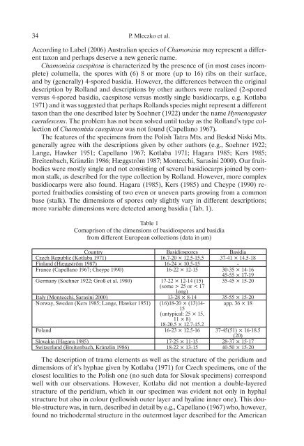
base (stalk). the dimensions <strong>of</strong> spores only slightly vary <strong>in</strong> different descriptions;<br />
more variable dimensions were detected among basidia (tab. 1).<br />
table 1<br />
comaprison <strong>of</strong> the dimensions <strong>of</strong> basidiospores and basidia<br />
from different european collections (data <strong>in</strong> μm)<br />
country Basidiospores Basidia<br />
czech republic (kotlaba 1971) 16.7-20 × 12.5-15.5 37-41 × 14.5-18<br />
F<strong>in</strong>land (hæggström 1987) 16-24 × 10.5-15 -<br />
France (capellano 1967; cheype 1990) 16-22 × 12-15 30-35 × 14-16<br />
45-55 × 17-19<br />
Germany (Soehner 1922; Groß et al. 1980) 17-22 × 12-14 (15)<br />
(some > 25 or < 17<br />
long)<br />
35-45 × 15-20<br />
italy (Montecchi, Saras<strong>in</strong>i 2000) 13-28 × 8-14 35-55 × 15-20<br />
norway, Sweden (Kers 1985; Lange, hawker 1951) (16)18-20 × (13)14-<br />
15<br />
(untypical: 25 × 15,<br />
11 × 8)<br />
18-20.5 × 12.7-15.2<br />
app. 36 × 18<br />
Poland 16-23 × 12.5-16 37-45(51) × 16-18.5<br />
(20)<br />
Slovakia (hagara 1985) 17-25 × 11-15 28-37 × 15-17<br />
Switzerland (Breitenbach, Kränzl<strong>in</strong> 1986) 18-22 × 13-15 40-50 × 15-20<br />
the description <strong>of</strong> trama elements as well as the structure <strong>of</strong> the peridium and<br />
dimensions <strong>of</strong> it’s hyphae given by kotlaba (1971) for czech specimens, one <strong>of</strong> the<br />
closest <strong>localities</strong> to the Polish one (no such data for Slovak specimens) correspond<br />
well with our observations. However, kotlaba did not mention a double-layered<br />
structure <strong>of</strong> the peridium, which <strong>in</strong> our specimen was evident not only <strong>in</strong> hyphal<br />
structure but also <strong>in</strong> colour (yellowish outer layer and hyal<strong>in</strong>e <strong>in</strong>ner one). this double-structure<br />
was, <strong>in</strong> turn, described <strong>in</strong> detail by e.g., capellano (1967) who, however,<br />
found no trichodermal structure <strong>in</strong> the outermost layer described for the american