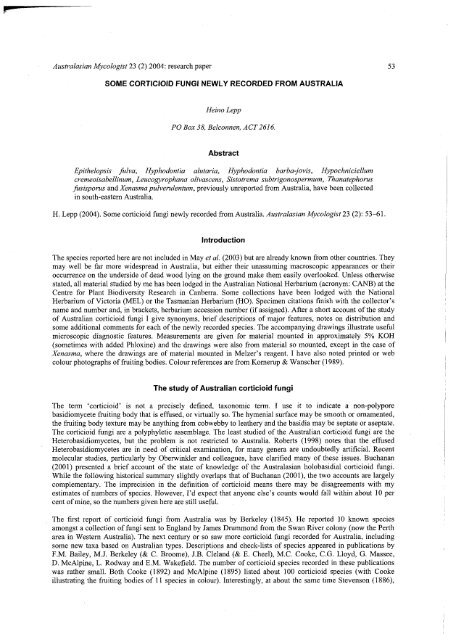
SOME CORTICIOID FUNGI NEWLY RECORDED FROM ...
SOME CORTICIOID FUNGI NEWLY RECORDED FROM ...
SOME CORTICIOID FUNGI NEWLY RECORDED FROM ...

You also want an ePaper? Increase the reach of your titles
YUMPU automatically turns print PDFs into web optimized ePapers that Google loves.
<strong>SOME</strong> <strong>CORTICIOID</strong> <strong>FUNGI</strong> <strong>NEWLY</strong> <strong>RECORDED</strong> <strong>FROM</strong> AUSTRALIA<br />
Heino Lepp<br />
PO Box 38, Belconnen, ACT2616.<br />
Abstract<br />
Epithelopsis fulva, Hyphodontia alutaria, Hyphodontia barba-jovis, Hypochniciellum<br />
cremeoisabellinum, Leucogyrophana olivascens, Sistotrema subtrigonospermum, Thanatephorus<br />
fusisporus and Xenasma pulverulentum, previously unreported from Australia, have been collected<br />
in south-eastern Australia.<br />
H. Lepp (2004). Some corticioid fungi newly recorded from Australia. Australasian Mycologist 23 (2): 53-61.<br />
Introduction<br />
The species reported here are not included in May et al. (2003) but are already known from other countries. They<br />
may well be far more widespread in Australia, but either their unassuming macroscopic appearances or their<br />
occurrence on the underside of dead wood lying on the ground make them easily overlooked. Unless otherwise<br />
stated, all material studied by me has been lodged in the Australian National Herbarium (acronym: CANB) at the<br />
Centre for Plant Biodiversity Research in Canberra. Some collections have been lodged with the National<br />
Herbarium of Victoria (MEL) or the Tasmanian Herbarium (HO). Specimen citations finish with the collector's<br />
name and number and, in brackets, herbarium accession number (if assigned). After a short account of the study<br />
of Australian corticioid fungi I give synonyms, brief descriptions of major features, notes on distribution and<br />
some additional comments for each of the newly recorded species. The accompanying drawings illustrate useful<br />
microscopic diagnostic features. Measurements are given for material mounted in approximately 5% KOH<br />
(sometimes with added Phloxine) and the drawings were also from material so mounted, except in the case of<br />
Xenasma, where the drawings are of material mounted in Melzer's reagent. I have also noted printed or web<br />
colour photographs of fruiting bodies. Colour references are from Kornerup & Wanscher (1989).<br />
The study of Australian corticioid fungi<br />
The term 'corticioid' is not a precisely defined, taxonomic term. I use it to indicate a non-polypore<br />
basidiomycete fruiting body that is effused, or virtually so. The hymenial surface may be smooth or ornamented,<br />
the fruiting body texture may be anything from cobwebby to leathery and the basidia may be septate or aseptate.<br />
The corticioid fungi are a polyphyletic assemblage. The least studied of the Australian corticioid fungi are the<br />
Heterobasidiomycetes, but the problem is not restricted to Australia. Roberts (1998) notes that the effused<br />
Heterobasidiomycetes are in need of critical examination, for many genera are undoubtedly artificial. Recent<br />
molecular studies, particularly by Oberwinkler and colleagues, have clarified many of these issues. Buchanan<br />
(2001) presented a brief account of the state of knowledge of the Australasian holobasidial corticioid fungi.<br />
While the following historical summary slightly overlaps that of Buchanan (2001), the two accounts are largely<br />
complementary. The imprecision in the definition of corticioid means there may be disagreements with my<br />
estimates of numbers of species. However, I'd expect that anyone else's counts would fall within about 10 per<br />
cent of mine, so the numbers given here are still useful.<br />
The first report of corticioid fungi from Australia was by Berkeley (1845). He reported 10 known species<br />
amongst a collection of fungi sent to England by James Drummond from the Swan River colony (now the Perth<br />
area in Western Australia). The next century or so saw more corticioid fungi recorded for Australia, including<br />
some new taxa based on Australian types. Descriptions and check-lists of species appeared in publications by<br />
F.M. Bailey, M.J. Berkeley (& C. Broome), J.B. Cleland (& E. Cheel), M.C. Cooke, C.G. Lloyd, G. Massee,<br />
D. McAlpine, L. Rodway and E.M. Wakefield. The number of corticioid species recorded in these publications<br />
was rather small. Both Cooke (1892) and McAlpine (1895) listed about 100 corticioid species (with Cooke<br />
illustrating the fruiting bodies of 11 species in colour). Interestingly, at about the same time Stevenson (1886),
using the same or very similar species concepts, recorded about 130 species in Britain. Brittlebank's (c. 1940)<br />
mimeographed catalogue listed about 150 species for Australia. Both McAlpine and Brittlebank based their lists<br />
on published papers by other authors rather than on critical examination of specimens.<br />
The New Zealander G.H. Cunningham has been the major contributor to our knowledge of Australian corticioid<br />
fungi. In the 1950s he published 12 lengthy papers about Australasian holobasidial corticioid fungi. These<br />
papers, supplemented with additional findings, were brought together in a posthumously published monograph<br />
(Cunningham 1963) with about 100 species recorded from Australia. The publications were based on<br />
examination of both existing herbarium collections and additional specimens sent to him by a wide range of<br />
correspondents, including many from Australia. He was also able to spend extended periods in the herbarium of<br />
the Royal Botanic Gardens at Kew, where he studied Kew's holdings of Australian fungi. His visits to Europe,<br />
and his northern hemisphere correspondents, also allowed him to study authentic material of diverse taxa. This<br />
background enabled him to bring a discerning eye to the Australian material and place it in a world context. Most<br />
importantly, Cunningham subjected his specimens to thorough microscopic examination. While Cunningham's<br />
monograph is now taxonomically dated and contains some dubious statements, it is still indispensable when used<br />
in conjunction with more modern literature relating to other parts of the world. There is no other monographic<br />
treatment of Australian corticioid fungi.<br />
From the mid-1950s other mycologists have added to knowledge of Australian corticioid fungi. In particular it is<br />
worth noting the contributions by P.K. Buchanan, W. Jiilich, R.A. Maas Geesteranus, D.A. Reid, J.A. Stalpers,<br />
P.H.B. Talbot and J.H. Warcup. Their papers, containing written descriptions and illustrations, gave new records,<br />
new taxa and re-assessments of Cunningham's types. A series of papers by Warcup and Talbot documented<br />
corticioid orchid mycorrhizas as well as some corticioid fungi isolated as mycelia from soil. Their work resulted<br />
in a significant addition to our knowledge, especially of those corticioids with insubstantial fruiting bodies—in<br />
genera such as Ceratobasidium, Thanatephorus and Tulasnella. Many of these taxa, because of their Rhizoctonia<br />
anamorphs, are dealt with by Roberts (1999), who included re-assessments of some Warcup and Talbot taxa. The<br />
studies reported by Price (1975) were done in Australia and the published account is based on her PhD thesis.<br />
Unfortunately, the published account contains no collecting localities. I understand collection localities are given<br />
in the thesis, but I have not seen the thesis. Apart from the mycologists noted by name many more have<br />
contributed in passing, for example, by citing Australian collections held in both Australian and overseas<br />
herbaria—and such contributions are recorded in May et al. (2003).<br />
Many corticioid species appear to be very widespread, with representatives from different continents showing<br />
virtually identical macro and micro features. This has naturally prompted the question as to whether such<br />
'species' are really single, widespread species or species complexes. There has been much investigation of this<br />
issue in the northern hemisphere, using mating tests and molecular studies, but much more needs to be done to<br />
see where the Australian representatives of such taxa fit in.<br />
The taxonomic and bibliographic catalogue by May et al. (2003) has greatly eased the problem of tracking down<br />
relevant literature. That catalogue records about 350 corticioid species for Australia—although without any<br />
guarantee as to the correctness of any record. As the authors warn, they have simply listed all the species that<br />
have been recorded, in print, for Australia. Taking the 350 at face value it is instructive to compare this with the<br />
1,320 species that Ginns (1998) lists for North America. Given these numbers, along with the abundance of<br />
habitats in Australia, it seems reasonable to suppose that there are at least another thousand species out in the<br />
Australian bush. Using the evidence in Ginns (1998), about 780 of those 1,320 North American species were<br />
added to the record during the period 1926-1998. This dwarfs the corresponding Australian increase.<br />
Notwithstanding the contributions by Cunningham and others and, while making allowances for factors such as<br />
changing species concepts and misidentifications of specimens, these figures still give some indication of the<br />
lack of attention given to Australian corticioid fungi by modern mycologists.<br />
Corticioid fungi feature very little in Australian field guides, with the norm being to have one or two species<br />
illustrated. Hood's (2003) semi-popular book contains brief descriptions, comments and black and white<br />
drawings of macroscopic and microscopic features of a dozen species. The fungal website of the Australian<br />
National Botanic Gardens (http://www.anbg.gov.au/fungi/) features photographs of about 45 Australian<br />
corticoid fungi (many not yet identified to species), and each photograph has an associated herbarium specimen<br />
at CANB. At present, the only way to access all of them is via taxon names in the photo list<br />
(http://www.anbg.gov.au/fungi/images-captions/index-list.html). While a few of these photos are used to
illustrate some points made in the website, most are not yet referred to. I intend to add a more detailed section<br />
about corticioid fungi to that website and so make it easier to find just the corticioid photos.<br />
Anecdotal evidence suggests that there is not a great number of herbarium collections of Australian corticioid<br />
fungi and that many of these are at least several decades old (with limited field notes and without the benefit of<br />
recent, critical study). My collections are held at CANB and most are less than a decade old. They have been<br />
gathered as a result of general collecting, aiming to sample widely, both in terms of geography and habitats, and<br />
making use of whatever opportunities have arisen. There is a small number of collections by other collectors,<br />
bringing the total in CANB to between 1,500 and 2,000 specimens. While useful, these constitute a small amount<br />
of material that must be far from representative of both the species in Australia and their distribution. The bulk of<br />
the material comes from south-east Australia (roughly in a quadrilateral formed by Newcastle, Lake Torrens,<br />
Adelaide and Hobart), with a small amount from the tropics and the inland areas in south-west Western<br />
Australia. Much of this material is not fully determined, but it is clear that CANB has examples of known<br />
species, that are presently unrecorded from Australia, as well as specimens I cannot immediately place in known<br />
species. Over time I hope to report on these finds, as well as studies based on collections in other herbaria. In this<br />
paper I record eight known species with immediately distinctive micro-characters. Though most are based on<br />
single collections I think it worthwhile to document the presence of these species in Australia sooner rather than<br />
later.<br />
Brief specimen descriptions and comments<br />
Epithelopsis fulva (G. Cunn.) Julich, Persoonia 8: 457 (1976). Figure 1<br />
Epithele fulva G. Cunn., Trans. Roy. Soc. New Zealand 83: 631 (1956).<br />
The fruiting body is effused, white to creamy and with a velvety appearance to the naked eye. A low power<br />
microscope (or a good hand lens) shows a strigose surface, owing to the abundant hyphal pegs. The specimen<br />
covered several square centimetres and was up to about 250 um thick. Hyphal system: dimitic; generative hyphae<br />
hyaline, thick-walled, with clamps, 2.4-4.8 um diameter, the walls to 1.2 um; skeletal hyphae hyaline, smooth,<br />
3.2-4.8 um diameter, the walls to 2 um. Spores: ellipsoidal, hyaline, thick-walled, smooth, inamyloid,<br />
cyanophilous, 10-12 x 6.4-7.2 um. Basidia: clavate, with 4 sterigmata, 24-28 x 6-8 um. Cystidia: scattered<br />
moniliform gloeocystidia—both embedded and reaching the hymenial surface. Hyphal pegs: the individual<br />
hyphae are almost always acuminate, rarely with obtuse apices. Occasional pegs are composed of as few as 4 or<br />
5 hyphae but mostly the pegs are dense bundles of numerous hyphae. The pegs arise from near the substrate,<br />
traverse the context and project from 90 to 160 um beyond the hymenium. The hyphae are aseptate, to 4.8 um<br />
diameter with walls to 1.6 um.<br />
Specimen studied: NSW, Kosciuszko National Park, Merritts Spur (near upper end of Easy Does It quad), on<br />
well-rotted wood by eucalypt-dominated creek side, with sparse understorey, 13 January 2002, H. Lepp 3584<br />
(CANB 649607).<br />
Comments: The species was described from New Zealand and is also known from Patagonia (Greslebin &<br />
Rajchenberg 2003). Ryvarden (1978) reported it from Africa, but Hjortstam, Manjon & Moreno (1988) opined<br />
that the African collection was a species of Epithele. Julich (1976) created the genus Epithelopsis to<br />
accommodate Epithele fulva (in his view an anomalous Epithele). Cunningham (1963) regarded this species as<br />
monomitic, with the hyphae of the pegs modified generative hyphae, each tapering from base to apex,<br />
aseptate save near the base, and with walls thickened to 1 um' but Julich (1976) found the hyphal system to be<br />
dimitic and recorded skeletal hyphae as 'loosely arranged' and 'often with secondary septa'. In the basal layer of<br />
the Australian specimen I found examples of what I consider to be skeletal hyphae with secondary septa.<br />
Interestingly, while the illustration in Cunningham (1963) shows clamped hyphae in the subhymenium, in the<br />
basal layer there are simply septate hyphae (which I interpret as secondarily septate skeletals). Currently there<br />
are two species in the genus, the other being Epithelopsis bosei A.B. De, with depressed cylindric spores, 15-21<br />
x 4.5-5.2 um (Boidin & Gilles 2000). Photograph: None.
Fig. 1 Epithelopsis fulva: a basidium; b spores; c gloeocystidia; d sporocarp section (empasizing hyphal pegs & basal<br />
layer. Fig. 2 Hyphodontia alutaria: a lagenocystidia; b haloed, capitate cystidia; c basidium; d smooth cystidium<br />
extending beyond hymenium. Fig. 3 Hyphodontia barba-jovis (JHWillis s.n.): a basidia; b apex of a tubular eystidium;<br />
c aculeal apex (margin dotted) showing tubular cystidia. Fig. 4 Hypochnicium cremeoisabellinum: basidium & spores.<br />
Fig. 5 Leucogyrophana olivascens: a basidium; b spores; c cystidium. Fig. 6 Sistotrema subtrigonospermum: a basidia;<br />
b loose spores; c octet of spores (as seated atop a basidium). Fig. 7 Thanatephorus tusisporus. a basidium;<br />
b spores (two germinating by repetition). Fig. 8 Xenasma pulverulentum: a spores; b capitate cystidia.<br />
m^tm—mm Scale bar = 10 um, except Fig. 1d (=100 um), Fig. 2d (=25 um) and Fig. 3c (=40 um).
Hyphodontia alutaria (Burt) J. Erikss. Symb. Bot. Upsal. 16: 104 (1958). Figure 2<br />
Peniophora alutaria Burt, Ann. Missouri Bot. Gard. 12: 332 (1925).<br />
Grandinia alutaria (Burt) Julich, Int. J. Mycol. Lichenol. 1:35 (1982).<br />
The fruiting body is pale yellow (2A4-3A4) and grandinioid. Hyphal system: monomitic; hyphae hyaline, thinwalled,<br />
with clamps, 2.4-4.0 um diameter. Spores: ellipsoidal, hyaline, thin-walled, smooth, inamyloid, 4.4-5.6<br />
x 3.2—4.0 um. Basidia: sub-urniform, with 4 sterigmata, 16-20 x<br />
4.0-4.8 um. Cystidia: of two types.<br />
(1) Lagenocystidia, with thickened walls, are abundant, 26-30 x 3.2—4.0 um. (2) smooth, thin-walled cystidia,<br />
mostly capitate to some degree, but not always. The latter type may grow to 96 um in length. A small percentage<br />
of short capitate cystidia have resinous haloes.<br />
Specimen studied: TASMANIA, Mt Field National Park, Lyrebird Nature Trail, on well-rotted wood in mixed<br />
rainforest, 7 April 2000, H. Lepp 2725 (CANB 627026).<br />
Comments: This is virtually a cosmopolitan species though Langer (1994) noted the lack of records from<br />
Australia and New Zealand. The abundance of lagenocystidia and septocystidia varies (Greslebin & Rajchenberg<br />
2000). In the cited specimen I saw none of the septate cystidia pictured by Eriksson & Ryvarden (1976) and only<br />
a few cystidia with intercalary swellings. Photograph: Breitenbach & Kranzlin (1986), as Grandinia alutaria.<br />
Hyphodontia barba-jovis (Bull.) J. Erikss., Symb. Bot. Upsal. 16: 104 (1958). Figure 3<br />
Hydnum barba-jovis Bull., Hist. Champ. France: 303 (1791).<br />
Odontia barba-jovis (Bull.) Fr., Epicrisis Syst. Mycol. 528 (1838).<br />
Kneiffia irpicoides P. Karst, Bidr. Kdnn. Finl. Nat. Folk 4%: 368 (1889).<br />
Grandinia barba-jovis (Bull.) Julich, Int. J. Mycol. Lichenol. 1: 35 (1982).<br />
Hyphodontia irpicoides (P. Karst.) Burds. & M.J. Larsen, Mycotaxon 17:515 (1983).<br />
Neither of the two collections studied came with descriptive notes of the fresh state. The dried specimens are<br />
pale brown and odontoid. The aculei are 0.5-2.5 mm long, dense, fimbriate and often merging to form<br />
compound teeth. Hyphal system: monomitic; hyphae hyaline, thin- to thick-walled, with clamps, 2.0-4.0 um<br />
diameter. Spores: ellipsoidal, hyaline, thin-walled, smooth, inamyloid, 4.0-5.2 x 3.2-4.0 um. Basidia:<br />
cylindrical but often with a slightly constricted waist, with 4 sterigmata, 10-16 x<br />
4-5.6 um. Cystidia: tubular<br />
cystidia arise from near the substrate, traverse the fruiting body and protrude well beyond the hymenium. These<br />
cystidia have thin-walled apices but are otherwise thick-walled. Bundles of these cystidia make up the core of<br />
each aculeus and the bulk of each bundle extends beyond the aculeal apex, with a small number of cystidia<br />
bending and protruding from the aculeus below the apex.<br />
Specimens studied: VICTORIA: Sherbrooke Plantation, Sherbrooke Forest Park, Dandenong Ranges, on<br />
Pims, 12 March 1977, N.H. Sinnott 2216 (CANB 608161). South Gippsland, Nyora, on bark and wood of<br />
dead fallen branches in open Eucalyptus obliqua forest with much wire grass, 28 November 1990; J.H. Willis<br />
s.n. (MEL 262624).<br />
Comments: This is a cosmopolitan species although Langer (1994) noted the lack of records from Australia. In<br />
the same monograph he recorded the spore sizes as 4.5-6 x 3.5^1.5 um. Cunningham (1959) recorded the<br />
species from New Zealand, noting that the New Zealand collections agreed with authentic specimens at Kew<br />
save that some are more brightly coloured and spores are slightly smaller'. He gave the spore sizes as 4-5 x<br />
3-3.5 um. The spore sizes in the two Australian collections are a mix of those two ranges. Photograph:<br />
Breitenbach & Kranzlin (1986), as Grandinia barba-jovis.<br />
Hypochniciellum cremeoisabellinum (Litsch.) Hjortstam, Mycotaxon 13: 125 (1981). Figure 4<br />
Corticium cremeoisabellinum Litsch., Ann. Mycol. 39: 117 (1941).<br />
Leucogyrophana cremeoisabellinum (Litsch.) Parmasto, Eesti NSV Tead. Akad. Toim., Biol., 16: 385 (1967).<br />
The fruiting body is soft, white and even. When fresh the surface is smooth, but short cracks appear on drying.<br />
The subiculum is a more open weave, more-or-less floccose. The specimen was irregularly spread over several
square centimetres and grew to about 0.5mm thick. Hyphal system: monomitic; hyphae hyaline, smooth, thin (to<br />
slightly thickened) walls, with clamps, 2.4-3.2 um diameter. Spores: ellipsoidal, hyaline, thick-walled, smooth,<br />
?amyloid (see the comments below), not cyanophilous, 5.6-6.4 * 3.6-4.4 um. Basidia: clavate, with (2-) 4<br />
sterigmata, 23-32 (-36) x 4.8-6.0 um. Cystidia: none.<br />
Specimen studied: NSW, Kosciuszko National Park, Merritts Spur (near upper end of Easy Does It quad), on<br />
well-rotted wood by eucalypt-dominated creek side, with sparse understorey, 13 January 2002, H. Lepp 3583<br />
(CANB 649606).<br />
Comments: Previously the species had been known from Europe (Eriksson & Ryvarden 1976), Tierra del Fuego<br />
(Hjortstam & Ryvarden 1985) and the Russian Far East (Kotiranta & Mukhin 1988). Eriksson & Ryvarden<br />
(1976), while reporting that the spores were a 'very light shadow of grey' in Melzer's, described them as<br />
inamyloid. In the specimen described above, the spores immediately turned dark grey-blue in Melzer's. When<br />
erected by Hjortstam & Ryvarden (1980) the genus held one species, with thick-walled, cyanophilous spores that<br />
were neither amyloid nor dextrinoid. When Hjortstam (1981) emended the genus, to include three anomalous<br />
Leucogyrophana species, he described the spores as 'thick-walled, cyanophilous, in some species with walls<br />
greyish in Melzer's reagent'. In the same paper Hjortstam noted that H. cremeoisabellinum was a little-known<br />
species and that possibly more than one taxon was involved. Rajchenberg & Wright (1987) compared<br />
Hypochniciellum iaganicum (Spegazzini) Rajchenberg & Wright, based on a type from a location given simply<br />
as 'Fuegia', with two Tierra del Fuegian collections of H. cremeoisabellinum which had been reported by<br />
Hjortstam & Ryvarden (1985). Rajchenberg & Wright (1987) stated that these three Fuegian collections all had<br />
acynophilous spores that showed a strong or dark greyish reaction in Melzer's. The only difference appeared to<br />
be in spore size ranges (6-8 x 4-5 urn for H. iaganicum, 5-7 x 3.5-5 um for the two H. cremeoisabellinum<br />
collections). While Rajchenberg & Wright (1987) noted the strong possibility that the two are conspecific (in<br />
which case H. iaganicum would take priority) they echoed Hjortstam's (1981) caution and noted the need for<br />
more collections and interfertility studies to delimit these species. Photograph: None.<br />
Leucogyrophana olivascens (Berk. & M.A. Curtis) Ginns & Weresub, Mem. N.Y. Bot. Gard. 28: 96<br />
(1976). Figure 5<br />
Corticium olivascens Berk. & M.A. Curtis, Grevillea 1: 179 (1873).<br />
The fresh fruiting body has a soft consistency. There is a white, arachnoid to floccose margin, then yellow (near<br />
2A8, 3A8) and towards the centre yellowish orange or greyish yellow (near 4B7, 4C7). The subiculum is white.<br />
The hymenium is tuberculate. When dry the hymenium becomes a very brittle crust over the subiculum. There<br />
are some weak, white cordons at the margins. The specimen examined is about 6 * 2 cm in area and less then<br />
1 mm thick. Hyphal system: monomitic; hyphae hyaline, smooth to densely encrusted, thin-walled, with clamps,<br />
4.0-6.0 um diameter. Spores: ovoid to broadly ellipsoidal, pale yellow-brown, slightly thickened walls, smooth,<br />
dextrinoid, a few cyanophilous, 5.6-7.2 x 4.0-4.8 um. Basidia: clavate, with 4 sterigmata, 22-28 x 4.4-5.6 um.<br />
Cystidia: hyaline, smooth, thin-walled, mostly cylindric, obtuse and with one clamped septum (very rarely 2 or<br />
3) but there are also a small number of clamped, subulate cystidia. The cystidia generally extend no more than<br />
40-60 um beyond the hymenial surface (very rarely to 100 um).<br />
Specimen studied: ACT, Canberra, eastern foot of Black Mountain, CSIRO grounds, on well-rotted eucalypt<br />
wood in a large, bare area of woodchip mulch around a large, long-dead eucalypt stump, 20 April 2000, H. Lepp<br />
2777 (CANB 627079).<br />
Comments: Ginns (1978) noted that the species was known from Canada, USA, Cuba, Bahama Islands, France,<br />
India and Okinawa (Japan). In the same paper he noted that the tuberculate hymenial surface readily<br />
distinguished L. olivascens from the other yellowish species in the genus. The other species have odontoid or<br />
meruliod hymenial surfaces. Photograph: None.<br />
Sistotrema subtrigonospermum D.P. Rogers, Iowa Univ. Stud. Nat. Hist. 17: 22 (1935). Figure 6<br />
Trechispora subtrigonosperma (D.P. Rogers) D.P. Rogers & H.S. Jacks., Farlowia 1: 328 (1943).
i<br />
Australasian Mycologist 23 (2) 2004: research paper 59<br />
The delicate and very thin fruiting body looks like little more than a pale grey bloom on the substrate, spread<br />
over an area of about 3*1 cm. When dry the fruiting body (on its pale substrate) is virtually invisible to the<br />
naked eye and is then best seen with a low power dissecting microscope. Hyphal system: monomitic; hyphae<br />
hyaline, clamped, 2.4-5.6 um in diameter. Spores: tetrahedral, hyaline, thin-walled, smooth, inamyloid, with<br />
sides from 3.6-AA urn long. Basidia: urniform, with 6-8 sterigmata, 14-20 * 4.8-5.6 um. Cystidia: none.<br />
Specimen studied: TASMANIA, approx 15 km WNW of Maydena, forestry track off Gordon River Road, on<br />
bleached, very rotten wood in mixed forest, 8 April 2000, H Lepp 2740 (CANB 627041).<br />
Comments: The species is widespread, being known from Europe (Eriksson et al 1984), North America (Ginns &<br />
Lefebvre 1993), Jamaica (Rogers 1944), Brazil (Rogers 1935), Reunion (Hjortstam & Larsson 1994) and<br />
Ethiopia (Hjortstam & Ryvarden 1996). Eriksson et al (1984) noted that the characteristic spores make this an<br />
easy species to identify. The combination of tetrahedral spores and 6-8 spored urniform basidia distinguishes<br />
this species from other corticioid fungi. Photograph: a photograph of the specimen cited above appears at:<br />
http://www.anbg.gov.au/fungi/images-captions/sistotrema- subtrigonospermum -0181 .html.<br />
Thanatephorus fusisporus (J. Schrot.) Hauerslev & P. Roberts, Nordic J. Bot. 16: 218 (1996).<br />
Figure 7<br />
Hypochnus fusisporus J. Schrot. in Cohn, Krypt.-Fl. Schlesien 3(1): 416 (1888).<br />
Zygodesmus limonisporus Ellis & Everh., Proc. Acad. Nat. Set Philadelphia 43: 87 (1891).<br />
Coniophora vaga Burt, Ann. Missouri Bot. GardA: 251 (1917).<br />
Corticium fenestratum Overholts, Mycologia 26: 510 (1934).<br />
Uthatobasidium fusisporum (J. Schrot.) Donk, Fungus 28: 22 (1958).<br />
The insubstantial fruiting body is grey and farinose/arachnoid, imperfectly covering an area of about 7x3 cm.<br />
Hyphal system: monomitic; hyphae hyaline, smooth, basal hyphae thick-walled (occasionally to 2 pm but mostly<br />
about 1 um) otherwise thin-walled, without clamps, 4.8-8.0 um in diameter. Spores: citriform, biapiculate,<br />
hyaline, thin-walled, smooth, inamyloid, repetitive, 7.6-12 x 4.4-6.4 um. Basidia: cylindric, with 4 sterigmata,<br />
12-24 x 8.0-12 um. Cystidia: none.<br />
Specimen studied: NSW, Monumea Gap State Forest, approx. 7 km SE of Bogan Gate, on a long-dead, fallen<br />
Callitris sapling in Callitris I Eucalyptus forest, 7 November 2001, H. Lepp 3354 (CANB 638070).<br />
Comments: The species is widespread, with Roberts (1999) noting it from various European countries, the<br />
Ukraine, Canada, Greenland and the USA. He also comments on the great variability in spore size (4—18 x 2.5-<br />
9.5 um) and shape (from almost globose to oblong) but notes that there is always a distinct apical projection.<br />
Photograph: print - Breitenbach & Kranzlin (1986), captioned as Hypochnus fusisporus; web —<br />
http://www.cbs.knaw.nl/images/Images_CBSFungi/CD/0001/0002/Uthatobasidium_fusisporuml.jpg<br />
Xenasma pulverulentum (Litsch.) Donk, Fungus 27: 25 (1957). Figure 8<br />
Corticium pulverulentum Litsch., Osterr. Bot. Zeitschr. 88: 112 (1939) [non Corticium pulverulentum (Lev.)<br />
Cooke, Grevillea 8: 89 (1880)].<br />
Ceratobasidium striisporum Rick, Lilloa 9: 219 (1943).<br />
Peniophorapulverulenta (Litsch.) H.S. Jacks., Can. J. Res., C 28: 532 (1950).<br />
The collector's field notes about the fruiting body are as follows: 'Grey, corticioid fungus with no apparent<br />
development of an abhymenial surface. The hymenial surface is uniformly coloured (grey or olivaceous-grey<br />
with a powdery, whitish bloom), thin (less than 1 mm thick, the texture membranous or rubbery, not stiff or<br />
parchment-like in any way. Identity of substrate uncertain.' The colour photograph accompanying the collection<br />
shows an irregularly wrinkled to tuberculate hymenial surface, with some very short cordons. When dry the<br />
fruiting body is hard and cartilaginous. Hyphal system: monomitic; hyphae hyaline, smooth, 2.4-3.6 um in<br />
diameter, a few clamps seen. Spores: ellipsoidal, hyaline, thin to thickened walls, striate, inamyloid, 7.2-8.8 *<br />
4.0-5.2 um. The striations disappear in KOH. Basidia: cylindric, pleurobasidia with 4 sterigmata, approx. 16-20
x 6-7 um (but not well seen). Cystidia: capitate, smooth, 10-25 um long, with the capitate apex to 4 um in<br />
diameter.<br />
Specimen studied: TASMANIA, Warra Long Term Ecological Research Area, Manuka Road, on rotten wood,<br />
2 November 2000, G. Gates, D. Ratkowsky & Z. Yuan 20001102C2 (CANB specimen not yet assigned a CANB<br />
number; part lodged in HO as HO520055).<br />
Comments: When dry the hyphae agglutinate strongly, making it very difficult to separate them in a rehydrated<br />
sample. Hence I didn't satisfactorily observe all the microscopic features but, given the spores and cystidia, I<br />
have no doubt about the identification. This is a widely distributed species, known from New Zealand<br />
(McKenzie et al. 2000), various European countries (Breitenbach & Kranzlin 1986, Hjortstam et al 1988,<br />
Jackson 1950, Zervakis et al. 2002), various North American locations (Ginns & Lefebvre 1993), Brazil (Roberts<br />
1999), and Ethiopia, Argentina, Colombia, Venezuela, Reunion and Morocco (Hjortstam & Ryvarden 1996).<br />
Hjortstam, Larsson & Ryvarden (1988) commented that this species is 'Easily recognized by the striate spores<br />
and the capitate cystidia' though they then note the similar species, Xenasma parvisporum Pouzar. Pouzar (1982)<br />
noted that its smaller spores (4.5-5.5 * 2.8-3.2 um) differentiated it from the closely related X. pulverulentum.<br />
I have seen published descriptions of only European and North American specimens of X. pulverulentum, with<br />
spore sizes in the range 8-12 x 4.5-6 um. However, Hjortstam, Larsson & Ryvarden (1988) noted in passing that<br />
they had seen Iranian specimens with smaller spores, 7.5-8.5 x 4.5-5 um, which is similar to the range seen in<br />
the Tasmanian collection. Several authors note that older spores may sometimes be dextrinoid, but I saw no<br />
dextrinoid spores. Photographs: print Breitenbach & Kranzlin (1986); web —<br />
http://aphyllo.com/spe/images.asp?id=1590100&ch=A&mn=img<br />
Acknowledgements<br />
I thank Genevieve Gates and David Ratkowsky for sending me part of their Xenasma collection, along with field<br />
notes and a photograph; Tom May and Cheryl Grgurinovic for allowing early access to Fungi of Australia,<br />
Volume 2B (during the manuscript stage); Tom May for permitting the loan of specimens from MEL and for<br />
some bibliographic information; Fungimap for paying for my travel to Tasmania in April 2000 and the NSW<br />
National Parks and Wildlife Service for supporting my participation in the Snowy Mountains Biodiversity Blitz<br />
in January 2002. I am obliged to the referee's helpful comments, including the suggestion to include a history<br />
section.<br />
References<br />
Berkeley, M.J. (1845). Decades of Fungi. Dec III—VII. Australian Fungi. London Journal of Botany 4,42-73 (as<br />
reprinted on pp. 20-51 in Berkeley, M.J., Decades of Fungi, Decas 1-62, A. Asher & Co, Amsterdam, 1969).<br />
Boidin, J. & Gilles, G. (2000). Basidiomycetes Aphyllophorales de file de La Reunion. XIX: le genre Epithele<br />
(Pat.) Pat. 1900. Bulletin Mensuelde la Societe linneene deLyon 69, 193-198.<br />
Breitenbach, J. & Kranzlin, F. (1986). Fungi of Switzerland, Volume 2. Verlag Mykologia, Lucerne.<br />
Brittlebank, C.C. (c. 1940). Catalogue of Australian Fungi. Published by the author.<br />
Buchanan, P.K. (2001). Aphyllophorales in Australasia. Australian Systematic Botany 14,417-437.<br />
Cooke, M.C. (1892) Handbook of Australian Fungi. Williams & Norgate, London.<br />
Cunningham, G.H. (1959). Hydnaceae of New Zealand, Part II—The Genus Odontia. Transactions of the Royal<br />
Society of New Zealand 86, 65-103.<br />
Cunningham, G.H. (1963). The Thelephoraceae of Australia and New Zealand. New Zealand Department of<br />
Scientific and Industrial Research Bulletin 145, 1-359.<br />
Eriksson, J., Hjortstam, K. & Ryvarden, L. (1984). The Corticiaceae of North Europe, Volume 7. Fungiflora,<br />
Oslo.<br />
Eriksson, J. & Ryvarden, L. (1976). The Corticiaceae of North Europe, Volume 4. Fungiflora, Oslo.<br />
Ginns. J. (1978). Leucogyrophana (Aphyllophorales): identification of species. Canadian Journal of Botany 56,<br />
1953-1973.<br />
Ginns, J. (1998). How many species are there? Folia Cryptogamica Estonica 33,29-33.<br />
Ginns, J. & Lefebvre, M.N. (1993). Lignicolous Corticioid Fungi (Basidiomycota) of North America. APS Press,<br />
St. Paul (= Mycologia Memoir 19).<br />
Greslebin, A.G. & Rajchenberg, M. (2000). The genus Hyphodontia in the Patagonian Andes forests of<br />
Argentina. Mycologia 92, 155-1165.
Greslebin, A.G. & Rajchenberg, M. (2003). Diversity of Corticiaceae sens. lat. in Patagonia, Southern Argentina.<br />
New Zealand Journal of Botany 41,437^146.<br />
Hjortstam, K. (1981). Notes on Corticiaceae (Basidiomycetes) FX. Three new combinations in Hypochniciellum.<br />
Mycotaxon 13, 124-126.<br />
Hjortstam, K. & Larsson, K.-H. (1994). Annotated check-list to genera and species of corticioid fungi<br />
(Aphyllophorales, Basidiomycotina) with special regards to tropical and subtropical areas. Windahlia 21,1-75.<br />
Hjortstam, K., Larsson, K.-H. & Ryvarden, L. (1988). The Corticiaceae of North Europe, Volume 8. Fungiflora,<br />
Oslo.<br />
Hjortstam, K., Manjon, J.L. & Moreno, G. (1988). Notes on select corticiaceous fungi from Spain and North<br />
Africa. Mycotaxon 33, 257—263.<br />
Hjortstam, K. & Ryvarden, L. (1996). New and interesting wood-inhabiting fungi (Basidiomycota—<br />
Aphyllophorales) from Ethiopia. Mycotaxon 60, 181-190.<br />
Hjortstam, K. & Ryvarden, L. (1980). Studies in Tropical Corticiaceae II. Mycotaxon 12, 168-184.<br />
Hjortstam, K. & Ryvarden, L. (1985). New and noteworthy Basidiomycetes (Aphyllophorales) from Tierra del<br />
Fuego, Argentina. Mycotaxon 22,159—167.<br />
Hood, I. (2003). An introduction to fungi on wood in Queensland. University of New England School of<br />
Environmental Sciences and Natural Resources Management, Armidale.<br />
Jackson, H.S. (1950). Studies of Canadian Thelephoraceae VI. The Peniophora rimicola group. Canadian<br />
Journal of Research 28(C), 525-534.<br />
Julich, W. (1976). Studies in hydnoid fungi—I: On some genera with hyphal pegs. Persoonia 8,447^158.<br />
Kotiranta, H. & Mukhin, V.A. (1988). Polyporaceae and Corticiaceae of an isolated forest of Abies nephrolepis<br />
in Kamchatka, Russian Far East. Karstenia 38, 69-80.<br />
Kornerup, A. & Wanscher, J.H. (1989). Methuen Handbook of Colour, 3rd edn, reprint. Methuen, London.<br />
Langer, E. (1994). Die Gattung Hyphodontia John Eriksson. J. Cramer, Berlin.<br />
May, T.W., Milne, J., Shingles, S. & Jones, R.H. (2003). Catalogue and Bibliography of Australian Fungi 2.<br />
Basidiomycota p.p. & Myxomycota p.p. Fungi of Australia 2B. ABRS/CSIRO, Melbourne.<br />
McAlpine, D. (1895). Systematic Arrangement of Australian Fungi, Robt. S. Brain, Government Printer,<br />
Melbourne.<br />
McKenzie, E.H.C., Buchanan, P.K. & Johnston, P.R. (2000). Checklist of fungi on Nothofagus species in New<br />
Zealand. New Zealand Journal of Botany 38, 635-720.<br />
Pouzar, Z. (1982). Taxonomic studies in resupinate fungi I. Ceskd Mykologie 36, 141-145.<br />
Price, LP. (1975). A study of cystidia in effused Aphyllophorales. Nova Hedwigia 24, 515-618.<br />
Rajchenberg, M. & Wright, J.E. (1987). Type studies of Corticiaceae and Polyporaceae (Aphyllophorales)<br />
described by C. Spegazzini. Mycologia 79, 246-264.<br />
Roberts, P. (1998). A revision of the genera Heterochaetella, Myxarium, Protodontia and Stypella<br />
(Heterobasidiomycetes). Mycotaxon 69, 209-248.<br />
Roberts, P. (1999). Rhizoctonia-forming fungi: A taxonomic guide. Royal Botanic Gardens, Kew.<br />
Rogers, D.P. (1935). Notes on the Lower Basidiomycetes. University of Iowa Studies in Natural History 17, 3—<br />
43.<br />
Rogers, D.P. (1944). The genera Trechispora and Galzinia (Thelephoraceae). Mycologia 36, 70-103.<br />
Ryvarden, L. (1978). Studies in the Aphyllophorales of Africa 6: Some species from eastern Central Africa.<br />
Bulletin du Jar din Botanique National de Belgique 48, 79-117.<br />
Stevenson, J. (1886). Hymenomycetes Britannici. British Fungi (Hymenomycetes), Volume II. William<br />
Blackwood and Sons, Edinburgh and London.<br />
Zervakis, G.I., Polemis, E. & Dimou, D.M. (2002). Mycodiversity studies in selected ecosystems of Greece: III<br />
Macrofungi recorded in Quercus forests from southern Peloponnese. Mycotaxon 84,141-162.