Abdominal Ultrasound - Livingston and Brighton ED
Abdominal Ultrasound - Livingston and Brighton ED
Abdominal Ultrasound - Livingston and Brighton ED

You also want an ePaper? Increase the reach of your titles
YUMPU automatically turns print PDFs into web optimized ePapers that Google loves.
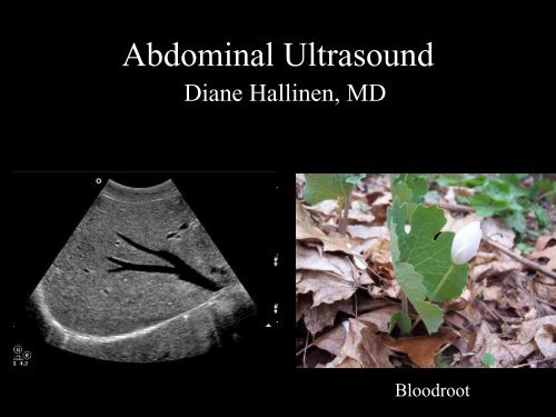
<strong>Abdominal</strong> <strong>Ultrasound</strong><br />
Diane Hallinen, MD<br />
Bloodroot
<strong>Abdominal</strong> <strong>Ultrasound</strong><br />
• Vasculature<br />
• Hepatobiliary<br />
• Spleen<br />
• Kidney<br />
• Bladder<br />
• Bowel
Where to put the probe?
Vasculature<br />
We are going to<br />
talk about …<br />
Celiac Trunk<br />
Superior<br />
Mesenteric<br />
Artery<br />
Portal Veins<br />
Hepatic Veins
Celiac, SMA
Celiac Trunk <strong>and</strong> SMA
Transverse Celiac
Transverse SMA-<br />
Bull’s Eye
Biliary Scanning - Longitudinal<br />
• For the gallbladder start with<br />
the probe marker pointing to<br />
the head, in the mid-clavicular<br />
line. Move the probe toward<br />
the midline until you find a<br />
gallbladder looking structure.<br />
• Rotate the probe so you get a<br />
long-axis view.
Transverse Approach<br />
from Superior to Inferior<br />
• Hepatic veins<br />
• Portal vein<br />
• Then locate the most<br />
anterior cystic<br />
structure… the<br />
gallbladder.
Some questions…<br />
• Who are you going to scan?<br />
• What are you going to do with the<br />
information? What if you get “no”<br />
information?<br />
• When are you going to do it? Before you<br />
order labs? After everything comes back?<br />
• Does everybody need a “formal exam?”<br />
• What are you going to say to the patient?
My Answers<br />
• I haul the machine in if the triage note makes<br />
me think gallbladder.<br />
• I do it right away since I may order lots or<br />
little based on what I see in conjunction with<br />
the H & P.<br />
• I’ll order a formal exam on everybody who<br />
needs to see a surgeon, or if I’m not sure.<br />
• I tell patients this is a focused exam, <strong>and</strong> they<br />
may need more “stuff done” later.
Biliary Colic Workup
Thickened Gallbladder Wall<br />
• Measure the<br />
anterior wall.<br />
• Normal less than<br />
2mm.<br />
• Need to be fasting.<br />
• Thick wall seen in<br />
jaundice, hepatitis,<br />
ascites, etc.
Thick Gallbladder Wall
Pericholecystic Fluid <strong>and</strong> other<br />
findings<br />
• http://www.med-ed.virginia.edu/courses/rad/edus/index6.html
• Stones send back<br />
all the sound waves<br />
to the transducer,<br />
thus they make a<br />
shadow…posterior<br />
shadowing.<br />
• Stones also move if<br />
they are not stuck.<br />
Posterior Shadow
A Layer of a Whole Bunch of<br />
• There is a<br />
multiple tiny<br />
stones producing<br />
this posterior<br />
shadowing.<br />
Small Stones
SONOGRAPHIC MURPHY<br />
• See liver, or bowel, or<br />
whatever.<br />
• Press on it.<br />
• Does it hurt?<br />
• See gallbladder.<br />
• Press on gallbladder.<br />
• See it move.<br />
• Does that hurt?
Sonographic Murphy’s Sign,<br />
• A positive sonographic Murphy’s sign,<br />
defined as the presence of maximal<br />
tenderness elicited by direct pressure of the<br />
transducer over a sonographically localized<br />
gallbladder, is present in most patients with<br />
acute cholecystitis.<br />
• The positive predictive value of sonographic<br />
Murphy’s sign combined with the presence<br />
of gallstones is reported to be 77% to 92%.
Negative Sonographic Murphys
Happens when the<br />
gallbladder is full of<br />
stones, or when it has<br />
one big one.<br />
Wall Echo Sign
Wall Echo Sign-Multiple Stones<br />
• Note the hypoechoic<br />
(black) stripe<br />
between the wall <strong>and</strong><br />
stones. This<br />
represents bile.<br />
• Important to know<br />
about since it can be<br />
confused with gas<br />
filled bowel.
Portal Vein
CBD<br />
anterior<br />
to Portal<br />
Vein
Portal Vein<br />
• Visualize the portal vein<br />
in a long axis view.<br />
• Anterior to the PV,<br />
(towards the skin) you<br />
should see the bile duct.<br />
• Portal vein has brighter<br />
(more echogenic) walls<br />
compared to hepatic<br />
veins.
Find that CBD<br />
Follow the<br />
gallbladder neck to<br />
the portal triad. The<br />
main lobar fissure<br />
should take you there<br />
(present in about<br />
70% of people).
Main Lobar Fissure
Porta Hepatis=Portal Triad<br />
• Rotating the probe<br />
on the portal vein<br />
should bring the<br />
common bile duct,<br />
hepatic artery <strong>and</strong><br />
portal vein into<br />
view.<br />
• The probe marker<br />
should be pointing<br />
to the patient’s<br />
right.
Mickey Mouse<br />
• If the CBD is<br />
dilated than the<br />
rodent has a swollen<br />
right ear.<br />
• If you forget which<br />
ear is which, you<br />
can doppler the big<br />
one.
Dilated Common Bile Duct
CBD- Measure the Internal<br />
Diameter.<br />
• Mean diameter is 4 mm, more than 7 mm is<br />
dilated.<br />
• CBD diameter increases with age, up to 10<br />
mm in the elderly .<br />
• In general it should be 1/10th the patient’s<br />
age.
Acute Cholecystitis<br />
• The solid arrows<br />
are pointing to the<br />
thickened<br />
gallbladder wall.<br />
• What are the open<br />
arrows pointing<br />
to?
Polyps<br />
• Non-mobile<br />
• No posterior<br />
shadowing
Gallbladder Sludge<br />
• Mobile<br />
• Nonshadowing<br />
• Dependent
Phrygian Cap<br />
Transverse<br />
congenital septum<br />
in the fundus
Pitfalls in Gallbladder Imaging<br />
• Misidentification of the gallbladder. A loop of<br />
bowel or the IVC can look like a gallbladder. Gut<br />
does peristalsis, IVC has flow on doppler. If<br />
possible you should show that the gallbladder<br />
communicates with main portal triad via the major<br />
lobar fissure.<br />
• Bowel gas, try left lateral decubitus position.<br />
• Contracted gallbladder in non-fasting patient.<br />
• Lack of power to penetrate adipose.
The Liver<br />
• Interesting findings can be seen in many<br />
patients…including<br />
– portal hypertension<br />
– cirrhosis<br />
– fatty liver<br />
– bile duct obstruction<br />
– cysts<br />
– tumors
Vena Cava <strong>and</strong> Hepatic Veins<br />
• There are three main<br />
hepatic veins, right, left <strong>and</strong><br />
middle.<br />
• They have thin, hypoechoic<br />
walls, <strong>and</strong> converge on the<br />
vena cava.<br />
• Remember that the portal<br />
vein has hyperechoic walls.
Bunny Sign<br />
• The rabbit’s head is the vena cava, the ears are the middle<br />
<strong>and</strong> left hepatic veins.<br />
• Use m-mode to observe the change in diameter of the vena<br />
cava with respiration
Transducer Parallel to the Costal<br />
Margin
• Usually seen in the<br />
setting of cirrhosis.<br />
• The portal vein is dilated<br />
to more than 15 mm.<br />
• Often seen with ascites<br />
<strong>and</strong> splenomegaly.<br />
Portal Hypertension<br />
Note the portal vein’s<br />
hyperechoic walls.
Cirrhosis-Early Stage<br />
• Loss of peripheral<br />
hepatic vessels<br />
• Increased echogenic<br />
wall of the portal vein.<br />
• Hepatomegaly
• Nodular liver<br />
contour.<br />
Cirrhosis Advanced Stage<br />
• Contracted liver.<br />
• Usually ascites
Fatty Liver<br />
• Results in increased echogenicity (brighter).<br />
Best compared to right kidney cortex.<br />
Focal Fatty Liver. Diffuse Fatty Liver
Bile Duct Obstruction
Liver Cysts-Benign<br />
• To be called a cyst<br />
it must have<br />
– echo free content<br />
– spherical shape<br />
– smooth outline<br />
– distal acoustic<br />
enhancement
Liver Tumors<br />
• The liver is a favorite<br />
site for metastatic<br />
tumors. GI tract,<br />
breast, lung, <strong>and</strong> the<br />
esophagus are the<br />
usual primaries.<br />
• Note the hypoechoic<br />
halo.
Renal <strong>Ultrasound</strong><br />
• Hydronephrosis<br />
• Cysts including polycystic kidney disease<br />
• Stones<br />
• Wilm’s tumor<br />
• Measuring Bladder Volume
• The echogenicity<br />
(brightness) of the<br />
renal cortex should<br />
be less than that of<br />
the liver or spleen.<br />
• In other words the<br />
kidney should be a<br />
tad darker than the<br />
liver.<br />
The Normal Kidney
Kidney Size<br />
• Size is best evaluated as<br />
length, which should be<br />
10 to 12 cm in adults.<br />
• Each kidney should be<br />
within 2 cm of each<br />
other in length.
• Use the liver as<br />
your window on the<br />
right.<br />
• The left can be<br />
harder.<br />
• Try prone.<br />
• Breath holding<br />
helps.<br />
Approach
Hydronephrosis Grades - Mild,<br />
I spraying<br />
II bear claw<br />
III severe, loss<br />
of renal<br />
parenchyma<br />
This is grade 2.<br />
Moderate, Severe
Bear Claw- Hydronephrosis
Simple Cyst Seen while doing a<br />
FAST
Polycystic Kidney
Nephrolithiasis<br />
• CT is better, but there<br />
is a place for doing a<br />
focused <strong>ED</strong> U.S.<br />
• Someone with known<br />
stones to look for<br />
obstruction.<br />
• Pregnancy.<br />
• When AAA is in the<br />
differential.
• Stones are<br />
rarely seen<br />
in the<br />
ureter, but<br />
they are<br />
visible in<br />
the kidney,<br />
bladder <strong>and</strong><br />
UVJ. Note<br />
distal<br />
shadowing<br />
Seeing the Stone
• Color Doppler probe is<br />
used to find urine<br />
squirting from the<br />
ureter into the bladder.<br />
• If the jet is present then<br />
there is no obstruction<br />
on that side.<br />
Ureteral Jets
• Younger than<br />
6.<br />
• Have a flank<br />
mass <strong>and</strong><br />
hematuria.<br />
• Mass is solid<br />
on U.S.<br />
Wilm’s tumor
Urinary Bladder<br />
• Female patient<br />
(note the uterus).<br />
• Top image is a<br />
longitudinal view.<br />
• Bottom is<br />
transverse.
Measure urine volume<br />
Why only one “up down” measurement <strong>and</strong> two “side<br />
to side”?
Measure Urine Volume<br />
Bladder volume<br />
is estimated by<br />
measuring<br />
maximal<br />
anteroposterior,<br />
coronal, <strong>and</strong><br />
craniocaudal<br />
measurements<br />
<strong>and</strong> multiplying<br />
by pi/6 (0.52)
Spleen Viewing Position<br />
Best seen from the<br />
a posteriolateral<br />
approach, with the<br />
probe position in<br />
between the ribs.
• A spleen looks like<br />
a crescent moon.<br />
• Usually best seen<br />
halfway between<br />
full inspiration <strong>and</strong><br />
expiration.<br />
Spleen
Splenomegaly<br />
• The “normal” spleen<br />
measures less than 12<br />
cm long, 5 cm thick<br />
<strong>and</strong> 7 cm in breadth.<br />
• It can be hard to get<br />
the whole spleen on<br />
the screen if your<br />
probe’s footprint isn’t<br />
big enough.
Bowel Obstruction/Ileus<br />
• I mention this because<br />
you will run into while<br />
looking for a AAA.<br />
• Usually bowel isn’t seen<br />
since gas disperses the<br />
beam. But in obstruction<br />
the bowel has a lot of<br />
fluid in it.
Bowel Obstruction
Bowel Obstruction
Appendicitis<br />
Greater than 6 mm in diameter
The image on the<br />
right is obtained by<br />
pressing down on<br />
the probe. Note the<br />
appendix does not<br />
compress.<br />
Press Down
Wall is greater than<br />
3 mm thick.<br />
Appendicitis
• Note the<br />
posterior<br />
shadowing.<br />
• Exam is done<br />
with a high<br />
frequency<br />
probe.<br />
Appendicolith
How to use this skill clinically.<br />
• 17 year old girl<br />
• C/O RLQ pain.<br />
• HCG –<br />
• Appendicitis<br />
versus tuboovarian<br />
abscess?
• Often seen in<br />
children after<br />
a viral illness.<br />
• Often<br />
confused with<br />
appendicitis<br />
on clinical<br />
exam.<br />
Mesenteric Adenitis
References:<br />
References<br />
<strong>Abdominal</strong> <strong>Ultrasound</strong> CD by Mark Deutchman<br />
(great resource)<br />
Emergency <strong>Ultrasound</strong> by Ma <strong>and</strong> Mateer<br />
<strong>Ultrasound</strong> in Emergency <strong>and</strong> Ambulatory Medicine<br />
by Simon <strong>and</strong> Snoey<br />
<strong>Ultrasound</strong> Teaching Manual by Hofer
Web Pages<br />
• http://www.gemedicalsystems.com/inen/rad<br />
/us/education/msucmehd.html<br />
• http://www3.medical.philips.com/enus/secure/photohome/index.asp