Clerodane and labdane diterpene glycosides from a Malagasy ...
Clerodane and labdane diterpene glycosides from a Malagasy ...
Clerodane and labdane diterpene glycosides from a Malagasy ...

Create successful ePaper yourself
Turn your PDF publications into a flip-book with our unique Google optimized e-Paper software.
Abstract<br />
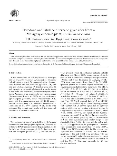
<strong>Clerodane</strong> <strong>and</strong> <strong>labdane</strong> <strong>diterpene</strong> <strong>glycosides</strong> <strong>from</strong> a<br />
<strong>Malagasy</strong> endemic plant, Cussonia racemosa<br />
R.R. Harinantenaina Liva, Ryoji Kasai, Kazuo Yamasaki*<br />
Institute of Pharmaceutical Sciences, Faculty of Medicine, Hiroshima University, 1-2-3 Kasumi, Minami-ku, Hiroshima, 734-8551, Japan<br />
Received 10 October 2001; received in revised form 4 January 2002<br />
Four clerodane <strong>glycosides</strong>, cussosides A–D, <strong>and</strong> one <strong>labdane</strong> glycoside, cussoside E were isolated <strong>from</strong> the dried leaves of Cussonia<br />
racemosa, along with two known flavonol <strong>glycosides</strong> identified as rutin <strong>and</strong> kaempferol rutinoside. The structures of the compounds<br />
were deduced on the basis of their physical <strong>and</strong> spectral data. # 2002 Elsevier Science Ltd. All rights reserved.<br />
Keywords: Araliaceae; Cussonia racemosa; Leaves; Cussosides A–E; <strong>Clerodane</strong>; Labdane; <strong>diterpene</strong> <strong>glycosides</strong>; <strong>Malagasy</strong> endemic plant<br />
1. Introduction<br />
In the continuation of our phytochemical investigation<br />
of Cussonia racemosa (Araliaceae), a <strong>Malagasy</strong><br />
endemic plant, seven (1–7) compounds were obtained,<br />
these were the four new clerodane <strong>glycosides</strong> (3–6) <strong>and</strong><br />
one new <strong>labdane</strong> glycoside (7) together with rutin (1)<br />
<strong>and</strong> kaempferol rutinoside (2) isolated <strong>from</strong> the leaves<br />
of the plant. The presence of clerodane <strong>diterpene</strong>s in the<br />
family Araliaceae is un-common. In our previous paper<br />
(Harinantenaina Liva et al., 2002), six new ent-kaurane<br />
type <strong>diterpene</strong> <strong>glycosides</strong>, named cussoracosides A–F,<br />
along with b-d-glucopyranosyl ent-16ß, 17-dihydroxykauran-19-oate<br />
(Cheng et al., 1993) <strong>and</strong> paniculoside IV<br />
(Yamasaki et al., 1977; Kaneda et al., 1978) were<br />
obtained. The present study deals with the structural<br />
elucidation of the new compounds.<br />
2. Results <strong>and</strong> discussion<br />
The methanol extract of the dried leaves of Cussonia<br />
racemosa on chromatographic separation, followed by<br />
systematic HPLC purification (see Experimental), led to<br />
the isolation of seven compounds (1–7), these were the<br />
five new <strong>diterpene</strong> <strong>glycosides</strong> (3–7) <strong>and</strong> the two fla-<br />
*Corresponding author. Tel.: +81-82-257-5285; fax: +81-82-257-<br />
5289.<br />
E-mail address: yamasaki@pharm.hiroshima-u.ac.jp (K. Yamasaki).<br />
Phytochemistry 60 (2002) 339–343<br />
0031-9422/02/$ - see front matter # 2002 Elsevier Science Ltd. All rights reserved.<br />
PII: S0031-9422(02)00064-X<br />
www.elsevier.com/locate/phytochem<br />
vonol <strong>glycosides</strong> rutin (1) <strong>and</strong> kaempferol rutinoside (2)<br />
(Harborne <strong>and</strong> Mabry, 1982), by comparison of physical<br />
data with literature <strong>and</strong> <strong>from</strong> spectroscopic analyses.<br />
Compound 3, was determined as C 26H 44O 8 by HR–<br />
FAB mass spectrometry. Inspection of the 1 H NMR<br />
spectrum (Table 1) exhibited signals ascribable to a<br />
bicyclic clerodane skeleton: three methyls at 0.72 (3H, s),<br />
0.75 (3H, d, J=5.7 Hz) <strong>and</strong> 1.93 (3H, s), methylene<br />
proton resonances at 4.35 <strong>and</strong> 4.10 (each 1H, d,<br />
J=9.8 Hz), 4.61 (2H, d, J =6.3 Hz) <strong>and</strong> 4.51 (2H, s),<br />
two olefinic protons at 5.32 (1H, br s)<strong>and</strong> 5.90 (2H, t,<br />
J=6.3 Hz), <strong>and</strong> an anomeric proton at 4.85 (d, J=7.8<br />
Hz). The 13 C NMR spectral data of 3 in CD 3OD<br />
(Table 2) indicated the signals of one b-glucopyranosyl<br />
unit together with 20 carbon signals for the aglycone<br />
moiety, which were very similar to those of jewenol A<br />
(3a) previously isolated <strong>from</strong> Portulaca cv Jewel (Ohsaki<br />
et al., 1988). However, the signal of the hydroxymethylene<br />
group at C-18 ( C 64.4) of 3a was replaced by<br />
a signal of one methyl group ( C 20.9) at the 4-position<br />
in 3. The other signals remained similar, except for the<br />
downfield shift of C-19 (+9.6 ppm) <strong>and</strong> the upfield shift<br />
of C-5 ( 0.9 ppm) due to the glucosylation. The complete<br />
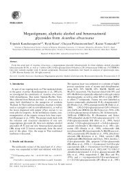
assignments were established by using HSQC,<br />
HMBC, <strong>and</strong> NOE analyses (Fig. 1). The relative configuration<br />
was confirmed as follows. In the NOESY<br />
spectrum, H-19a <strong>and</strong> H-19b were correlated with the<br />
methyl group at C-9 <strong>and</strong> the latter with the methyl group<br />
at C-8. No NOE correlation was observed between H-10<br />
<strong>and</strong> the CH 2-19. Therefore, cussoside A, (3) was<br />
deduced as shown.
340 R.R. Harinantenaina Liva et al. / Phytochemistry 60 (2002) 339–343<br />
Table 1<br />
1 H NMR spectral data for compounds 3–6 (400 MHz in pyridine-d5)<br />
H 3 4 5 6<br />
3 5.32 br s 5.31 br s 5.38 br s 5.27 br s<br />
6 1.45–1.65 a 1.49–1.62 a 1.42–1.60 a 1.46–1.65 a<br />
7a 1.31 m 1.35 m 1.29 m 1.29 m<br />
7b 1.45–1.65 a 1.49–1.62 a 1.42–1.60 a 1.46–1.65 a<br />
8 1.45–1.65 a 1.49–1.62 a 1.42–1.60 a 1.46–1.65 a<br />
10 1.45–1.65 a 1.49–1.62 a 1.42–1.60 a 1.46–1.65 a<br />
11a 1.01–1.04 a 0.95–1.07 a 1.10–1.16 a 1.02–1.05 a<br />
11b 1.01–1.04 a 0.95–1.07 a 1.10–1.16 a 1.02–1.05 a<br />
14 5.90 t (6.3) 5.70 t (6.3) 5.71 t (6.3) 5.80 t(6.3)<br />
15a,b 4.61 d (6.3) 4.60 d (6.3) 4.60 d (6.3) 4.61 d(6.3)<br />
16a,b 4.51 s 4.45 s 4.48 s 4.49 s<br />
17 0.75 d (5.7) 0.74 d (5.8) 0.76 d (5.7) 0.75 d (5.8)<br />
18 1.93 s 1.92 s 1.87 s 1.91 s<br />
19a 4.10 d (9.8) 4.08 d (9.8) 4.09 d (10.5) 4.07 d(10.0)<br />
19b 4.35 d (9.8) 4.32 d (9.8) 4.22 d (10.5) 4.17 d(10.0)<br />
20 0.72 s 0.71 s 0.80 s 0.79 s<br />
19-O -Glc<br />
1 0 4.85 d (7.8) 4.93 d (7.8) 4.90 d (7.8) 4.77 d (7.6)<br />
15-O-Glc 15-O-Api Api<br />
1 00 4.82 d (7.8) 5.49 d (1.8) 5.68 d (1.6)<br />
a Overlapped signals.<br />
Compounds 4 <strong>and</strong> 5 (cussosides B, <strong>and</strong> C) have the<br />
molecular formula C32H54O13 <strong>and</strong> C31H52O12, respectively,<br />
as determined by HR–FAB-mass spectrometry.<br />
The 1 H <strong>and</strong> 13 C NMR spectral data of 4 <strong>and</strong> 5 were<br />
correlated with those of 3, except for the presence of<br />
additional signals due to a b-glucopyranosyl unit ( 4.82,<br />
d, J=7.8 Hz) in 4 <strong>and</strong> a b-apiofuranosyl unit ( 5.49, d,<br />
J=1.8 Hz) in 5. The 13 C NMR spectra of 4 <strong>and</strong> 5<br />
showed the signal of the hydroxymethylene at C-15<br />
shifted downfield ( 65.3 <strong>and</strong> 64.4, respectively) while<br />
that of C-14 was shifted upfield at 122.5, 122.9,<br />
respectively, <strong>and</strong> compared to those of 3. This suggested<br />
that the additional glucopyranosyl or apiofuranosyl<br />
unit must be attached at C-15. HSQC, HMBC, <strong>and</strong><br />
NOESY experiments were carried out in order to confirm<br />
the connectivities <strong>and</strong> the relative stereochemistry.<br />
Thus, the structures of 4 <strong>and</strong> 5 were determined as<br />
shown.<br />
Compound 6, named cussoside D, has the same<br />
molecular formula as 5. The 1 H <strong>and</strong> 13 C NMR spectroscopic<br />
data of 6, which are very similar to 3, suggested<br />
that 6 is a clerodane <strong>diterpene</strong> glycoside with a terminal<br />
b-apiofuranose <strong>and</strong> a 6-linked b-glucopyranose. The<br />
13 C NMR spectral data of the aglycone of 6 are superimposable<br />
with those of 3. However, differences were<br />
observed in the signals due to the sugar moiety. On<br />
going <strong>from</strong> 3 to 6, the carbon signal due to C-6 of the<br />
glucopyranosyl unit was shifted by +6.1 ppm <strong>and</strong> that<br />
of C-5 by 1.0 ppm. The biose was confirmed to be at C-<br />
19 by the observation of a HMBC correlation between<br />
the anomeric proton signal of b-glucopyranose at 4.77<br />
(d, J =7.6 Hz) <strong>and</strong> C-19 carbon signal at 74.9. Based on<br />
these results, 6 could be formulated as shown.<br />
Compound 7, was determined as C31H52O11 by HR–<br />
FAB mass spectrometry. The 1 H NMR (Table 1) spectrum<br />
exhibited signals due to one vinyl group, two tertiary<br />
methyl groups ( 0.75 <strong>and</strong> 1.14), an exocyclic<br />
methylene ( 4.84 <strong>and</strong> 4.54, each brs), a hydroxymethylene<br />
group attached to a quaternary carbon, two<br />
anomeric protons of b-glucopyranosyl <strong>and</strong> b-apiofuranosyl<br />
[at 4.75 (d, J =7.8 Hz) <strong>and</strong> 5.72 (d, J =1.7<br />
Hz), respectively]. The 13 C NMR spectral data indicated<br />
the presence of eleven signals ascribable to one terminal<br />
b-apiofuranosyl <strong>and</strong> one 6-linked b-glucopyranosyl<br />
units as observed in 6, together with twenty signals for<br />
the aglycone, which suggested being a <strong>labdane</strong> <strong>diterpene</strong>.<br />
On comparison of the 13 C NMR spectral data of 7<br />
<strong>and</strong> ent-labda-8(17),13-diene-15,18-diol (8) (Tanaka et<br />
al., 1984), differences were recognized in the chemical<br />
shifts of C-5, C-18, C-19, except the signals due to the<br />
sugars. This data assumed that 7 <strong>and</strong> 8 are C-4 epimer.<br />
Moreover the observation of NOE correlation between<br />
the two methyls at 0.75 <strong>and</strong> 1.14 allowed us to conclude<br />
that the orientation of the hydroxymethylene in 7 was<br />
equatorial, hence a. The allocation of the primary<br />
hydroxyl group to be at C-14 <strong>and</strong> the exocyclic methylene<br />
at C-8(17) was deduced by HSQC, HMBC, <strong>and</strong> NOE<br />
experiments. Based on the above data, the structure of<br />
cussoside E(7) was established as shown.<br />
The absolute configurations of all new isolated compounds<br />
were not determined because of the small<br />
amount of samples. Interestingly, the ent-kaurane <strong>diterpene</strong><br />
<strong>glycosides</strong> previously isolated <strong>from</strong> C. racemosa<br />
could be biogenetically synthesized <strong>from</strong> <strong>labdane</strong>, as<br />
proposed in the literature (Railton et al., 1984), since<br />
compound 7 has been found in this species.
3. Experimental<br />
3.1. General<br />
NMR spectra ( 1 H, 13 C, HSQC, HMBC) were recorded<br />
in pyridine-d 5 using a Jeol JNM A-400 spectrometer<br />
(400 MHz for 1 H NMR <strong>and</strong> 100 MHz for 13 C NMR).<br />
MS were recorded on a Jeol JMS-SX 102 spectrometer.<br />
R.R. Harinantenaina Liva et al. / Phytochemistry 60 (2002) 339–343 341<br />
Fig. 1. Important HMBC ( ) <strong>and</strong> NOE ( ) correlations of 3.<br />
Optical rotations were measured with Union PM-1 digital<br />
polarimeter. Preparative HPLC was carried out on columns<br />
of ODS (150 20 mm i.d., YMC) with a Tosoh<br />
refraction index (RI-8) detector, flow rate 6 ml/min. For<br />
CC, silica gel G 60 (Merck), RP-18 (50 mm, YMC) <strong>and</strong><br />
highly porous copolymer of styrene <strong>and</strong> divinylbenzene<br />
(Mitsubishi Chem. Ind. Co. Ltd) were used. The solvent<br />
systems were: (I) CH2Cl2–MeOH–H2O (17:4:0.5 to 17:8:
342 R.R. Harinantenaina Liva et al. / Phytochemistry 60 (2002) 339–343<br />
2), (II) 20–100% MeOH, (III) 30% CH3CN, (IV) 45%<br />
CH3CN, (V) 50% MeOH, (VI) 70% MeOH. The spray<br />
reagent used for TLC was 10% H2SO4 in 50% EtOH.<br />
3.2. Plant material<br />
Plant material was collected in March 2000 <strong>from</strong><br />
Ranomafana-Ifanadiana, Madagascar. The identity of<br />
the plant was confirmed by Dr. Arm<strong>and</strong> Rakotozafy<br />
<strong>from</strong> Institut Malgache de Recherches Applique´ es. A<br />
voucher specimen (CUSSRAC-LI01) has been deposited<br />
in the Herbarium of the Institute of Pharmaceutical<br />
Sciences, Hiroshima University Faculty of Medicine.<br />
3.3. Extraction <strong>and</strong> isolation<br />
The dried leaves (1.75 kg) of C. racemosa were<br />
extracted with hot MeOH. After removal of the solvent<br />
by evaporation, the residue (200 g) was suspended in<br />
water <strong>and</strong> extracted with hexane <strong>and</strong> EtOAc successively.<br />
The aqueous layer (45.0 g) was subjected to a<br />
column of highly porous copolymer of styrene <strong>and</strong><br />
divinylbenzene, <strong>and</strong> eluted with H 2O, 30% MeOH,<br />
MeOH <strong>and</strong> Me2CO, successively. The fraction eluted<br />
with MeOH was applied to a column of silica gel (system<br />
I), affording eight fractions. Fraction 1 was subjected<br />
to a CC on RP-18 <strong>and</strong> ODS-HPLC using systems<br />
II <strong>and</strong> III, respectively to afford compounds 3 (13.4<br />
mg), 5 (7.4 mg) <strong>and</strong> 7 (12 mg). Fraction 2 was further<br />
separated on column of RP-18 using system II to give 18<br />
fractions. Fractions 2-5 <strong>and</strong> 2-8 were similarly purified<br />
by ODS–HPLC (system V <strong>and</strong> IV, respectively) to<br />
afford compounds 2 (7 mg) <strong>and</strong> 4 (13.7 mg). Fractions<br />
2–11 were purified by ODS–HPLC (system VI) to afford<br />
compound 6 (16.2 mg). Fraction 8 was subjected to a<br />
CC on RP-18 using system II to afford compound 1<br />
(35.1 mg).<br />
3.4. Cussoside A (3)<br />
Amorphous powder. [ ]D 29 = 60.7 (MeOH; c 0.8); 1 H<br />
NMR: Table 1; 13 C NMR: Table 2; Negative HR–<br />
FABMS, m/z: [M–H] 483.2932 (C26H43O8 requires<br />
483.2957).<br />
3.5. Cussoside B (4)<br />
Amorphous powder. [ ]D 29 = 91.8 (MeOH; c 0.7); 1 H<br />
NMR: Table 1; 13 C NMR: Table 2; Negative H–R-<br />
FABMS, m/z: [M–H] 645.3448 (C32H53O13 requires<br />
645.3485).<br />
3.6. Cussoside C (5)<br />
Amorphous powder. [ ] D 29 = 66.6 (MeOH; c 0.2); 1 H<br />
NMR: Table 1 <strong>and</strong> 13 C NMR: Table 2; Negative H–R–<br />
Table 2<br />
13 C NMR spectral data for compounds 3–8 (100 MHz in pyridine-d5)<br />
C 3a a 3 a 3 4 5 6 7 8<br />
1 18.2 18.6 18.0 18.0 18.0 18.0 39.1 38.9<br />
2 27.2 27.5 26.8 26.8 26.9 26.7 19.3 19.3<br />
3 128.8 123.3 122.4 122.4 122.3 122.6 36.3 36.0<br />
4 146.3 143.7 142.9 142.9 142.9 142.6 38.5 38.5<br />
5 43.9 43.0 42.2 42.2 42.5 42.1 56.6 48.5<br />
6 32.1 32.9 32.6 32.6 32.9 32.8 24.7 24.5<br />
7 28.0 28.5 27.9 27.9 27.9 27.9 38.8 38.9<br />
8 37.5 37.7 36.6 36.6 36.7 36.6 148.6 149.0<br />
9 39.8 39.8 38.9 38.9 38.9 38.9 56.3 56.7<br />
10 47.6 47.9 46.8 46.8 46.7 46.8 39.8 39.8<br />
11 38.3 38.4 37.7 37.5 37.5 37.7 22.3 22.4<br />
12 29.1 29.1 28.5 28.3 28.4 28.4 38.7 38.5<br />
13 143.4 143.7 143.0 145.6 145.6 142.6 137.4 137.5<br />
14 127.2 127.3 127.2 122.5 122.9 127.2 125.9 126.0<br />
15 58.6 58.7 58.4 65.3 64.4 58.4 58.9 59.0<br />
16 60.0 60.2 60.0 60.0 60.0 60.0 16.4 16.5<br />
17 16.1 16.3 16.1 16.1 16.1 16.1 106.5 106.5<br />
18 64.4 20.9 21.1 21.2 20.7 21.1 28.1 71.4<br />
19 65.6 75.2 74.4 74.4 74.8 74.9 72.2 18.2<br />
20 19.3 19.2 18.9 18.9 18.8 18.9 15.6 15.2<br />
19-O -Glc<br />
1 0 105.5 105.9 106.0 105.3 105.4 104.9<br />
2 0 75.4 75.5 75.5 75.0 75.3 75.2<br />
3 0 77.9 78.4 78.4 78.4 78.7 78.6<br />
4 0 71.6 71.6 71.6 71.5 71.5 71.8<br />
5 0 78.2 78.7 78.7 78.5 77.7 77.0<br />
6 0 62.7 62.7 62.8 62.6 68.8 68.8<br />
15-O- or6 0 -O- Glc Api Api Api<br />
1 00 103.5 109.3 111.0 110.9<br />
2 00 75.2 78.3 77.7 77.8<br />
3 00 78.4 80.0 80.4 80.4<br />
4 00 71.9 75.0 75.1 75.0<br />
5 00 78.5 65.7 65.7 65.7<br />
6 00 62.8<br />
a Measured in CD3OD.<br />
FABMS, m/z: [M–H] 615.3411 (C 31H 51O 12 requires<br />
615.3380).<br />
3.7. Cussoside D (6)<br />
Amorphous powder. [ ]D 29 = 83.8 (MeOH; c 1.0); 1 H<br />
NMR: Table 1 <strong>and</strong> 13 C NMR: Table 2; Negative HR–<br />
FABMS, m/z: [M–H] 615.3411 (C31H51O12 requires<br />
615.3380).<br />
3.8. Cussoside E (7)<br />
Amorphous powder. [ ]D 29 = 30.5 (MeOH; c 1.6); 1 H<br />
NMR spectral data (pyridine-d5): 5.75 (1H, t, J =6.3<br />
Hz, H-14), 5.72 (1H, d, J =1.7 Hz, H-1 00 ), 4.84 (1H, brs,<br />
H-17a), 4.75 (1H, d, J =7.8 Hz, H-1 0 ), 4.54 (1H, brs, H-<br />
17b), 4.47 (2H, d, J =6.3 Hz, H-15), 4.37 (1H, d, J =9.5<br />
Hz, H-19a), 3.45 (1H, d, J =9.5 Hz, H-19b), 1.67 (3H,<br />
s, 16-CH 3), 1.58 (1H, brd, J =10.25 Hz, H-5), 1.16 (1H,<br />
m, H-9), 1.14 (3H, s, 18-CH3), 0.75 (3H, s, 20-CH3). 13 C
NMR: (Table 2). Negative HR–FABMS, m/z: [M–H]<br />
599.3456 (C31H51O11 requires 599.3431).<br />
Acknowledgements<br />
H.L.R.R. is grateful to the Japanese Ministry of<br />
Education, Culture, Sport, Science <strong>and</strong> Technology for<br />
the award of a scholarship to study for a PhD at the<br />
Institute of Pharmaceutical Sciences, Hiroshima University,<br />
Japan. We also thank the Research Center for<br />
Molecular Medicine, Hiroshima University, School of<br />
Medicine, Japan, for the use of NMR facilities.<br />
References<br />
Cheng, D.-L., Cao, X.-P., Wei, H.-X., He, L., 1993. Kaurane diterpenoids<br />
<strong>from</strong> Aster ageratoides. Phytochemistry 33, 1181–1183.<br />
R.R. Harinantenaina Liva et al. / Phytochemistry 60 (2002) 339–343 343<br />
Harborne, J.B., Mabry, T.J., 1982. The Flavonoids: Advances in<br />
Research. Chapman & Hall, London.<br />
Harinantenaina Liva, R.R., Kasai, R., Yamasaki, K., 2002. Entkaurane<br />
diterpenoid <strong>glycosides</strong> <strong>from</strong> the leaves of Cussonia racemosa,<br />
a <strong>Malagasy</strong> endemic plant. Chemical <strong>and</strong> Pharmaceutical<br />
Bulletin 50, 268–271.<br />
Kaneda, N., Kohda, H., Yamasaki, K., Tanaka, O., Nishi, K., 1978.<br />
Paniculosides-IV, <strong>diterpene</strong>-glucosides <strong>from</strong> Stevia ovata Lag. Chemical<br />
<strong>and</strong> Pharmaceutical Bulletin 26, 2266–2267.<br />
Ohsaki, A., Ohno, N., Shibata, K., Tokoroyama, T., Kubota, T.,<br />
Hirotsu, K., Higuchi, T., 1988. Minor diterpenoids <strong>from</strong> Portulaca<br />
cv Jewel. Phytochemistry 27, 2171–2173.<br />
Railton, I.D., Fellows, B., West, C.A., 1984. Ent-kaurene synthesis in<br />
chloroplasts <strong>from</strong> higher plants. Phytochemistry 23, 1261–1267.<br />
Tanaka, T., Kawamura, K., Kitahara, T., Kohda, H., Tanaka, O.,<br />
1984. Ent-<strong>labdane</strong>-type <strong>diterpene</strong> glucosides <strong>from</strong> leaves of Rubus<br />
chingii. Phytochemistry 23, 615–621.<br />
Yamasaki, K., Kohda, H., Kobayashi, T., Kaneda, N., Kasai, R.,<br />
Tanaka, O., Nishi, K., 1977. Application of 13 C NMR spectroscopy<br />
to chemistry of <strong>glycosides</strong>: Structures of Paniculosides-I, -II, -III,<br />
-IV, <strong>and</strong> -V, <strong>diterpene</strong> glucosides of Stevia paniculata Lac. Chemical<br />
<strong>and</strong> Pharmaceutical Bulletin 25, 2895–2899.