The Characterization of Hyalochlorella marina gen. et ... - Microbiology
The Characterization of Hyalochlorella marina gen. et ... - Microbiology
The Characterization of Hyalochlorella marina gen. et ... - Microbiology

Create successful ePaper yourself
Turn your PDF publications into a flip-book with our unique Google optimized e-Paper software.
<strong>Characterization</strong> <strong>of</strong> <strong>Hyalochlorella</strong> <strong>marina</strong> I79<br />
cytokinesis, one would expect the curves to be non-superimposable with a high<br />
narrow peak in nuclear distribution at one nucleus. <strong>The</strong> distribution curves were<br />
roughly superimposable (Fig. 2), indicating that nuclear division did indeed accompany<br />
growth.<br />
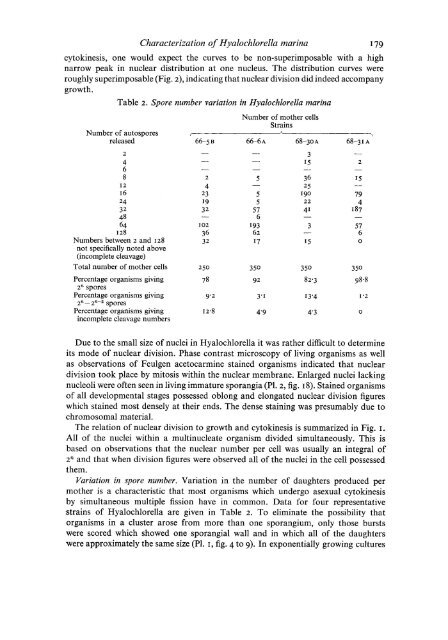
Table 2. Spore number variation in <strong>Hyalochlorella</strong> <strong>marina</strong><br />
Number <strong>of</strong> mother cells<br />
Strains<br />
Number <strong>of</strong> autospores I h -l<br />
released 66-5 B 66-6 A 68-30 A 68-31 A<br />
2<br />
4<br />
6<br />
8<br />
I2<br />
16<br />
24<br />
32<br />
48<br />
64<br />
I 28<br />
Numbers b<strong>et</strong>ween 2 and 128<br />
not specifically noted above<br />
(incompl<strong>et</strong>e cleavage)<br />
2<br />
4<br />
23<br />
19<br />
32<br />
-<br />
I02<br />
36<br />
32<br />
5<br />
-<br />
5<br />
5<br />
57<br />
6<br />
I93<br />
62<br />
I7<br />
3<br />
I5<br />
2<br />
I5<br />
-<br />
79<br />
4<br />
187<br />
Total number <strong>of</strong> mother cells 250 350 350 3 50<br />
Percentage organisms giving<br />
2n spores<br />
78 92 82.3 98.8<br />
Percentage organisms giving 9.2 3-1 13'4 1'2<br />
2n - 2n-2 spores<br />
Percentage organisms giving I 2.8 4'9 4'3 0<br />
incompl<strong>et</strong>e cleavage numbers<br />
Due to the small size <strong>of</strong> nuclei in <strong>Hyalochlorella</strong> it was rather difficult to d<strong>et</strong>ermine<br />
its mode <strong>of</strong> nuclear division. Phase contrast microscopy <strong>of</strong> living organisms as well<br />
as observations <strong>of</strong> Feul<strong>gen</strong> ac<strong>et</strong>ocarmine stained organisms indicated that nuclear<br />
division took place by mitosis within the nuclear membrane. Enlarged nuclei lacking<br />
nucleoli were <strong>of</strong>ten seen in living immature sporangia (Pl. 2, fig. IS). Stained organisms<br />
<strong>of</strong> all developmental stages possessed oblong and elongated nuclear division figures<br />
which stained most densely at their ends. <strong>The</strong> dense staining was presumably due to<br />
chromosomal material.<br />
<strong>The</strong> relation <strong>of</strong> nuclear division to growth and cytokinesis is summarized in Fig. I.<br />
All <strong>of</strong> the nuclei within a multinucleate organism divided simultaneously. This is<br />
based on observations that the nuclear number per cell was usually an integral <strong>of</strong><br />
2n and that when division figures were observed all <strong>of</strong> the nuclei in the cell possessed<br />
them.<br />
Variation in spore number. Variation in the number <strong>of</strong> daughters produced per<br />
mother is a characteristic that most organisms which undergo asexual cytokinesis<br />
by simultaneous multiple fission have in common. Data for four representative<br />
strains <strong>of</strong> <strong>Hyalochlorella</strong> are given in Table 2. To eliminate the possibility that<br />
organisms in a cluster arose from more than one sporangium, only those bursts<br />
were scored which showed one sporangial wall and in which all <strong>of</strong> the daughters<br />
were approximately the same size (Pl. I, fig. 4 to 9). In exponentially growing cultures<br />
57<br />
6<br />
0