Peptide substrates for chymosin (rennin)
Peptide substrates for chymosin (rennin)
Peptide substrates for chymosin (rennin)

Create successful ePaper yourself
Turn your PDF publications into a flip-book with our unique Google optimized e-Paper software.
Biochem. J. (1987) 244, 553-558 (Printed in Great Britain)<br />
<strong>Peptide</strong> <strong>substrates</strong> <strong>for</strong> <strong>chymosin</strong> (<strong>rennin</strong>)<br />
Interaction sites in i-casein-related sequences located outside the (103-108)-hexapeptide region that<br />
fits into the enzyme's active-site cleft<br />
Servaas VISSER,* Charles J. SLANGEN and Peter J. VAN ROOIJEN<br />
Department of Biophysical Chemistry, Netherlands Institute <strong>for</strong> Dairy Research, P.O. Box 20, 6710 BA Ede,<br />
Netherlands<br />
The role of individual amino acid residues in the 98-102 and 111-112 regions of bovine K-casein in its<br />
interaction with the milk-clotting enzyme <strong>chymosin</strong> (<strong>rennin</strong>) was investigated. To this end the tryptic 98-112<br />
fragment of K-casein was modified in its N- and/or C-terminal part by chemical (guanidation,<br />
ethoxy<strong>for</strong>mylation, repeated Edman degradation) and enzymic (carboxypeptidase) treatments. Further, use<br />
was made of short synthetic K-casein analogues in which His-102 had been replaced by Pro or Lys. All<br />
peptides and their derivatives were tested comparatively at various pH values <strong>for</strong> their ability to act as<br />
<strong>chymosin</strong> <strong>substrates</strong> via specific cleavage of the peptide bond at position 105-106. The results indicate that<br />
in the alternating 98-102 sequence (His-Pro-His-Pro-His) the His as well as the Pro residues contribute to<br />
the substrate activity with no predominant role of any one of these groups. Another interaction site is <strong>for</strong>med<br />
by the Lys residue at position 111 of the substrate. A model of the enzyme-substrate complex is proposed.<br />
Herein the 103-108 fragment of the substrate, to be accommodated within the enzyme's active-site cleft, is<br />
brought into position by electrostatic binding (via His-98, His-100, His-102 and Lys-1 11) near the entrance<br />
of the cleft. These interactions are strongly supported by Pro residues at positions 99, 101, 109 and 110 of<br />
the substrate, which act as stabilizers of the proper con<strong>for</strong>mation of the substrate in the enzyme-substrate<br />
complex.<br />
INTRODUCTION<br />
The specific cleavage of K-casein by <strong>chymosin</strong> (<strong>rennin</strong>,<br />
EC 3.4.23.4), which <strong>for</strong>ms the first step in the<br />
milk-clotting process, has been the subject of numerous<br />
investigations (<strong>for</strong> a review see Dalgleish, 1982).<br />
Valuable in<strong>for</strong>mation as to the importance of individual<br />
amino acid side chains in the vicinity of the <strong>chymosin</strong>sensitive<br />
Phe-Met bond of K-casein was obtained from<br />
kinetic studies carried out at pH 4.7 with short, mostly<br />
synthetic, peptide <strong>substrates</strong> (Schattenkerk et al., 1971;<br />
Raymond et al., 1972; Visser et al., 1976, 1977, 1980;<br />
Raymond & Bricas, 1979; Visser, 1981). Computermodelling<br />
studies of the three-dimensional structure of<br />
<strong>chymosin</strong> not only supported the general conclusions of<br />
these studies, but also provided more detailed in<strong>for</strong>mation<br />
on the possible way of positioning of the various<br />
substrate residues within the enzyme's active-site cleft<br />
(B. L. Sibanda & T. L. Blundell, unpublished work). The<br />
amino acid sequence of the region around the cleavable<br />
Phe-Met bond at position 105-106 of bovine K-casein is<br />
as follows (Mercier et al., 1973):<br />
at pH 4.7 and 30 °C). Lengthening of the peptide chain<br />
at the C-terminal end did increase the substrate capacity<br />
to some extent [<strong>for</strong> K(103-112) kcat./Km = 67 mm-' s-.],<br />
but a 30-fold improvement was obtained by addition of<br />
the 98-102 region to the sequence, bringing the substrate<br />
properties of K(98-112) close to those of whole K-casein<br />
(Visser et al., 1980). In those studies no attempts were<br />
made to assign individual residues within this 98-102<br />
sequence as being particularly responsible <strong>for</strong> the large<br />
jump in substrate quality.<br />
Results from chemical modification and photooxidation<br />
experiments carried out on whole K-casein<br />
(Hill & Laing, 1965; Kaye & Jolles, 1978) suggested a<br />
particular role of one or more of the three His residues<br />
(which are all located in the 98-102 fragment) in the<br />
<strong>for</strong>mation of the enzyme-substrate complex. From the<br />
results of ionic-strength-dependence experiments done<br />
with whole K-casein (Payens & Both, 1980) and with the<br />
K(98-112) fragment (Visser et al., 1980), it was<br />
postulated that the positive charges at both sides of the<br />
Phe-Met bond (i.e. His and Lys residues) could be of<br />
-His-Pro-His-Pro-His-Leu-Ser-Phe-Met-Ala-Ile-Pro-Pro-Lys-Lys-<br />
98 100 102 104 106 108 110 112<br />
From kinetic studies by Visser et al. (1976) it was<br />
concluded that K(103-108)OMe represents the smallest<br />
peptide analogue of K-casein with good substrate<br />
properties towards <strong>chymosin</strong> (kcat /Km = 22 mM-' s-1<br />
Vol. 244<br />
importance <strong>for</strong> electrostatic enzyme-substrate interactions<br />
(see also Payens & Visser, 1981).<br />
In the present study we have focused on the<br />
Abbreviations used: K(nfl-nfi), the ni-ntj sequence of bovine K-casein; OMe, methyl ester; Har, homoarginine; Nle, norleucine; NAc-His,<br />
NO-acetylhistidine; DEP, diethyl pyrocarbonate (ethoxy<strong>for</strong>mic anhydride).<br />
* To whom correspondence should be addressed.<br />
553
554<br />
contribution to the <strong>for</strong>mation of the enzyme-substrate<br />
complex of individual residues in the 98-102 region of the<br />
substrate and also of the Lys residues at positions 111<br />
and 112. The latter <strong>for</strong>m the nearest charged residues at<br />
the C-terminal side of the Phe-Met bond in K-casein. In<br />
this study we used small synthetic K-casein fragments<br />
having substitutions at the 102 position and also the<br />
98-112 tryptic fragment (Visser et al., 1980) that had<br />
been chemically or enzymically modified in the Nand/or<br />
C-terminal part. Both the 98-102 and the<br />
111-112 sequences are expected to include potential sites<br />
<strong>for</strong> interaction with counterparts outside but near the<br />
edge of the enzyme's active-site cleft.<br />
EXPERIMENTAL<br />
Materials<br />
The peptide <strong>substrates</strong> K(101-108), K(102-108),<br />
K(103-108), K(103-110) and K(103-112) or analogues of<br />
these sequences were synthesized by 'solution methods'<br />
as described by Schattenkerk et al. (1973). They were<br />
found to be homogeneous by t.l.c. The tryptic K(98-111)<br />
and K(98-112) fragments, isolated as described by Visser<br />
et al. (1980), were used as starting material <strong>for</strong> the<br />
modification experiments described below.<br />
The <strong>chymosin</strong> preparation was the same as the one<br />
used in previous studies (Visser et al., 1976, 1977, 1980);<br />
its proteolytic activity towards Leu-Ser-Phe-Nle-Ala-<br />
IleOMe as determined by u.v. spectrophotometry at<br />
230 nm under specified conditions (Visser & Rollema,<br />
1986) amounted to 540 mkat/kg.<br />
C-Terminal chain-shortening: preparation of K(98-110)<br />
A solution of 3.2 mg of carboxypeptidase B (71<br />
units/mg; Worthington Biochemical Corp.) in 2.3 ml of<br />
0.02 M-ammonium bicarbonate buffer, pH 8.2, was<br />
incubated with 5 1l of di-isopropyl phosphorofluoridate<br />
(Fluka) <strong>for</strong> 24 h at room temperature. Then a solution of<br />
100 mg of K(98-l 11) or K(98-112) in 20 ml of the above<br />
pH 8.2 buffer was added and the reaction mixture was<br />
kept at 30 °C <strong>for</strong> 24 h. After being freeze-dried, the<br />
product was taken up with 20 ml of water and the<br />
solution was centrifuged. The supernatant was, in<br />
approx. 0.5 ml portions, applied to a number of Sep-Pak<br />
C18 cartridges (Waters Associates) that had been<br />
pre-washed with methanol and water. The Sep-Pak<br />
columns were eluted with 5 ml each of 15%, 50 %,9 750%<br />
and 1000% methanol successively. The combined 50% -<br />
methanol and 75% -methanol fractions were, after<br />
evaporation and freeze-drying, re-chromatographed on<br />
Sep-Pak C18 in the same way. The fraction eluted with<br />
5000 methanol or (dependent on the recovery and the<br />
purity judged by t.l.c.) the combined 500% -methanol and<br />
75 O%-methanol fractions were used <strong>for</strong> analysis and <strong>for</strong><br />
the preparation of other <strong>substrates</strong> via N-terminal<br />
chain-shortening.<br />
N-Terminal chain-shortening: preparation of K(99-110),<br />
K(100-110), K(101-110) and K(102-110)<br />
The K(98- 1I0) tridecapeptide (10-15 mg) obtained by<br />
the above procedure was used as starting material <strong>for</strong><br />
one, two, three or four Edman degradation steps. Each<br />
step consisted of the following procedures.<br />
Coupling. A solution of the peptide in 1 ml of buffer<br />
(benzyldimethylamine/propan-l-ol/water, 3:10:12, by<br />
S. Visser, C. J. Slangen and P. J. van Rooijen<br />
vol., adjusted to pH 9.4 with acetic acid and kept under<br />
N2) was flushed with N2 in a vessel as described by Tarr<br />
(1975, 1977). Then 155 4td of phenyl isothiocyanate<br />
(Sequenal grade; Pierce Chemical Co.) was added at<br />
50 °C and the solution was kept at this temperature <strong>for</strong><br />
40 min with occasional shaking (every 5 min). Finally,<br />
the reaction mixture was dried at 50 °C by flushing with<br />
N2. Cleavage. The phenyl isothiocyanate derivative was<br />
taken up with 1 ml of anhydrous trifluoroacetic acid<br />
(Eastman). After 10 min at 50 °C the reaction mixture<br />
was dried at 50 °C by flushing with N2.<br />
Extraction. The residue of the cleavage reaction was<br />
taken up with a mixture of 200,l of water and 100 ,l of<br />
pyridine, and extracted twice with 500,1 of benzene/<br />
ethyl acetate (1: 1, v/v) or heptane/ethyl acetate<br />
(1: 1, v/v). The aqueous layer was evaporated to dryness.<br />
After the last degradation step the product obtained<br />
was purified. To this end the residue of the extraction<br />
procedure was taken up with 1 ml of water and applied<br />
to a Sep-Pak C18 cartridge. Elution was then carried out<br />
with water (1 ml) and with 5-6 ml each of 25%, 5000,<br />
75% and 100% methanol successively. The methanol<br />
fractions were evaporated to dryness under vacuum and<br />
checked <strong>for</strong> purity by t.l.c. The 50%-methanol fraction<br />
was chosen <strong>for</strong> amino acid analysis and kinetic<br />
measurements.<br />
Modification of lysine residues: preparation of<br />
K(98-112, Harl,' 112)<br />
2-Methylisourea (in the <strong>for</strong>m of its sulphate; Fluka)<br />
was converted into its free base by adding 875 mg in<br />
small portions to 20 ml of a saturated aqueous solution<br />
of Ba(OH)2 with stirring, which resulted in a pH of 10.3.<br />
The precipitate <strong>for</strong>med was centrifuged off and discarded.<br />
To 2.6 ml of the freshly prepared reagent was added<br />
20 mg of K(98-112) and the solution was kept at 5 °C <strong>for</strong><br />
3 days (Kimmel, 1967). The reaction mixture was then<br />
acidified to pH 5.4 by the addition of acetic acid, and<br />
the guanidated peptide was purified by ion-exchange<br />
chromatography on a 13 cm x 1 cm column of SP-<br />
(sulphopropyl-)Sephadex C-25 (Pharmacia) by using<br />
a gradient from 0.30 to 0.75 M-ammonium acetate<br />
buffer, pH 5.4, essentially as described <strong>for</strong> K(98-112) by<br />
Visser et al. (1980). After repeated freeze-drying of the<br />
appropriate column fraction, the purity of the end<br />
product (yield 14 mg) was checked by t.l.c. and by amino<br />
acid analysis.<br />
Modification of histidine residues: preparation of<br />
ethoxy<strong>for</strong>mylated K(98-112)<br />
Ethoxy<strong>for</strong>mylation was per<strong>for</strong>med by reaction with<br />
DEP (Sigma Chemical Co.) and monitoring the progress<br />
of the reaction via the increase in absorbance at 240 nm<br />
(Ova'di et al., 1967; Miihlrad et al., 1967). The<br />
concentration of DEP in a stock solution was determined<br />
by its reaction (approx. 60 min at room temperature)<br />
with an excess of NAc-His dissolved in 0.05 M-sodium<br />
acetate buffer, pH 5.5. The molar absorptivity of the<br />
reaction product at 240 nm was derived from a series of<br />
incubations of NAc-His with increasing concentrations<br />
of DEP (DEP/NAc-His molar ratio 0.7-14: 1). The<br />
absorbance measured after 15 h at room temperature in<br />
the above pH 5.5 buffer was <strong>for</strong> each incubation<br />
essentially the same as the one obtained after 1.5 h of<br />
reaction. The molar absorptivity was found to reach a<br />
1987
<strong>Peptide</strong> <strong>substrates</strong> <strong>for</strong> <strong>chymosin</strong> (<strong>rennin</strong>)<br />
- maximum of approx. 4.0 x 10³ M-1 cm-1 at a DEP/NAc-<br />
His molar ratio of 12-14:1.<br />
Modification of K(98-112) was carried out as follows.<br />
To 6.5 ml portions of the peptide solution (67 /M) in<br />
0.05 M-sodium acetate buffer, pH 5.5, was added 152 ,ul<br />
of ethanolic DEP solution of appropriate concentration.<br />
The final DEP/K(98-112) molar ratio varied from 2.2: 1<br />
to 45: 1, which corresponds to 0.7-15 mol of DEP per<br />
equivalent of His. After 1.5 h and again after 15 h at<br />
room temperature, duplicate samples were drawn <strong>for</strong> the<br />
determination of the amino acid content, the percentage<br />
modification and the initial cleavage by the action of<br />
<strong>chymosin</strong>.<br />
T.l.c.<br />
Samples were applied to ready-to-use silica gel (Merck<br />
F254, thickness 0.25 mm) or cellulose (Merck, thickness<br />
0.1 mm) plates. After development in butan-1-ol/acetic<br />
acid/pyridine/water (15:3:10:12, by vol.) the chromatograms<br />
were dried and sprayed with ninhydrin/collidine<br />
or with diazotized sulphanilic acid (Pauly reagent). The<br />
latter staining procedure is specific <strong>for</strong> histidine residues<br />
in the peptides concerned.<br />
Amino acid analysis<br />
Samples were hydrolysed with 6 M-HCI at 110 °C <strong>for</strong><br />
24 h and 96 h in sealed evacuated tubes. Duplicate runs<br />
were made on a JEOL-5AH amino acid analyser with<br />
L-norleucine and L-2-amino-3-guanidinopropionic acid<br />
as internal standards <strong>for</strong> analysis on the long column and<br />
the short column respectively.<br />
Kinetic measurements<br />
The kinetics of enzymic cleavage were determined at<br />
three pH values: (a) at pH 4.7, in order to facilitate<br />
comparison with results obtained previously (Visser,<br />
1981) with other <strong>substrates</strong>, (b) at pH 6.6, i.e. the pH of<br />
milk, and (c) at an intermediate pH expected to be<br />
Table 1. Amino acid composition of some peptides tested as <strong>substrates</strong> <strong>for</strong> <strong>chymosin</strong><br />
situated at or close to the optimum value <strong>for</strong> the cleavage<br />
of the substrate in question ('apparent pH optimum').<br />
To check the general per<strong>for</strong>mance and reproducibility<br />
of the method, the substrate Leu-Ser-Phe-Nle-Ala-<br />
IleOMe was included as internal standard in each series<br />
of kinetic measurements (pH 4.7).<br />
Initial-rate measurements and data processing were<br />
per<strong>for</strong>med as described in previous papers (Visser et al.,<br />
1976, 1980; Vreeman et al., 1977). Apparent pH optima<br />
were derived from theoretical bell-shaped curves fitted to<br />
experimental data expressing initial reaction rates as a<br />
function of pH determined at relatively low substrate<br />
concentrations (i.e. below the Km value).<br />
RESULTS<br />
Analysis of purity of <strong>substrates</strong><br />
The results of amino acid analysis and t.l.c., used as a<br />
check of the efficiency of preparative Edman degradation,<br />
guanidation and ethoxy<strong>for</strong>mylation, are shown in Table<br />
1 and Fig. 1 respectively. It is seen that according to these<br />
criteria the various <strong>substrates</strong> are sufficiently pure, except<br />
K(102-1 10), which is obviously contaminated with some<br />
remaining K(I0l-11 0). Nevertheless, we have included<br />
this preparation in our comparative investigations of the<br />
effect of chain-shortening on the kinetics of <strong>chymosin</strong><br />
action.<br />
Kinetic parameters<br />
Influence of chain-shortening and of guanidation. The<br />
kinetic parameters of <strong>chymosin</strong> action on enzymically or<br />
chemically modified K-casein fragments are listed in<br />
Table 2. With this series of experiments it was aimed to<br />
study the influence of chain-shortening at either end of<br />
K(98-112) on the kinetics. It was presumed that <strong>for</strong> a<br />
proper comparison of kinetic parameters at any pH (<strong>for</strong><br />
instance at pH 4.7 or at the 'physiological' pH of 6.6) the<br />
(apparent) pH optima <strong>for</strong> the <strong>substrates</strong> in question<br />
Data are averaged results from 24 h and 96 h hydrolysates (6 M-HCI, 110 °C) except <strong>for</strong> serine and isoleucine, <strong>for</strong> which values<br />
from 24 h and 96 h hydrolysates respectively are reported. Numbers <strong>for</strong> guanidated K(98-112) were from a single analysis (24 h<br />
hydrolysate).<br />
Amino acid composition (mol of residue/mol)<br />
K(98-112)<br />
Amino ethoxy- K(98-1 12)<br />
acid K(98-l 12)* <strong>for</strong>mylatedt guanidated K(98-111)* K(98-1 10) K(99-110) K(100- 110) K(101-1 10) K(102-1 10)<br />
Ser<br />
Pro<br />
Ala<br />
Met<br />
Ile<br />
Leu<br />
Phe<br />
Lys<br />
His<br />
Har<br />
1.0<br />
4.0<br />
1.0<br />
1.0<br />
1.0<br />
1.0<br />
1.1<br />
2.0<br />
2.9<br />
0.9 0.9 0.9 0.9 0.9 0.9 0.9<br />
4.0 3.9 3.8 4.0 2.9 3.0 2.5<br />
1.0 0.9 1.0 1.0 1.0 1.0 1.0<br />
0.9 0.9 1.0 1.0 1.0 0.9 1.0<br />
1.0 0.9 1.0 1.0 1.0 1.0 1.0<br />
1.0 1.0 1.0 1.0 1.0 1.1 1.0<br />
0.9 0.9 1.0 1.0 1.0 1.0 1.0<br />
2.0 2.0 - 1.1 - - - - -<br />
2.9 2.1 3.0 3.0 2.9 1.9 1.9 1.2 1.0<br />
- - - 2.1<br />
* Used as starting material. Data are from Visser et al. (1980).<br />
t Modified as described in the Experimental section, taking DEP/His ratios of 2.2:1 (left-hand column) and 15:1 (right-hand<br />
column). Only runs on the short column of the analyser (resulting in Lys and His values) were made.<br />
Vol. 244<br />
555
556<br />
; ~ ~~~~~~~~ ~<br />
.... .. .... .... .. ..................<br />
.. ... ..<br />
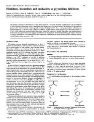
Fig. 1. Silica-gel t.l.c. patterns of peptide fragments obtained<br />
from bovine ic-asein by enzymic and chemical<br />
procedures<br />
Staining was with Pauly reagent. For further experimental<br />
details see the text. Lane 1, K(98-l 12, Har'11'112); lane 2,<br />
K(98-ll2); lane 3, K(98-lll); lane 4, K(98-llO); lane 5,<br />
K(99-llO); lane 6, K(lOO-llO); lane 7, K(lOl-llO); lane 8,<br />
K(lO2-l1 0).<br />
should be about the same. If the latter is not the case, a<br />
comparison of parameters exclusively near the optimum<br />
pH itself seems a better criterion. From Table 2 it is seen<br />
that guanidation of Lys-l11 and Lys-1 12 (leading to a<br />
lengthening and a higher pK value of the side chain)<br />
does not greatly influence the kinetics of<strong>chymosin</strong> action.<br />
The same holds <strong>for</strong> the removal of Lys-1 12 from<br />
the C-terminal position. The presence of Lys-1 11 in the<br />
peptide chain seems to be more important, since<br />
elimination of this residue clearly decreases the substrate<br />
capacity (as mentioned above, in this case one should<br />
rather take into account exclusively the results obtained<br />
at the apparent pH optimum). It cannot be excluded,<br />
however, that, if we had replaced Lys-1 11 in K(98-1 12)<br />
by a non-positive residue, its binding function would<br />
have been (partly) taken over by Lys-1 12. Further<br />
chain-shortening from the N-terminal side, i.e. going<br />
from K(98-110) to K(102-1 10) in Table 2, results in a<br />
gradual decrease of kcat./Km, predominantly caused by<br />
an increasing Km. The proteolytic constant of<br />
320 mM-' *s-. found <strong>for</strong> K(102-1 10) at pH 4.7 approaches<br />
the value of 105 mm-' s-' obtained previously (Visser<br />
et al., 1976) <strong>for</strong> the synthetic substrate K(103-1 IO)OMe.<br />
The latter substrate represents one of the two longest and<br />
most susceptible peptide <strong>substrates</strong> found in that study<br />
under the same experimental conditions as the present<br />
ones. The results given in Table 2 do not suggest a<br />
predominant effect of one particular residue in the<br />
98-101 region. It rather seems that the two His and the<br />
two Pro residues that were chemically removed from the<br />
peptide chain contribute to roughly the same extent to<br />
the overall substrate capacity.<br />
t<br />
Role of His-102. The possible role of His-102 in the<br />
enzyme-substrate complex was studied by testing some<br />
synthetic <strong>substrates</strong> in which Lys or Pro had been<br />
substituted at this position. The results of comparative<br />
measurements, carried out at pH 4.7 as well as at the<br />
apparent pH optimum, are shown in Table 3. In these<br />
experiments it was presumed that the effect on the<br />
kinetics at pH 4.7 of a Met -- Nle replacement is the same<br />
<strong>for</strong> the hepta- and octa-peptide esters (Table 3, nos. 3<br />
and 7) as <strong>for</strong> the hexapeptide esters (cf. nos. 1 and 2). It<br />
can be concluded that the replacement of His by Pro at<br />
I 0<br />
w .a<br />
C<br />
ci<br />
*1<br />
=<br />
vi<br />
;-b<br />
no<br />
0<br />
n<br />
I<br />
a<br />
0I' 4lb'<br />
0<br />
vi<br />
I<br />
*g<br />
a<br />
*<br />
+l<br />
vi<br />
4<br />
4)<br />
._<br />
0<br />
0<br />
.0<br />
0<br />
0<br />
-0 10<br />
0<br />
0<br />
0<br />
*5<br />
et<br />
c0<br />
cd<br />
0<br />
0d<br />
.0<br />
.<br />
"0<br />
cd<br />
$<br />
0<br />
4)<br />
30<br />
0^<br />
04<br />
0~<br />
0.<br />
0<br />
ao4<br />
0. sd<br />
S. Visser, C. J. Slangen and P. J. van Rooijen<br />
4)A Ct<br />
Cd<br />
.0<br />
U,<br />
0 _<br />
+1 +1<br />
88<br />
- _<br />
_ -4<br />
0<br />
0 o<br />
66<br />
+1 +1<br />
C7% en<br />
O0<br />
+1+l<br />
cn C<br />
L _ ,<br />
0 80<br />
en - " 00 enf-<br />
+1 +1 +1 +1 +1 +1 +1<br />
0 0N_t-t en tr -<br />
- -Ol-N t0%<br />
_n _- ~e<br />
0<br />
+1<br />
00<br />
o.<br />
0<br />
6 +1<br />
Il<br />
666 6C56o<br />
+l +1 +1 +1 +1 +1<br />
O NCl C 00 Cl 0<br />
o 0% Co -<br />
'( '4<br />
00Q<br />
N- '4% '1 0 '% all<br />
-; Cl -4 Cl Cl 0<br />
+l +1 +1 +1 +1 +1<br />
N*0 *Ile I e<br />
sQ t- w 0 0~ wv<br />
11 'It t I'l en<br />
r- r- r- r - - qT "<br />
r 00 L0<br />
_ d en<br />
+l +1 +1+1++<br />
o.~~~~~~~~~~~~~W<br />
o~~~~~<br />
8<br />
+1<br />
-<br />
0-<br />
W)<br />
0<br />
't _1 -.. _ _<br />
S" 00 00<br />
0 0000o<br />
0 0000_<br />
+1 +l +l +l +l<br />
0 0CoooNZZ<br />
o -Clo-Sf<br />
- Cl -" C el N<br />
+1 +I +I+I+I+I aj<br />
W'4) Wf) It W4% en<br />
01%<br />
00<br />
o O~<br />
++.I+-<br />
"t en _<br />
+l +1 +1 +1 +1<br />
Sf)<br />
N Cb 1.0 N 00 00_O-<br />
.- Cl4<br />
Cl4 Cl4<br />
+l +1 +1 +1 +1<br />
0 a en e 00<br />
66600(<br />
+l +1 +1 +1 +1<br />
CO 0 00 o<br />
O_- ( C<br />
00 N 00 N N<br />
00 00 _ _-<br />
"0<br />
5-4<br />
10<br />
"0<br />
00<br />
Cl<br />
00<br />
0N<br />
ON C) s CDI<br />
Sf 00ClN'0 -<br />
O<br />
0<br />
+l<br />
0000' O 0CO, en 0C<br />
_4 00 % _ C<br />
+l +l +l +l +l +l<br />
No _<br />
W r O oN<br />
1-i<br />
<strong>Peptide</strong> <strong>substrates</strong> <strong>for</strong> <strong>chymosin</strong> (<strong>rennin</strong>)<br />
Table 3. Kinetic parameters (±S.E.L) of chyMosin action on synthetic analogues of the K-casein sequences 103-108, 102-108 and<br />
101-108: influence of substitutions at position 102 of the sequence<br />
Measurements were carried out at pH 4.7 and at the individual 'apparent' pH optimum (30 °C, I 0.05).<br />
Substrate<br />
[SI0 kcat Km kcat./Km<br />
No. 101 102 103 104 105 106 107 108 pH (mM) (s-') (mM) (mM-1 s-1)<br />
2<br />
3<br />
4<br />
5<br />
His -<br />
[His-<br />
Lys -<br />
Leu - Ser - Phe - Met - Ala - IleOMe*<br />
Leu - Ser - Phe - Nle - Ala - IeOMet<br />
Leu -<br />
Leu -<br />
Leu -<br />
Ser - Phe -<br />
Ser - Phe -<br />
Ser - Phe -<br />
Met -<br />
Nle -<br />
Nle -<br />
Ala - IleOMe*<br />
Ala - IleOMe<br />
Ala - IleOMe<br />
6 Pro- Leu - Ser - Phe - Nle - Ala - IleOMe<br />
7 Pro- His- Leu - Ser - Phe - Met- Ala - IleOMe*<br />
8 [Pro- His- Leu - Ser - Phe - Nle - Ala - IleOMe<br />
9 Pro- Pro- Leu - Ser - Phe - Nle - Ala - IleOMe<br />
4.7<br />
4.7<br />
3.9t<br />
4.7<br />
4.7<br />
4.7<br />
5.7t<br />
4.7<br />
4.2t<br />
4.7<br />
5.1t<br />
4.7<br />
4.7<br />
4.1t<br />
0.10-0.80 18.3+0.9<br />
0.09-0.57 23.7+0.7<br />
0.08-0.69 29.4+ 1.1<br />
0.09-0.65 16.0+0.8<br />
21<br />
0.12-0.76 6.1 +0.2<br />
0.10-0.84 9.1+0.3<br />
0.13-1.08 32.1+0.8<br />
0.12-0.93 15.5+0.2<br />
0.09-0.71 32.3 +0.5<br />
0.15-0.62 43.6+0.6<br />
42<br />
0.03-0.24 52.0+1.4<br />
0.04-0.30 57.1 +0.6<br />
* Results obtained at pH 4.7 taken from Visser et al. (1976).<br />
t Apparent pH optimum (see the text).<br />
t Also used as internal standard substrate in this series (pH 4.7). See also Table 2.<br />
§ Kinetic parameters assumed to differ from those of the preceding substrate by a factor equal to that obtained <strong>for</strong> the Met-* Nle<br />
replacement in the hexapeptide ester (cf. <strong>substrates</strong> nos. 1 and 2).<br />
position 102 does not decrease the kcat./Km value but at<br />
both pH values results in good to excellent substrate<br />
properties (Table 3, nos. 6 and 9). In contrast,<br />
replacement of His-102 by Lys leads to a diminished<br />
substrate behaviour (Table 3, no. 5).<br />
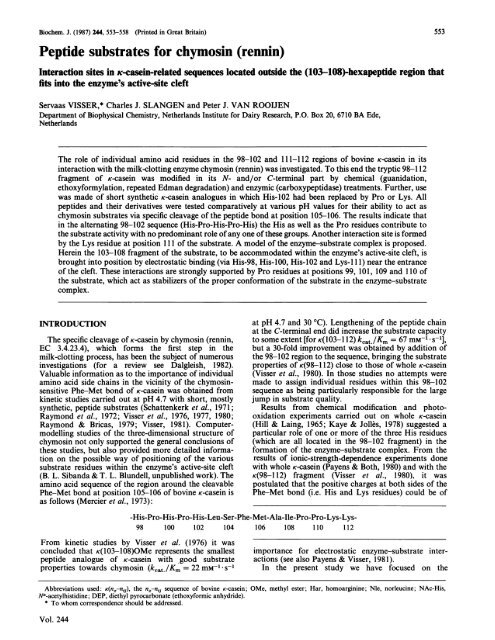
Ethoxy<strong>for</strong>mylation<br />
To investigate further the possible effect of the side<br />
chains of the various His residues on the substrate<br />
activity, we have modified these positions in K(98-112) by<br />
ethoxy<strong>for</strong>mylation. The extent of modification (being<br />
virtually the same <strong>for</strong> 1.5 h and 15 h reaction periods)<br />
was calculated by taking a molar absorptivity of<br />
4.0 x 10³ M-1 cm-' <strong>for</strong> the reaction product at 240 nm.<br />
The effect of ethoxy<strong>for</strong>mylation on the substrate activity<br />
of K(98-112) at pH 5.5 is illustrated by Fig. 2. It is seen<br />
that, by an increasing (average) modification of His<br />
residues (effected by increasing DEP/His ratios in the<br />
reaction mixture), the substrate activity gradually<br />
decreases, but is not completely destroyed even when all<br />
three histidine residues have been ethoxy<strong>for</strong>mylated.<br />
DISCUSSION<br />
To find out if and to what extent individual amino acid<br />
residues more remote from the cleavable Phe-Met bond<br />
of K-casein contribute to the <strong>for</strong>mation of the enzymesubstrate<br />
complex, we have chosen the approach of<br />
substrate modification. This was done in three ways: (a)<br />
use of synthetic K-casein analogues in which one of the<br />
crucial residues had been replaced; (b) chain-shortening<br />
of K(98-112) by chemical or enzymic procedures; (c)<br />
chemical modification of one or more amino acid side<br />
chains in K(98-112). All these modifications resulted in<br />
well-defined products, the purity of which could be easily<br />
checked. A possible exception is the specific modification<br />
of His residues by ethoxy<strong>for</strong>mylation. It is generally<br />
Vol. 244<br />
0.85 +0.05<br />
0.32+0.02<br />
0.32+0.02<br />
0.52+0.03<br />
0.20<br />
0.47+0.02<br />
0.41 +0.02<br />
0.35 +0.02<br />
0.12+0.01<br />
0.31 +0.1<br />
0.26+0.01<br />
0.12<br />
0.10+0.01<br />
0.12+0.01<br />
557<br />
21.6 +0.7<br />
75 + 3<br />
91+5<br />
30.8+ 1.4<br />
105]§<br />
13.0+0.4<br />
22.1 +0.9<br />
92+4<br />
131 +6<br />
104+3<br />
168+4<br />
350]§<br />
520+20<br />
475 +8<br />
known (Miles, 1977) that results obtained via this<br />
procedure should be interpreted with caution, because of<br />
the possibility of simultaneous modification of residues<br />
other than His, and the risk of irreversible ring-opening<br />
of His side chains when a large excess of modifying<br />
reagent is used (Loosemore & Pratt, 1976). The latter<br />
leads to an overestimation of the ethoxy<strong>for</strong>mylated His<br />
residues as measured at 240 nm. However, the occurrence<br />
of this side reaction can be tested by measuring the loss<br />
of His residues via amino acid analysis. Non-selectivity<br />
of the reaction of DEP with whole K-casein was reported<br />
by Reimerdes & Klostermeyer (1973). By carrying out<br />
the ethoxy<strong>for</strong>mylation with a relatively short peptide<br />
having no or only a limited number of reactive groups<br />
other than histidine, one can avoid or at least better<br />
control this kind of side reaction. The modification of<br />
K(98-112), per<strong>for</strong>med. by carefully increasing the<br />
DEP/His ratio in the reaction mixture, showed a<br />
destruction of His residues only at the highest DEP/His<br />
ratios used (cf. Table 1). The molar absorptivity of<br />
4.0 x 10³ M-1 cm-' at 240 nm found <strong>for</strong> the reaction<br />
product of DEP and NAc-His is in agreement with the<br />
value of 3.9 x 10³ M-1 cm- (at 242 nm) reported by<br />
Choong et al. (1977) and is only a little higher than the<br />
3.6 x 10³ M-1 cm-' (at 240 nm) found by Holbrook &<br />
Ingram (1973). The results of the experiment with<br />
ethoxy<strong>for</strong>mylated K(98-112) disclose the involvement of<br />
His side chains of the substrate in its reaction with the<br />
enzyme. However, no predominant role of one particular<br />
His residue, as was suggested <strong>for</strong> whole K-casein by Kaye<br />
& Jolles (1978), could be established by our experiment.<br />
The apparent modification of more than the maximum<br />
of three His residues at higher DEP/His ratios (cf. Fig.<br />
2) is in line with results obtained by Kaye & Jolles (1978)<br />
with whole K-casein. It should probably be ascribed to<br />
the above-mentioned destruction of imidazole groups.<br />
Also, by the other modification procedures products
558 S. Visser, C. J. Slangen and P. J. van Rooijen<br />
DEP/His molar ratio<br />
0 5 10 15 20<br />
100<br />
80<br />
60<br />
-2 40<br />
0~~~~~~~~<br />
200<br />
0 1 2 3 4<br />
Modified His residues (mol/mol)<br />
Fig. 2. Susceptibility of Kc(98-112) towards <strong>chymosin</strong> action at<br />
pH 5.5 as a function of the extent of modification by<br />
ethoxy<strong>for</strong>mylation of histidine residues (0) and as a<br />
function of the excess of DEP reagent used (0)<br />
For experimental details see the text.<br />
were obtained in which His residues appeared to have a<br />
binding function in the enzyme-substrate complex, as<br />
reflected by the Km values measured. It should be noted,<br />
however, that interjacent Pro residues at positions 99 and<br />
101 are at least as important in this respect and probably<br />
act in the same way as Pro- 109 and Pro-i 10 as promoters<br />
of a proper positioning of the substrate part in the<br />
enzyme-substrate complex. Even the introduction of a<br />
Pro residue at the position of His-102 seems to stabilize<br />
the con<strong>for</strong>mation of the enzyme-substrate complex<br />
equally well, whereas a Lys residue in this position <strong>for</strong>ms<br />
an interfering factor.<br />
In the enzyme-substrate complex the 103-108 sequence<br />
of the substrate is, as an extended structure (Raap et al.,<br />
1983), accommodated within the enzyme's active-site<br />
cleft. The hydrophobic Phe-105 and Met-106 side chains<br />
of the substrate as well as those of Leu- 103 and Ile-108<br />
are directed towards hydrophobic pockets along the wall<br />
of the active-site cleft, whereas the hydroxy group<br />
of Ser-104 <strong>for</strong>ms part of a hydrogen bridge with some<br />
counterpart of the enzyme (Visser et al., 1976, 1977;<br />
B. L. Sibanda & T. L. Blundell, unpublished work). The<br />
substrate parts immediately adjacent to the 103-108<br />
fragment (i.e. the regions 98-102 and 109-111) are<br />
<strong>for</strong>ming fl-turns (Raap et al., 1983) located around the<br />
edge of the active-site cleft in the enzyme-substrate<br />
complex. As was also put <strong>for</strong>ward by Payens and his<br />
co-workers (Payens & Both, 1980; Payens & Visser,<br />
Received 28 July 1986/27 November 1986; accepted 24 February 1987<br />
1981), these parts contribute strongly to a firm and<br />
efficient binding to the enzyme molecule, thus making the<br />
Phe-Met bond of the substrate easily accessible to<br />
cleavage. It would be interesting to investigate further (<strong>for</strong><br />
instance by minimizing the free energy of interaction<br />
in computer-modelling experiments) which negative<br />
charges on the enzyme part of the enzyme-substrate<br />
complex are candidates <strong>for</strong> interaction with the positive<br />
groups in the substrate regions discussed.<br />
We are much indebted to Dr. Lynn Sibanda and Dr. Tom<br />
Blundell (University of London) <strong>for</strong> the in<strong>for</strong>mation on their<br />
computer model of <strong>chymosin</strong>, and to Mrs. Cecile Schattenkerk<br />
(University of Leiden) <strong>for</strong> making the synthetic peptide<br />
samples available. We thank Dr. Henk Vreeman from this<br />
Institute <strong>for</strong> helpful discussions and critical remarks.<br />
REFERENCES<br />
Choong, Y. S., Shepherd, M. G. & Sullivan, P. A. (1977)<br />
Biochem. J. 165, 385-393<br />
Dalgleish, D. G. (1982) in Developments in Dairy Chemistry<br />
(Fox, P. F., ed.), part 1, pp. 157-187, Applied Science<br />
Publishers, Barking<br />
Hill, R. D. & Laing, R. R. (1965) J. Dairy Res. 32, 193-201<br />
Holbrook, J. J. & Ingram, V. A. (1973) Biochem. J. 131,<br />
729-738<br />
Kaye, N. M. C. & Jolles, P. (1978) Biochim. Biophys. Acta 536,<br />
329-340<br />
Kimmel, J. R. (1967) Methods Enzymol. 11, 584-589<br />
Loosemore, M. J. & Pratt, R. F. (1976) FEBS Lett. 72, 155-158<br />
Mercier, J. C., Brignon, G. & Ribadeau Dumas, B. (1973) Eur.<br />
J. Biochem. 35, 222-235<br />
Miles, E. W. (1977) Methods Enzymol. 47, 431-442<br />
Miihlrad, A., Hegyi, G. & Toth, G. (1967) Acta Biochim.<br />
Biophys. Acad. Sci. Hung. 2, 19-29<br />
Ovadi, J., Libor, S. & El6di, P. (1967) Acta Biochim. Biophys.<br />
Acad. Sci. Hung. 2, 455-458<br />
Payens, T. A. J. & Both, P. (1980) Adv. Chem. Ser. 188,<br />
129-141<br />
Payens, T. A. J. & Visser, S. (1981) Neth. Milk Dairy J. 35,<br />
387-389<br />
Raap, J., Kerling, K. E. T., Vreeman, H. J. & Visser, S. (1983)<br />
Arch. Biochem. Biophys. 221, 117-124<br />
Raymond, M. N. & Bricas, E. (1979) J. Dairy Sci. 62,<br />
1719-1725<br />
Raymond, M. N., Garnier, J., Bricas, E., Cilianu, S., Blasnic,<br />
M., Chaix, A. & Lefrancier, P. (1972) Biochimie 54, 145-154<br />
Reimerdes, E. H. & Klostermeyer, H. (1973) Milchwissenschaft<br />
28, 558-564<br />
Schattenkerk, C., Holtkamp, I., Hessing, J. G. M., Kerling,<br />
K. E. T. & Havinga, E. (1971) Recl. Trav. Chim. Pays-Bas<br />
90, 1320-1322<br />
Schattenkerk, C., Voskuyl-Holtkamp, I. & Bokhorst, R. (1973)<br />
Recl. Trav. Chim. Pays-Bas 92, 92-116<br />
Tarr, G. E. (1975) Anal. Biochem. 63, 361-370<br />
Tarr, G. E. (1977) Methods Enzymol. 47, 335-357<br />
Visser, S. (1981) Neth. Milk Dairy J. 35, 65-88<br />
Visser, S. & Rollema, H. S. (1986) Anal. Biochem. 153,235-241<br />
Visser, S., van Rooijen, P. J., Schattenkerk, C. & Kerling,<br />
K. E. T. (1976) Biochim. Biophys. Acta 438, 265-272<br />
Visser, S., van Rooijen, P. J., Schattenkerk, C. & Kerling,<br />
K. E. T. (1977) Biochim. Biophys. Acta 481, 171-176<br />
Visser, S., van Rooijen, P. J. & Slangen, K. J. (1980) Eur. J.<br />
Biochem. 108, 415-421<br />
Vreeman, H. J., van Rooijen, P. J. & Visser, S. (1977) Anal.<br />
Biochem. 77, 251-264<br />
1987