J. Vacuum Sci. technol.B _Vol.19_No.6_2001_pp.2045-2049.pdf
J. Vacuum Sci. technol.B _Vol.19_No.6_2001_pp.2045-2049.pdf
J. Vacuum Sci. technol.B _Vol.19_No.6_2001_pp.2045-2049.pdf
Create successful ePaper yourself
Turn your PDF publications into a flip-book with our unique Google optimized e-Paper software.
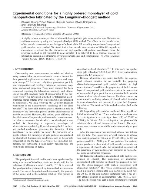
Experimental conditions for a highly ordered monolayer of gold<br />
nanoparticles fabricated by the Langmuir–Blodgett method<br />
Shujuan Huang, a) Gen Tsutsui, Hiroyuki Sakaue, Shoso Shingubara,<br />
and Takayuki Takahagi<br />
Department of Electrical Engineering, Hiroshima University, Kagamiyama 1-4-1,<br />
Higashi-Hiroshima 739-8527, Japan<br />
Received 14 December 2000; accepted 20 August <strong>2001</strong><br />
A highly ordered monolayer film of alkanethiol-encapsulated gold nanoparticles was fabricated on<br />
a silicon substrate by using the Langmuir–Blodgett LB method. The effects on the particle order,<br />
of the particle concentration and the type of solvent of the LB spreading suspension of encapsulated<br />
gold particles, were studied. We found that a low particle concentration of 0.06–0.3 mg/mL in<br />
chloroform is optimal for the fabrication of high quality gold particle monolayers. Since the<br />
proposed method is not restricted to gold particles, it is believed to be a practical process for<br />
fabricating quantum dot structures of various particle sizes and compositions. © <strong>2001</strong> American<br />
<strong>Vacuum</strong> Society. DOI: 10.1116/1.1410943<br />
I. INTRODUCTION<br />
Constructing new nanostructured materials and devices<br />
using nanoparticles has attracted much research interest for<br />
their potential applications in electronics, optics, and magnetic<br />
storage. 1–5 As known, only three parameters, particle<br />
size, composition, and topology, determine electronic, magnetic,<br />
and optical properties. Thus, much research has been<br />
conducted regarding the fabrication, assembly, and utilization<br />
of nanodot structures made of nanoparticles. In our previous<br />
work, 6,7 we developed a method for fabricating a selforganized<br />
planar ordered array of gold particles encapsulated<br />
by alkanethiol. We have observed the Coulomb blockade<br />
phenomena in the nanostructures consisting of 9-nm-diam<br />
particles. 8 The fabrication method plays a significant role in<br />
the study of the fabrication process and the characteristics of<br />
nanodot structures. However, this method poses difficulty in<br />
the fabrication of large-scale, well-controlled nanostructures.<br />
In order to overcome this drawback, we developed a new<br />
method for fabricating a large-scale monolayer of<br />
alkanethiol-encapsulated gold particles using the LB method,<br />
and studied mechanism governing the formation of this<br />
monolayer. 9 In this article, we report the fabrication of a<br />
highly ordered LB monolayer of gold particles encapsulated<br />
by alkanethiol. Experimental conditions, such as the particle<br />
concentration and the type of solvent of LB spreading suspension,<br />
for fabricating a high-quality nanostructure are<br />
studied and discussed in detail.<br />
II. EXPERIMENT<br />
The gold particles used in this work were synthesized by<br />
using a mixture of trisodium citrate and tannic acid for the<br />
reduction of chloroauric acid (HAuCl4). 10,11 It is reported<br />
that gold particles synthesized by this method are homodispersed.<br />
The size of the particles is determined by the quantity<br />
of the tannic acid in the reducing solution. This method is<br />
a Electronic mail: sjhuang@hiroshima-u.ac.jp<br />
described in detail elsewhere. 10,11 In this work, we synthesized<br />
gold colloids of 8.30.7 and 171.6 nm in diameter to<br />
prepare the LB monolayer.<br />
Because alkanethiols are water insoluble, the aqueous<br />
gold colloidal solution is not suitable for preparing<br />
alkanethiol-encapsulated gold particle solution in high<br />
concentrations. 6 In addition, the preparation of the LB monolayer<br />
of encapsulated gold particles requires the suspension<br />
of encapsulated gold particles in a water-insoluble volatile<br />
solvent such as chloroform or benzene. For these reasons, we<br />
made use of a mediating solvent of ethanol, which is soluble<br />
in water, chloroform, and benzene, to prepare the LB spreading<br />
solution. The details of this method are described in the<br />
following:<br />
1 Suspending gold particles in ethanol. First, a gold colloidal<br />
solution of 8.3 or 17 nm in diameter was dissociated<br />
by centrifugation at a centrifugal force CF of 25 000 or<br />
12 000 g for 30 min. After centrifugation, two phases of the<br />
solutions, dark red, and transparent had formed. The supernatant<br />
was removed as carefully and as completely as possible.<br />
After the supernatant was removed, ethanol was infused<br />
into the tube. This suspension of gold particles in ethanol<br />
was centrifugalized for 30 min at CF25 000 g for 8.3 nm<br />
particles and CF12 000 g for 17 nm particles. The centrifugation<br />
produced a black spot of gold particle precipitate and<br />
a supernatant of ethanol. After the supernatant was removed,<br />
the precipitate of gold particles was dispersed in fresh ethanol<br />
using an ultrasonic processor.<br />
2 Preparing alkanethiol-encapsulated gold particle suspension<br />
in ethanol. The suspension of alkanethiolencapsulated<br />
gold particles in ethanol was prepared by mixing<br />
the above-prepared gold particle suspension with<br />
alkanethiol solution. 6,7 The typical experimental conditions<br />
used in preparing encapsulated gold particles included mixing<br />
20 mL of the gold particle suspension with 5 mL of 1<br />
mM ethanol solution of dodecanethiol CH 3–CH 2 11–SH.<br />
The mixed solution was kept at room temperature overnight.<br />
2045 J. Vac. <strong>Sci</strong>. Technol. B 19„6…, NovÕDec <strong>2001</strong> 1071-1023Õ<strong>2001</strong>Õ19„6…Õ2045Õ5Õ$18.00 ©<strong>2001</strong> American <strong>Vacuum</strong> Society<br />
2045
2046 Huang et al.: Experimental conditions for gold nanoparticles 2046<br />
FIG. 1. Surface pressure–area per particle (-A) isotherm for 8.3-nm-diam<br />
gold particles encapsulated by dodecanethiol.<br />
The mixed solution was then centrifugalized for 30 min at<br />
CF12 000 g for 8.3 nm particles and CF6000 g for 17 nm<br />
particles in order to remove the unreacted dodecanethiol<br />
molecules.<br />
3 Suspending encapsulated gold particles in chloroform<br />
or benzene. The above centrifugation caused the<br />
dodecanethiol-encapsulated gold particles to precipitate at<br />
the bottom of the tube, which could be dispersed in chloroform<br />
or benzene using an ultrasonic processor. The concentration<br />
of gold particles in the suspension was determined by<br />
the amount of solvent infused.<br />
The LB instrument used in this work was a KSV minitrough<br />
manufactured by KSV Instruments. To spread the<br />
sample, minute droplets 3 L of the encapsulated gold<br />
particle suspension prepared above were very carefully cast<br />
on the surface of the pure water in the LB trough at intervals<br />
of 30 s. After the solvent evaporated, the hydrophobic<br />
dodecanethiol-encapsulated gold particles remained on the<br />
water’s surface. These particles were then compressed by<br />
moving the barriers at a speed of 5 mm/min. The surface<br />
pressure isotherm was recorded throughout the compression.<br />
The gold particles were transferred by retracting a hydrogenterminated<br />
silicon substrate, 12 which was immersed vertically<br />
in the pure water in the trough before the particle suspension<br />
was spread. The retracting speed was 0.5 mm/min.<br />
The temperature of the pure water in the trough was about<br />
24 °C.<br />
In the present work, scanning electron microscopy SEM<br />
observations of the transferred gold particles of 8.3 and 17<br />
nm in diameter were performed on S-5000 and S-800 Hitachi,<br />
respectively.<br />
III. RESULTS AND DISCUSSION<br />
A. LB monolayer of alkanethiol-encapsulated gold<br />
particles<br />
Figure 1 displays a smooth surface pressure–area per particle<br />
(-A) isotherm for the 8.3-nm-diam dodecanethiolencapsulated<br />
gold particle suspension with a particle concentration<br />
of 0.3 mg/mL in chloroform, which provides rich<br />
J. Vac. <strong>Sci</strong>. Technol. B, Vol. 19, No. 6, NovÕDec <strong>2001</strong><br />
information on the formation of a stable monolayer of the<br />
encapsulated gold particles. At the beginning of the LB compression,<br />
the average distance between particles was quite<br />
large, so that the surface pressure was very low. As the barrier<br />
moved, the interparticle distance decreased and the surface<br />
pressure increased slightly. From the point of about 140<br />
nm 2 , the surface pressure apparently started to increase. This<br />
was because the interparticle distance became very small so<br />
that the interaction of the particles increased remarkably.<br />
When the barrier compressed further, the surface pressure<br />
increased abruptly and a densely packed monolayer of the<br />
particles tended to form. We found that a surface pressure of<br />
10 mN/m is the optimal condition for a good quality<br />
monolayer of 8.3-nm-diam gold particles. At this point, the<br />
area occupied per particle is about 91 nm 2 . According to our<br />
previous study, 6<br />
the estimated area per dodecanethiol-<br />
encapsulated particle is about 78 nm 2 for a close-packed<br />
monolayer. Thus, the particle coverage at this pressure is<br />
about 85%. We have reported that the unoccupied area was<br />
caused by the poor accommodation of the domains and the<br />
arrangement defects of the particles. 9 When the gold particles<br />
were compressed beyond 10 mN/m, some patches of<br />
bilayer had formed and the monolayer collapsed.<br />
The monolayer of the gold particles was deposited onto a<br />
hydrogen-terminated silicon substrate over an area of 1 cm 2 .<br />
SEM observations demonstrated that it was composed of<br />
two-dimensional domains of ordered, close-packed gold particles.<br />
The sizes of these domains varied, some of which<br />
were more than 100 m 2 . Figure 2a shows the SEM image<br />
of part of a domain of 8.3-nm-diam gold particles transferred<br />
at 10 mN/m. The inset shows a high-magnification SEM<br />
image. Figures 2b and 2c display fast Fourier transformation<br />
FFT images. The high-magnification SEM image demonstrates<br />
that a very high level of orderliness of the particle<br />
arrangement had formed locally. Furthermore, its FFT image<br />
Fig. 2c reveals clear first and second FFT orders, which<br />
also indicates a highly ordered close-packed arrangement of<br />
gold particles. However, at longer ranges, as shown in Fig.<br />
2a, it can be seen that the gold particles have not formed a<br />
perfect order. The particle arrangement looks like a polycrystal,<br />
which has many ‘‘crystal grains.’’ Some particle vacancies<br />
and dislocations were observed in SEM observations.<br />
Correspondingly, the FFT image tends toward the shape of a<br />
ring rather than hexagonal spots. This is due to arrangement<br />
defects that are discussed in Sec. III B.<br />
B. Effect of the spreading suspension’s particle<br />
concentration on the formation of the<br />
particle monolayer<br />
In order to determine the most suitable conditions for the<br />
fabrication of high quality monolayers of alkanethiolencapsulated<br />
gold particles, experimental study was carried<br />
out under different particle concentrations of the LB spreading<br />
suspension from chloroform. Figure 3 shows the SEM<br />
images of LB monolayers of 8.3-nm-diam gold particles fabricated<br />
from particle concentrations of 0.06 mg/mL (1<br />
10 13 /mL) and 0.6 mg/mL (110 14 /mL). Both monolayers
2047 Huang et al.: Experimental conditions for gold nanoparticles 2047<br />
FIG. 2.a SEM image of a LB monolayer of dodecanethiol-encapsulated gold particles 8.3 nm in diameter. The inset shows a high-magnification SEM image,<br />
b and c show the FFT images of the SEM image in a and the inset, respectively.<br />
were transferred at 10 mN/m. Obviously, the encapsulated<br />
gold particles formed in quite different arrangements.<br />
In the case of a low particle concentration of 0.06 mg/mL,<br />
the gold particle monolayer is similar to that made from a<br />
medium concentration of 0.3 mg/mL, as shown in Fig. 2. The<br />
films take on a crystal structure with some defects including<br />
particle vacancies Fig. 4a, ‘‘edge dislocations’’ Fig.<br />
4b, and grain boundaries. However, in the LB monolayer<br />
made from a high particle concentration of 0.6 mg/mL, many<br />
JVSTB-MicroelectronicsandNanometer Structures<br />
narrow gaps or voids have formed between the ‘‘crystal<br />
grains’’ in addition to particle vacancies and dislocations.<br />
Moreover, the FFT image of the LB monolayer made from<br />
the low particle concentration shows hexagonal spots,<br />
whereas the FFT image of the monolayer made from the high<br />
concentration is more like a ring rather than six clear spots.<br />
This indicates that the degree of long-range order of the<br />
monolayer made from the low concentration is better than<br />
that made from the high concentration. In other words, a low
2048 Huang et al.: Experimental conditions for gold nanoparticles 2048<br />
FIG. 3. SEM images of 8.3-nm-diam gold particle monolayers prepared<br />
from LB spreading suspensions of a a low particle concentration of 0.06<br />
mg/mL and b a high particle concentration of 0.6 mg/mL. The insets show<br />
the corresponding FFT images.<br />
particle concentration of 0.06–0.3 mg/mL is more suitable<br />
than a high concentration of 0.6 mg/ml for the LB preparation<br />
of gold particle monolayers.<br />
We propose that the difference between the LB monolayers<br />
made from the low- and high-particle-concentration suspensions<br />
is caused by the different forming processes. In our<br />
previous study of the forming mechanism of gold particle LB<br />
monolayers, 9 we found that the ordered particle domains<br />
were a result of a self-organization process induced by the<br />
evaporation of the sample suspension, rather than the compression<br />
of the barriers. More specifically, when a droplet of<br />
the encapsulated gold suspension was cast onto the surface<br />
of the water in a trough, the solvent’s water insolubility<br />
FIG. 4. Defects in the LB monolayer of the encapsulated gold particles. a<br />
A particle vacancy and b a dislocation indicated by the white line.<br />
J. Vac. <strong>Sci</strong>. Technol. B, Vol. 19, No. 6, NovÕDec <strong>2001</strong><br />
FIG. 5. Schematics of the formation of the defects in LB monolayers prepared<br />
from a a low particle concentration and b a high particle concentration<br />
of LB spreading suspensions.<br />
caused it to spread into a thin layer. As the solvent evaporated<br />
and became a thin layer with a thickness equal to the<br />
particle size, the attractive force between particles induced<br />
by the surface tension of the solvent increased significantly<br />
and caused the particles to pack together into ordered arrays.<br />
In the case of low particle concentration, when the sample<br />
first began to spread, the particle density on the water’s surface<br />
was quite low, so that only small domains of closepacked<br />
particles had formed due to the surface tension of the<br />
solvent. As the particle suspension was cast onto the water’s<br />
surface in succession, the new incoming particles around<br />
these initial domains were transported to them by the same<br />
interaction as seen in the formation of the small domains.<br />
Consequently, the domains of the ordered particles grew<br />
larger by agglomeration. However, when a particle with a<br />
much larger or smaller size was transported to the particle<br />
array, as shown in Fig. 5a, the arrangement of the particles<br />
would be changed, forming a defect in the particle arrangement.<br />
Therefore, we can conclude that the main cause of the<br />
defects in the particle arrangement in this case is the variation<br />
of the particle sizes.<br />
On the other hand, in the case of the high particle concentration,<br />
as a droplet of the particle suspension was spread and<br />
evaporated, many small particle domains had already formed<br />
initially, as shown in Fig. 5b. Meanwhile, since the particle<br />
concentration was quite high, some domains were quite close<br />
to each other. As a result, the interaction between these domains,<br />
caused by the solvent’s surface tension, was large<br />
enough to drive them to coalesce together. Due to the poor<br />
accommodation of the particle domains, many narrow gaps<br />
between the ‘‘crystal grains’’ formed.<br />
C. Effect of solvent on particle order<br />
As known, the solvent of the particle suspension is another<br />
important condition for fabricating a high-quality
2049 Huang et al.: Experimental conditions for gold nanoparticles 2049<br />
FIG. 6. SEM images of 17-nm-diam gold particle monolayers prepared from<br />
a chloroform suspension and b benzene suspension.<br />
monolayer of alkanethiol-encapsulated gold particles. 6 This<br />
is due to the dependence of the evaporation rate of the<br />
sample, which plays a significant role in the formation of the<br />
ordered particle domains, on the solvent. In this work, we<br />
prepared dodecanethiol-encapsulated gold particle suspensions<br />
from chloroform and from benzene, respectively. The<br />
diameter of the gold colloidal particles is 17 nm. The particle<br />
concentrations of these two suspensions were the same,<br />
0.06 mg/ml. Figure 6 shows the LB monolayers prepared<br />
from these two kinds of samples. It is noticed that the monolayer<br />
prepared from the particle suspension in chloroform is<br />
more ordered. The only arrangement defects were particle<br />
vacancies and dislocations. However, the monolayer prepared<br />
from the particle suspension in benzene reveals a<br />
lesser ordering. Many gaps formed between particles, as<br />
shown by arrow 1 in Fig. 6b. In addition, some particle<br />
aggregations, as indicated by arrow 2, had also formed,<br />
which demonstrates that the alkanethiol-encapsulated gold<br />
particle suspension from benzene is not as stable as that from<br />
chloroform.<br />
JVSTB-MicroelectronicsandNanometer Structures<br />
In regard to the gaps between particles, we consider that<br />
they are due to benzene’s slower evaporation rate. As known,<br />
benzene evaporates more slowly than does chloroform. In<br />
the slower evaporation process of the gold particle spreading<br />
suspension from benzene, on the one hand, the small particle<br />
domains were first formed in the same way as the particle<br />
suspension from chloroform. On the other hand, unlike the<br />
fast evaporation of the suspension from chloroform, there<br />
was enough time for these small domains to coalesce together<br />
under the above-mentioned interaction, during benzene’s<br />
slower evaporation. As a result, many gaps between<br />
particles had formed due to the poorer accommodation of the<br />
small particle domains.<br />
IV. CONCLUSION<br />
In this article, we reported the fabrication of a highly<br />
ordered wafer-scale monolayer of alkanethiol-encapsulated<br />
gold particles using the LB method. The effects of the particle<br />
concentration and the solvent, from which the LB<br />
spreading suspensions of the encapsulated gold particles<br />
were made, were studied experimentally. The results demonstrate<br />
that a low particle concentration of 0.06–0.3 mg/mL<br />
resulted in a highly ordered monolayer of gold particles. In<br />
addition, using chloroform as the solvent produced a more<br />
stable particle suspension and a more ordered monolayer<br />
than the use of benzene.<br />
1 B. Korgel, S. Fullam, S. Connolly, and D. Fitzmaurice, J. Phys. Chem. B<br />
102, 8379 1998.<br />
2 G. Markovich, D. V. Leff, S.-W. Chung, H. M. Soyez, B. Dunn, and J. R.<br />
Heath, Appl. Phys. Lett. 70, 3107 1997.<br />
3 J.-P. Bourgoin, C. Kergueris, E. Lefevre, and S. Palacin, Thin Solid Films<br />
327, 515 1998.<br />
4 D. B. Janes, V. R. Kolagunta, R. G. Osifchin, W. J. Mahoney, J. D.<br />
Bielefeld, R. P. Andres, and J. I. Henderson, Superlattices Microstruct. 18,<br />
2275 1995.<br />
5 S. Sun, C. B. Murray, D. Weller, L. Folks, and A. Moser, <strong>Sci</strong>ence 287,<br />
1989 2000.<br />
6 S. Huang, H. Sakaue, S. Shingubara, and T. Takahagi, Jpn. J. Appl. Phys.,<br />
Part 1 37, 7198 1998.<br />
7 S. Huang, G. Tsutsui, H. Sakaue, S. Shingubara, and T. Takahagi, Jpn. J.<br />
Appl. Phys., Part 2 38, L473 1999.<br />
8 S. Huang, G. Tsutsui, H. Sakaue, S. Shingubara, and T. Takahagi, J. Vac.<br />
<strong>Sci</strong>. Technol. B 18, 2653 2000.<br />
9 S. Huang, G. Tsutsui, H. Sakaue, S. Shingubara, and T. Takahagi, J. Vac.<br />
<strong>Sci</strong>. Technol. B 19, 115<strong>2001</strong>.<br />
10 J. W. Slot and H. J. Geuze, Eur. J. Cell Biol. 38, 871985.<br />
11 G. Tsutsui, S. Huang, H. Sakaue, S. Shingubara, and T. Takahagi, Jpn. J.<br />
Appl. Phys., Part 1 40, 346 <strong>2001</strong>.<br />
12 T. Takahagi, I. Nagai, A. Ishitani, and H. Kuroda, J. Appl. Phys. 64, 3516<br />
1988.


