download the full PDF issue - Australian Prescriber
download the full PDF issue - Australian Prescriber
download the full PDF issue - Australian Prescriber

Create successful ePaper yourself
Turn your PDF publications into a flip-book with our unique Google optimized e-Paper software.
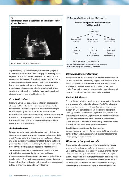
Fig. 2<br />
Transthoracic image of vegetation on <strong>the</strong> anterior leaflet<br />
of <strong>the</strong> mitral valve<br />
AMVL anterior mitral valve leaflet<br />
vegetations (Fig. 2). Transoesophageal echocardiography is<br />
more sensitive than transthoracic imaging for detecting small<br />
vegetations, abscess cavities and leaflet perforation, and is<br />
superior for <strong>the</strong> imaging of pros<strong>the</strong>tic valves. 3 Indications for<br />
transoesophageal echocardiography include a diagnostically<br />
inadequate transthoracic echocardiogram, a negative<br />
transthoracic echocardiogram despite ongoing high clinical<br />
suspicion of endocarditis, pros<strong>the</strong>tic valve involvement and<br />
staphylococcal (or suspected) bacteraemia.<br />
Pros<strong>the</strong>tic valves<br />
Pros<strong>the</strong>tic valves are susceptible to infection, degeneration,<br />
stenosis and thrombosis. They are routinely checked with<br />
transthoracic echocardiography (see box). Transoesophageal<br />
echocardiography is indicated when <strong>the</strong> assessment of<br />
pros<strong>the</strong>tic valve function is hampered by acoustic shadowing, or<br />
<strong>the</strong> detection of vegetations is made difficult by o<strong>the</strong>r artifacts.<br />
It is essential when evaluating complicated endocarditis in<br />
patients with pros<strong>the</strong>tic valves.<br />
Embolic disease<br />
vegetation<br />
Echocardiography plays a very important role in finding <strong>the</strong><br />
source of an embolus following a stroke or peripheral embolic<br />
event. Younger patients or those who have suffered occlusion<br />
of a large peripheral vessel are more likely to have suffered a<br />
purely cardiac embolic event. Older patients are more likely to<br />
have intrinsic cerebrovascular disease or atrial fibrillation.<br />
Transthoracic echocardiography is easier, carries negligible<br />
risk and is less expensive, but has a lower yield than<br />
transoesophageal echocardiography. Sources of emboli that are<br />
usually better defined by transoesophageal echocardiography<br />
include left atrial appendage thrombus, small vegetations, septal<br />
defects or aneurysm and aortic arch a<strong>the</strong>roma.<br />
136 | VOLUME 29 | NUMBER 5 | OCTOBER 2006<br />
Follow-up of patients with pros<strong>the</strong>tic valves<br />
Baseline postoperative transthoracic study<br />
(within 4 weeks)<br />
Mechanical<br />
valves<br />
TTE at 3, 5,<br />
10 years<br />
TTE transthoracic echocardiography<br />
From: Guidelines of <strong>the</strong> Prince Charles Hospital<br />
Echocardiography Laboratory, Brisbane<br />
Cardiac masses and tumour<br />
Patients in whom <strong>the</strong> diagnosis of an intracardiac mass should<br />
be considered are those with cryptogenic stroke or o<strong>the</strong>r embolic<br />
events, those with atrial fibrillation, dilated cardiomyopathy,<br />
anteroapical infarction, bacteraemia or fever of unknown<br />
origin. Echocardiography can accurately diagnose primary and<br />
secondary cardiac tumours, thrombi and vegetations.<br />
Pericardial disease<br />
TTE at 1 year<br />
Biopros<strong>the</strong>tic<br />
aortic valves<br />
TTE at 3, 5,<br />
7 years<br />
<strong>the</strong>n annually<br />
Echocardiography is <strong>the</strong> investigation of choice for <strong>the</strong> diagnosis<br />
and evaluation of a pericardial effusion (Fig. 3). The effusion's<br />
presence, size, haemodynamic significance and response to<br />
<strong>the</strong>rapy are well demonstrated. Echocardiographic signs in<br />
keeping with tamponade include right atrial invagination at <strong>the</strong><br />
onset of systole (sensitive), right ventricular collapse in diastole<br />
(specific) and marked respiratory variation in transvalvular<br />
inflow velocities. Transthoracic echocardiography assists in <strong>the</strong><br />
planning and <strong>the</strong> execution of pericardiocentesis.<br />
Constrictive pericarditis may be diagnosed on<br />
echocardiography, however <strong>the</strong> assessment of <strong>the</strong> pericardium<br />
can be difficult and investigation such as magnetic resonance<br />
imaging may be more helpful.<br />
Diseases of <strong>the</strong> great vessels<br />
Biopros<strong>the</strong>tic<br />
mitral valves<br />
TTE at 3 and<br />
5 years<br />
<strong>the</strong>n annually<br />
Transthoracic echocardiography shows <strong>the</strong> main pulmonary<br />
arteries as far as <strong>the</strong> proximal main branches, <strong>the</strong> hepatic<br />
veins as <strong>the</strong>y drain into <strong>the</strong> inferior vena cava, and <strong>the</strong> inferior<br />
vena cava as it emerges from <strong>the</strong> liver and enters <strong>the</strong> right<br />
atrium. Three of <strong>the</strong> four pulmonary veins can usually be seen<br />
transthoracically where <strong>the</strong>y connect with <strong>the</strong> left atrium, but<br />
for a complete examination of pulmonary venous drainage,<br />
transoesophageal echocardiography is usually required. The