The interpretation of arterial blood gases - Australian Prescriber
The interpretation of arterial blood gases - Australian Prescriber
The interpretation of arterial blood gases - Australian Prescriber

Create successful ePaper yourself
Turn your PDF publications into a flip-book with our unique Google optimized e-Paper software.
Abnormal laboratory results<br />
<strong>The</strong> <strong>interpretation</strong> <strong>of</strong> <strong>arterial</strong> <strong>blood</strong> <strong>gases</strong><br />
Abhishek K Verma, Resident, General Medicine, and Paul Roach, Consultant Respiratory and<br />
Sleep Medicine Physician, Gosford Hospital, New South Wales<br />
Summary<br />
Arterial <strong>blood</strong> gas analysis is used to measure<br />
the pH and the partial pressures <strong>of</strong> oxygen and<br />
carbon dioxide in <strong>arterial</strong> <strong>blood</strong>. <strong>The</strong> investigation<br />
is relatively easy to perform and yields<br />
information that can guide the management <strong>of</strong><br />
acute and chronic illnesses. This information<br />
indicates a patient's acid–base balance, the<br />
effectiveness <strong>of</strong> their gas exchange and the state<br />
<strong>of</strong> their ventilatory control. Interpretation <strong>of</strong><br />
an <strong>arterial</strong> <strong>blood</strong> gas result should not be done<br />
without considering the clinical findings. <strong>The</strong><br />
results change as the body compensates for the<br />
underlying problem. Factors relating to sampling<br />
technique, specimen processing and environment<br />
may also influence the results.<br />
Key words: acid–base disorders, pulmonary function tests.<br />
(Aust Prescr 2010;33:124–9)<br />
Introduction<br />
Arterial <strong>blood</strong> gas analysis is a common investigation in<br />
emergency departments and intensive care units for monitoring<br />
patients with acute respiratory failure. It also has some<br />
application in general practice, such as assessing the need for<br />
domiciliary oxygen therapy in patients with chronic obstructive<br />
pulmonary disease. An <strong>arterial</strong> <strong>blood</strong> gas result can help in the<br />
assessment <strong>of</strong> a patient's gas exchange, ventilatory control and<br />
acid–base balance. However, the investigation does not give<br />
a diagnosis and should not be used as a screening test. It is<br />
imperative that the results are considered in the context <strong>of</strong> the<br />
patient's symptoms.<br />
Arterial puncture<br />
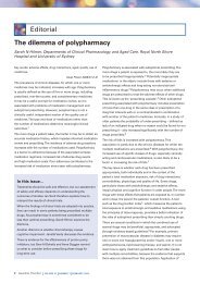
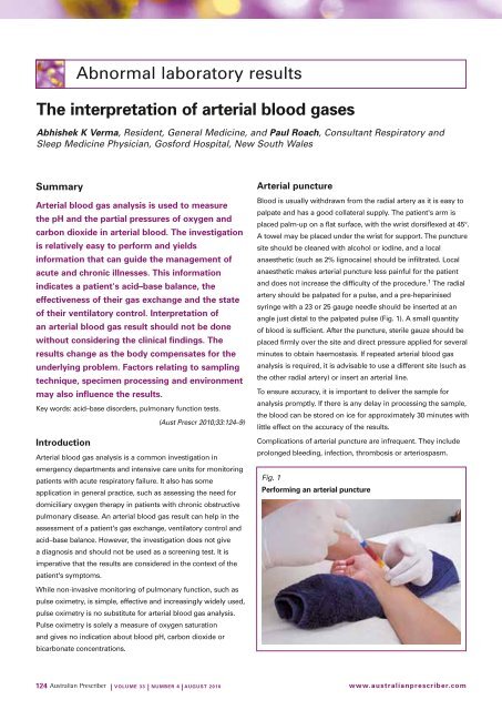
Blood is usually withdrawn from the radial artery as it is easy to<br />
palpate and has a good collateral supply. <strong>The</strong> patient's arm is<br />
placed palm-up on a flat surface, with the wrist dorsiflexed at 45°.<br />
A towel may be placed under the wrist for support. <strong>The</strong> puncture<br />
site should be cleaned with alcohol or iodine, and a local<br />
anaesthetic (such as 2% lignocaine) should be infiltrated. Local<br />
anaesthetic makes <strong>arterial</strong> puncture less painful for the patient<br />
and does not increase the difficulty <strong>of</strong> the procedure. 1 <strong>The</strong> radial<br />
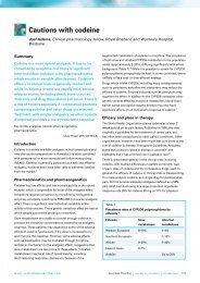
artery should be palpated for a pulse, and a pre-heparinised<br />
syringe with a 23 or 25 gauge needle should be inserted at an<br />
angle just distal to the palpated pulse (Fig. 1). A small quantity<br />
<strong>of</strong> <strong>blood</strong> is sufficient. After the puncture, sterile gauze should be<br />
placed firmly over the site and direct pressure applied for several<br />
minutes to obtain haemostasis. If repeated <strong>arterial</strong> <strong>blood</strong> gas<br />
analysis is required, it is advisable to use a different site (such as<br />
the other radial artery) or insert an <strong>arterial</strong> line.<br />
To ensure accuracy, it is important to deliver the sample for<br />
analysis promptly. If there is any delay in processing the sample,<br />
the <strong>blood</strong> can be stored on ice for approximately 30 minutes with<br />
little effect on the accuracy <strong>of</strong> the results.<br />
Complications <strong>of</strong> <strong>arterial</strong> puncture are infrequent. <strong>The</strong>y include<br />
prolonged bleeding, infection, thrombosis or arteriospasm.<br />
Fig. 1<br />
Performing an <strong>arterial</strong> puncture<br />
While non-invasive monitoring <strong>of</strong> pulmonary function, such as<br />
pulse oximetry, is simple, effective and increasingly widely used,<br />
pulse oximetry is no substitute for <strong>arterial</strong> <strong>blood</strong> gas analysis.<br />
Pulse oximetry is solely a measure <strong>of</strong> oxygen saturation<br />
and gives no indication about <strong>blood</strong> pH, carbon dioxide or<br />
bicarbonate concentrations.<br />
124 | Volume 33 | NumBeR 4 | AuGuST 2010 www.australianprescriber.com
Interpreting a <strong>blood</strong> gas result<br />
<strong>The</strong> automated analysers measure the pH and the partial<br />
pressures <strong>of</strong> oxygen (PaO 2<br />
) and carbon dioxide (PaCO 2<br />
) in<br />
<strong>arterial</strong> <strong>blood</strong>. Bicarbonate (HCO 3ˉ) is also calculated (Box 1).<br />
<strong>The</strong>se measurements should be considered with the patient's<br />
clinical features (Table 1).<br />
pH<br />
<strong>The</strong> pH determines the presence <strong>of</strong> acidaemia or alkalaemia. If<br />
the body has compensated for the disorder, the pH may be in<br />
the normal range.<br />
PaCO 2<br />
<strong>The</strong> PaCO 2<br />
reflects the state <strong>of</strong> alveolar ventilation. An elevated<br />
PaCO 2<br />
reflects alveolar hypoventilation, whereas a decreased<br />
PaCO 2<br />
reflects alveolar hyperventilation. Acute changes in PaCO 2<br />
will alter the pH. As a general rule, a low pH with a high PaCO 2<br />
suggests a respiratory acidosis, while a low pH with a low<br />
PaCO 2<br />
suggests a metabolic acidosis.<br />
Box 1<br />
Reference ranges for <strong>arterial</strong> <strong>blood</strong> <strong>gases</strong><br />
pH 7.35 – 7.45<br />
PaO 2<br />
80 – 100* mmHg 10.6 – 13.3 kPa<br />
PaCO 2<br />
35 – 45 mmHg 4.7 – 6.0 kPa<br />
HCO 3ˉ<br />
22 – 26 mmol/L<br />
Base excess –2 – +2 mmol/L<br />
Reference ranges for venous <strong>blood</strong> <strong>gases</strong><br />
pH 7.32 – 7.43<br />
PvO 2<br />
25 – 40 mmHg<br />
PvCO 2<br />
41 – 50 mmHg<br />
HCO 3ˉ<br />
23 – 27 mmol/L<br />
* age and altitude dependent (see text)<br />
Kilopascals: to convert pressures to kPa, divide mmHg by 7.5<br />
Table 1<br />
Correlating <strong>arterial</strong> <strong>blood</strong> gas results with clinical features<br />
metabolic imbalances Respiratory imbalances<br />
metabolic acidosis metabolic alkalosis Respiratory acidosis Respiratory alkalosis<br />
pH ↓ ↑ ↓ ↑<br />
PaCO 2<br />
N (uncompensated)<br />
↓ (compensated)<br />
N (uncompensated)<br />
↑ (compensated)<br />
↑<br />
↓<br />
HCO 3ˉ ↓ ↑ N (uncompensated)<br />
↑ (compensated)<br />
N (uncompensated)<br />
↓ (compensated)<br />
Base excess ↓ ↑ N/↑ N/↓<br />
Clinical features Kussmaul-type<br />
breathing (deeper, faster<br />
respiration), shock, coma<br />
Paraesthesia, tetany,<br />
weakness<br />
Acute: air hunger,<br />
disorientation<br />
Common<br />
causes<br />
With raised anion gap:<br />
diabetic ketoacidosis,<br />
lactic acidosis, poisons<br />
(e.g. ethylene glycol),<br />
drug overdoses<br />
(paracetamol, aspirin,<br />
isoniazid, alcohol)<br />
With normal anion<br />
gap: diarrhoea,<br />
secretory adenomas,<br />
ammonium chloride<br />
poisoning, interstitial<br />
nephritis, renal tubular<br />
acidosis, acetazolamide<br />
administration<br />
Vomiting, prolonged<br />
therapy with potassiumwasting<br />
diuretics or<br />
steroids, Cushing's<br />
disease, ingestion/<br />
overdose <strong>of</strong> sodium<br />
bicarbonate (e.g.<br />
antacids)<br />
N = within normal range ↑ = increased ↓ = decreased<br />
Chronic: hypoventilation,<br />
hypoxia, cyanosis<br />
Hypoventilation –<br />
chronic lung disease<br />
with CO 2<br />
retention,<br />
e.g. chronic obstructive<br />
pulmonary disease,<br />
respiratory depression<br />
from drugs (e.g. opioids,<br />
sedatives), severe<br />
asthma, pulmonary<br />
oedema<br />
Acute: hyperventilation,<br />
paraesthesia, lightheadedness<br />
Chronic: hyperventilation,<br />
latent tetany<br />
Hyperventilation –<br />
anxiety, pain, febrile<br />
illness, hypoxia,<br />
pulmonary embolism,<br />
pregnancy, sepsis<br />
www.australianprescriber.com<br />
| Volume 33 | NumBeR 4 | AuGuST 2010 125
<strong>The</strong>re is a delayed response <strong>of</strong> PaCO 2<br />
to an acute change.<br />
Increases in PaCO 2<br />
occur relatively slowly, as the body's overall<br />
CO 2<br />
stores are very large (approximately 20 L) and the volume<br />
<strong>of</strong> CO 2<br />
generated by metabolism (200 mL/min) makes little<br />
overall difference. For instance, during a breath-hold, the PaCO 2<br />
rises at a rate <strong>of</strong> only 2–3 mmHg per minute, hence patients<br />
with a very high PaCO 2<br />
usually have a long-standing disorder.<br />
Accordingly, even when treated the PaCO 2<br />
may take a long time<br />
to return to normal.<br />
PaO 2<br />
<strong>The</strong> state <strong>of</strong> <strong>arterial</strong> <strong>blood</strong> oxygenation is determined by the PaO 2<br />
.<br />
This reflects gas exchange in the lungs and normally the PaO 2<br />
decreases with age. This is due to decreased elastic recoil in the<br />
lungs in the elderly, thereby yielding a greater ventilation-perfusion<br />
mismatch. <strong>The</strong> expected PaO 2<br />
when breathing air at sea level<br />
can be calculated with the equation PaO 2<br />
= 100 – (age x 0.25).<br />
Consequently, a PaO 2<br />
<strong>of</strong> 75 mmHg, which may be <strong>of</strong> concern in a<br />
young person, is usually unremarkable in an 85-year-old.<br />
A PaO 2<br />
that is less than expected indicates hypoxaemia. This<br />
can result from hypoventilation or a mismatch <strong>of</strong> ventilation and<br />
perfusion. If alveolar ventilation is adequate (that is, PaCO 2<br />
is<br />
normal), then the hypoxaemia is almost certainly caused by a<br />
ventilation-perfusion disturbance. <strong>The</strong> nature <strong>of</strong> the hypoxaemia<br />
can be further assessed by the difference between the alveolar<br />
and <strong>arterial</strong> oxygen tensions.<br />
<strong>The</strong> alveolar–<strong>arterial</strong> oxygen tension difference<br />
If an <strong>arterial</strong> <strong>blood</strong> gas result shows hypoxaemia (low<br />
PaO 2<br />
) and inadequate alveolar ventilation (high PaCO 2<br />
), it<br />
must be determined whether the hypoxaemia is related to<br />
hypoventilation, or is secondary to a disturbance in ventilationperfusion,<br />
or both. This is assessed by calculating the difference<br />
between the alveolar (PAO 2<br />
) and <strong>arterial</strong> (PaO 2<br />
) oxygen tensions<br />
(see Box 2).<br />
<strong>The</strong> alveolar–<strong>arterial</strong> difference, or gradient, can be estimated<br />
only if the oxygen fraction <strong>of</strong> inspired air (FiO 2<br />
, usually 0.21 on<br />
room air), barometric pressure and water vapour pressure<br />
Box 2<br />
<strong>The</strong> alveolar–<strong>arterial</strong> oxygen gradient<br />
P(A–a)O 2<br />
= PAO 2<br />
– PaO 2<br />
PaO 2<br />
= <strong>arterial</strong> oxygen tension<br />
PAO 2<br />
= alveolar oxygen tension<br />
PAO 2<br />
= FiO 2<br />
(P B<br />
– P H2O<br />
) – 1.2(PaCO 2<br />
)<br />
FiO 2<br />
P B<br />
= oxygen fraction in inspired air<br />
= barometric pressure (760 mmHg at sea level)<br />
P H2O<br />
= water vapour tension (47 mmHg at 37° C)<br />
Normal value
into account the appropriateness <strong>of</strong> the metabolic response for<br />
any given disorder, thus limiting its utility when interpreting<br />
results.<br />
Anion gap<br />
<strong>The</strong> anion gap assists with the diagnosis <strong>of</strong> metabolic acidosis<br />
(Box 3). This difference between the concentrations <strong>of</strong> measured<br />
anions and cations increases with dehydration and decreases<br />
with hypoalbuminaemia. <strong>The</strong> gap also widens if there is an<br />
increase in the concentration <strong>of</strong> unmeasured anions such as<br />
ketones and lactate.<br />
Factors influencing <strong>blood</strong> gas results<br />
A number <strong>of</strong> sampling and environmental factors may affect<br />
the result <strong>of</strong> the analysis. Delayed processing <strong>of</strong> the sample<br />
may yield a falsely low PaO 2<br />
, as the delay allows leucocytes to<br />
consume oxygen. This can be avoided by prompt transport <strong>of</strong><br />
the sample on ice.<br />
Air bubbles introduced when performing the <strong>arterial</strong> puncture<br />
can also cause a falsely high PaO 2<br />
and a falsely low PaCO 2<br />
. 2<br />
This can be avoided by gently removing air bubbles within the<br />
specimen immediately after collection without agitating the<br />
sample.<br />
Body temperature can also affect <strong>arterial</strong> <strong>blood</strong> gas tensions.<br />
This is relevant in febrile or hypothermic patients, so body<br />
temperature should be recorded at the time <strong>of</strong> collection. 3<br />
Box 3<br />
<strong>The</strong> anion gap concept<br />
n<br />
n<br />
n<br />
n<br />
n<br />
n<br />
n<br />
the anion gap is an artificial concept that may indicate<br />
the cause <strong>of</strong> a metabolic acidosis<br />
it represents the disparity between the major measured<br />
plasma cations (sodium and potassium) and the anions<br />
(chloride and bicarbonate)<br />
when calculating the anion gap, potassium is usually<br />
omitted from the calculation thus:<br />
Gap = Na + – (Clˉ + HCO 3ˉ)<br />
the anion gap is normally between 8 and 16 mmol/L<br />
a raised anion gap indicates an increased concentration<br />
<strong>of</strong> lactate, ketones or renal acids and is seen in<br />
starvation and uraemia<br />
a raised anion gap is seen in overdoses <strong>of</strong> paracetamol,<br />
salicylates, methanol or ethylene glycol<br />
a normal anion gap is seen if a metabolic acidosis is<br />
due to diarrhoea or urinary loss <strong>of</strong> bicarbonate<br />
Mixed acid–base disorders<br />
It is possible to have a mixed respiratory and metabolic disorder<br />
that makes <strong>interpretation</strong> <strong>of</strong> an <strong>arterial</strong> <strong>blood</strong> gas result difficult.<br />
As a general rule, when a normal pH is accompanied by an<br />
abnormal PaCO 2<br />
or HCO 3ˉ then a mixed metabolic-respiratory<br />
disorder exists. Table 2 provides some common clinical<br />
examples <strong>of</strong> mixed respiratory and metabolic disturbances, and<br />
Table 2<br />
examples <strong>of</strong> mixed acid–base disorders<br />
mixed metabolic/<br />
respiratory disturbance<br />
Respiratory acidosis and<br />
metabolic acidosis<br />
Respiratory alkalosis and<br />
metabolic alkalosis<br />
Respiratory acidosis and<br />
metabolic alkalosis<br />
Respiratory alkalosis and<br />
metabolic acidosis<br />
Metabolic acidosis and<br />
metabolic alkalosis<br />
example<br />
A patient with acute pulmonary oedema after an acute myocardial infarct<br />
Mechanism: poor cardiac circulation (causing a lactic acidosis – metabolic acidosis) with concurrent poor<br />
alveolar ventilation (due to pulmonary oedema) – causing CO 2 retention and a concomitant respiratory acidosis<br />
A patient with hepatic cirrhosis who is given diuretics<br />
Mechanism: patients with hepatic cirrhosis can experience the phenomenon <strong>of</strong> the hepatopulmonary<br />
syndrome where the major symptom is dyspnoea (causing a respiratory alkalosis), while diuretics can<br />
cause a decrease in <strong>blood</strong> volume, which stimulates the renin-angiotensin-aldosterone system, increasing<br />
the exchange between Na + and K + or H + at the distal tubule, resulting in an increase in bicarbonate<br />
concentration and a metabolic alkalosis<br />
A patient with long-standing chronic obstructive pulmonary disease who is given diuretics for concomitant<br />
heart failure<br />
Mechanism: long-standing air flow limitation may cause chronic hypercapnia and respiratory acidosis via<br />
impaired CO 2 excretion, while diuretics can cause a decrease in <strong>blood</strong> volume, which stimulates the reninangiotensin-aldosterone<br />
system, increasing the exchange between Na + and K + or H + at the distal tubule,<br />
resulting in an increase in bicarbonate concentration and a metabolic alkalosis<br />
A patient with chronic renal failure who begins to hyperventilate secondary to anxiety<br />
Mechanism: chronic renal failure causes a metabolic acidosis by uraemia and failure to excrete acids while<br />
the respiratory alkalosis results from blowing <strong>of</strong>f excess CO 2 due to alveolar hyperventilation<br />
A patient with chronic renal failure who suffers from severe intractable vomiting<br />
Mechanism: chronic renal failure causes a metabolic acidosis by uraemia and failure to excrete acids while a<br />
concurrent metabolic alkalosis results from the depletion in the body stores <strong>of</strong> H + and Cl - through vomiting<br />
www.australianprescriber.com<br />
| Volume 33 | NumBeR 4 | AuGuST 2010 127
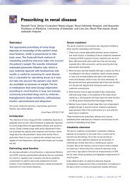
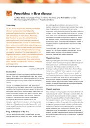
Fig. 2<br />
Interpreting acidaemia on an <strong>arterial</strong> <strong>blood</strong> gas result<br />
pH<br />
Units<br />
PaCO 2<br />
(mmHg)<br />
Units<br />
Base excess<br />
(mmol/L)<br />
Interpretation<br />
Positive<br />
(>+2)<br />
Primary respiratory<br />
acidosis with renal<br />
compensation<br />
High<br />
(>45)<br />
Normal<br />
(–2 – +2)<br />
Primary respiratory<br />
acidosis<br />
Negative<br />
(+2)<br />
Mixed respiratory and<br />
metabolic alkalosis<br />
low<br />
(
Fig. 2 and Fig. 3 are algorithms for the consideration <strong>of</strong> primary<br />
and mixed acid–base disorders. 4<br />
limitations <strong>of</strong> <strong>blood</strong> gas analysis<br />
<strong>The</strong> <strong>blood</strong> gas analysis cannot yield a specific diagnosis. A<br />
patient with asthma may have similar values to another patient<br />
with pneumonia. Alternatively, a patient with chronic obstructive<br />
pulmonary disease and respiratory failure may have similar<br />
results to a patient with pulmonary oedema.<br />
<strong>The</strong> analysis does not reflect the degree to which an<br />
abnormality actually affects a patient. A low PaO 2<br />
does not<br />
necessarily indicate tissue hypoxia, nor does a normal PaO 2<br />
indicate adequate tissue oxygenation. Oxygen utilisation<br />
is influenced by other factors such as regional <strong>blood</strong> flow,<br />
haemoglobin affinity for oxygen and cardiac output.<br />
Blood gas analysis cannot be used as a screening test for early<br />
pulmonary disease. Severe disease may be present before<br />
significant changes are seen in <strong>blood</strong> <strong>gases</strong>.<br />
Venous <strong>blood</strong> <strong>gases</strong><br />
It is easier to obtain a venous sample than an <strong>arterial</strong> sample. In<br />
some situations analysis <strong>of</strong> venous <strong>blood</strong> can provide enough<br />
information to assist in clinical decisions. In general, the pH,<br />
CO 2<br />
and HCO 3ˉ values are similar in venous and <strong>arterial</strong> <strong>blood</strong><br />
(Box 1). <strong>The</strong> main difference is the partial pressure <strong>of</strong> oxygen<br />
in venous <strong>blood</strong> is less than half that <strong>of</strong> <strong>arterial</strong> <strong>blood</strong>. Venous<br />
<strong>blood</strong> should not therefore be used to assess oxygenation.<br />
Conclusion<br />
Measuring <strong>arterial</strong> <strong>blood</strong> <strong>gases</strong> can be a useful adjunct to the<br />
assessment <strong>of</strong> patients with either acute or chronic diseases.<br />
<strong>The</strong> results show if the patient is acidaemic or alkalaemic and<br />
whether the cause is likely to have a respiratory or metabolic<br />
component. <strong>The</strong> PaCO 2<br />
reflects alveolar ventilation and the<br />
PaO 2<br />
reflects the oxygenation <strong>of</strong> <strong>arterial</strong> <strong>blood</strong>. When combined<br />
with a patient's clinical features, <strong>blood</strong> gas analysis can facilitate<br />
diagnosis and management.<br />
References<br />
1. Lightowler JV, Elliot MW. Local anaesthetic infiltration<br />
prior to <strong>arterial</strong> puncture for <strong>blood</strong> gas analysis: a survey<br />
<strong>of</strong> current practice and a randomised double blind placebo<br />
controlled trial. J R Coll Physicians Lond 1997;31:645-6. [R]<br />
2. Harsten A, Berg B, Inerot S, Muth L. Importance <strong>of</strong> correct<br />
handling <strong>of</strong> samples for the results <strong>of</strong> <strong>blood</strong> gas analysis.<br />
Acta Anaesthesiol Scand 1988;32:365-8.<br />
3. Williams AJ. ABC <strong>of</strong> oxygen: assessing and interpreting<br />
<strong>arterial</strong> <strong>blood</strong> <strong>gases</strong> and acid-base balance. BMJ<br />
1998;317:1213-6.<br />
4. Drage S, Wilkinson D. Acid base balance. Update 13. 2001.<br />
World Federation <strong>of</strong> Societies <strong>of</strong> Anaesthesiologists.<br />
http://update.anaesthesiologists.org/wp-content/<br />
uploads/2009/09/Acid-Base-Balance-Update-13.pdf<br />
[cited 2010 Jul 7]<br />
Further reading<br />
Martin L. All you really need to know to interpret <strong>arterial</strong> <strong>blood</strong><br />
<strong>gases</strong>. 2nd ed. Philadelphia, PA: Lippincott Williams & Wilkins;<br />
1999.<br />
Conflict <strong>of</strong> interest: none declared<br />
Self-test questions<br />
<strong>The</strong> following statements are either true or false<br />
(answers on page 131)<br />
7. <strong>The</strong> partial pressure <strong>of</strong> carbon dioxide in <strong>arterial</strong> <strong>blood</strong><br />
(PaCO 2<br />
) is inversely related to alveolar ventilation.<br />
8. <strong>The</strong> partial pressure <strong>of</strong> oxygen in <strong>arterial</strong> <strong>blood</strong> (PaO 2<br />
)<br />
reflects the gas exchange function <strong>of</strong> the lungs.<br />
New drugs<br />
Some <strong>of</strong> the views expressed in the following notes on newly approved products should be regarded as tentative, as there may be limited published<br />
data and little experience in Australia <strong>of</strong> their safety or efficacy. However, the Editorial Executive Committee believes that comments made in good<br />
faith at an early stage may still be <strong>of</strong> value. As a result <strong>of</strong> fuller experience, initial comments may need to be modified. <strong>The</strong> Committee is prepared<br />
to do this. Before new drugs are prescribed, the Committee believes it is important that full information is obtained either from the manufacturer's<br />
approved product information, a drug information centre or some other appropriate source.<br />
Certolizumab<br />
Cimzia (UCB)<br />
pre-filled syringe containing 200 mg in 1 mL <strong>of</strong> liquid<br />
Approved indication: rheumatoid arthritis<br />
<strong>Australian</strong> Medicines Handbook section 15.2.1<br />
Certolizumab, like adalimumab, etanercept and infliximab, is<br />
a tumour necrosis factor inhibitor indicated for rheumatoid<br />
arthritis. It is a recombinant humanised antibody Fab' fragment<br />
which has been pegylated to extend its plasma half-life to that<br />
<strong>of</strong> the whole antibody.<br />
Peak plasma concentrations are reached between 54 and 171<br />
hours after subcutaneous administration and its bioavailability is<br />
approximately 80%. <strong>The</strong> terminal elimination half-life is around<br />
14 days. However, the presence <strong>of</strong> antibodies to certolizumab<br />
increases its clearance and appears to correlate with reduced<br />
patient responses. Giving methotrexate concomitantly with<br />
certolizumab reduces the formation <strong>of</strong> anti-certolizumab<br />
antibodies.<br />
www.australianprescriber.com<br />
| Volume 33 | NumBeR 4 | AuGuST 2010 129