Australasian Mycologis - First Year Biology
Australasian Mycologis - First Year Biology
Australasian Mycologis - First Year Biology

You also want an ePaper? Increase the reach of your titles
YUMPU automatically turns print PDFs into web optimized ePapers that Google loves.
countries including Brazil (Sunhede 1989) and South<br />
Africa (Dyer 1948, Sunhede 1989) from the<br />
Southern Hemisphere. It is described in detail here<br />
to assist with the mapping of possible rare and<br />
endangered species in Australia, and its distribution<br />
may provide evidence as to whether the species is<br />
native to Australia or localized to an area where it<br />
may have been introduced with exotic plant material<br />
and from where it is slowly expanding.<br />
Materials and Methods<br />
Tissue was examined by bright field microscopy in<br />
5% W/V KOH, with the later addition of 1% W/V<br />
Congo Red for contrast as described previously<br />
(Rees et al. 2001). Colour description numbers for<br />
features of the fruitbody follow Kornerup &<br />
Wanscher (1981). Preparations were also examined<br />
in Melzer's Iodine to determine colour change in<br />
iodine. Measures for spore sizes do not include spore<br />
ornamentation, but as spore ornamentation is so<br />
distinctive a feature in Myriostoma coliforme,<br />
figures have also been provided which include<br />
ornamentation. Spore size range is given for 20<br />
spores.<br />
Results<br />
Myriostoma coliforme (With. : Pers.) Corda, in<br />
Anleit. Stud. Mykol. 81 (1842).<br />
Lycoperdon coliforme With, in Bot. arr. veg. Gr.<br />
Brit. Ed. 1,2: 783 (1776).<br />
Geastrum coliforme (With.) Pers. in Synops. Meth.<br />
Fung. 131 (1801).<br />
Myriostoma anglicum Desv., J. Bot. (Morot) 2: 104<br />
(1809).<br />
Geaster columnatum Lev., Ann. Sci. Nat. Bot. Sir. 3,<br />
5: 161 (1846).<br />
The 'Pepper Pot'.<br />
Basidiomata 40-55 mm wide, epigeous, consisting<br />
of an outer exoperidium splitting unevenly into 9-11<br />
lacerated rays recurving at the tips, mounted on a<br />
short pedicel (1-2 mm) with cottony, white, basal<br />
mycelium. The rays have three layers, an outer, pale<br />
ochraceous, mycelial layer tending to crack in<br />
places, revealing the middle fibrous layer consisting<br />
of densely packed, hyaline, hyphae (1.0-2.5 um)<br />
wide. The uppermost cellular, pseudoparenchymatous<br />
layer is pale beige initially,<br />
becoming more yellow to brown with age. It tends to<br />
wear away in places, leaving dark brown,<br />
pulverulent patches, which contain thick-walled,<br />
overlapping, spherical cells, 25-30 um in diameter,<br />
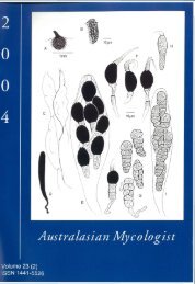
forming a cellular, pigmented layer (Fig. 1C).<br />
Surmounting the rays, on laterally flattened, short,<br />
cream stalks 3^1 mm high which tend to fuse near<br />
the top, is the flattened, globose to oval<br />
endoperidium, 20-30 mm wide with a punctate,<br />
shiny, gun-metal grey-brown surface broken by one<br />
to six stomata which have fimbriate margins. The<br />
greyish brown glebal contents are hairy, containing<br />
several columellae, continuous with the flattened<br />
stalks at the base of the endoperidium. Attached to<br />
the columellae and the peridial walls, are aseptate,<br />
thick-walled, unbranched, yellow-brown, capillitial<br />
threads 3.6-6.0 um wide with tapering ends (Fig.<br />
IB). The columellae are not connected to the<br />
stomata, but terminate at various points in the glebal<br />
mass some distance from the stomata. Basidiospores<br />
(20) 3.9-4.8 x 3.9-4.8 um excluding ornamentation,<br />
5.4-7.0 x 5.4-7.0 um including conspicuous,<br />
irregular, flaring ornamentation up to 2 um high<br />
(Fig. 1 A), are globose to marginally subglobose,<br />
yellow-brown, and exhibit no reaction in Melzer's<br />
Iodine. Basidia and clamp connections were not<br />
observed. Hyphae of the endoperidial wall (2.6-<br />
4.0 um wide) are aseptate and pigmented, except at<br />
the margins of the stomata, where there are densely<br />
packed, hyaline, thin-walled, narrow hyphae, 1.0—<br />
2.6 um in width, similar to the fibrous layer of the<br />
exoperidium, accompanied by small spherical to<br />
oval cells (Fig. ID). These structures may be the<br />
remains of the pseudoparenchymatous, exoperidial<br />
layer from which they detached as the basidiome<br />
expanded.<br />
Habit and habitat: In the Sydney region,<br />
Myriostoma coliforme occurs in groups on sandy<br />
soil in association with a range of plants in well<br />
mulched, but well-drained, urban garden plots with a<br />
sunny aspect, close to marine foreshores.<br />
Material examined: Sydney. Royal Botanic Gardens,<br />
on leaf litter on garden beds, R.G. Coveny, 3.vi.l978<br />
(F172); 27.vi.1980 (R.G.Coveny 61); 13.iv. 1981<br />
(R.G.Coveny 49); 25.iii.1982 (R.G.Coveny 1982); on<br />
soil under Ficus virens Aiton., Bed 29, F. Taeker,<br />
24.V.1985, (UNSW 85/478); Bed 16, on soil under<br />
Ficus virens, R. Coveny, 18.viii.2003, (UNSW<br />
03/23).<br />
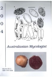
Plate 1 (top right). Myriostoma coliforme (UNSW<br />
85/478) with numerous stomatal openings.<br />
Photography F. Taeker.<br />
Figure 1 (bottom right). Myriostoma coliforme<br />
(UNSW 03/23). A. Basidiospores. B. Capillitial<br />
threads with tapering ends. C. Cells from the<br />
pseudoparenchymatous layer of the exoperidium. D.<br />
Cells from the single stomatal opening in the smallest<br />
fruitbody. Scale 1 mm = 0.5 um for A, 2.0 um for B<br />
and 1 um for C. and D.