10 Identification guide to common periphyton in New ... - NIWA
10 Identification guide to common periphyton in New ... - NIWA
10 Identification guide to common periphyton in New ... - NIWA

You also want an ePaper? Increase the reach of your titles
YUMPU automatically turns print PDFs into web optimized ePapers that Google loves.
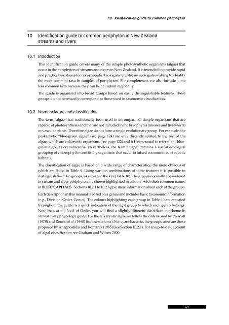
<strong>10</strong> <strong>Identification</strong> <strong>guide</strong> <strong>to</strong> <strong>common</strong> periphy<strong>to</strong>n<br />
<strong>10</strong> <strong>Identification</strong> <strong>guide</strong> <strong>to</strong> <strong>common</strong> periphy<strong>to</strong>n <strong>in</strong> <strong>New</strong> Zealand<br />
streams and rivers<br />
<strong>10</strong>.1 Introduction<br />
This identification <strong>guide</strong> covers many of the simple pho<strong>to</strong>synthetic organisms (algae) that<br />
occur <strong>in</strong> the periphy<strong>to</strong>n of streams and rivers <strong>in</strong> <strong>New</strong> Zealand. It is <strong>in</strong>tended <strong>to</strong> provide rapid<br />
and practical assistance for non-specialist biologists and stream ecologists wish<strong>in</strong>g <strong>to</strong> identify<br />
the most <strong>common</strong> taxa <strong>in</strong> samples of periphy<strong>to</strong>n. For completeness we also <strong>in</strong>clude some<br />
less <strong>common</strong> taxa because they can be abundant regionally.<br />
The <strong>guide</strong> is organised <strong>in</strong><strong>to</strong> broad groups based on easily dist<strong>in</strong>guishable features. These<br />
groups do not necessarily correspond <strong>to</strong> those used <strong>in</strong> taxonomic classification.<br />
<strong>10</strong>.2 Nomenclature and classification<br />
The term “algae” has traditionally been used <strong>to</strong> encompass all simple organisms that are<br />
capable of pho<strong>to</strong>synthesis and that are not <strong>in</strong>cluded <strong>in</strong> the bryophytes (mosses and liverworts)<br />
or vascular plants. Therefore algae do not form a s<strong>in</strong>gle evolutionary group. For example, the<br />
prokaryotic “blue-green algae” (see page 124) are only distantly related <strong>to</strong> the rest of the<br />
algae, which are eukaryotic organisms (see page 122) and it is now usual <strong>to</strong> refer <strong>to</strong> the bluegreen<br />
algae as cyanobacteria. Nevertheless, the term “algae” rema<strong>in</strong>s a useful ecological<br />
group<strong>in</strong>g of chlorophyll a-conta<strong>in</strong><strong>in</strong>g organisms that occur <strong>in</strong> mixed communities <strong>in</strong> aquatic<br />
habitats.<br />
The classification of algae is based on a wide range of characteristics, the more obvious of<br />
which are listed <strong>in</strong> Table 9. Us<strong>in</strong>g various comb<strong>in</strong>ations of these features it is possible <strong>to</strong><br />
dist<strong>in</strong>guish the ma<strong>in</strong> groups, as shown <strong>in</strong> the key (Table <strong>10</strong>). The groups normally encountered<br />
<strong>in</strong> stream and river periphy<strong>to</strong>n are shown highlighted <strong>in</strong> colours, with their <strong>common</strong> names<br />
<strong>in</strong> BOLD CAPITALS. Sections <strong>10</strong>.2.1 <strong>to</strong> <strong>10</strong>.2.6 give more <strong>in</strong>formation about each of the groups.<br />
Each description <strong>in</strong> this manual is based on a genus and <strong>in</strong>cludes basic taxonomic <strong>in</strong>formation<br />
(e.g., Division, Order, Genus). The colours highlight<strong>in</strong>g each group <strong>in</strong> Table <strong>10</strong> are repeated<br />
throughout the <strong>guide</strong> as a quick <strong>in</strong>dication of the algal group <strong>to</strong> which each genus belongs.<br />
Note that, at the level of Order, you will f<strong>in</strong>d a slightly different classification scheme <strong>in</strong><br />
almost every phycology <strong>guide</strong>. For the eukaryotic algae we follow the orders used by Prescott<br />
(1978) and Round at al. (1990) (for the dia<strong>to</strong>ms). For cyanobacteria, the groups used are those<br />
proposed by Anagnostidis and Komàrek (1985) (see Section <strong>10</strong>.2.1). For an up-<strong>to</strong>-date account<br />
of algal classification see Graham and Wilcox 2000.<br />
121<br />
121
122<br />
Stream Periphy<strong>to</strong>n Moni<strong>to</strong>r<strong>in</strong>g Manual<br />
Table 9: Characteristics of algae used <strong>in</strong> classification and identification<br />
Characteristic Examples<br />
Cellular The simple pho<strong>to</strong>synthetic organisms found <strong>in</strong> periphy<strong>to</strong>n are<br />
organisation: either:<br />
• “prokaryotes”, <strong>in</strong> which the cell <strong>in</strong>clusions are not bound by<br />
membranes and pigments are distributed throughout the cell,<br />
giv<strong>in</strong>g a diffuse appearance (these are the “blue-greens”<br />
(Cyanobacteria)); or<br />
• “eukaryotes”, <strong>in</strong> which cell <strong>in</strong>clusions such as nucleus,<br />
mi<strong>to</strong>chondria and chloroplasts are dist<strong>in</strong>guishable and are<br />
enclosed by membranes.<br />
Cell pigments All possess the pho<strong>to</strong>synthetic pigment chlorophyll a, but<br />
chlorophylls b and c, and phycobil<strong>in</strong>s also occur <strong>in</strong> certa<strong>in</strong> groups.<br />
Flagella Flagella (s<strong>in</strong>gular: flagellum) are long, th<strong>in</strong> flexible appendages that<br />
allow many algal cells <strong>to</strong> move around. Some algae have no<br />
flagella; sometimes they occur only at the reproductive stages; <strong>in</strong><br />
some cases they are always present and there may be one, two or<br />
four. Where there are two or more flagella they may be the same or<br />
different lengths. Flagellated species are ma<strong>in</strong>ly plank<strong>to</strong>nic and are<br />
rarely encountered <strong>in</strong> stream periphy<strong>to</strong>n.<br />
Colour Cell colour ranges from red <strong>to</strong> brown through yellow and green <strong>to</strong><br />
blue-green and even blue. The major taxonomic groups are named<br />
on the basis of colour but, because many taxa conta<strong>in</strong> a mixture of<br />
pigments, colour is not always a reliable <strong>guide</strong> <strong>to</strong> the groups.<br />
Colour may vary with environmental conditions and may also vary<br />
accord<strong>in</strong>g <strong>to</strong> the type and duration of preservation.<br />
Food reserves Some of these organisms have food reserves of starch and<br />
therefore a positive reaction <strong>to</strong> the iod<strong>in</strong>e starch test. Others s<strong>to</strong>re<br />
food as leucos<strong>in</strong> or oil and do not react <strong>to</strong> the starch test.<br />
Cell wall type Cyanobacteria often possess a muco-polysaccharide sheath;<br />
green algae have cellulose cell walls laid down <strong>in</strong> a criss-cross<br />
pattern; dia<strong>to</strong>ms have rigid walls made of silica.<br />
Reproduction Some algae and cyanobacteria reproduce by cell division, each<br />
new cell be<strong>in</strong>g <strong>in</strong>dependent of all others. Most algae also produce<br />
specialised reproductive bodies, or spores, and <strong>in</strong> some groups the<br />
form of these is the ma<strong>in</strong> character that dist<strong>in</strong>guishes species.<br />
Sexual reproduction is also <strong>common</strong>.<br />
Gross This is the group of characteristics that is of most practical use <strong>in</strong><br />
structure identify<strong>in</strong>g algae. For example, is the alga s<strong>in</strong>gle-celled,<br />
filamen<strong>to</strong>us, colonial or sheet-like? Does the alga have different<br />
types of cells – e.g., heterocysts? Many of the same structural<br />
characteristics occur <strong>in</strong> different groups, and thus do not reflect<br />
natural relationships.<br />
Ecology Aquatic algae <strong>in</strong>clud<strong>in</strong>g cyanobacteria are capable of <strong>in</strong>habit<strong>in</strong>g<br />
almost all damp <strong>to</strong> wet habitats <strong>in</strong> existence, from sea water<br />
through <strong>to</strong> hot spr<strong>in</strong>gs. However, many types occur only with<strong>in</strong> a<br />
certa<strong>in</strong> range of conditions.
Table <strong>10</strong>: Key <strong>to</strong> the major groups of <strong>common</strong> freshwater periphy<strong>to</strong>n<br />
<strong>10</strong> <strong>Identification</strong> <strong>guide</strong> <strong>to</strong> <strong>common</strong> periphy<strong>to</strong>n<br />
1a Cells without chloroplasts (though granules <strong>in</strong> the cells could be mistaken for chloroplasts),<br />
diffuse blue-green, olive or red-brown. Cells often very small.<br />
Division: Cyanobacteria (BLUE-GREENS) [see page 124]<br />
1b Cells with chloroplasts (discrete structure <strong>in</strong> which the cell pigment is concentrated). 2<br />
2a Chloroplasts pale <strong>to</strong> deep grass green. 3<br />
2b Chloroplasts some other colour. 4<br />
3a S<strong>in</strong>gle-celled, filamen<strong>to</strong>us or colonial form, normally with one or two chloroplasts arranged as<br />
sheets <strong>in</strong>side the cell wall or along the length of the cell. Sometimes with many chloroplasts.<br />
3b<br />
Where flagella are present there are two or four and their lengths are equal. Positive reaction <strong>to</strong><br />
starch test.<br />
Division: Chlorophyta; Class: Chlorophyceae (GREEN ALGAE) [see page 124]<br />
Large, erect plants, typically 4-50 cm long, with regular branches. Rooted by rhizoids <strong>in</strong> soft<br />
sediment. Cells large with numerous chloroplasts.<br />
Division: Chlorophyta; Class: Charophyceae<br />
3c S<strong>in</strong>gle-celled, colonial or filamen<strong>to</strong>us, normally with two or more small, discoid chloroplasts per<br />
cell. Where flagella are present there are two, of unequal length. Negative reaction <strong>to</strong> starch<br />
test.<br />
Division: Xanthophyta (YELLOW-GREEN ALGAE) [see page 125]<br />
3d S<strong>in</strong>gle cells with one long, thick flagellum emerg<strong>in</strong>g from a depression at the end of the cell.<br />
Two-<strong>to</strong>-many discoid chloroplasts. Cell wall can be elastic and striated.<br />
Division: Euglenophyta<br />
4a Cells with a rigid, ornamented silica wall composed of two halves, sometimes form<strong>in</strong>g filaments.<br />
Often motile, but no flagella. One <strong>to</strong> many brownish chloroplasts.<br />
Division: Bacillariophyta (DIATOMS) [see page 126]<br />
4b Cells not as above. 5<br />
5a Cells with two flagella, partly with<strong>in</strong> two furrows on the cell surface, one around the cell, the<br />
other at right angles. Cell walls of smooth or angular plates, flat or with project<strong>in</strong>g horns.<br />
Positive reaction <strong>to</strong> starch test.<br />
5b Cells without two deep furrows<br />
Division: D<strong>in</strong>ophyta (= Pyrrophyta {d<strong>in</strong>oflagellates})<br />
6<br />
6a Chloroplasts pale yellow <strong>to</strong> brown, usually 1 or 2 per cell. Cells s<strong>in</strong>gle, colonial or (rarely)<br />
filamen<strong>to</strong>us. If flagella are present they are either one long one, or one long and one short.<br />
Division: Chrysophyta (GOLDEN-BROWN ALGAE) [see page 128]<br />
6b Chloroplasts some other colour, no cysts, flagella (if present) not strongly unequal. 7<br />
7a S<strong>in</strong>gle-celled, bean-shaped with two slightly unequal flagella aris<strong>in</strong>g from a small depression of<br />
furrow. One or two chloroplasts, coloured olive, red or blue.<br />
7b<br />
Division: Cryp<strong>to</strong>phyta<br />
Plants filamen<strong>to</strong>us and frequently with complex structure, or flattened and encrust<strong>in</strong>g, or (rarely)<br />
s<strong>in</strong>gle-celled. One or more chloroplasts per cell, coloured olive, red or blue. Usually attached <strong>to</strong><br />
rocks and mosses <strong>in</strong> streams. No flagella.<br />
Division: Rhodophyta (RED ALGAE) [see page 128]<br />
Throughout the idenification <strong>guide</strong>, the above colours are used <strong>to</strong> <strong>in</strong>dicate the major<br />
algal group <strong>to</strong> which each genus belongs.<br />
123<br />
123
124<br />
Stream Periphy<strong>to</strong>n Moni<strong>to</strong>r<strong>in</strong>g Manual<br />
<strong>10</strong>.2.1 Cyanobacteria<br />
Cyanobacteria are easily dist<strong>in</strong>guished from other algae by the absence of chloroplasts – the<br />
discrete organelles that conta<strong>in</strong> the pho<strong>to</strong>synthetic pigments <strong>in</strong> eukaryotes. Instead, these<br />
pigments are diffused throughout the cell pro<strong>to</strong>plasm. Additional pigments found <strong>in</strong> this<br />
group are responsible for the range of colours they exhibit, most notably the blue-green<br />
appearance. The orders of cyanobacteria represented <strong>in</strong> this <strong>guide</strong> are listed below. These<br />
groups are those used by Anagnostidis and Komàrek (1985), with details <strong>in</strong> Anagnostidis<br />
and Komàrek (1988a, b) and Komàrek and Anagnostidis (1986, 1989). This classification system<br />
follows the traditional botanical approach, be<strong>in</strong>g based on “Geitler's” system (see Geitler<br />
1925, 1932, 1942) but now <strong>in</strong>corporat<strong>in</strong>g morphological, cy<strong>to</strong>logical and ecophysiological<br />
characters. Note that several other classification schemes have been proposed based on the<br />
bacteriological approach (Stanier 1977 and Rippka et al. 1979) or on “Drouet's” system (Drouet<br />
and Daily 1956 and Drouet 1981), where the number of taxa was reduced considerably.<br />
Chroococcales: Unicells, colonies, pseudoparenchyma<strong>to</strong>us colonies or<br />
pseudofilamen<strong>to</strong>us colonies. Trichomes, heterocysts and ak<strong>in</strong>etes are<br />
lack<strong>in</strong>g. Cell division <strong>in</strong> one, two or more perpendicular planes.<br />
Examples <strong>in</strong>clude Gloeothece and Chamaesiphon.<br />
Oscilla<strong>to</strong>riales: Cells form<strong>in</strong>g trichomes (a s<strong>in</strong>gle row of cells connected by cross<br />
walls); false branch<strong>in</strong>g, gas vesicles and sheaths lack<strong>in</strong>g or<br />
facultatively present. Heterocysts, ak<strong>in</strong>etes and true branch<strong>in</strong>g absent.<br />
Reproduction occurs by “hormogonia” formation through trichome<br />
fragmentation. Examples are Phormidium, Oscilla<strong>to</strong>ria and Lyngbya.<br />
Nos<strong>to</strong>cales: Cells form<strong>in</strong>g trichomes with a wide or narrow mucilag<strong>in</strong>ous sheath.<br />
Trichomes unbranched or falsely branched (<strong>in</strong>itiated at a heterocyst or<br />
between two vegetative cells). Specialised nitrogen-fix<strong>in</strong>g cells<br />
(heterocysts) and spore cells (ak<strong>in</strong>etes) may be present. Reproduction<br />
ma<strong>in</strong>ly by hormogonia or hormocysts. Examples are Nos<strong>to</strong>c,<br />
Tolypothrix, Calothrix and Rivularia.<br />
Stigonematales: Cells form<strong>in</strong>g true trichomes, sometimes comb<strong>in</strong>ed with<br />
pseudotrichomes. True branch<strong>in</strong>g always present while false branch<strong>in</strong>g<br />
may occur. Ak<strong>in</strong>etes rarely present while heterocysts occur<br />
facultatively <strong>in</strong> several genera. Reproduction ma<strong>in</strong>ly by hormogonia<br />
and hormocysts. Best known example is Stigonema.<br />
<strong>10</strong>.2.2 The green algae (Division: Chlorophyta)<br />
The orders listed below belong <strong>to</strong> the class Chlorophyceae and are represented <strong>in</strong> the periphy<strong>to</strong>n<br />
of <strong>New</strong> Zealand streams and rivers. These are traditional orders (largely as used <strong>in</strong><br />
Prescott 1973). See Graham and Wilcox (2000) for recent classification schemes.<br />
Tetrasporales: These occur <strong>in</strong> a non-motile vegetative form, usually <strong>in</strong> colonies held<br />
<strong>to</strong>gether by mucilage (e.g., Gloeocystis, Palmella). The cells can reproduce<br />
by simple cell division. The chloroplasts are usually described as<br />
cup-like – that is, they are curved so that they l<strong>in</strong>e part of the <strong>in</strong>side of<br />
the cell.<br />
Chlorococcales: These can look similar <strong>to</strong> the Tetrasporales (e.g., Oocystis). The ma<strong>in</strong><br />
difference is that the cells do not reproduce by simple cell division <strong>in</strong><br />
the vegetative state, though they do divide <strong>to</strong> form spores. S<strong>in</strong>gle-
<strong>10</strong> <strong>Identification</strong> <strong>guide</strong> <strong>to</strong> <strong>common</strong> periphy<strong>to</strong>n<br />
Ulotrichales:<br />
celled or colonial. Examples found <strong>in</strong> periphy<strong>to</strong>n are Pediastrum,<br />
Ankistrodesmus, Scenedesmus.<br />
Unbranched simple filaments with mostly cyl<strong>in</strong>drical cells conta<strong>in</strong><strong>in</strong>g<br />
a s<strong>in</strong>gle band-like chloroplast similar <strong>to</strong> that <strong>in</strong> the Chlorococcales.<br />
Most species are attached when young, but become free-float<strong>in</strong>g later.<br />
The best known filamen<strong>to</strong>us alga <strong>in</strong> this order is Ulothrix. Others<br />
<strong>in</strong>clude Gem<strong>in</strong>ella.<br />
Ulvales Many cells <strong>in</strong> a sheet-like arrangement, e.g., Enteromorpha, <strong>in</strong> which<br />
the sheets form hollow filaments.<br />
Microsporales: Unbranched filaments of cyl<strong>in</strong>drical cells with walls <strong>in</strong> sections with a<br />
cell wall at the centre – so that broken-up filaments comprise Hshaped<br />
pieces. The chloroplast covers the whole wall of the cell and<br />
may be th<strong>in</strong> or dense. The only genus <strong>in</strong> this order is Microspora.<br />
Cyl<strong>in</strong>drocapsales: Another order with only one genus – Cyl<strong>in</strong>drocapsa. Usually<br />
filamen<strong>to</strong>us with dense, large chloroplasts.<br />
Chae<strong>to</strong>phorales: Branched filaments that arise from a holdfast. Cells form<strong>in</strong>g branches<br />
often smaller than those nearer the base. Chloroplasts are parietal,<br />
sometimes completely cover<strong>in</strong>g the cell wall. Examples are<br />
Chae<strong>to</strong>phora, Draparnaldia and Stigeoclonium.<br />
Cladophorales: (also known as Siphoncladales) Repeatedly branched filaments,<br />
Oedogoniales:<br />
cyl<strong>in</strong>drical cells, thick walls. Chloroplast parietal and net-like <strong>in</strong><br />
young, healthy specimens, but sometimes appear<strong>in</strong>g as many small<br />
disks. Often the cell walls are very thick and the filaments frequently<br />
carry many dia<strong>to</strong>m epiphytes, e.g., Cladophora, Rhizoclonium.<br />
There are both unbranched and branched forms <strong>in</strong> this order, which is<br />
characterised by occasional r<strong>in</strong>g-like scars at the front end of cells,<br />
caused by cell division. Genera described <strong>in</strong> this <strong>guide</strong> are Oedogonium<br />
(unbranched) and Bulbochaete (branched).<br />
Zygnematales: Unbranched filaments of long or short cyl<strong>in</strong>drical cells with a cellulose<br />
cell wall; end walls are separated by a middle lamella. The<br />
chloroplasts are large, with 1–2 per cell usually. Species <strong>in</strong> this order<br />
have no motile reproductive cells. Instead, cells transform <strong>in</strong><strong>to</strong><br />
amoeboid gametes, two of which fuse <strong>to</strong> form the zygospore (via<br />
“conjugation” of cells). Genera represented <strong>in</strong> this <strong>guide</strong> are<br />
Mougeotia, Spirogyra and Zygnema.<br />
Desmidiales: The desmids are s<strong>in</strong>gle-celled forms (occasionally <strong>in</strong> filaments) related<br />
<strong>to</strong> the Zygnematales through hav<strong>in</strong>g a similar mode of reproduction.<br />
Most desmids are divided <strong>in</strong><strong>to</strong> two equal halves. The chloroplasts are<br />
variable and can be complex. Examples found <strong>in</strong> periphy<strong>to</strong>n are<br />
Cosmarium, Closterium and Staurastrum.<br />
<strong>10</strong>.2.3 The yellow-green algae (Division: Xanthophyta)<br />
The yellow-green algae can be difficult <strong>to</strong> dist<strong>in</strong>guish from the green algae. The ma<strong>in</strong> features<br />
that separate the two divisions are a predom<strong>in</strong>ance of yellowish pigments (e.g., carotenoids)<br />
<strong>in</strong> the yellow-greens, and leucos<strong>in</strong> or oils as food reserves, rather than starch. Just<br />
two orders are <strong>in</strong>cluded <strong>in</strong> this manual.<br />
125<br />
125
126<br />
Stream Periphy<strong>to</strong>n Moni<strong>to</strong>r<strong>in</strong>g Manual<br />
Tribonematales Branched or unbranched filaments, e.g., Tribonema.<br />
Vaucheriales Branched, cyl<strong>in</strong>drical filaments, no cross walls, e.g., Vaucheria.<br />
<strong>10</strong>.2.4 The dia<strong>to</strong>ms (Division: Bacillariophyta)<br />
The structure of dia<strong>to</strong>ms means that it is relatively easy <strong>to</strong> identify many of them <strong>to</strong> species<br />
or even variety. In this <strong>guide</strong>, we <strong>in</strong>clude descriptions of many <strong>common</strong> species – particularly<br />
those that are useful <strong>in</strong>dica<strong>to</strong>rs of environmental conditions. This should enable you <strong>to</strong> dist<strong>in</strong>guish<br />
them from other species that are similar <strong>in</strong> appearance, but that you won’t necessarily<br />
be able <strong>to</strong> identify beyond genus level. Some identifications should be possible us<strong>in</strong>g<br />
fresh or frozen material.<br />
For detailed studies on dia<strong>to</strong>ms it is necessary <strong>to</strong> exam<strong>in</strong>e acid-cleaned specimens at magnifications<br />
of up <strong>to</strong> x<strong>10</strong>00, us<strong>in</strong>g an oil-immersion objective on a light microscope <strong>in</strong> order <strong>to</strong><br />
see the ornamentation of the cell walls. In addition, you need an understand<strong>in</strong>g of their structure<br />
and a comprehensive collection of specialised dia<strong>to</strong>m taxonomy texts because, as yet,<br />
there is no complete <strong>guide</strong> <strong>to</strong> dia<strong>to</strong>ms <strong>in</strong> <strong>New</strong> Zealand.<br />
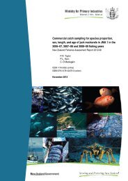
Briefly, the dia<strong>to</strong>m cell wall is made of<br />
silica and the basic construction is of two<br />
Structure of a generalised dia<strong>to</strong>m frustule<br />
halves (valves) that fit <strong>to</strong>gether with one<br />
raphe<br />
stigma<br />
half overlapp<strong>in</strong>g the other so that the<br />
striae, made<br />
whole structure resembles a chocolate<br />
box. The two valves <strong>to</strong>gether are called<br />
the frustule. Classification is based on the<br />
up of alveolae<br />
(or puncta)<br />
arrangement of various surface features<br />
and etch<strong>in</strong>gs on the frustule. A major<br />
valves<br />
girdle,<br />
made up<br />
of girdle<br />
feature is the raphe, a pair of slits <strong>in</strong> the<br />
valve face. When present, the raphe may<br />
be on both valves or on one valve only.<br />
bands<br />
The two valves may be connected by a<br />
(draw<strong>in</strong>g: Liz Bergey)<br />
girdle or a series of girdle bands. The valve faces may carry rows (called striae) of open<strong>in</strong>gs<br />
or depressions (known as puncta or areolae). Larger holes are called “stigmata” (s<strong>in</strong>gular:<br />
stigma). Most descriptions of dia<strong>to</strong>ms are based on the view from the <strong>to</strong>p (“valve view”).<br />
The view from the side (“girdle view”) can look quite different.<br />
The structure of the chloroplasts and other cell <strong>in</strong>clusions may also be considered <strong>in</strong><br />
descriptions. For more details about the f<strong>in</strong>e structure of dia<strong>to</strong>ms refer <strong>to</strong> Barber and Haworth<br />
(1981), Round, Crawford and Mann (1990) and Cox (1996).<br />
As for the algae <strong>in</strong> general, various classification systems have been proposed for dia<strong>to</strong>ms<br />
(see Bourelly 1981, Krammer and Lange-Bertalot 1987–1997, Round et al. 1990) and we follow<br />
the system of orders of Round et al. 1990. For descriptive purposes, these orders can be<br />
placed <strong>in</strong> seven groups (as used by E. S<strong>to</strong>ermer, pers. comm.) that are convenient though not<br />
necessarily natural (i.e., made up of related organisms). The illustrations below show<br />
representative valve and girdle views <strong>in</strong> each group.<br />
Centrics: Radially symmetric valves. No raphe system.<br />
Thalassiosirales: Cyclotella;<br />
Melosirales: Melosira; Aulacosirales: Aulacoseira.
Araphid Usually symmetrical along the all axes of the<br />
pennates: valve (exceptions: Meridion, Asterionella). No raphe<br />
system on either valve, but usually a “pseudoraphe”<br />
present (i.e., a break <strong>in</strong> the striae, as if a<br />
raphe were present).<br />
Fragilariales: Fragilaria, Fragilariaforma, Staurosira,<br />
Dia<strong>to</strong>ma, Meridion, Synedra;<br />
Tabellariales: Tabellaria.<br />
Monoraphid Usually symmetrical along at least two axes. A<br />
pennates: raphe system present on one valve only. The second<br />
valve may or many not have a pseudoraphe.<br />
The ornamentation on the two valves can be quite<br />
different. (Illustration shows raphe valve, girdle<br />
view and pseudoraphe valve.)<br />
Achnanthales: Achnanthes, Achnanthidium,<br />
Cocconeis.<br />
Eunotioids: A rudimentary raphe system on both valves, with<br />
the two valves usually similar. There may be asymmetry<br />
along all three axes.<br />
Eunotiales: Eunotia, Eunophora, Act<strong>in</strong>ella.<br />
Biraphid Symmetric naviculoids: These are usually more<br />
pennates: or less symmetric along both the long and crossvalve<br />
axes (exception: some P<strong>in</strong>nularia species). A<br />
fully developed raphe system present on both<br />
valves. The two valves are usually identical.<br />
Mas<strong>to</strong>gloiales: Mas<strong>to</strong>gloia;<br />
Naviculales: Brachysira, Frustulia, Navicula,<br />
Neidium, P<strong>in</strong>nularia, Stauroneis.<br />
Biraphid Asymmetric: Lack<strong>in</strong>g symmetry along at least one<br />
pennates: axis. A raphe system present on both valves. The<br />
two valves are usually identical (an exception is<br />
Rhoicosphenia).<br />
Cymbellales: Cymbella, Rhoicosphenia, Encyonema,<br />
Gomphonema, Gomphoneis, Reimeria;<br />
Thalassiophysales: Amphora.<br />
Biraphid “External” raphe: The raphe lies partly or wholly<br />
pennates: along the edge of the valve, sometimes with<strong>in</strong> a<br />
raised canal. The structure can be quite complex.<br />
Some dia<strong>to</strong>ms <strong>in</strong> this group are nitrogen-fix<strong>in</strong>g<br />
because of a symbiotic relationship with cyanophytes,<br />
and can therefore <strong>to</strong>lerate nitrogen-limited<br />
situations. Bacillariales: Nitzschia;<br />
Rhopalodales: Epithemia, Rhopalodia;<br />
Surirellales: Surirella, Stenopterobia.<br />
<strong>10</strong> <strong>Identification</strong> <strong>guide</strong> <strong>to</strong> <strong>common</strong> periphy<strong>to</strong>n<br />
127<br />
127
128<br />
Stream Periphy<strong>to</strong>n Moni<strong>to</strong>r<strong>in</strong>g Manual<br />
<strong>10</strong>.2.5 The golden-brown algae (Division: Chrysophyta)<br />
Mostly motile cells, though the division <strong>in</strong>cludes some filamen<strong>to</strong>us and sheet-like forms.<br />
This Division is more <strong>common</strong>ly found <strong>in</strong> lake habitats than <strong>in</strong> flow<strong>in</strong>g waters. Chrysophytes<br />
are characterised by possess<strong>in</strong>g a very large chloroplast, and by their particular comb<strong>in</strong>ation<br />
of pigments. The one example described is D<strong>in</strong>obryon (Order: Chrysomonadales).<br />
<strong>10</strong>.2.6 The red algae (Division: Rhodophyta)<br />
The red algae are dist<strong>in</strong>guished from the other algal divisions by their methods of reproduction<br />
and life his<strong>to</strong>ries. The “red” comes from the predom<strong>in</strong>ance of the red pigment phycoerythr<strong>in</strong><br />
<strong>in</strong> many species. Most red algae are mar<strong>in</strong>e. The examples <strong>in</strong> this manual represent<br />
the two subclasses of the Rhodophyta. See Bourelly (1985) for a version of classification f<strong>in</strong>er<br />
than this. See Entwistle and Kraft (1984) for an account of freshwater red algae <strong>in</strong> south-east<br />
Australia, <strong>in</strong>clud<strong>in</strong>g notes on the <strong>New</strong> Zealand flora.<br />
Bangiophycidae Freshwater forms have a branched, unbranched or plate-like thallus,<br />
with thick cell walls. Asexual reproduction is by non-motile spores.<br />
Sexual reproduction rare. Represented by Chroodactylon, Compsopogon.<br />
Florideophycidae The more “advanced” group, with a wide range of morphologiesfrom<br />
slightly branched filaments <strong>to</strong> more complex growths. The female sex<br />
organs (carpogonia) are characteristic. Representatives are:<br />
Audou<strong>in</strong>ella, Batrachospermum, Bostrychia.<br />
<strong>10</strong>.3 How <strong>to</strong> use this <strong>guide</strong><br />
To aid identification, each taxon described <strong>in</strong> this <strong>guide</strong> is assigned <strong>to</strong> a group on the basis of<br />
cell type and growth form. These groups do not necessarily correspond <strong>to</strong> the traditional<br />
classification outl<strong>in</strong>ed above.<br />
Pho<strong>to</strong>synthetic periphy<strong>to</strong>n may be subdivided <strong>in</strong><strong>to</strong> two cell types:<br />
• Conta<strong>in</strong><strong>in</strong>g “organelles” (dist<strong>in</strong>guishable structures like chloroplasts and a nucleus),<br />
but may not have cell walls. These are the green, yellow-green and red algae, and the<br />
dia<strong>to</strong>ms.<br />
• Cell contents diffuse, granular, with no dist<strong>in</strong>guishable organelles (but take care not <strong>to</strong><br />
<strong>in</strong>terpret granules as chloroplasts). These are the cyanobacteria.<br />
These groups can then each be divided <strong>in</strong><strong>to</strong> four broad growth forms that are easily dist<strong>in</strong>guished:<br />
• flamen<strong>to</strong>us, unbranched;<br />
• filamen<strong>to</strong>us, branched;<br />
• s<strong>in</strong>gle-celled;<br />
• colonial or multi-celled (e.g., sheets).<br />
S<strong>in</strong>ce dia<strong>to</strong>ms are readily recognised from their silica frustules they are placed <strong>in</strong> groups on<br />
their own.<br />
A further obvious feature of algae is colour. In periphy<strong>to</strong>n this can be:<br />
• green (various shades);<br />
• brown – golden-brown;<br />
• pale bluish green, olive-green or p<strong>in</strong>kish.
<strong>10</strong> <strong>Identification</strong> <strong>guide</strong> <strong>to</strong> <strong>common</strong> periphy<strong>to</strong>n<br />
However, even though colour is used <strong>to</strong> name the major taxonomic groups, the colour of a<br />
particular algal genus or species can vary quite a lot depend<strong>in</strong>g on, for example, the<br />
environmental conditions of the habitat, the health of the cells, preservatives used, and the<br />
light source and filters on your microscope. Therefore, you should be cautious <strong>in</strong> us<strong>in</strong>g colour<br />
as a diagnostic feature. Just remember that it is often helpful, but sometimes can be mislead<strong>in</strong>g.<br />
The groups used <strong>in</strong> this <strong>guide</strong> are as follows.<br />
(a) Group C: cells conta<strong>in</strong><strong>in</strong>g chloroplasts (eukaryotic algae)<br />
1. C: unbranched filaments. All of these are green algae, except one representative from<br />
the yellow-greens. 11 genera<br />
2. C: branched filaments. This is a more diverse group, <strong>in</strong>clud<strong>in</strong>g genera from the green,<br />
yellow-green and red algae. <strong>10</strong> genera<br />
3. C: unbranched filaments, or as s<strong>in</strong>gle cells (dia<strong>to</strong>ms). This group <strong>in</strong>cludes dia<strong>to</strong>ms<br />
that grow <strong>in</strong> filaments. They are grouped separately from the unbranched filaments<br />
above because all of them are also frequently seen <strong>in</strong> periphy<strong>to</strong>n as s<strong>in</strong>gle cells. 7<br />
genera, some with <strong>in</strong>dividual species described.<br />
4. C: s<strong>in</strong>gle cells (dia<strong>to</strong>ms). Because dia<strong>to</strong>ms are relatively easy <strong>to</strong> recognise from their<br />
solid “skele<strong>to</strong>ns”, and because many species are found <strong>in</strong> periphy<strong>to</strong>n, they are all<br />
grouped <strong>to</strong>gether. 35 genera, with <strong>in</strong>dividual species described for most.<br />
5. C: s<strong>in</strong>gle cells (non-dia<strong>to</strong>m). This much smaller group <strong>in</strong>cludes s<strong>in</strong>gle-celled green<br />
algae. 3 genera.<br />
6. C: colonial. Included here are cells that are always found grow<strong>in</strong>g <strong>in</strong> colonies of 4 cells<br />
or more. 6 genera (5 green, 1 golden-brown).<br />
7. C: colonial (sheet-like). This <strong>in</strong>cludes just one dist<strong>in</strong>ctive green alga.<br />
(b) Group BG: cells with diffuse cell contents (prokaryotic algae: Cyanobacteria)<br />
8. BG: unbranched filaments. Filaments grow<strong>in</strong>g <strong>in</strong> masses or s<strong>in</strong>gly. mixed with other<br />
algae. 7 genera.<br />
9. BG: filaments <strong>in</strong> gelat<strong>in</strong>ous masses. These are filamen<strong>to</strong>us forms that always grow <strong>in</strong><br />
cohesive gelat<strong>in</strong>ous clumps. 2 genera.<br />
<strong>10</strong>. BG: branched filaments. Filaments grow<strong>in</strong>g <strong>in</strong> masses or s<strong>in</strong>gly, mixed with other<br />
algae. 5 genera.<br />
11. BG: s<strong>in</strong>gle cells. One example of a s<strong>in</strong>gle-celled blue-green alga <strong>in</strong> periphy<strong>to</strong>n.<br />
12. BG: colonies. Two examples of colonial cyanobacteria occasionally found <strong>in</strong><br />
periphy<strong>to</strong>n.<br />
To use the <strong>guide</strong>, decide on the category <strong>to</strong> which your specimen belongs, then look for<br />
possible matches <strong>in</strong> the relevant section. All genera <strong>in</strong>cluded are listed on page 131.<br />
The coloured bars alongside the descriptions refer <strong>to</strong> the algal group (e.g., Division) <strong>to</strong> which<br />
the genus belongs (see Table 11 on page 123 for a key <strong>to</strong> the groups).<br />
As noted above, each description refers <strong>in</strong>itially <strong>to</strong> a genus. In each section the genera are<br />
usually <strong>in</strong> alphabetical order. Most green, yellow-green and red algae require detailed studies<br />
– <strong>in</strong>clud<strong>in</strong>g exam<strong>in</strong>ation of the reproductive bodies – <strong>in</strong> order <strong>to</strong> get past genus level. Many<br />
dia<strong>to</strong>ms, on the other hand, can be identified <strong>to</strong> species relatively easily. Common species<br />
with<strong>in</strong> many of the genera are described (<strong>in</strong> words and/or pho<strong>to</strong>graphs), with the more<br />
129<br />
129
130<br />
Stream Periphy<strong>to</strong>n Moni<strong>to</strong>r<strong>in</strong>g Manual<br />
Def<strong>in</strong>itions<br />
frequently encountered species first – though these may not be the most <strong>common</strong> <strong>in</strong> all<br />
samples. In environmental studies species identification can be important because some<br />
dia<strong>to</strong>m species are known <strong>to</strong> have particular environmental preferences that are not shared<br />
by other species <strong>in</strong> the same genus. Their identification can add considerable value <strong>to</strong> the<br />
<strong>in</strong>terpretation of taxonomic data.<br />
Notes on macroscopic appearance and microscopic details are <strong>in</strong>cluded. The former is <strong>in</strong>cluded<br />
only where a taxon has some recognisable feature, rather than be<strong>in</strong>g part of a mixed periphy<strong>to</strong>n<br />
community. For microscopic exam<strong>in</strong>ation we assume the use of a good quality light microscope<br />
capable of magnifications of at least 400x. An oil-immersion objective allow<strong>in</strong>g magnifications<br />
of ><strong>10</strong>00↔ is also desirable for exam<strong>in</strong><strong>in</strong>g the smaller algal taxa. Ideally you will have an<br />
eyepiece micrometer on your microscope so that you can measure the dimensions of your<br />
specimens. This should be calibrated us<strong>in</strong>g a stage micrometer.<br />
A typical size range is given for most taxa.<br />
For a few genera we <strong>in</strong>clude notes on the appearance of algae follow<strong>in</strong>g freez<strong>in</strong>g, with illustrations<br />
where the changes are particularly noticeable. Many types of algae – particularly dia<strong>to</strong>ms –<br />
are not greatly altered by freez<strong>in</strong>g other than some shr<strong>in</strong>kage of the chloroplasts.<br />
Although exam<strong>in</strong>ation of frozen material is not ideal, it is often impractical <strong>to</strong> exam<strong>in</strong>e fresh<br />
material <strong>in</strong> rout<strong>in</strong>e environmental moni<strong>to</strong>r<strong>in</strong>g programmes. Normally part of the sample<br />
needs <strong>to</strong> be analysed for chlorophyll a (a measure of <strong>to</strong>tal live biomass). Unless this can be<br />
done immediately, the samples must be temporarily preserved. Freez<strong>in</strong>g is the most effective<br />
way of do<strong>in</strong>g this. Preservatives such as glutaraldeyde reta<strong>in</strong> the cellular structure of algae,<br />
but <strong>in</strong>terfere with the chlorophyll content (see Section 8).<br />
For each taxon there are notes on typical habitat and environmental preferences (if known),<br />
as well as observations on abundance and distribution. These comments are based on<br />
published notes on algal distributions <strong>in</strong> <strong>New</strong> Zealand (Cassie 1984a,b, Biggs and Price 1987),<br />
on <strong>in</strong>formation <strong>in</strong> the literature on habitat preferences (Cassie 1989, Cox 1996, W<strong>in</strong>ter and<br />
Duthie 2000), and on personal observations.<br />
We also note taxa that might be confused with the one be<strong>in</strong>g described. In addition, for some<br />
of the dia<strong>to</strong>ms we <strong>in</strong>clude notes on recent name changes.<br />
All the pho<strong>to</strong>graphs <strong>in</strong> this <strong>guide</strong> are of periphy<strong>to</strong>n from <strong>New</strong> Zealand rivers and streams.<br />
Magnifications are approximate.<br />
Follow<strong>in</strong>g the illustrated <strong>guide</strong>, Sections <strong>10</strong>.5 <strong>to</strong> <strong>10</strong>.8 comprise:<br />
• A glossary expla<strong>in</strong><strong>in</strong>g the technical terms used <strong>in</strong> the descriptions.<br />
• A table summaris<strong>in</strong>g recent new dia<strong>to</strong>m genera that are becom<strong>in</strong>g generally accepted.<br />
These genera do not appear <strong>in</strong> older identification texts, though many of the species<br />
can be found under their traditional genus name.<br />
• A list of general references used for identification of algae. (See Section 11 for a full list<br />
of literature cited.)<br />
• A list of the dia<strong>to</strong>m species described, with authorities, and reference(s) <strong>to</strong> one or more<br />
source(s) from which the identification may be made.<br />
Conductivity: high, >200 µS/cm<br />
medium, 80–200<br />
low < 80<br />
LM = light microscope<br />
SEM = scann<strong>in</strong>g electron microscope<br />
µm = micrometre (1 mm = <strong>10</strong>00 µm)<br />
cf. = “compare with” (i.e., looks like)
<strong>10</strong>.4 Guide <strong>to</strong> periphy<strong>to</strong>n genera<br />
Contents (names <strong>in</strong> parentheses <strong>in</strong>dicate a partial description only)<br />
C: unbranched filaments (pages 132–139)<br />
Cyl<strong>in</strong>drocapsa 132<br />
Gem<strong>in</strong>ella 132<br />
Klebsormidium 133<br />
Microspora 133<br />
C: branched filaments (pages 140–147)<br />
Audou<strong>in</strong>ella 140<br />
Batrachospermum 141<br />
(Bostrychia) 143<br />
Bulbochaete 142<br />
Aulacoseira 148<br />
Dia<strong>to</strong>ma 149<br />
Eunotia 150<br />
(Encyonema) 155<br />
Achnanthes 156<br />
Achnanthidium 157<br />
Act<strong>in</strong>ella 159<br />
Amphora 160<br />
Asterionella 159<br />
Brachysira 161<br />
Cocconeis 162<br />
Cyclotella 163<br />
Cyclostephanos 163<br />
Cymbella 164<br />
Dia<strong>to</strong>mella 165<br />
Diploneis 166<br />
Encyonema 166<br />
C: s<strong>in</strong>gle cells (non-dia<strong>to</strong>m) (pages 189–190)<br />
Cosmarium 189 Closterium 189<br />
C: colonial (pages 190–193)<br />
Ankistrodesmus 190<br />
(Chrysocapsa) 191<br />
(Crucigenia) 193<br />
Mougeotia 134<br />
Oedogonium 135<br />
Rhizoclonium 134<br />
(occasional branches)<br />
Chae<strong>to</strong>phora 142<br />
Chroodactylon 143<br />
Cladophora 144<br />
Compsopogon 143<br />
D<strong>in</strong>obryon 191<br />
Gloeocystis 191<br />
(Palmella) 191<br />
BG: filaments <strong>in</strong> gelat<strong>in</strong>ous masses (pages 199–200)<br />
Nos<strong>to</strong>c 199 Rivularia 200<br />
<strong>10</strong> <strong>Identification</strong> <strong>guide</strong> <strong>to</strong> <strong>common</strong> periphy<strong>to</strong>n<br />
C:unbranched filaments, or as s<strong>in</strong>gle cells (dia<strong>to</strong>ms) (pages 148–155)<br />
C: s<strong>in</strong>gle cells (dia<strong>to</strong>ms) (pages 156–188)<br />
C: colonial (sheet-like) (page 193)<br />
Enteromorpha 193<br />
Anabaena 194<br />
Calothrix 194<br />
(occasionally branched)<br />
Fragilaria 151<br />
Fragilariforma 152<br />
(Frustulia) 155<br />
Melosira 153<br />
Epithemia 168<br />
(Eunophora) 160<br />
Frustulia 169<br />
Gomphoneis 170<br />
Gomphonema 171<br />
Gyrosigma 173<br />
(Hantzschia) 177<br />
Mas<strong>to</strong>gloia 173<br />
Meridion 174<br />
Navicula 174<br />
Neidium 176<br />
Nitzschia 177<br />
BG: unbranched filaments (pages 194–198)<br />
Dichothrix 200<br />
Coleodesmium 201<br />
BG: s<strong>in</strong>gle cells (page 203)<br />
Chamaesiphon 203<br />
BG: colonies (page 204)<br />
Aphanocapsa 204<br />
Cyl<strong>in</strong>drospermum 195<br />
(Heteroleible<strong>in</strong>ia) 196<br />
Lep<strong>to</strong>lyngbya 195<br />
BG: branched filaments (pages 200–203)<br />
(Fischerella) 202<br />
Scy<strong>to</strong>nema 202<br />
Merismopedia 204<br />
Spirogyra 136<br />
Tribonema 137<br />
Ulothrix 138<br />
Zygnema 139<br />
Draparnaldia 145<br />
Stigeoclonium 146<br />
Vaucheria 147<br />
(Pseudostaurosira) 152<br />
Tabellaria 154<br />
see also Staurosira 184<br />
Staurosirella 185<br />
P<strong>in</strong>nularia 179<br />
Placoneis 181<br />
Reimeria 181<br />
Rhoicosphenia 182<br />
Rhopalodia 182<br />
Sellaphora 183<br />
Stauroneis 184<br />
Staurosira 184<br />
Staurosirella 185<br />
Stenopterobia 185<br />
Surirella 186<br />
Synedra 187<br />
Staurastrum 190<br />
Pediastrum 192<br />
Scenedesmus 192<br />
Tetrastrum 193<br />
Lyngbya 196<br />
Oscilla<strong>to</strong>ria 197<br />
Phormidium 198<br />
Stigonema 202<br />
Tolypothrix 203<br />
131
132<br />
Stream Periphy<strong>to</strong>n Moni<strong>to</strong>r<strong>in</strong>g Manual<br />
Cyl<strong>in</strong>drocapsa C: unbranched filaments<br />
Division: Chlorophyta, Order: Cyl<strong>in</strong>drocapsales<br />
How <strong>to</strong> recognise the genus:<br />
Macroscopic: Cyl<strong>in</strong>drocapsa grows as dark<br />
green-brown masses.<br />
Microscopic: Thick cell wall with a manylayered<br />
clear gelat<strong>in</strong>ous sheath between<br />
cells. Each cell conta<strong>in</strong>s a s<strong>in</strong>gle large<br />
dense chloroplast. In the example<br />
illustrated the cells have a dist<strong>in</strong>ctive<br />
purple colouration, which occurs dur<strong>in</strong>g<br />
reproduction. Normally cells are green.<br />
The filaments are about 25 µm across<br />
Habitat and distribution: The genus<br />
Cyl<strong>in</strong>drocapsa is found mostly <strong>in</strong> boggy<br />
areas but also <strong>in</strong> slow-flow<strong>in</strong>g shallow<br />
streams downstream of wetlands. It has<br />
been reported rarely <strong>in</strong> <strong>New</strong> Zealand<br />
and we have found it only <strong>in</strong> high<br />
altitude, prist<strong>in</strong>e areas. The specimen<br />
pho<strong>to</strong>graphed was collected <strong>in</strong> the<br />
Arthur’s Pass region, South Island.<br />
Possible confusion: Dist<strong>in</strong>ctive, therefore<br />
unlikely <strong>to</strong> be confused.<br />
Gem<strong>in</strong>ella C: unbranched filaments<br />
Division: Chlorophyta, Order: Ulotrichales<br />
How <strong>to</strong> recognise the genus:<br />
Macroscopic: Gem<strong>in</strong>ella has no dist<strong>in</strong>ctive<br />
macroscopic features.<br />
Microscopic: Small, separate cells, each<br />
with a s<strong>in</strong>gle folded chloroplast, embedded<br />
<strong>in</strong> a thick mucilag<strong>in</strong>ous sheath <strong>to</strong><br />
form filaments. Individual cells are<br />
about 8 µm across and <strong>10</strong> µm long. The<br />
entire sheath is around 30 µm <strong>in</strong> diameter.<br />
Habitat and distribution: Gem<strong>in</strong>ella is typically<br />
a lake-dwell<strong>in</strong>g genus but has been<br />
found <strong>in</strong> the periphy<strong>to</strong>n of lake-fed<br />
rivers, usually close <strong>to</strong> the lake outlet.<br />
Possible confusion: Dist<strong>in</strong>ctive, therefore<br />
unlikely <strong>to</strong> be confused.<br />
Cyl<strong>in</strong>drocapsa filaments. <strong>to</strong>p, x 200, bot<strong>to</strong>m, x 800.<br />
Gem<strong>in</strong>ella. Note the sheath, just visible. x 800
How <strong>to</strong> recognise the genus:<br />
Macroscopic: Microspora forms long,<br />
deep-green filaments, often trail<strong>in</strong>g <strong>in</strong><br />
water and entangled around other algae<br />
or vascular water plants.<br />
Microscopic: Unbranched filaments of<br />
regular, oblong or, usually, squarish<br />
cells; the chloroplast is reticulate (netlike)<br />
and parietal (arranged aga<strong>in</strong>st the<br />
cell walls). Through the microscope the<br />
chloroplast appears as a dense layer<br />
around the <strong>in</strong>side of the cell wall.<br />
The clearest dist<strong>in</strong>guish<strong>in</strong>g feature of<br />
this genus is the H-shaped jo<strong>in</strong> between<br />
cells, although this is not always<br />
obvious. Try <strong>to</strong> f<strong>in</strong>d the end of a filament;<br />
the cells normally break at the end<br />
of the H rather than at the divid<strong>in</strong>g wall.<br />
Under high power (<strong>10</strong>00x) it should be<br />
possible <strong>to</strong> see the where successive<br />
sections of cell wall overlap. Filaments<br />
range from about <strong>10</strong> <strong>to</strong> 40 µm across.<br />
Habitat and distribution: Microspora is found<br />
<strong>in</strong> a range of conditions, but often <strong>in</strong><br />
clean, gently flow<strong>in</strong>g streams. The genus<br />
is <strong>common</strong> and widespread. It may<br />
proliferate <strong>in</strong> enriched cold streams.<br />
Possible confusion: Tribonema (see page 137).<br />
This also has H-jo<strong>in</strong>s. However, the two<br />
genera are easy <strong>to</strong> dist<strong>in</strong>guish as<br />
Tribonema has several discoid<br />
chloroplasts rather than a dense net.<br />
<strong>10</strong> <strong>Identification</strong> <strong>guide</strong> <strong>to</strong> <strong>common</strong> periphy<strong>to</strong>n<br />
Klebsormidium C: unbranched filaments<br />
Division: Chlorophyta Class: Charophyceae<br />
How <strong>to</strong> recognise the genus:<br />
Microscopic: Narrow filaments (less than<br />
<strong>10</strong> µm across). The curved chloroplast<br />
covers a relatively small proportion of<br />
the cell wall.<br />
Habitat and distribution: Not certa<strong>in</strong>.<br />
Possible confusion: The chloroplasts <strong>in</strong><br />
Ulothrix (page 138) are similarly curved<br />
but occupy much more of the cell wall.<br />
Note: Klebsormidium was recently placed <strong>in</strong> the<br />
Charophyceae (see Margulis et al. 1990)<br />
Klebsormidium. x 550 (pho<strong>to</strong>: Stephen Moore, Otago<br />
Regional Council)<br />
Microspora C: unbranched filaments<br />
Division: Chlorophyta Order: Microsporales<br />
from <strong>to</strong>p:<br />
Microspora grow<strong>in</strong>g amongst macrophytes <strong>in</strong> a<br />
spr<strong>in</strong>g-fed stream.<br />
Microspora filaments (with dia<strong>to</strong>ms). x 150<br />
Fragment of Microspora <strong>in</strong> which the cells are<br />
divid<strong>in</strong>g. Note the new H-shapes alternat<strong>in</strong>g with the<br />
older (outer) pieces. x 350<br />
Microspora sp. x 680<br />
133
134<br />
Stream Periphy<strong>to</strong>n Moni<strong>to</strong>r<strong>in</strong>g Manual<br />
Mougeotia C: unbranched filaments<br />
Division: Chlorophyta, Order: Zygnematales<br />
How <strong>to</strong> recognise the genus:<br />
Macroscopic: Mougeotia grows <strong>in</strong> light<br />
green cot<strong>to</strong>ny-look<strong>in</strong>g masses that feel<br />
slimy and are often mixed with other<br />
types of algae.<br />
Microscopic: Unbranched filaments of<br />
oblong cells, usually much longer than<br />
they are wide. The chloroplast is a<br />
ribbon-like and runs along the cell. It<br />
may be twisted <strong>in</strong> places. Pyrenoids<br />
often visible at regular <strong>in</strong>tervals along<br />
the chloroplast. In many cases, between<br />
each cell is a well-def<strong>in</strong>ed lens-shaped<br />
(or “rice-gra<strong>in</strong>” shaped) area.<br />
Typically filaments are 25–40 µm across,<br />
with cells up <strong>to</strong> 150 µm long.<br />
Habitat and distribution: Mougeotia is typically<br />
found <strong>in</strong> moderately enriched <strong>to</strong> highly<br />
enriched slow flow<strong>in</strong>g streams. It has<br />
been observed as the dom<strong>in</strong>ant taxon<br />
dur<strong>in</strong>g low flows <strong>in</strong> an enriched lowland<br />
river <strong>in</strong> Canterbury.<br />
Possible confusion: After freez<strong>in</strong>g, Mougeotia<br />
and Spirogyra (see page 136) can be<br />
confused, though the lens-shape<br />
between cells (if present) ream<strong>in</strong>s clear.<br />
The two genera are easily<br />
dist<strong>in</strong>guishable when fresh.<br />
from <strong>to</strong>p:<br />
Mougeotia from the Waiau River. Note the sheet-like<br />
chloroplasts, <strong>in</strong> some cases folded over. x 500<br />
Mougeotia filament from the Waipara River,<br />
Canterbury. x 650<br />
Filament follow<strong>in</strong>g freez<strong>in</strong>g. x 650<br />
Rhizoclonium C: unbranched filaments<br />
Division: Chlorophyta, Order: Cladophorales<br />
How <strong>to</strong> recognise the genus:<br />
Macroscopic: Long, coarse, <strong>to</strong>ugh filaments,<br />
dull-green coloured.<br />
Microscopic: Cells from <strong>10</strong> <strong>to</strong> over 50 µm<br />
wide, usually much longer than wide.<br />
Chloroplast is a dense or loose network.<br />
Habitat and distribution: Rhizoclonium is found<br />
only <strong>in</strong> high conductivity, warm rivers<br />
and streams, Encountered most often <strong>in</strong><br />
Hawkes Bay (usually very large species).<br />
Also found <strong>in</strong> tidal rivers <strong>in</strong> Otago<br />
(Stephen Moore, pers. comm.)<br />
Possible confusion: Possibly with Cladophora,<br />
which is branched, though sometimes<br />
Rhizoclonium puts out rhizoids.<br />
Rhizoclonium sp. x 450 (pho<strong>to</strong>: Stephen Moore,<br />
Otago Regional Council)
How <strong>to</strong> recognise the genus:<br />
Macroscopic: Oedogonium sp. grows <strong>in</strong><br />
dull green masses and can form massive<br />
blooms under warm, stable, low-flow<br />
conditions.<br />
Microscopic: Unbranched filaments<br />
attached <strong>to</strong> the substrate at one end. The<br />
cells are fairly regular and square <strong>to</strong><br />
oblong and sometimes slightly barrelshaped.<br />
The chloroplast is a light green<br />
parietal network with a lacy appearance.<br />
Pyrenoids are scattered throughout the<br />
network.Look for the scarr<strong>in</strong>g that<br />
occurs as a result of cell division – one or<br />
more r<strong>in</strong>gs encircl<strong>in</strong>g the outer cell wall<br />
at the end of some cells. This, with the<br />
non-branch<strong>in</strong>g form, is diagnostic for the<br />
genus.<br />
In frozen material, the chloroplast<br />
shr<strong>in</strong>ks away from the cell wall but<br />
rema<strong>in</strong>s attached at the perimeter at each<br />
end of the cell. Although it looks quite<br />
different from the fresh cells, it is fairly<br />
dist<strong>in</strong>ctive.<br />
There are many species of Oedogonium.<br />
To dist<strong>in</strong>guish them it is necessary <strong>to</strong><br />
consider a comb<strong>in</strong>ation of size of the<br />
vegetative cells, and the size and form of<br />
the reproductive bodies. Filaments range<br />
from about <strong>10</strong> <strong>to</strong> 40 µm across.<br />
Habitat and distribution: Oedogonium is<br />
normally associated with fairly enriched<br />
conditions but may also occur <strong>in</strong> streams<br />
with stable, low flows, sometimes with<br />
little apparent enrichment. It is extremely<br />
<strong>common</strong> and widespread and<br />
occasionally forms blooms.<br />
Possible confusion: Usually there is no<br />
difficulty <strong>in</strong> identify<strong>in</strong>g this genus,<br />
although the branched genus Bulbochaete<br />
(closely related) has similar cells (see<br />
page 142).<br />
<strong>10</strong> <strong>Identification</strong> <strong>guide</strong> <strong>to</strong> <strong>common</strong> periphy<strong>to</strong>n<br />
Oedogonium C: unbranched filaments<br />
Division: Chlorophyta, Order: Oedogoniales<br />
from <strong>to</strong>p:<br />
Oedogonium bloom <strong>in</strong> the Hakataramea River, North<br />
Otago, 1996.<br />
Fresh Oedogonium filament. Nore the scarr<strong>in</strong>g on<br />
the left hand cell. Specimen from clean, foothillls<br />
stream, Canterbury. x 400<br />
A different Oedogonium species, from an enriched<br />
lowland river. Note the lacy chloroplasts and cellscarr<strong>in</strong>g.<br />
x 600<br />
Filament follow<strong>in</strong>g freez<strong>in</strong>g. This is the typical<br />
appearance: cell walls collapse and the chloroplast<br />
shr<strong>in</strong>ks <strong>to</strong> the middle of the cell. x 300<br />
135
136<br />
Stream Periphy<strong>to</strong>n Moni<strong>to</strong>r<strong>in</strong>g Manual<br />
Spirogyra C: unbranched filaments<br />
Division: Chlorophyta, Order: Zygnematales<br />
How <strong>to</strong> recognise the genus:<br />
Macroscopic: Clouds of f<strong>in</strong>e, bright green<br />
filaments often with a foamy look<br />
(bubbles at the water surface). Very<br />
slimy <strong>to</strong> <strong>to</strong>uch.<br />
Microscopic: Smooth-sided filaments with<br />
cells vary<strong>in</strong>g from almost square <strong>to</strong> very<br />
much longer than they are wide. Cells<br />
up <strong>to</strong> 70 µm wide and <strong>10</strong>0 µm long, or<br />
more. With<strong>in</strong> the cells, one or more long<br />
ribbon-like chloroplasts are arranged <strong>in</strong><br />
a spiral or series of overlapp<strong>in</strong>g spirals.<br />
Pyrenoids are dotted along the chloroplasts.<br />
The walls between successive cells are<br />
usually more or less flat; sometimes<br />
somewhat convex (<strong>in</strong><strong>to</strong> the cell). Some<br />
species have “replicate” end walls that<br />
are evident under the microscope as an<br />
H-shape on the divid<strong>in</strong>g wall.<br />
In frozen material the spiral chloroplasts<br />
shr<strong>in</strong>k <strong>to</strong>gether <strong>to</strong> form a dense mass at<br />
the centre of each cell.<br />
Habitat and distribution: Spirogyra is found<br />
most often <strong>in</strong> slow-flow<strong>in</strong>g backwaters<br />
<strong>in</strong> open (unshaded) situations <strong>in</strong> a range<br />
of environments from prist<strong>in</strong>e mounta<strong>in</strong><br />
rivers <strong>to</strong> lowland streams and often<br />
appears <strong>in</strong> response <strong>to</strong> po<strong>in</strong>t sources of<br />
nutrients such as from groundwater<br />
<strong>in</strong>puts. The genus is extremely <strong>common</strong><br />
and widespread. Species with replicate<br />
ends seem <strong>to</strong> occur mostly <strong>in</strong> lakes.<br />
Possible confusion: With Mougeotia (when<br />
frozen) (see page 134). In both cases the<br />
chloroplast shr<strong>in</strong>ks <strong>to</strong> the centre of the<br />
cell.<br />
from <strong>to</strong>p:<br />
Spirogyra grow<strong>in</strong>g along the marg<strong>in</strong>s of the Ashley<br />
River, Canterbury.<br />
Dense Spirogyra from Siverstream, Canterbury. x135<br />
Three species with different spiral patterns. Note the<br />
pyrenoids (first specimen). nuclei (second and third<br />
specimens) and replicate ends (third specimen) x<br />
270<br />
Spirogyra filaments follow<strong>in</strong>g freez<strong>in</strong>g.
How <strong>to</strong> recognise the genus:<br />
Macroscopic: Light green masses of<br />
filaments.<br />
Microscopic: Cells longer than wide,<br />
usually slightly <strong>in</strong>flated at the central<br />
part of the cell (barrel-shaped). The cell<br />
walls are th<strong>in</strong> and successive cells are<br />
jo<strong>in</strong>ed at the central area (rather than at<br />
the cell divid<strong>in</strong>g walls) where cell walls<br />
overlap. Thus when the cells dissociate,<br />
they break <strong>in</strong><strong>to</strong> H-shaped pieces – halves<br />
of adjacent cells. There are several discshaped<br />
pale green chloroplasts dotted<br />
through each cell.<br />
Tribonema filaments are usually quite<br />
f<strong>in</strong>e, rang<strong>in</strong>g <strong>in</strong> size from 3 <strong>to</strong> <strong>10</strong> µm <strong>in</strong><br />
diameter and 15 <strong>to</strong> 50 µm long.<br />
Habitat and distribution: This genus occurs <strong>in</strong><br />
open situations <strong>in</strong> a range of conditions,<br />
from clean headwater streams (mixed<br />
with filamen<strong>to</strong>us dia<strong>to</strong>ms, dur<strong>in</strong>g lowflow<br />
conditions), <strong>to</strong> lowland highconductivity<br />
streams.<br />
Possible confusion: Microspora also has Hshaped<br />
cell walls. However, <strong>in</strong><br />
Microspora the chloroplasts are much<br />
denser and are s<strong>in</strong>gle sheets rather than<br />
several separate bodies (see page 133).<br />
<strong>10</strong> <strong>Identification</strong> <strong>guide</strong> <strong>to</strong> <strong>common</strong> periphy<strong>to</strong>n<br />
Tribonema C: unbranched filaments<br />
Division: Xanthophyta, Order: Tribonematales<br />
from <strong>to</strong>p:<br />
A f<strong>in</strong>e filament of Tribonema, show<strong>in</strong>g the disc-like<br />
chloroplasts. Cell jo<strong>in</strong>s not obvious. x 500.<br />
Tribonema filament clearly show<strong>in</strong>g H-jo<strong>in</strong>s between<br />
calls, and also the yellow-green colour of the disk-like<br />
chloroplasts. x 500.<br />
Higher power pho<strong>to</strong> of a Tribonema cell separat<strong>in</strong>g at<br />
the H-jo<strong>in</strong>. x 1400<br />
137
138<br />
Stream Periphy<strong>to</strong>n Moni<strong>to</strong>r<strong>in</strong>g Manual<br />
Ulothrix C: unbranched filaments<br />
Division: Chlorophyta, Order: Ulotricales<br />
How <strong>to</strong> recognise the genus:<br />
Macroscopic: Vivid green ske<strong>in</strong>s of f<strong>in</strong>e<br />
filaments.<br />
Microscopic: Unbranched filaments of<br />
cells rang<strong>in</strong>g <strong>in</strong> length from noticeably<br />
shorter than they are wide <strong>to</strong> (occasionally)<br />
longer. Each cell has a s<strong>in</strong>gle<br />
chloroplast that forms either a complete<br />
or partial r<strong>in</strong>g around the <strong>in</strong>side of the<br />
cell wall. The width of the r<strong>in</strong>g varies<br />
from almost as long as the cell <strong>to</strong> very<br />
narrow. The filaments may be attached<br />
<strong>to</strong> the stream substrate with a holdfast.<br />
Cells of Ulothrix zonata – a very <strong>common</strong><br />
species <strong>in</strong> <strong>New</strong> Zealand – may be up <strong>to</strong><br />
45 µm <strong>in</strong> diameter. Other species are<br />
smaller – up <strong>to</strong> 20 µm <strong>in</strong> diameter.<br />
Habitat and distribution: Ulothrix sp. (especially<br />
U. zonata) is very widespread.<br />
Much growth occurs <strong>in</strong> spr<strong>in</strong>g and,<br />
under suitable flow conditions (i.e., low,<br />
stable flows) blooms of Ulothrix are<br />
<strong>common</strong> <strong>in</strong> many lowland rivers. It also<br />
dom<strong>in</strong>ates the periphy<strong>to</strong>n communities<br />
of many mounta<strong>in</strong> streams <strong>in</strong> spr<strong>in</strong>g and<br />
late summer.<br />
Possible confusion: Perhaps could be confused<br />
with sparsely branched types of<br />
Stigeoclonium (see page 145), <strong>in</strong> which<br />
the chloroplasts are similar.<br />
from <strong>to</strong>p:<br />
Spr<strong>in</strong>g growth of Ulothrix sp. <strong>in</strong> a steep headwater<br />
stream (Avoca catchment, Canterbury).<br />
Three examples of Ulothrix (probably U. zonata).<br />
Note the air bubbles <strong>in</strong> the cells <strong>in</strong> the <strong>to</strong>p two<br />
pho<strong>to</strong>graphs. x 270<br />
Two examples of Ulothrix at high power show<strong>in</strong>g the<br />
different extent of the chloroplast. x 680
How <strong>to</strong> recognise the genus:<br />
Macroscopic: Light green slimy filaments.<br />
Microscopic: Unbranched filaments of<br />
cyl<strong>in</strong>drical cells with straight or rounded<br />
end walls. The cells vary <strong>in</strong> length from<br />
almost square <strong>to</strong> oblong. There are two<br />
star-shaped chloroplasts per cell,<br />
arranged side-by side. Sometimes this<br />
arrangement is extremely clear. Where<br />
the chloroplasts more or less fill the cell,<br />
the arrangement is not quite as obvious.<br />
In fresh material, often the cell nucleus<br />
can be seen between the chloroplasts, as<br />
a greyish, more-or-less circular body.<br />
In frozen material the chloroplasts<br />
shr<strong>in</strong>k and lose their star-like appearance<br />
but it is usually still possible <strong>to</strong><br />
discern the double side-by-side arrangement.<br />
Filaments vary from about 20 <strong>to</strong> 50 µm<br />
wide.<br />
Habitat and distribution: Zygnema is usually<br />
found <strong>in</strong> relatively still waters <strong>in</strong> lakes,<br />
rather than rivers. However it has been<br />
collected from slow-mov<strong>in</strong>g backwater<br />
areas <strong>in</strong> rivers <strong>in</strong> a range of environmental<br />
conditions: prist<strong>in</strong>e <strong>to</strong> lowland. This<br />
genus is widespread but is not <strong>common</strong>ly<br />
found <strong>in</strong> periphy<strong>to</strong>n.<br />
Possible confusion: Zygnema is usually easy <strong>to</strong><br />
recognise though there may be confusion<br />
with Spirogyra <strong>in</strong> frozen material (see<br />
page 136) as the chloroplasts clump<br />
<strong>to</strong>gether <strong>in</strong> both genera.<br />
<strong>10</strong> <strong>Identification</strong> <strong>guide</strong> <strong>to</strong> <strong>common</strong> periphy<strong>to</strong>n<br />
Zygnema C: unbranched filaments<br />
Division: Chlorophyta, Order: Zygnematales<br />
from <strong>to</strong>p:<br />
Zygnema grow<strong>in</strong>g <strong>in</strong> the Waipawa River, Hawkes Bay<br />
Filaments with various densities of cell contents: note<br />
the muclag<strong>in</strong>ous cover<strong>in</strong>g <strong>to</strong> the filaments <strong>in</strong> the <strong>to</strong>p<br />
two filaments. x 270<br />
Lower pho<strong>to</strong>: x 600.<br />
139
140<br />
Stream Periphy<strong>to</strong>n Moni<strong>to</strong>r<strong>in</strong>g Manual<br />
Audou<strong>in</strong>ella C: branched filaments<br />
How <strong>to</strong> recognise the genus:<br />
Macroscopic: P<strong>in</strong>k–brown patches, like<br />
felt, sometimes form<strong>in</strong>g a cont<strong>in</strong>uous,<br />
firmly attached mat, on rocky substrates.<br />
Microscopic: Tufts of branched filaments<br />
grow<strong>in</strong>g from a basal holdfast. Thick cell<br />
walls and square <strong>to</strong> oblong cells, sometimes<br />
with constrictions at the cell walls.<br />
The filaments branch repeatedly with<br />
often no clear “ma<strong>in</strong>” filament; tips<br />
usually rounded or slightly taper<strong>in</strong>g.<br />
The chloroplasts are a dull greyish green<br />
sometimes with a p<strong>in</strong>k or purple t<strong>in</strong>ge.<br />
There are no pyrenoids.<br />
The most <strong>common</strong> species <strong>in</strong> <strong>New</strong><br />
Zealand is Audou<strong>in</strong>ella hermanii. Cells are<br />
<strong>10</strong>–25 µm <strong>in</strong> diameter.<br />
Habitat and distribution: Audou<strong>in</strong>ella can be<br />
very <strong>common</strong> <strong>in</strong> clean foothills and<br />
lowland rivers, where the reddish brown<br />
mats may be visible particularly on very<br />
stable substrates like bedrock and large<br />
s<strong>to</strong>nes. Also found <strong>in</strong> shady forest<br />
streams. Widespread.<br />
Possible confusion: Usually none, though note<br />
that Batrachospermum (page 141) goes<br />
through a young stage that resembles<br />
Audou<strong>in</strong>ella.<br />
Division: Rhodophyta, Subclass: Florideophycidae<br />
from <strong>to</strong>p:<br />
Audou<strong>in</strong>ella grow<strong>in</strong>g amongst moss and green algae,<br />
recently dried out.<br />
Audou<strong>in</strong>ella from the Cass River, Canterbury. x 220<br />
(pho<strong>to</strong>: Nelson Boustead)<br />
Audou<strong>in</strong>ella. x 680
How <strong>to</strong> recognise the genus:<br />
Macroscopic: Red–brown <strong>to</strong> green–grey<br />
fronds grow<strong>in</strong>g <strong>in</strong> stream<strong>in</strong>g brown<br />
jelly-like masses up <strong>to</strong> about 15 cm long.<br />
The ma<strong>in</strong> branches are clearly visible <strong>to</strong><br />
the naked eye and have a beaded<br />
appearance.<br />
Microscopic: Batrachospermum comprises<br />
a thick central filament bear<strong>in</strong>g closely<br />
spaced whorls of much branched<br />
filaments. Individual cells are bulbous<br />
with rounded cells at the branch tips.<br />
The central filament is also branched.<br />
Habitat and distribution: Clean, cool streams,<br />
often spr<strong>in</strong>g fed, or shady forest streams.<br />
Possible confusion: Unlikely, though note that<br />
Batrachospermum goes through a young<br />
stage that resembles Audou<strong>in</strong>ella (page<br />
140). If you see both genera <strong>in</strong> a sample,<br />
there is a good chance that it is all<br />
Batrachospermum. However, without<br />
cultur<strong>in</strong>g the sample you cannot be<br />
certa<strong>in</strong>.<br />
<strong>10</strong> <strong>Identification</strong> <strong>guide</strong> <strong>to</strong> <strong>common</strong> periphy<strong>to</strong>n<br />
Batrachospermum C: branched filaments<br />
Division: Rhodophyta, Subclass: Florideophycidae<br />
from <strong>to</strong>p:<br />
Batrachospermum sp. from a spr<strong>in</strong>g-fed creek,<br />
Canterbury. x 70<br />
A different species of Batrachospermum sp. from the<br />
same spr<strong>in</strong>g-fed creek. x 135<br />
As above. x 270<br />
141
142<br />
Stream Periphy<strong>to</strong>n Moni<strong>to</strong>r<strong>in</strong>g Manual<br />
Bulbochaete C: branched filaments<br />
How <strong>to</strong> recognise the genus:<br />
Macroscopic: Green filaments with no<br />
particular dist<strong>in</strong>guish<strong>in</strong>g features.<br />
Microscopic: Bulbochaete has branched<br />
filaments of somewhat bulbous cells,<br />
most bear<strong>in</strong>g a long hair – a “seta”<br />
(thugh these can detach). Cells are 20–30<br />
µm across. Bulbochaete is closely related<br />
<strong>to</strong> Oedogonium (see page 135) and has a<br />
similar chloroplast – a lacy network<br />
l<strong>in</strong><strong>in</strong>g the cell.<br />
Habitat and distribution: Bulbochaete is encountered<br />
most often <strong>in</strong> lakes and pools,<br />
however it does occur <strong>in</strong> the periphy<strong>to</strong>n<br />
of outlet streams and rivers.<br />
Possible confusion: Usually none; a very<br />
dist<strong>in</strong>ctive genus.<br />
Chae<strong>to</strong>phora C: branched filaments<br />
Division: Chlorophyta, Order: Chae<strong>to</strong>phorales<br />
How <strong>to</strong> recognise the genus:<br />
Macroscopic: Bright green firm jelly-like<br />
blobs up <strong>to</strong> <strong>10</strong> mm across, or elongated<br />
branch<strong>in</strong>g fronds <strong>in</strong> mucilage.<br />
Microscopic: Chae<strong>to</strong>phora is closely related<br />
<strong>to</strong> Stigeoclonium and is dist<strong>in</strong>guished by<br />
be<strong>in</strong>g enclosed <strong>in</strong> firm mucilage (hence<br />
its macroscopic appearance) and by its<br />
much denser branch<strong>in</strong>g. Each tuft arises<br />
from a flattened mass of cells. The<br />
chloroplast is a parietal band cover<strong>in</strong>g<br />
most of the cell wall.<br />
Habitat and distribution: Chae<strong>to</strong>phora seems <strong>to</strong><br />
prefer cool, clean and moderate- <strong>to</strong> fastflow<strong>in</strong>g<br />
streams. It is not often found <strong>in</strong><br />
periphy<strong>to</strong>n but may possibly be more<br />
<strong>common</strong> than recorded because of<br />
confusion with Stigeoclonium (see below).<br />
Possible confusion: Maybe with Stigeoclonium,<br />
though growth form differs: Chae<strong>to</strong>phora<br />
grows <strong>in</strong> compact masses, Stigeoclonium<br />
<strong>in</strong> stream<strong>in</strong>g tufts. The growth form of<br />
Chae<strong>to</strong>phora can be preserved quite well<br />
even after samples have been blended<br />
and frozen.<br />
Division: Chlorophyta, Order: Oedogoniales<br />
from <strong>to</strong>p:<br />
Bulbochaete, from a lake outlet, Canterbury. x 135<br />
Bulbochaete with setae miss<strong>in</strong>g Note the<br />
resemblance of the cells <strong>to</strong> Oedogonium. x 270.<br />
from <strong>to</strong>p:<br />
Chae<strong>to</strong>phora cf. <strong>in</strong>crassata (branch<strong>in</strong>g fronds). x 70<br />
Chae<strong>to</strong>phora, a type that forms firm globules. x 80
<strong>10</strong> <strong>Identification</strong> <strong>guide</strong> <strong>to</strong> <strong>common</strong> periphy<strong>to</strong>n<br />
Chroodactylon C: branched filaments<br />
Division: Rhodophyta; Subclass: Bangiophycidae<br />
How <strong>to</strong> recognise the genus:<br />
Microscopic: S<strong>in</strong>gle rows of vivid bluegreen,<br />
oval cells, usually isolated from<br />
each other, with<strong>in</strong> a thick, clear sheath.<br />
Branch<strong>in</strong>g is irregular. The chloroplast is<br />
star-shaped, though this is not always<br />
evident. .<br />
Habitat and distribution: Chroodactylon is<br />
found occasionally <strong>in</strong> periphy<strong>to</strong>n, and<br />
can be <strong>common</strong> with<strong>in</strong> <strong>in</strong>dividual<br />
samples. Distribution not known.<br />
Possible confusion: Dist<strong>in</strong>ctive. The genus<br />
could be mistaken for a cyanobacteria<br />
because of its colour. However, discrete<br />
chloroplasts are usually obvious.<br />
Compsopogon C: branched filaments<br />
How <strong>to</strong> recognise the genus:<br />
Macroscopic: Forms coarse, str<strong>in</strong>gy red–<br />
brown or bleached filaments, sometimes<br />
very long (“like fish<strong>in</strong>g l<strong>in</strong>e”).<br />
Microscopic: Ma<strong>in</strong> filaments can be 400<br />
µm wide or more and comprise a<br />
“cortex” (layer) of angular cells overly<strong>in</strong>g<br />
a central axis of large, clear cells.<br />
Branches reduce <strong>to</strong> a s<strong>in</strong>gle row of disclike<br />
cells at the tips.<br />
Habitat and distribution: Usually reported<br />
from tropical areas. In <strong>New</strong> Zealand,<br />
most records of Compsopogon have been<br />
from lowland rivers <strong>in</strong> the North Island.<br />
Possible confusion: Dist<strong>in</strong>ctive, though it<br />
could possibly be confused with another<br />
red alga, Bostrychia (Subclass:<br />
Florideophycidae) which also has multicellular<br />
branched filaments (see pho<strong>to</strong>).<br />
In Bostrychia the cells are arranged <strong>in</strong><br />
longitud<strong>in</strong>al l<strong>in</strong>es (i.e., a bundle of<br />
several filaments with the outer layer<br />
form<strong>in</strong>g a cortex of flattened cells),<br />
clearest <strong>in</strong> young filaments. The branch<br />
tips tend <strong>to</strong> curl. In Compsopogon, the<br />
cells are not clearly <strong>in</strong> rows. Bostrychia is<br />
ma<strong>in</strong>ly a mar<strong>in</strong>e genus but one species<br />
occurs <strong>in</strong> fresh water.<br />
Chroodactylon, show<strong>in</strong>g branch<strong>in</strong>g (<strong>to</strong>p, x250) and<br />
the strik<strong>in</strong>g blue-green colour (bot<strong>to</strong>m, x 450).<br />
(Pho<strong>to</strong>s: Nelson Boustead)<br />
Division: Rhodophyta; Subclass: Bangiophycidae<br />
Compsopogon. Top: SEM of ma<strong>in</strong> stem show<strong>in</strong>g the<br />
cortex of flattened, angular cells overly<strong>in</strong>g rounded<br />
axial cells. x 120. Bot<strong>to</strong>m: draw<strong>in</strong>g show<strong>in</strong>g ma<strong>in</strong><br />
stem and branch.<br />
Bostrychia. x 70 (pho<strong>to</strong>: Nelson Boustead)<br />
143
144<br />
Stream Periphy<strong>to</strong>n Moni<strong>to</strong>r<strong>in</strong>g Manual<br />
Cladophora C: branched filaments<br />
Division: Chlorophyta, Order: Cladophorales<br />
How <strong>to</strong> recognise the genus:<br />
Macroscopic: Deep or dull green <strong>to</strong><br />
brown, <strong>to</strong>ugh filaments <strong>in</strong> mats or<br />
masses; <strong>in</strong>dividual filaments sometimes<br />
thick, str<strong>in</strong>gy and khaki brown because<br />
of epiphytes.<br />
Microscopic: Large branched filaments<br />
with very long cells. Branches orig<strong>in</strong>ate<br />
from cell cross walls and are usually<br />
smaller than the ma<strong>in</strong> stem. Filaments<br />
taper slightly <strong>to</strong>wards rounded ends.<br />
The cell contents appear granular – a<br />
comb<strong>in</strong>ation of the lace-like chloroplast<br />
l<strong>in</strong><strong>in</strong>g the <strong>in</strong>side of the cell walls (often<br />
appear<strong>in</strong>g as many small chloroplasts)<br />
and numerous pyrenoids.<br />
There is no mucilag<strong>in</strong>ous layer on the<br />
outside of the cell walls, account<strong>in</strong>g for<br />
the coarse feel and appearance of<br />
Cladophora, and the ease of colonisation<br />
of the filaments by epiphytes – ma<strong>in</strong>ly<br />
dia<strong>to</strong>ms.<br />
In frozen material the cell walls may<br />
collapse and become dis<strong>to</strong>rted.<br />
In <strong>New</strong> Zealand, Cladophora glomerata<br />
has been identified as the most <strong>common</strong><br />
species. Cells on the ma<strong>in</strong> axes may be<br />
up <strong>to</strong> 75 – <strong>10</strong>0 µm across, on branches<br />
they are about half that size and also<br />
shorter.<br />
Habitat and distribution: Cladophora is often<br />
found <strong>in</strong> high-conductivity streams and<br />
rivers and can form large blooms <strong>in</strong><br />
summer and autumn under stable flow<br />
conditions. The taxon is widespread <strong>in</strong><br />
soft-rock areas of North Island (naturally<br />
high conductivity) and occurs elsewhere<br />
<strong>in</strong> response <strong>to</strong> other sources of enrichment.<br />
Possible confusion: Rhizoclonium (see page<br />
134). Rhizoclonium has large, long,<br />
cyl<strong>in</strong>drical cells, very occasionally<br />
branched, but not with the tree-like<br />
pattern <strong>in</strong> Cladophora. There is a slight<br />
chance that Cladophora filaments could<br />
be confused with Vaucheria, which lacks<br />
cross walls (see page 147).<br />
from <strong>to</strong>p:<br />
Cladophora, Wairoa River, Hawkes Bay.<br />
Cladophora sp.: <strong>to</strong>p: x 70; bot<strong>to</strong>m: x 270. Note the<br />
pattern of branch<strong>in</strong>g.<br />
Cladophora glomerata, with epiphytes (dia<strong>to</strong>ms). x<br />
270
How <strong>to</strong> recognise the genus:<br />
Macroscopic: Light green, soft masses, up<br />
<strong>to</strong> about 5 mm across, slimy because of<br />
copious mucilage.<br />
Microscopic: Draparnaldia is closely<br />
related <strong>to</strong> Stigeoclonium and Chae<strong>to</strong>phora.<br />
It is dist<strong>in</strong>guished by the thick central<br />
filament that carries densely branch<strong>in</strong>g<br />
tufts of filaments whose ends taper <strong>to</strong><br />
f<strong>in</strong>e hairs. The large central cells have a<br />
s<strong>in</strong>gle parietal chloroplast that extends<br />
part way around the cell and is much<br />
narrower than the cell. The central stem<br />
is up <strong>to</strong> <strong>10</strong>0 µm across. Branch cells are<br />
up <strong>to</strong> <strong>10</strong> µm across, and longer than they<br />
are wide.<br />
Habitat and distribution: Draparnaldia occurs<br />
mostly <strong>in</strong> clean, cool streams, often<br />
spr<strong>in</strong>g-fed. Distribution is uncerta<strong>in</strong>.<br />
Possible confusion: Usually no problem<br />
identify<strong>in</strong>g this genus.<br />
<strong>10</strong> <strong>Identification</strong> <strong>guide</strong> <strong>to</strong> <strong>common</strong> periphy<strong>to</strong>n<br />
Draparnaldia C: branched filaments<br />
Division: Chlorophyta, Order: Chae<strong>to</strong>phorales<br />
from <strong>to</strong>p:<br />
Draparnaldia from a spr<strong>in</strong>g-fed stream, Canterbury.<br />
x 80<br />
Note the very wide ma<strong>in</strong> branches. x 200<br />
145
146<br />
Stream Periphy<strong>to</strong>n Moni<strong>to</strong>r<strong>in</strong>g Manual<br />
Stigeoclonium C: branched filaments<br />
Class: Chlorophyceae, Order: Chae<strong>to</strong>phorales<br />
How <strong>to</strong> recognise the genus:<br />
Macroscopic: Bright green tufts grow<strong>in</strong>g<br />
on s<strong>to</strong>ny substrates, often dotted<br />
amongst growths of brown algae<br />
(dia<strong>to</strong>ms).<br />
Microscopic: Branched alga with small,<br />
squarish <strong>to</strong> elongated cells, sometimes<br />
<strong>in</strong>flated at the centre and constricted at<br />
the cross walls. The cross-walls are<br />
straight. With<strong>in</strong> the cells, the chloroplast<br />
partly l<strong>in</strong>es the <strong>in</strong>side of the cell wall.<br />
Each tuft is attached <strong>to</strong> the stream<br />
substrate by a holdfast structure of<br />
similar cells <strong>in</strong> a sheet-like arrangement.<br />
Branch<strong>in</strong>g varies among species and can<br />
be quite sparse. The filaments taper<br />
either <strong>to</strong> a po<strong>in</strong>t or <strong>to</strong> a more blunt end.<br />
Cell diameter is typically up <strong>to</strong> about 15<br />
µm for ma<strong>in</strong> axes and less for the<br />
branches.<br />
Stigeoclonium does not change greatly <strong>in</strong><br />
overall appearance follow<strong>in</strong>g freez<strong>in</strong>g.<br />
The branch<strong>in</strong>g habit and taper<strong>in</strong>g<br />
filaments of this genus make it easy <strong>to</strong><br />
recognise, fresh or frozen.<br />
Habitat and distribution:<br />
Stigeocloneum seems <strong>to</strong> prefer moderate<strong>to</strong><br />
fast-flow<strong>in</strong>g streams <strong>in</strong> a range of<br />
environments, from clean <strong>to</strong> enriched<br />
waters. The genus is <strong>common</strong> and<br />
widespread and may dom<strong>in</strong>ate the<br />
periphy<strong>to</strong>n.<br />
Possible confusion:<br />
In very sparsely branched specimens the<br />
filaments may be mistaken for Ulothrix,<br />
because of the similar arrangement of<br />
the chloroplast. If you identify a doubtful<br />
Ulothrix, check along the filaments <strong>to</strong><br />
see if there is any branch<strong>in</strong>g. Also watch<br />
out for confusion with Chae<strong>to</strong>phora,<br />
which forms firm mucilag<strong>in</strong>ous clumps<br />
(see page 143).<br />
from <strong>to</strong>p:<br />
Tuft of Stigeoclonium with dia<strong>to</strong>ms.<br />
Stigeoclonium from the Hokitika River, West Coast. x<br />
70.<br />
Stigeoclonium from the Cust River, Canterbury. x 270
How <strong>to</strong> recognise the genus:<br />
Macroscopic: Dark green round or oval<br />
“cushions” – up <strong>to</strong> about 20 cm across –<br />
attached <strong>to</strong> s<strong>to</strong>nes or other stable<br />
substrate. When squeezed free of water<br />
the cushion has a <strong>to</strong>ugh “woolly” feel.<br />
Microscopic: Large branched filaments<br />
with rounded tips and no cell cross<br />
walls. Multiple small, disc-like<br />
chloroplasts l<strong>in</strong>e the cell wall. Dia<strong>to</strong>m<br />
epiphytes are often present.<br />
In frozen material, the cell walls collapse<br />
and become dis<strong>to</strong>rted and the chloroplasts<br />
clump <strong>to</strong>gether.<br />
The filaments are up <strong>to</strong> 150 µm <strong>in</strong><br />
diameter and decrease <strong>in</strong> size with<br />
branch<strong>in</strong>g. Note that branch<strong>in</strong>g can be<br />
<strong>in</strong>frequent and the clearest diagnostic<br />
feature is the lack of cross walls.<br />
Habitat and distribution: Found <strong>in</strong> a wide<br />
range of conditions, from clean, cool<br />
headwater and spr<strong>in</strong>g-fed streams, <strong>to</strong><br />
more enriched lowland locations.<br />
Possible confusion: The macroscopic appearance<br />
of Vaucheria is very characteristic. It<br />
is possible that isolated filaments under<br />
the microscope could be mistaken for<br />
Cladophora. Therefore <strong>in</strong> cells with<br />
apparently multiple small chloroplasts,<br />
you should always check for the presence<br />
of cross walls.<br />
<strong>10</strong> <strong>Identification</strong> <strong>guide</strong> <strong>to</strong> <strong>common</strong> periphy<strong>to</strong>n<br />
Vaucheria C: branched filaments<br />
Division: Xanthophyta, Order: Vaucheriales<br />
from <strong>to</strong>p:<br />
Vaucheria grow<strong>in</strong>g <strong>in</strong> tufts.<br />
Vaucheria from the Avon River, Christchurch. x 70<br />
Show<strong>in</strong>g branch<strong>in</strong>g, x 135<br />
Show<strong>in</strong>g many small chloroplasts. x 270<br />
147
148<br />
Stream Periphy<strong>to</strong>n Moni<strong>to</strong>r<strong>in</strong>g Manual<br />
Aulacoseira C: unbranched filaments/s<strong>in</strong>gle cells (dia<strong>to</strong>ms)<br />
Division: Bacillariophyta Order: Centrales<br />
How <strong>to</strong> recognise the genus:<br />
Microscopic: Aulacoseira forms dist<strong>in</strong>ctive short filaments <strong>in</strong> which the ornamentation of the<br />
valves is often visible even under relatively low power (e.g., x 400). Several irregular<br />
chloroplasts.<br />
Possible confusion: Usually none.<br />
Aulacoseira species<br />
The most <strong>common</strong> species of Aulacoseira<br />
<strong>in</strong> <strong>New</strong> Zealand is probably A. granulata<br />
(and its varieties). The species is is<br />
characterised by widely spaced long<br />
sp<strong>in</strong>es l<strong>in</strong>k<strong>in</strong>g successive valves. These<br />
cannot be seen easily <strong>in</strong> live material.<br />
The valves range from 5 <strong>to</strong> 25 µm wide<br />
and can be short and fat or very elongated<br />
(A. granulata var. angustissima).<br />
The specimens illustrated are tentatively<br />
identified as this species.<br />
Habitat and distribution: Aulacoseira species<br />
are most often found <strong>in</strong> lakes as part of<br />
the phy<strong>to</strong>plank<strong>to</strong>n. However they may<br />
appear occasionally <strong>in</strong> the periphy<strong>to</strong>n of<br />
lake-fed streams and rivers.<br />
Note: The old genus name Aulacoseira has<br />
recently been resurrected <strong>to</strong> <strong>in</strong>clude<br />
several species previously placed <strong>in</strong><br />
Melosira, but separated because of their<br />
very different cell wall structure (see<br />
Round et al. 1990).<br />
from <strong>to</strong>p:<br />
Aulacoseira filament, Tarawera River. x 270<br />
Cleaned specimen, Waiau River. x 680<br />
Aulacoseira sp. SEM. Cells separated at the girdle.
Dia<strong>to</strong>ma hiemale var. mesodon<br />
Short ribbon-like filaments of square<br />
cells each conta<strong>in</strong><strong>in</strong>g several irregular,<br />
brown-coloured chloroplasts; sometimes<br />
seen from the <strong>to</strong>p. Often seen as s<strong>in</strong>gle<br />
cells. Two or 3 ribs across the valve face.<br />
Cells 15–20 µm long and 5–12 µm across<br />
(valve view).<br />
Habitat and distribution: Common and<br />
widespread; “occasional” or less <strong>in</strong><br />
many periphy<strong>to</strong>n samples. This species<br />
often dom<strong>in</strong>ates the periphy<strong>to</strong>n <strong>in</strong> cold,<br />
clean headwater streams.<br />
<strong>10</strong> <strong>Identification</strong> <strong>guide</strong> <strong>to</strong> <strong>common</strong> periphy<strong>to</strong>n<br />
Dia<strong>to</strong>ma C: unbranched filaments/s<strong>in</strong>gle cells (dia<strong>to</strong>ms)<br />
Division: Bacillariophyta, Order: Fragilariales<br />
How <strong>to</strong> recognise the genus:<br />
Macroscopic: Brown sh<strong>in</strong>y, slimy cover<strong>in</strong>g on rocks, though often Dia<strong>to</strong>ma species are mixed<br />
with other algae – both dia<strong>to</strong>ms and green algae.<br />
Microscopic: Dia<strong>to</strong>ma species form ribbon-like filaments or zig-zag or star-like colonies.<br />
They are also seen as solitary cells. Each cell conta<strong>in</strong>s several chloroplasts. In cleaned<br />
material Dia<strong>to</strong>ma cells lie <strong>in</strong> either valve or girdle view. Thick ribs of silica across the valve<br />
face (“costae”) are visible as dark l<strong>in</strong>es (valve view) or knobs at the sides (girdle view).<br />
Possible confusion: The two Dia<strong>to</strong>ma species described below are fairly dist<strong>in</strong>ctive.<br />
Dia<strong>to</strong>ma tenuis<br />
Solitary or <strong>in</strong> zig-zag colonies; cells<br />
elongated. In valve view, the two ends<br />
may bulge out slightly (sometimes <strong>in</strong><br />
different directions). In girdle view you<br />
see a rectangle with dark dots down<br />
each long edge. Cells are typically 15–30<br />
µm long and up <strong>to</strong> 4 µm wide.<br />
Habitat and distribution: D. tenuis has been<br />
identified ma<strong>in</strong>ly from the periphy<strong>to</strong>n of<br />
southern South Island rivers, from the<br />
lowlands <strong>to</strong> the headwaters. This species<br />
has been recorded as dom<strong>in</strong>ant <strong>in</strong> the<br />
periphy<strong>to</strong>n, e.g., <strong>in</strong> some locations on the<br />
Waiau River, Southland, <strong>in</strong> summer<br />
2000.<br />
from <strong>to</strong>p:<br />
Filaments of D. hiemale grow<strong>in</strong>g <strong>in</strong> a backwater of a<br />
prist<strong>in</strong>e mounta<strong>in</strong> stream. (The green is Spirogyra.)<br />
Live Dia<strong>to</strong>ma hiemale cha<strong>in</strong>s. x 450<br />
Dia<strong>to</strong>ma hiemale valve and girdle views. x <strong>10</strong>00<br />
Jo<strong>in</strong>ed cells of Dia<strong>to</strong>ma tenuis, girdle view, plus valve<br />
view (right). x 1200<br />
149
150<br />
Stream Periphy<strong>to</strong>n Moni<strong>to</strong>r<strong>in</strong>g Manual<br />
Eunotia C: unbranched filaments/s<strong>in</strong>gle cells (dia<strong>to</strong>ms)<br />
Division: Bacillariophyta, Order: Eunotiales<br />
How <strong>to</strong> recognise the genus:<br />
Microscopic: In valve view Eunotia species are mostly variations on a crescent shape; the<br />
girdle view is rectangular, sometimes rhomboidal. Each part of the raphe system is very<br />
short. Often all four raphe ends may be seen <strong>in</strong> girdle view. Some species of Eunotia form<br />
ribbon-like filaments <strong>in</strong> which they lie <strong>in</strong> girdle view. There are two sheet-like chloroplasts<br />
<strong>in</strong> each cell. Species range <strong>in</strong> size from <strong>10</strong> µm up <strong>to</strong> 150 µm long.<br />
Possible confusion: S<strong>in</strong>gle cells of Eunotia are usually recognisable, especially if ly<strong>in</strong>g <strong>in</strong> valve<br />
view. In filaments, there could be confusion with Fragilaria. Look for s<strong>in</strong>gle cells <strong>in</strong> valve<br />
view that are likely <strong>to</strong> be the same species as the filaments (from their size, etc). In wet<br />
samples you can agitate the sample <strong>to</strong> get cells <strong>to</strong> turn over. Check the chloroplasts:<br />
Fragilaria has irregular plates; Eunotia usually has two larger sheets per cell. Under high<br />
power LM, focus through <strong>to</strong> f<strong>in</strong>d the raphe system <strong>in</strong> Eunotia.<br />
Eunotia species<br />
<strong>Identification</strong> of many Eunotia species is<br />
difficult without access <strong>to</strong> a good<br />
microscope and <strong>to</strong> clean<strong>in</strong>g and mount<strong>in</strong>g<br />
facilities. Therefore we do not<br />
provide any <strong>in</strong>dividual descriptions. For<br />
characteris<strong>in</strong>g periphy<strong>to</strong>n it is more<br />
important <strong>to</strong> be able <strong>to</strong> dist<strong>in</strong>guish<br />
between filaments of Fragilaria and<br />
Eunotia.<br />
Habitat and distribution: Eunotia species tend<br />
<strong>to</strong> be very <strong>common</strong> <strong>in</strong> low-conductivity,<br />
acid still waters, e.g., <strong>in</strong> lakes and<br />
upland bogs and tarns. The genus can<br />
also be an important part of stream<br />
periphy<strong>to</strong>n, especially as filaments <strong>in</strong><br />
relatively clean, spr<strong>in</strong>g-fed lowland<br />
streams, or mounta<strong>in</strong>-fed streams, e.g.<br />
Eunotia cf. serpent<strong>in</strong>a (illustrated).<br />
from <strong>to</strong>p:<br />
Eunotia filaments from a spr<strong>in</strong>g-fed stream,<br />
Canterbury. Note the chloroplasts. This is probably<br />
Eunotia serpent<strong>in</strong>a, which has undulat<strong>in</strong>g valve<br />
marg<strong>in</strong>s when seen <strong>in</strong> valve view, discernible here as<br />
fa<strong>in</strong>t shadows runn<strong>in</strong>g along the filament. x 375<br />
Girdle view of a s<strong>in</strong>gle valve of Eunotia cf.<br />
serpent<strong>in</strong>a. x 800<br />
Valve view, Eunotia serpent<strong>in</strong>a. x 800<br />
Valve view of Eunotia cf. <strong>in</strong>cisa (from a small stream,<br />
Stewart Island). x 2000<br />
SEM show<strong>in</strong>g structure of valve of Eunotia. Note<br />
short raphes extend<strong>in</strong>g over the edge of the valve<br />
face.
Fragilaria vaucheriae<br />
Small sp<strong>in</strong>dle-shaped cells, sometimes<br />
with capitate ends, rectangular <strong>in</strong> girdle<br />
view. Often seen as s<strong>in</strong>gle cells but also<br />
<strong>in</strong> filaments. The species is dist<strong>in</strong>guished<br />
by the presence of a slightly swollen<br />
area, without striae, <strong>to</strong> one side of the<br />
valve centre. There are two narrow<br />
chloroplasts. Up <strong>to</strong> 30 µm long and 6 µm<br />
wide.<br />
Habitat and distribution: Very <strong>common</strong>; seems<br />
<strong>to</strong> occur <strong>in</strong> a wide range of conditions.<br />
Fragilaria capuc<strong>in</strong>a<br />
Pencil-shaped cells generally longer than<br />
F. vaucheriae (up <strong>to</strong> over <strong>10</strong>0 µm long, but<br />
often only 25 µm). Central area spans the<br />
whole width of the valve, clear axial area.<br />
F<strong>in</strong>e striae usually, though some varieties<br />
have a coarser arrangement with striae<br />
clearly alternat<strong>in</strong>g on either side of the<br />
valve (see pho<strong>to</strong>).<br />
Habitat and distribution: Probably widespread.<br />
When not <strong>in</strong> cha<strong>in</strong>s, some<br />
varieties of this species may be identified<br />
as Synedra rumpens (see page 188).<br />
<strong>10</strong> <strong>Identification</strong> <strong>guide</strong> <strong>to</strong> <strong>common</strong> periphy<strong>to</strong>n<br />
Fragilaria C: unbranched filaments/s<strong>in</strong>gle cells (dia<strong>to</strong>ms)<br />
Division: Bacillariophyta, Order: Fragilariales<br />
How <strong>to</strong> recognise the genus:<br />
Microscopic: Fragilaria species form ribbon-like colonies <strong>in</strong> which cells lie <strong>in</strong> girdle view.<br />
Some species are <strong>common</strong>ly seen as s<strong>in</strong>gle cells. An elongated chloroplasts lies aga<strong>in</strong>st the<br />
face of each valve. Currently, the genus Fragilaria is reserved for species that have certa<strong>in</strong><br />
structural features that cannot usually be seen under LM (<strong>in</strong>clud<strong>in</strong>g, <strong>in</strong> many species,<br />
sp<strong>in</strong>es around the marg<strong>in</strong>s of the valve face, which allow them <strong>to</strong> form cha<strong>in</strong>s).<br />
Possible confusion: S<strong>in</strong>gle cells of some Fragilaria species cannot be dist<strong>in</strong>guished from Synedra<br />
under LM. Some Synedra species also have sp<strong>in</strong>es around the valve edge and form cha<strong>in</strong>s<br />
(page 187). Fragilaria filaments could be confused with Eunotia (see page 150).<br />
from <strong>to</strong>p:<br />
Fragilaria filaments grow<strong>in</strong>g over a thick dia<strong>to</strong>m mat.<br />
Filament of Fragilaria vaucheriae. Fresh material. x<br />
500.<br />
Fragilaria vaucheriae, cleaned frustules, valve view<br />
(left), girdle view (right). x 2000<br />
Fragilaria capuc<strong>in</strong>a (left); Fragilaria capuc<strong>in</strong>a var.<br />
distans (right – note the widely-spaced striae). x 2000<br />
SEM , Fragilaria capuc<strong>in</strong>a. Note the small sp<strong>in</strong>es at<br />
the valve marg<strong>in</strong>.<br />
151
152<br />
Stream Periphy<strong>to</strong>n Moni<strong>to</strong>r<strong>in</strong>g Manual<br />
Fragilariforma C: unbranched filaments/s<strong>in</strong>gle cells (dia<strong>to</strong>ms)<br />
Division: Bacillariophyta, Order: Fragilariales<br />
How <strong>to</strong> recognise the genus:<br />
Microscopic: Fragilariforma is a new genus based on Fragilaria viriscens. It is dist<strong>in</strong>guished<br />
from Fragilaria by the type of puncta and its very narrow pseudoraphe, i.e., striae almost<br />
span the whole valve width. Other diagnostic features are visible only under SEM.<br />
Fragilariforma species form ribbon-like colonies <strong>in</strong> which they lie <strong>in</strong> girdle view.<br />
Possible confusion: With other Fragilaria species and also with Eunotia species <strong>in</strong> cha<strong>in</strong>s (see page<br />
150). Always try <strong>to</strong> f<strong>in</strong>d a valve view <strong>in</strong> the sample.<br />
Fragilariforma viriscens<br />
Small elongated cells (<strong>10</strong>–20 µm long by<br />
about <strong>10</strong> µm wide) usually seen as<br />
filaments. The cells have f<strong>in</strong>e striae, a<br />
very narrow axial area and no central<br />
area.<br />
Habitat and distribution: Occasionally found<br />
<strong>in</strong> South Island periphy<strong>to</strong>n samples.<br />
Reported <strong>to</strong> prefer low-conductivity<br />
spr<strong>in</strong>g and stream waters (Cox 1996).<br />
Note on the re-classification of Fragilaria<br />
The genus Fragilaria has recently been<br />
downsized by splitt<strong>in</strong>g off newly<br />
established genera <strong>in</strong>clud<strong>in</strong>g<br />
Fragilariforma. The other new genera<br />
represented <strong>in</strong> this <strong>guide</strong> <strong>in</strong>clude species<br />
that are most often seen as s<strong>in</strong>gle cells,<br />
though they may also form short<br />
filaments. They are covered on pages 184<br />
(Staurosira, based on Fragilaria construens)<br />
and 185 (Staurosirella, based on Fragilaria<br />
lapponica).<br />
Pseudostaurosira (based on Fragilaria<br />
brevistriata) is also seen occasionally (see<br />
pho<strong>to</strong>, right)<br />
See Williams and Round (1987, 1988) for<br />
a full account of the new genera.<br />
from <strong>to</strong>p:<br />
Fragilariforma cf. viriscens: live filament. x <strong>10</strong>00<br />
Cleaned material, valve and girdle views; from a<br />
South Island West Coast stream. x 850.<br />
Pseudostaurosira brevistriata valve view and short<br />
cha<strong>in</strong>, Okuku River, Canterbury. x 1300
Melosira varians<br />
This is the only Melosira species known<br />
<strong>in</strong> <strong>New</strong> Zealand. Cleaned material<br />
viewed under high power shows barrelshaped<br />
frustules with little ornamentation.<br />
Easy <strong>to</strong> identify. M. varians ranges<br />
from <strong>10</strong> <strong>to</strong> 40 µm <strong>in</strong> diameter.<br />
Habitat and distribution: Melosira varians is<br />
found throughout the country <strong>in</strong> slow <strong>to</strong><br />
medium flow<strong>in</strong>g open lowland streams.<br />
It can dom<strong>in</strong>ate the periphy<strong>to</strong>n community<br />
<strong>in</strong> moderately enriched situations. It<br />
is reported as both a “cleanwater<br />
species” and “moderately polluted water<br />
species” <strong>in</strong> Cassie (1989).<br />
<strong>10</strong> <strong>Identification</strong> <strong>guide</strong> <strong>to</strong> <strong>common</strong> periphy<strong>to</strong>n<br />
Melosira C: unbranched filaments/s<strong>in</strong>gle cells (dia<strong>to</strong>ms)<br />
Division: Bacillariophyta, Order: Centrales<br />
How <strong>to</strong> recognise the genus:<br />
Macroscopic: Medium brown fluffy masses, though often Melosira is mixed with other algae<br />
– both dia<strong>to</strong>ms and green algae. Can sometimes form whitish <strong>to</strong> iridescent green mats as a<br />
monoculture.<br />
Microscopic: Cyl<strong>in</strong>drical filaments with cells approximately as long as they are wide. There<br />
is no ornamentation on the cell walls either <strong>in</strong> valve or girdle view. Several <strong>to</strong> many<br />
irregular, brown-coloured, chloroplasts l<strong>in</strong>e the cell walls. Sometimes the chloroplasts fit<br />
<strong>to</strong>gether <strong>in</strong> a neat jigsaw pattern. Cells are loosely jo<strong>in</strong>ed and the filaments break up easily<br />
<strong>in</strong><strong>to</strong> s<strong>in</strong>gle cells, especially follow<strong>in</strong>g blend<strong>in</strong>g. It is <strong>common</strong> <strong>to</strong> see Melosira only as s<strong>in</strong>gle<br />
cells unless the sample is dom<strong>in</strong>ated by the genus.<br />
Possible confusion: Aulacoseira (page 148) forms similar cha<strong>in</strong>s but the valves have noticeable<br />
ornamentation. If Melosira is not recognised as a dia<strong>to</strong>m filament, the species may be<br />
confused with a range of non-dia<strong>to</strong>m filaments.<br />
Note: Other species of Melosira listed <strong>in</strong> some<br />
texts have been transferred <strong>to</strong> the genus<br />
Aulacoseira, notably A. granulata (see<br />
page 148).<br />
from <strong>to</strong>p:<br />
Masses of Melosira varians grow<strong>in</strong>g on a stream bed.<br />
Melosira varians-dom<strong>in</strong>ated periphy<strong>to</strong>n community,<br />
pho<strong>to</strong>graphed follow<strong>in</strong>g s<strong>to</strong>rage (freez<strong>in</strong>g). x 400<br />
Healthy Melosira varians filament show<strong>in</strong>g<br />
chloroplasts <strong>in</strong> two planes of focus. x 480<br />
153
154<br />
Stream Periphy<strong>to</strong>n Moni<strong>to</strong>r<strong>in</strong>g Manual<br />
Tabellaria C: unbranched filaments/s<strong>in</strong>gle cells (dia<strong>to</strong>ms)<br />
Division: Bacillariophyta, Order: Tabellariales<br />
How <strong>to</strong> recognise the genus:<br />
Microscopic: Tabellaria species form zig-zag or straight filaments and are also often seen as<br />
s<strong>in</strong>gle cells. The cells usually fall <strong>in</strong> girdle view – oblong <strong>to</strong> square with prom<strong>in</strong>ent dark<br />
projections runn<strong>in</strong>g <strong>to</strong>wards the centre of the cell (“septa” – sheets of silica partly fill<strong>in</strong>g <strong>in</strong><br />
the girdle bands). Valve views – narrow with swollen ends and centre – are usually seen <strong>in</strong><br />
cleaned preparations where frustules have broken apart <strong>in</strong><strong>to</strong> the two valves and several<br />
girdle bands. This is an araphid genus. There are several small chloroplasts throughout the<br />
cell.<br />
Possible confusion: Unlikely.<br />
Tabellaria flocculosa<br />
Tabellaria flocculosa forms zig-zag cha<strong>in</strong>s<br />
of valves <strong>in</strong> girdle view. Each valve is<br />
square <strong>to</strong> oblong <strong>in</strong> girdle view and<br />
usually about <strong>10</strong>–40 µm wide by 30–<strong>10</strong>0<br />
µm long. There are several (up <strong>to</strong> 7 or 8)<br />
irregular look<strong>in</strong>g septa at each side,<br />
usually with short rudimentary septa<br />
opposite. In valve view there is a<br />
prom<strong>in</strong>ent central bulge and expanded<br />
ends.<br />
Habitat and distribution: This is the only<br />
Tabellaria species found widely <strong>in</strong> <strong>New</strong><br />
Zealand periphy<strong>to</strong>n. Although widespread,<br />
it is not often <strong>common</strong> <strong>in</strong> stream<br />
periphy<strong>to</strong>n communities. It seems <strong>to</strong> do<br />
best <strong>in</strong> low-conductivity, clean waters.<br />
(see Cassie (1989), p. 18). We have found<br />
the elongated form illustrated mostly <strong>in</strong><br />
clean, high-altitude outlets from tarns.<br />
Note: The elongated Tabellaria species<br />
illustrated is most likely a form of<br />
Tabellaria flocculosa. A widespread species<br />
that has the same shape <strong>in</strong> girdle view is<br />
Tabellaria fenestrata. However, <strong>in</strong> T.<br />
fenestrata there are usually four septa<br />
only and no rudimentary septa. Some<br />
the the cells shown here appear <strong>to</strong> have<br />
both rudimentary septa and sometimes<br />
more than four full-sized septa.<br />
from <strong>to</strong>p:<br />
Cha<strong>in</strong>s of Tabellaria flocculosa, typical cells, more or<br />
less square (<strong>to</strong>p), and elongated oblong cells<br />
(bot<strong>to</strong>m). x 350<br />
Tabellaria flocculosa, valve views. Left: typical form. x<br />
1500. Right: elongated form. x 700.
<strong>10</strong> <strong>Identification</strong> <strong>guide</strong> <strong>to</strong> <strong>common</strong> periphy<strong>to</strong>n<br />
The follow<strong>in</strong>g two genera form “filaments” <strong>in</strong> mucilage tubes <strong>in</strong> which the cells are not jo<strong>in</strong>ed<br />
<strong>in</strong><strong>to</strong> true filaments. Both genera are frequently seen <strong>in</strong> s<strong>in</strong>gle-celled form, and are described <strong>in</strong><br />
more detail under C: s<strong>in</strong>gle cells (dia<strong>to</strong>ms)<br />
Encyonema C: unbranched filaments/s<strong>in</strong>gle cells (dia<strong>to</strong>ms)<br />
How <strong>to</strong> recognise the genus:<br />
Microscopic: See page 166 for a full<br />
description. Some species of Encyonema<br />
typically form long “filaments” of<br />
somewhat irregularly arranged frustules<br />
<strong>in</strong> mucilage tubes.<br />
Possible confusion: S<strong>in</strong>gle cells of Encyonema<br />
are easily confused with Cymbella,<br />
though the latter tend <strong>to</strong> grow on stalks<br />
(like Gomphonema or Gomphoneis) and do<br />
not occur <strong>in</strong> mucilage tubes.<br />
Division: Bacillariophyta, Order: Cymbellales<br />
Frustulia C: unbranched filaments/s<strong>in</strong>gle cells (dia<strong>to</strong>ms)<br />
Division: Bacillariophyta, Order: Naviculales<br />
How <strong>to</strong> recognise the genus:<br />
Microscopic: See page 169 for a full<br />
description. Frustulia may occur<br />
scattered <strong>in</strong> long mucilage tubes, but is<br />
normally encountered as s<strong>in</strong>gle cells.<br />
Possible confusion: S<strong>in</strong>gle cells of Frustulia<br />
may be confused with other naviculoid<br />
dia<strong>to</strong>ms but this is the only naviculoid<br />
genus you will f<strong>in</strong>d <strong>in</strong> mucilage tubes.<br />
Encyonema caespi<strong>to</strong>sa packed <strong>in</strong><strong>to</strong> a mucilage tube,<br />
form<strong>in</strong>g a filament. x 400<br />
Frustulia cells <strong>in</strong> a mucilage tube. x400<br />
155
156<br />
Stream Periphy<strong>to</strong>n Moni<strong>to</strong>r<strong>in</strong>g Manual<br />
Achnanthes C: s<strong>in</strong>gle cells (dia<strong>to</strong>ms)<br />
Division: Bacillariophyta, Order: Achnanthales<br />
How <strong>to</strong> recognise the genus:<br />
Microscopic: This is a monoraphid genus. In girdle view the valve is slightly bent <strong>in</strong><strong>to</strong> a Vshape<br />
with the raphe valve on the <strong>in</strong>side of the V (i.e., concave). The raphe and<br />
pseudoraphe are somewhat off-centre and the areolae are large and clearly seen at <strong>10</strong>00 x.<br />
Possible confusion: Unlikely. Most representatives of this genus are mar<strong>in</strong>e, and the freshwater<br />
species are large and dist<strong>in</strong>ctive.<br />
Achnanthes <strong>in</strong>flata<br />
Microscopic: The only <strong>common</strong> representative<br />
of Achnanthes <strong>in</strong> periphy<strong>to</strong>n <strong>in</strong><br />
<strong>New</strong> Zealand is A. <strong>in</strong>flata, which is<br />
recognised from its large size (up <strong>to</strong> 80<br />
µm long) and dist<strong>in</strong>ctive shape.<br />
Habitat and distribution: Normally found<br />
ma<strong>in</strong>ly <strong>in</strong> clean, cool, shaded streams<br />
and never <strong>in</strong> great numbers. This species<br />
seems <strong>to</strong> be able <strong>to</strong> withstand some saltwater<br />
<strong>in</strong>trusion (Patrick and Reimer<br />
1966) and has been reported as <strong>common</strong><br />
<strong>in</strong> an Otago estuary (Steven Moore,<br />
Otago Regional Council, pers. comm.)<br />
A note on the re-classification of Achnanthes<br />
Until recently many monraphid dia<strong>to</strong>m<br />
species with a “bent” valve were<br />
<strong>in</strong>cluded <strong>in</strong> Achnanthes, but many<br />
freshwater species have now been<br />
transferred <strong>to</strong> the genus Achnanthidium<br />
[and subsequently <strong>to</strong> other genera, see<br />
Ross et al. 1990] (see page 157). The<br />
genus Achnanthes is currently reserved<br />
for species with a particular type of<br />
areola and raphe structure. The chloroplast<br />
is also different.<br />
from <strong>to</strong>p:<br />
Achnanthes cf. <strong>in</strong>flata, valve view (raphless valve). x<br />
<strong>10</strong>00<br />
Achnanthes cf. <strong>in</strong>flata, live cells <strong>in</strong> girdle view. x <strong>10</strong>00
Achnanthidium m<strong>in</strong>utissimum<br />
A very small species (5–25 µm long),<br />
narrow with slightly p<strong>in</strong>ched ends. In<br />
girdle view the V is flattened out a little<br />
at each end.<br />
Habitat and distribution: Common and<br />
widespread <strong>in</strong> a range of ecological<br />
conditions but does best <strong>in</strong> clean, lowconductivity<br />
streams. The species often<br />
dom<strong>in</strong>ates the periphy<strong>to</strong>n follow<strong>in</strong>g<br />
flood<strong>in</strong>g, grow<strong>in</strong>g <strong>in</strong> dense masses<br />
attached <strong>to</strong> substrates. In more stable<br />
conditions other species take over.<br />
Achnanthidium lanceolatum (now<br />
transferred <strong>to</strong> Planothidium lanceolatum, see<br />
note page 158)<br />
Planothidium lanceolatum has an obvious<br />
“empty area” on the pseudoraphe valve<br />
on one side of the central area, formed<br />
by a flap of silica. The cells can be<br />
relatively large (up <strong>to</strong> 35 µm long)<br />
compared with other species <strong>in</strong> this<br />
genus. Each stria is made up of more<br />
than one row of puncta (visible only<br />
under SEM).<br />
Habitat and distribution: P. lanceolatum is<br />
widespread and occasionally abundant.<br />
It occurs <strong>in</strong> the periphy<strong>to</strong>n of a wide<br />
range of river and stream types, is<br />
<strong>to</strong>lerant of moderate levels of organic<br />
pollution and is reported <strong>to</strong> favour<br />
alkal<strong>in</strong>e waters (e.g., see Round and<br />
Bukhtiyarova 1996).<br />
<strong>10</strong> <strong>Identification</strong> <strong>guide</strong> <strong>to</strong> <strong>common</strong> periphy<strong>to</strong>n<br />
Achnanthidium C: s<strong>in</strong>gle cells (dia<strong>to</strong>ms)<br />
Division: Bacillariophyta, Order: Achnanthales<br />
How <strong>to</strong> recognise the genus:<br />
Macroscopic: Achnanthidium is usually part of a mixed community of periphy<strong>to</strong>n. When<br />
dom<strong>in</strong>ant these dia<strong>to</strong>ms form a dense, dark brown slimy layer.<br />
Microscopic: A monoraphid genus. Most Achnanthidium species are very small and live<br />
attached by mucilage <strong>to</strong> substrates or aquatic plants. The valve is bent so that <strong>in</strong> girdle<br />
view they are slightly V-shaped. In valve view they are oval, elongated or captitate (i.e.,<br />
with a “head” at the ends). In many species the raphe and pseudoraphe valves differ <strong>in</strong><br />
their ornamentation. There is one chloroplast.<br />
Possible confusion: Other small dia<strong>to</strong>ms. Look for the V-shape <strong>in</strong> girdle view.<br />
from <strong>to</strong>p:<br />
Dense brown coat<strong>in</strong>g of dia<strong>to</strong>ms on a river s<strong>to</strong>ne.<br />
Achnanthidium m<strong>in</strong>utissimum dom<strong>in</strong>ated this<br />
community from the Selwyn River, Canterbury.<br />
Achnanthidium m<strong>in</strong>utissimum, valve and girdle views<br />
(g.v. <strong>in</strong> centre). x 1400<br />
Planothidium lanceolatum, show<strong>in</strong>g the raphe valve<br />
(left) and rapheless valve (right). Note the silica<br />
“flap”. x 1<strong>10</strong>0<br />
157
158<br />
Stream Periphy<strong>to</strong>n Moni<strong>to</strong>r<strong>in</strong>g Manual<br />
Achnanthidium l<strong>in</strong>earis (now transferred<br />
<strong>to</strong> Rossithidium l<strong>in</strong>earis, see note below)<br />
A very small dia<strong>to</strong>m (<strong>10</strong>–20 µm long),<br />
elongated oval <strong>in</strong> shape and often seen<br />
<strong>in</strong> girdle view as a straight-sided shallow<br />
“V”. Striae are quite close <strong>to</strong>gether and<br />
usually parallel, with no break at the<br />
centre except sometimes a slight gap on<br />
the raphe valve.<br />
Habitat and distribution: Common, widespread<br />
and seems <strong>to</strong> <strong>to</strong>lerate a wide<br />
range of conditions.<br />
The follow<strong>in</strong>g related species have yet <strong>to</strong> be<br />
formally transferred <strong>to</strong> an appropriate genus.<br />
Achnanthes oblongella (= A. saxonica <strong>in</strong><br />
Foged 1979)<br />
Another small species, <strong>10</strong>–20 µm long. The<br />
raphless valve has quite prom<strong>in</strong>ent and<br />
slightly irregular striae.<br />
Habitat and distribution: Widespread but not<br />
usually <strong>common</strong>.<br />
Achnanthes exigua<br />
Small, <strong>10</strong>–20 µm long, with noticeably<br />
p<strong>in</strong>ched ends.<br />
Habitat and distribution: Not very <strong>common</strong>.<br />
Habitat unknown.<br />
A note on the re-classification of<br />
Achnanthidium<br />
Some species <strong>in</strong>cluded <strong>in</strong> Achnanthidium<br />
have been re-assigned <strong>to</strong> new genera,<br />
proposed by Round and Bukhtiyarova<br />
(1996) on the basis that the orig<strong>in</strong>al<br />
genus lacked any unify<strong>in</strong>g diagnostic<br />
characters except for the “bent” frustule<br />
and for be<strong>in</strong>g monoraphid. The genera<br />
<strong>common</strong>ly found <strong>in</strong> the <strong>New</strong> Zealand<br />
periphy<strong>to</strong>n are represented by the first<br />
three species described above:<br />
Achnanthidium (e.g., A. m<strong>in</strong>utissima)<br />
Planothidium (e.g., A. lanceolata)<br />
Rossithidium (e.g., A. l<strong>in</strong>earis)<br />
from <strong>to</strong>p:<br />
Rossithidium l<strong>in</strong>earis, valve view (left) and girdle view<br />
(right). x 1400<br />
Achnanthes oblongella, raphe valve (left) and<br />
raphless valve (right). x 1400<br />
Achnanthes exigua, rapheless valve. x 1400
<strong>10</strong> <strong>Identification</strong> <strong>guide</strong> <strong>to</strong> <strong>common</strong> periphy<strong>to</strong>n<br />
Act<strong>in</strong>ella C: s<strong>in</strong>gle cells (dia<strong>to</strong>ms)<br />
Division: Bacillariophyta, Order: Eunotiales<br />
How <strong>to</strong> recognise the genus<br />
Microscopic: Elongated cells expanded at one end, and liv<strong>in</strong>g attached <strong>to</strong> plants or substrate<br />
by the other (narrow) end. Some species are wedge-shaped <strong>in</strong> girdle view (like<br />
Gomphonema). Closely related <strong>to</strong> Eunotia, Act<strong>in</strong>ella species also usually have an <strong>in</strong>conspicuous<br />
raphe system that lies <strong>to</strong> one side of the valve.<br />
Possible confusion: Act<strong>in</strong>ella species are often very dist<strong>in</strong>ctive, however small forms that are<br />
wedge-shaped <strong>in</strong> girdle view could easily be mistaken for Gomphonema under low power<br />
LM. Check for raphe end<strong>in</strong>gs at the centre of the valve <strong>in</strong> Gomphonema (visible as a slightly<br />
thickened area <strong>in</strong> the middle of each long side <strong>in</strong> girdle view, see page 171).<br />
Act<strong>in</strong>ella species<br />
Until recently Act<strong>in</strong>ella had been rarely<br />
reported <strong>in</strong> <strong>New</strong> Zealand. The genus has<br />
now been found <strong>in</strong> several locations and<br />
these f<strong>in</strong>ds <strong>in</strong>clude new species that are<br />
still be<strong>in</strong>g described.<br />
Habitat and distribution: Act<strong>in</strong>ella seems <strong>to</strong> be<br />
conf<strong>in</strong>ed <strong>to</strong> clean, acid habitats. It can be<br />
extremely <strong>common</strong> locally. Distribution<br />
not certa<strong>in</strong>. Most recent f<strong>in</strong>d<strong>in</strong>gs have<br />
been <strong>in</strong> the South Island and Stewart<br />
Island.<br />
Act<strong>in</strong>ella sp. valve and girdle views. From the outlet<br />
stream of a high altitude tarn, South Island. x 1400<br />
Asterionella C: s<strong>in</strong>gle cells (dia<strong>to</strong>ms)<br />
Division: Bacillariophyta, Order: Eunotiales<br />
How <strong>to</strong> recognise the genus<br />
Microscopic: An araphid genus with long th<strong>in</strong> cells <strong>in</strong>flated at one or both ends, often<br />
form<strong>in</strong>g star-shaped colonies.<br />
Possible confusion: Should be no problem recognis<strong>in</strong>g this genus.<br />
Asterionella formosa<br />
Long cells, <strong>in</strong>flated at both ends, more at<br />
one end than the other. Often seen jo<strong>in</strong>ed<br />
at one end <strong>to</strong> make a star-shape.<br />
Habitat and distribution: Asterionella formosa is<br />
most <strong>common</strong> <strong>in</strong> lake plank<strong>to</strong>n, but can<br />
be <strong>common</strong> <strong>in</strong> lake-fed rivers and<br />
streams.<br />
Asterionella formosa. Left: s<strong>in</strong>gle cell from<br />
periphy<strong>to</strong>n (Waiau River). x 600. Right: draw<strong>in</strong>g of<br />
part of star-shaped colony.<br />
159
160<br />
Stream Periphy<strong>to</strong>n Moni<strong>to</strong>r<strong>in</strong>g Manual<br />
Amphora C: s<strong>in</strong>gle cells (dia<strong>to</strong>ms)<br />
Amphora veneta var. capitata<br />
A very small, f<strong>in</strong>e dia<strong>to</strong>m <strong>in</strong> which the<br />
valve mark<strong>in</strong>gs are difficult <strong>to</strong> see. Size:<br />
20–40 µm long and up <strong>to</strong> 15 µm wide<br />
(whole valve).<br />
Habitat and distribution: A. veneta has been<br />
reported as <strong>common</strong> <strong>in</strong> a moderately<br />
enriched North Island stream and has<br />
occurred occasionally elsewhere. It is<br />
reported (Cox 1996) <strong>to</strong> occur <strong>in</strong> enriched<br />
waters and <strong>to</strong> be pollution <strong>to</strong>lerant.<br />
Amphora ovalis<br />
Much larger, 30–<strong>10</strong>0 µm long and up <strong>to</strong><br />
50 µm wide.<br />
Habitat and distribution: Not seen very often<br />
<strong>to</strong> date. We have found some <strong>in</strong> the<br />
Waiau River. This species is reported <strong>to</strong><br />
be widespread <strong>in</strong> medium conductivity<br />
waters overseas.<br />
Division: Bacillariophyta, Order: Thalassiophysales<br />
How <strong>to</strong> recognise the genus:<br />
Microscopic: An asymmetrical biraphid dia<strong>to</strong>m. Amphora species <strong>in</strong> valve view are roughly<br />
crescent-moon-shaped, like Cymbella. However, because the girdle band is much wider on<br />
the convex side of the valve than on the concave side, the two valves are usually visible<br />
side-by-side, i.e., the girdle and valve views are the same. S<strong>in</strong>gle valves are seen when the<br />
frustule has broken apart. There is an H-shaped chloroplast. In many Amphora species, the<br />
ornamentations on the valve are <strong>to</strong>o f<strong>in</strong>e <strong>to</strong> be seen except under very high power.<br />
Possible confusion: The valve views of Amphora and Cymbella/Encyonema are similar <strong>in</strong> shape:<br />
Amphora tends <strong>to</strong> be f<strong>in</strong>er. In Cymbella and Encyonema the girdle may be also wider at the<br />
<strong>to</strong>p (curved) side of the valve than at the bot<strong>to</strong>m so that <strong>in</strong> girdle view, both valve faces are<br />
visible obliquely. You’ll need <strong>to</strong> look for cells <strong>in</strong> valve view <strong>to</strong> be sure of your identification.<br />
Also note that Eunophora (see pho<strong>to</strong> below) looks superficially like Amphora, but is actually<br />
closely related <strong>to</strong> Eunotia. Eunophora (a recently described genus, see Vyverman et al. 1998)<br />
is <strong>common</strong> <strong>in</strong> prist<strong>in</strong>e, high-altitude lakes but is rarely found <strong>in</strong> streams and therefore is<br />
not described separately <strong>in</strong> this manual.<br />
from <strong>to</strong>p:<br />
Amphora veneta var. capitata, s<strong>in</strong>gle valve (left) and<br />
whole frustule. Cleaned material, from the Waitekauri<br />
River, Coromandel. x <strong>10</strong>00<br />
Amphora ovalis, from the Waiau River, Southland. x<br />
<strong>10</strong>00<br />
Eunophora sp. (Order: Eunotiales). As <strong>in</strong> Amphora<br />
the valves lie side by side; however the genus has<br />
four <strong>to</strong> many chloroplasts and features <strong>in</strong> <strong>common</strong><br />
with Eunotia. Left: live cell , x 700 ; right: cleaned<br />
frustule x <strong>10</strong>00<br />
Compare with Eunophora:
<strong>10</strong> <strong>Identification</strong> <strong>guide</strong> <strong>to</strong> <strong>common</strong> periphy<strong>to</strong>n<br />
Brachysira C: s<strong>in</strong>gle cells (dia<strong>to</strong>ms)<br />
Division: Bacillariophyta, Order: Naviculales<br />
How <strong>to</strong> recognise the genus:<br />
Microscopic: A symmetrical biraphid dia<strong>to</strong>m. Brachysira species are naviculoid <strong>in</strong> valve<br />
view and more or less rectangular <strong>in</strong> girdle view. Instead of the regular radial striae seen <strong>in</strong><br />
Navicula species, the valve decorations are more irregular, generally appear<strong>in</strong>g as dashes<br />
that l<strong>in</strong>e up <strong>to</strong> form wavy l<strong>in</strong>es down the valve face. There is s<strong>in</strong>gle, lobed chloroplast.<br />
Possible confusion: The shape is similar <strong>to</strong> that of Navicula (see page 174). However, <strong>in</strong> the latter,<br />
the paired chloroplasts should be dist<strong>in</strong>ctive. Under medium <strong>to</strong> high power (over 400x) it<br />
should be possible <strong>to</strong> see clearly the irregular mark<strong>in</strong>gs on cleaned frustules of Brachysira.<br />
Brachysira vitrea (= Anomoeoneis vitrea)<br />
Slender naviculoid shape with p<strong>in</strong>ched<br />
ends, 15-40 µm long and about 5 µm<br />
wide. Striations not clear on the valve<br />
face, even at high power.<br />
Habitat and distribution: Widespread mostly<br />
<strong>in</strong> low <strong>to</strong> medium conductivity waters.<br />
Most <strong>common</strong>ly found <strong>in</strong> lakes and<br />
tarns, but has has been found <strong>in</strong> periphy<strong>to</strong>n<br />
samples from lake-fed streams<br />
and rivers, e.g., it is quite <strong>common</strong> <strong>in</strong> the<br />
Waiau River <strong>in</strong> Southland.<br />
Brachysira serians (= Anomoeoneis<br />
serians)<br />
Cells larger: 50–<strong>10</strong>0 µm long and <strong>10</strong>–20<br />
µm wide. Pronounced naviculoid shape.<br />
Variety acuta is smaller and has drawn<br />
out ends. <strong>Identification</strong> not certa<strong>in</strong>.<br />
Habitat and distribution: Less often seen <strong>in</strong><br />
periphy<strong>to</strong>n. More likely <strong>to</strong> occur <strong>in</strong> the<br />
bot<strong>to</strong>m sediments of clean, acid-water<br />
lakes and tarns.<br />
Note: Until recently, Brachysira species were<br />
<strong>in</strong>cluded <strong>in</strong> the genus Anomoeoneis.<br />
Species rema<strong>in</strong><strong>in</strong>g <strong>in</strong> the latter genus are<br />
typically found <strong>in</strong> high conductivity<br />
waters. A. sphaerophora has been recorded<br />
<strong>in</strong> <strong>New</strong> Zealand but has not been<br />
noted <strong>in</strong> any of our periphy<strong>to</strong>n samples.<br />
from <strong>to</strong>p:<br />
Brachysira vitrea, from the Waiau River. x 1400<br />
Brachysira cf serians var. acuta, live specimen<br />
show<strong>in</strong>g the form of the chloroplast (compare with<br />
Navicula); also note the large oil droplets (food<br />
s<strong>to</strong>rage). x <strong>10</strong>00<br />
Brachysira sp. (unknown), Waiau River. x 1400<br />
SEM (Brachysira sp.) show<strong>in</strong>g mark<strong>in</strong>gs on the valve<br />
face.<br />
161
162<br />
Stream Periphy<strong>to</strong>n Moni<strong>to</strong>r<strong>in</strong>g Manual<br />
Cocconeis C: s<strong>in</strong>gle cells (dia<strong>to</strong>ms)<br />
Division: Bacillariophyta, Order: Achnanthales<br />
How <strong>to</strong> recognise the genus:<br />
Microscopic: A monoraphid dia<strong>to</strong>m. Cocconeis species are oval <strong>in</strong> shape and virtually always<br />
seen <strong>in</strong> valve view. Commonly epiphytic. There is a s<strong>in</strong>gle flattened or lobed chloroplast,<br />
and the puncta – especially around the edge of the frustule – are often clearly visible.<br />
Exam<strong>in</strong><strong>in</strong>g cleaned material under high power (x <strong>10</strong>00) should allow you <strong>to</strong> see both the<br />
raphe and rapheless valves, which can be quite different.<br />
Possible confusion: Usually none<br />
Cocconeis placentula<br />
A oval-shaped dia<strong>to</strong>m that varies<br />
considerably <strong>in</strong> size – from <strong>10</strong> <strong>to</strong> 90 µm<br />
long and 8 <strong>to</strong> 40 µm wide. Look for the<br />
dist<strong>in</strong>ctive ridge around the perimeter of<br />
the raphe valve. The ornamentation of<br />
the raphless valve varies <strong>in</strong> different<br />
varieties.<br />
Habitat and distribution: C. placentula is<br />
<strong>common</strong> and widespread, occurr<strong>in</strong>g <strong>in</strong> a<br />
range of conditions from clean <strong>to</strong><br />
moderately enriched <strong>to</strong> very enriched<br />
waters. This species sometimes dom<strong>in</strong>ates<br />
the periphy<strong>to</strong>n follow<strong>in</strong>g high<br />
flows because its flattened shape allows<br />
it <strong>to</strong> withstand turbulent conditions.<br />
Found on s<strong>to</strong>nes or grow<strong>in</strong>g as an<br />
epiphyte on wood, filamen<strong>to</strong>us algae or<br />
vascular plants.<br />
Cocconeis pediculus<br />
C. pediculus differs from C. placentula <strong>in</strong><br />
that it is dist<strong>in</strong>ctly curved (so that it is<br />
like a bowl) and lacks an extra r<strong>in</strong>g<br />
around the outer edge of the raphe<br />
valve. Its size range is smaller: <strong>10</strong> <strong>to</strong><br />
about 30 µm long and up <strong>to</strong> 20 µm wide.<br />
Habitat and distribution: Grows as an<br />
epiphyte on filamen<strong>to</strong>us algae (e.g.,<br />
Cladophora) or on vascular plants. Tends<br />
<strong>to</strong> occur <strong>in</strong> high conductivity waters. Not<br />
as widespread or as <strong>common</strong> as C.<br />
placentula. All of our examples have<br />
come from periphy<strong>to</strong>n from North<br />
Island rivers.<br />
from <strong>to</strong>p:<br />
A filament of the green alga Cladophora colonised by<br />
Cocconeis as an epiphyte. x 300<br />
Cocconeis placentula, raphe valve (left), rapheless<br />
valve (right). x 1200<br />
Cocconeis cf. pediculus. x 1200
<strong>10</strong> <strong>Identification</strong> <strong>guide</strong> <strong>to</strong> <strong>common</strong> periphy<strong>to</strong>n<br />
Cyclotella C: s<strong>in</strong>gle cells (dia<strong>to</strong>ms)<br />
Cyclotella cf. stelligera<br />
Microscopic: Cyclotella stelligera is typically<br />
5–40 µm <strong>in</strong> diameter and circular <strong>in</strong><br />
valve view, with radial striae around the<br />
marg<strong>in</strong> and a few raised bumps <strong>in</strong> the<br />
centre – the overall effect a bit like a<br />
daisy. In girdle view the cells are<br />
rectangular with rounded edges.<br />
Habitat and distribution: Cyclotella is normally<br />
plank<strong>to</strong>nic, <strong>in</strong> lakes. C. stelligera is seen<br />
occasionally <strong>in</strong> the periphy<strong>to</strong>n of lakefed<br />
streams and rivers.<br />
Division: Bacillariophyta, Order: Thalassiosirales<br />
How <strong>to</strong> recognise the genus:<br />
Microscopic: This is a centric dia<strong>to</strong>m. In valve view it is round with radial striae around the<br />
edge that differ from the pattern <strong>in</strong> the middle. The girdle view is rectangular (with<br />
rounded corners). There are numerous small chloroplasts.<br />
Possible confusion: Many small centric dia<strong>to</strong>ms are difficult <strong>to</strong> identify. However, the <strong>common</strong><br />
Cyclotella species (below) is one of just a few that turn up regularly <strong>in</strong> periphy<strong>to</strong>n samples,<br />
and it is fairly easy <strong>to</strong> recognise.<br />
from <strong>to</strong>p:<br />
Cyclotella stelligera <strong>in</strong> two planes of focus. x 1200<br />
Cyclotella cf. menegh<strong>in</strong>iana, another species found <strong>in</strong><br />
habitats similar <strong>to</strong> those of C. stelligera. x 1400<br />
Cyclostephanos C: s<strong>in</strong>gle cells (dia<strong>to</strong>ms)<br />
Division: Bacillariophyta, Order: Thalassiosirales<br />
How <strong>to</strong> recognise the genus:<br />
Microscopic: Another centric dia<strong>to</strong>m. It differs from Cyclotella (above) <strong>in</strong> hav<strong>in</strong>g a pattern of<br />
radiat<strong>in</strong>g mark<strong>in</strong>gs on the valve face that is cont<strong>in</strong>uous between the edge and centre,<br />
though you’ll need <strong>to</strong> view cleaned specimens <strong>in</strong> different planes of focus. Other ultrastructural<br />
details separate the two genera <strong>in</strong>clud<strong>in</strong>g the presence of sp<strong>in</strong>es <strong>in</strong><br />
Cyclostephanos.<br />
Possible confusion: Other centric dia<strong>to</strong>ms (see above); however this genus is clearly<br />
dist<strong>in</strong>guishable from Cyclotella (above).<br />
Cyclostephanos sp. <strong>in</strong> two planes of focus; note that<br />
the striae are cont<strong>in</strong>uous from the edge <strong>to</strong> the centre<br />
of the valve but the valve is not flat. Common <strong>in</strong> the<br />
Moawhango River, North Island. x 1200<br />
163
164<br />
Stream Periphy<strong>to</strong>n Moni<strong>to</strong>r<strong>in</strong>g Manual<br />
Cymbella C: s<strong>in</strong>gle cells (dia<strong>to</strong>ms)<br />
Division: Bacillariophyta, Order: Cymbellales<br />
How <strong>to</strong> recognise the genus:<br />
Macroscopic: Cymbella kappii can accumulate as mats of light brown-green streaky slime.<br />
Microscopic: An biraphid dia<strong>to</strong>m that is asymmetric across the valve. Most species are more<br />
or less half-moon shaped <strong>in</strong> valve view. Some grow on mucilage stalks.<br />
Possible confusion: Cymbella resembles Encyonema (page 166), and the differences are obvious<br />
only under high power (<strong>10</strong>00 x). In Cymbella the raphe ends turn upwards at their outer<br />
ends and are deflected downwards <strong>in</strong> the middle of the valve. In Encyonema, it is the other<br />
way around. Features of the chloroplast are also reversed <strong>in</strong> the two genera.<br />
Other half-moon shaped genera <strong>in</strong>clude Epithemia (page 168) and Amphora (page 160).<br />
These are fairly easy <strong>to</strong> separate from Cymbella.<br />
Cymbella kappii<br />
Cymbella kappii is typically 25–35 µm<br />
long and about <strong>10</strong> µm wide. Because this<br />
species is so <strong>common</strong> <strong>in</strong> <strong>New</strong> Zealand<br />
periphy<strong>to</strong>n, you can be reasonably<br />
confident from the shape alone that the<br />
identification is correct. If <strong>in</strong> doubt,<br />
check that there are two stigmata<br />
(isolated holes) <strong>in</strong> the central area, below<br />
the raphe end<strong>in</strong>gs (see pho<strong>to</strong>).<br />
Habitat and distribution: Very widespread <strong>in</strong><br />
<strong>New</strong> Zealand and has been recorded as<br />
dom<strong>in</strong>ant or abundant <strong>in</strong> several<br />
lowland streams and rivers.<br />
Cymbella aspera<br />
C. aspera is a large, easily recognised<br />
dia<strong>to</strong>m, typically 70 <strong>to</strong> 150 µm long and<br />
20 <strong>to</strong> 45 µm wide. The striae and puncta<br />
are large enough <strong>to</strong> dist<strong>in</strong>guish and<br />
count easily. There are no stigmata <strong>in</strong> the<br />
central area of the valve.<br />
Habitat and distribution: This species is seen<br />
<strong>in</strong> the periphy<strong>to</strong>n fairly often, and can be<br />
abundant <strong>in</strong> low-conductivity streams.<br />
Widespread.<br />
from <strong>to</strong>p:<br />
Cymbella slime <strong>in</strong> the Wangapeka River, near Nelson<br />
left: Cymbella kappii. x <strong>10</strong>00; right: Cymbella sp.,<br />
show<strong>in</strong>g girdle view. x 800<br />
Cymbella aspera: live specimen show<strong>in</strong>g<br />
chloroplast.Note the “fold” at the dorsal (<strong>to</strong>p) side of<br />
the cell. In Encyonema, this is at the bot<strong>to</strong>m. x 350<br />
Cymbella aspera, cleaned specimen. Note lack of<br />
stigmata. x 350
Cymbella tumida<br />
A larger, chunkier dia<strong>to</strong>m than C. kappii,<br />
typically 40–80 µm long and 15–20 µm<br />
wide. Slightly bulbous ends; striae<br />
radiat<strong>in</strong>g from a round central area with<br />
a s<strong>in</strong>gle stigma.<br />
Habitat and distribution: Cymbella tumida<br />
seems <strong>to</strong> occur <strong>in</strong> rivers with moderate<br />
<strong>to</strong> high conductivity. It has been noted as<br />
<strong>common</strong> <strong>in</strong> some North Island samples.<br />
Other Cymbella species<br />
Stream periphy<strong>to</strong>n may conta<strong>in</strong> various<br />
other species of Cymbella. To dist<strong>in</strong>guish<br />
these from the three <strong>common</strong> species<br />
described, exam<strong>in</strong>e cleaned material<br />
under high power and look for: overall<br />
shape and size, density and direction of<br />
the striae, shape and size of the central<br />
area and number of stigmata present.<br />
from <strong>to</strong>p:<br />
Cymbella tumida, from the Maowhango River, North<br />
Island. x 1200<br />
Cymbella cistula var gracilis. Note 4 stigmata. x 700<br />
Cymbella sp. (unknown). x <strong>10</strong>00<br />
<strong>10</strong> <strong>Identification</strong> <strong>guide</strong> <strong>to</strong> <strong>common</strong> periphy<strong>to</strong>n<br />
Dia<strong>to</strong>mella C: s<strong>in</strong>gle cells (dia<strong>to</strong>ms)<br />
Division: Bacillariophyta, Order: Naviculales<br />
How <strong>to</strong> recognise the genus:<br />
Microscopic: A symmetric biraphid genus with noticeable “septa” present, each a plate<br />
with three large holes. In girdle view the solid sept show up as short thick l<strong>in</strong>es.<br />
Possible confusion: A dist<strong>in</strong>ctive genus.<br />
Dia<strong>to</strong>mella balfouriana<br />
Dia<strong>to</strong>mella parva<br />
Dia<strong>to</strong>mella balfouriana is up <strong>to</strong> 30 µm<br />
long; D. parva around <strong>10</strong> µm.<br />
Habitat and distribution: Possibly conf<strong>in</strong>ed <strong>to</strong><br />
clean mounta<strong>in</strong>-fed streams. Distribution<br />
not certa<strong>in</strong>. Specimens found <strong>in</strong> a stream<br />
<strong>in</strong> Abel Tasman National Park (D.<br />
balfouriana) and <strong>in</strong> the Waiau River,<br />
Southland (D. parva).<br />
Dia<strong>to</strong>mella balfouriana, valve view focussed <strong>to</strong> show<br />
the raphe (lop) and the septum (bot<strong>to</strong>m). x 1400<br />
Dia<strong>to</strong>mella parva, girdle view. x 1400<br />
165
166<br />
Stream Periphy<strong>to</strong>n Moni<strong>to</strong>r<strong>in</strong>g Manual<br />
Diploneis C: s<strong>in</strong>gle cells (dia<strong>to</strong>ms)<br />
Encyonema C: s<strong>in</strong>gle cells (dia<strong>to</strong>ms)<br />
Division: Bacillariophyta, Order: Cymbellales<br />
How <strong>to</strong> recognise the genus:<br />
Macroscopic: Some species of Encyonema grow <strong>in</strong> mucilage tubes (see page 155) and can<br />
accumulate as extensive mats of light brown slime.<br />
Microscopic: A biraphid dia<strong>to</strong>m, asymmetric across the valve with most species roughly<br />
half-moon shaped. Look for upward-po<strong>in</strong>t<strong>in</strong>g central raphe end<strong>in</strong>gs and downwardpo<strong>in</strong>t<strong>in</strong>g<br />
term<strong>in</strong>al end<strong>in</strong>gs. The chloroplast <strong>in</strong> valve view is more or less H-shaped.<br />
Possible confusion: Cymbella (see page 164).<br />
Division: Bacillariophyta, Order: Naviculales<br />
How <strong>to</strong> recognise the genus:<br />
Microscopic: A symmetric biraphid dia<strong>to</strong>m. Usually oval <strong>in</strong> shape, but sometimes with a<br />
constriction <strong>in</strong> the middle of the valve. The raphe lies <strong>in</strong> a thick rib of silica. Obvious rows<br />
of spots (holes) run parallel <strong>to</strong> the outer edges of this rib. The striae are easy <strong>to</strong> see even<br />
under low power. Two chloroplasts.<br />
Possible confusion: A fairly dist<strong>in</strong>ctive dia<strong>to</strong>m genus.<br />
Diploneis elliptica<br />
Diploneis elliptica is basically oval,<br />
usually between 20 and 80 µm long. It is<br />
dist<strong>in</strong>guished from other species by its<br />
shape, rounded central area and the<br />
coarse striae and puncta. Diploneis ovalis<br />
is similar <strong>in</strong> shape but has f<strong>in</strong>er striae<br />
and puncta.<br />
Habitat and distribution: Usually <strong>in</strong>habits<br />
lakes, though we have found this dia<strong>to</strong>m<br />
<strong>in</strong> the periphy<strong>to</strong>n of lake-fed rivers; can<br />
be occasional <strong>to</strong> <strong>common</strong>.<br />
Encyonema m<strong>in</strong>utum (= Cymbella<br />
m<strong>in</strong>uta = Cymbella ventricosa)<br />
Small (15–30 µm long, 5–7 µm wide)<br />
with a more or less flat ventral (bot<strong>to</strong>m)<br />
side and a strongly rounded dorsal side.<br />
The striae can be seen clearly at 400↔ and<br />
the raphe runs close <strong>to</strong> the ventral<br />
(bot<strong>to</strong>m) marg<strong>in</strong>. There are no stigmata.<br />
Habitat and distribution: E. m<strong>in</strong>utum is<br />
<strong>common</strong> and widespread throughout<br />
<strong>New</strong> Zealand. It has been recorded as<br />
dom<strong>in</strong>ant or abundant <strong>in</strong> periphy<strong>to</strong>n<br />
from streams dra<strong>in</strong><strong>in</strong>g moderately<br />
developed catchments.<br />
Diploneis cf. elliptica. x 1800<br />
Encyonema m<strong>in</strong>utum, cleaned specimens show<strong>in</strong>g<br />
range of size. x 1500
Encyonema gracile (= Cymbella gracilis =<br />
Cymbella lunata)<br />
Narrow cells, slightly rounded on the<br />
dorsal (upper) side and more or less flat<br />
beneath, from 30 <strong>to</strong> 50 µm long and 5 <strong>to</strong><br />
7 µm wide. The rounded ends sometimes<br />
seem <strong>to</strong> po<strong>in</strong>t slightly downwards.<br />
F<strong>in</strong>e striae.<br />
Habitat and distribution: This species seems <strong>to</strong><br />
occur mostly <strong>in</strong> the periphy<strong>to</strong>n of cleanwater<br />
lake-fed rivers. It has also been<br />
recorded <strong>in</strong> the periphy<strong>to</strong>n of high<br />
altitude lakes and tarns (acid conditions?).<br />
Most examples seen <strong>in</strong> South<br />
Island samples.<br />
Encyonema prostratum (= Cymbella<br />
prostrata)<br />
Chunky cells with prom<strong>in</strong>ent mark<strong>in</strong>gs,<br />
often seen as filaments <strong>in</strong> mucilage<br />
tubes. Cells are 40–80 µm long and 20–30<br />
µm wide. The striae cont<strong>in</strong>e right around<br />
the ends of the valve.<br />
Habitat and distribution: Seems <strong>to</strong> occur<br />
usually <strong>in</strong> relatively high conductivity<br />
streams. This species does not appear <strong>to</strong><br />
be widespread but it has been reported<br />
<strong>in</strong> abundance <strong>in</strong> the Sho<strong>to</strong>ver River (S.<br />
Moore, pers. comm.) and <strong>in</strong> a stream<br />
near Murchison.<br />
Encyonema caespi<strong>to</strong>sum (= Cymbella<br />
caespi<strong>to</strong>sa)<br />
Microscopic: Smaller than E. prostratum,<br />
with more po<strong>in</strong>ted ends; 20–40 µm long<br />
and <strong>10</strong>–15 µm wide. The cells often have<br />
a somewhat asymmetrical appearance.<br />
Aga<strong>in</strong> the striae are quite prom<strong>in</strong>ent.<br />
Occurs as filaments <strong>in</strong> mucilage tubes<br />
(see illustration page 155).<br />
Habitat and distribution: Found <strong>in</strong> the periphy<strong>to</strong>n<br />
of large rivers. Distribution<br />
unknown.<br />
Note: The old genus Encyonema has now been<br />
expanded <strong>to</strong> <strong>in</strong>clude many species<br />
placed <strong>in</strong> Cymbella, <strong>in</strong>clud<strong>in</strong>g the<br />
examples above. (See Round et al. 1990.)<br />
<strong>10</strong> <strong>Identification</strong> <strong>guide</strong> <strong>to</strong> <strong>common</strong> periphy<strong>to</strong>n<br />
from <strong>to</strong>p:<br />
Encyonema gracile. x 1400<br />
Encyonema prostratum cells <strong>in</strong> a mucilage tube. x<br />
350<br />
Encyonema prostratum. x 1400<br />
Encyonema caespi<strong>to</strong>sum. x 1400.<br />
167
168<br />
Stream Periphy<strong>to</strong>n Moni<strong>to</strong>r<strong>in</strong>g Manual<br />
Epithemia C: s<strong>in</strong>gle cells (dia<strong>to</strong>ms)<br />
Division: Bacillariophyta, Order: Rhopalodiales<br />
How <strong>to</strong> recognise the genus:<br />
Microscopic: Epithemia has an “external” raphe. In valve view the cells are typically shaped<br />
like an orange segment (side view) or half-moon; the girdle view is roughly rectangular.<br />
Thickened areas (costae) on the valve wall show up as obvious dark l<strong>in</strong>es under LM. The<br />
raphe runs <strong>in</strong> a “canal” along the ventral (i.e., the bot<strong>to</strong>m, concave) edge of the valve,<br />
curv<strong>in</strong>g up at the centre. A s<strong>in</strong>gle deeply lobed sheet-like chloroplast lies at the ventral side<br />
of the valve. All cells conta<strong>in</strong> symbiotic cyanobacteria (visible as small spheres ma<strong>in</strong>ly <strong>in</strong><br />
the girdle view of live specimens).<br />
Possible confusion: Possibly Cymbella and Encyonema (see pages 164, 167). However, the<br />
structure of Epithemia is quite different from either of these genera.<br />
Epithemia sorex<br />
Epithemia sorex is 20–60 µm long and 6–<br />
16 µm wide. In valve view it looks like a<br />
“Napolean’s Hat”. In girdle view it is<br />
oval with squared-off ends (because of<br />
the way the valve bulges out at the<br />
sides). In valve view the entire raphe<br />
canal is visible.<br />
Habitat and distribution: Common, found <strong>in</strong><br />
lowland (enriched) streams and rivers<br />
throughout <strong>New</strong> Zealand, often<br />
grow<strong>in</strong>g attached <strong>to</strong> plants and other<br />
algae. It occasionally dom<strong>in</strong>ates<br />
periphy<strong>to</strong>n, usually <strong>in</strong> moderately<br />
enriched streams after prolonged low<br />
flows.<br />
Epithemia cf. adnata (= Epithemia zebra)<br />
Larger than Epithemia sorex, up <strong>to</strong> 150<br />
µm, with rounded ends, coarser mark<strong>in</strong>gs<br />
and costae usually more widely and<br />
less regularly spaced. Rectangular <strong>in</strong><br />
girdle view.<br />
Habitat and distribution: Seems <strong>to</strong> be widespread,<br />
though not usually <strong>common</strong> <strong>in</strong><br />
periphy<strong>to</strong>n. Often occurs with Epithemia<br />
sorex. Reported <strong>to</strong> prefer moderate <strong>to</strong><br />
high conductivity streams, possibly<br />
extend<strong>in</strong>g <strong>to</strong> brackish water (Cox 1996).<br />
from <strong>to</strong>p:<br />
Epithemia sorex, girdle view (live cell). x 850, valve<br />
view (cleaned specimen). x 1400<br />
SEM show<strong>in</strong>g <strong>in</strong>ternal “costae” and raphe canal.<br />
Epithemia cf. adnata, girdle view (live). x 500; valve<br />
view (cleaned specimen). x <strong>10</strong>00
<strong>10</strong> <strong>Identification</strong> <strong>guide</strong> <strong>to</strong> <strong>common</strong> periphy<strong>to</strong>n<br />
Frustulia C: s<strong>in</strong>gle cells (dia<strong>to</strong>ms)<br />
Division: Bacillariophyta, Order: Naviculales<br />
How <strong>to</strong> recognise the genus:<br />
Microscopic: Frustulia is a symmetrical biraphid naviculoid genus, rectangular <strong>in</strong> girdle<br />
view. The raphe is dist<strong>in</strong>ctive: <strong>in</strong>stead of a simple slit (or l<strong>in</strong>e), there is a ridge at either side<br />
(actually on the <strong>in</strong>side of the valve) so that the raphe appears <strong>to</strong> lie <strong>in</strong> a channel. The striae<br />
are very f<strong>in</strong>e, barely visible under LM. Two chloroplasts, as <strong>in</strong> Navicula, but usually<br />
extend<strong>in</strong>g futher across the valve face.<br />
Possible confusion: Other naviculoid genera. Look for the form of the raphe, especially the outer<br />
ends, and the extent of the chloroplasts. In Navicula (see page 174) the two lateral<br />
chloroplasts do not extend the width of the valve.<br />
Frustulia rhomboides<br />
A fairly large species, somewhat angular<br />
<strong>in</strong> shape, from 70 <strong>to</strong> 150 µm long and <strong>to</strong><br />
30 µm wide (at the widest po<strong>in</strong>t).<br />
Habitat and distribution: Usually occurs only<br />
occasionally <strong>in</strong> stream periphy<strong>to</strong>n, but<br />
this species can be abundant. It can also<br />
be very <strong>common</strong> <strong>in</strong> clean-water lake<br />
sediments. Widespread.<br />
Frustulia rhomboides var. crass<strong>in</strong>erva<br />
Smaller than Frustulia rhomboides, 30–50<br />
µm long. This variety has capitate<br />
(p<strong>in</strong>ched) ends and very slightly<br />
undulat<strong>in</strong>g marg<strong>in</strong>s.<br />
Habitat and distribution: Not certa<strong>in</strong>. Aga<strong>in</strong><br />
may be abundant <strong>in</strong> streams <strong>in</strong> some<br />
areas (e.g., West Coast). May be more<br />
<strong>common</strong> <strong>in</strong> cleaner (acid) waters.<br />
Note: Other varieties of Frustulia rhomboides<br />
have more rounded sides, or are longer<br />
and more slender.<br />
Frustulia vulgaris<br />
Up <strong>to</strong> 60 µm long, 12 µm wide. The cell<br />
has barely capitate ends and slightly<br />
rounded sides. The valve face sometimes<br />
looks slightly asymmetrical, with the<br />
raphe tend<strong>in</strong>g <strong>to</strong> one side.<br />
Habitat and distribution: This species occurs<br />
quite often <strong>in</strong> periphy<strong>to</strong>n samples over a<br />
range of conditions; can be <strong>common</strong>.<br />
Note: Some species of Frustulia form “filaments”<br />
of cells <strong>in</strong> mucilage tubes. See<br />
page 155.<br />
from <strong>to</strong>p:<br />
Frustulia rhomboides, live specimen. x 450<br />
Frustulia cf. rhomboides var. crass<strong>in</strong>erva. x 1300<br />
Frustulia cf. vulgaris. x 1300<br />
169
170<br />
Stream Periphy<strong>to</strong>n Moni<strong>to</strong>r<strong>in</strong>g Manual<br />
Gomphoneis C: s<strong>in</strong>gle cells (dia<strong>to</strong>ms)<br />
Division: Bacillariophyta, Order: Cymbellales<br />
How <strong>to</strong> recognise the genus:<br />
Macroscopic: Gomphoneis can form thick, glisten<strong>in</strong>g, light-brownish mats on river substrates.<br />
Microscopic: An asymmetric biraphid dia<strong>to</strong>m, Gomphoneis is club-shaped <strong>in</strong> valve view,<br />
with the raphe ly<strong>in</strong>g along the centre of the long axis. Under high power (<strong>in</strong> cleaned<br />
specimens) there seems <strong>to</strong> be a discont<strong>in</strong>uity <strong>in</strong> the density of the striae about half way<br />
between the valve marg<strong>in</strong> and the raphe. This occurs where the puncta go right through <strong>to</strong><br />
the <strong>in</strong>side (see SEM below). Double rows of striae may just be visible. In girdle view the<br />
genus is wedge-shaped. Gomphoneis grows attached <strong>to</strong> the substrate by long branch<strong>in</strong>g<br />
mucilage stalks. The chloroplast seems <strong>to</strong> be H-shaped, with sheets flattened aga<strong>in</strong>st each<br />
valve face and jo<strong>in</strong>ed at the centre.<br />
Possible confusion: Gomphonema spp. (page 171). Look for the larger size, double striae (barely<br />
visible under LM) and long stalks <strong>in</strong> Gomphoneis.<br />
Gomphoneis m<strong>in</strong>uta var. cassieae<br />
Gomphoneis m<strong>in</strong>uta var. cassieae ranges <strong>in</strong><br />
size from 60 <strong>to</strong> <strong>10</strong>0 µm long and about 20<br />
µm wide (at the widest po<strong>in</strong>t).<br />
Habitat and distribution: This species occurs<br />
all over <strong>New</strong> Zealand and often dom<strong>in</strong>ates<br />
periphy<strong>to</strong>n communities <strong>in</strong><br />
moderately enriched <strong>to</strong> enriched waters.<br />
Note: This species has formerly been identified<br />
as Gomphoneis herculeana and was reassigned<br />
by Kociolek and S<strong>to</strong>ermer 1988.<br />
We assume here that the <strong>New</strong> Zealand<br />
form of Gomphoneis is G. m<strong>in</strong>uta var.<br />
cassieae. However, the difference between<br />
the two species is subtle.<br />
from <strong>to</strong>p:<br />
Thick Gomphoneis growth on a s<strong>to</strong>ne, with Fragilaria<br />
(the darker strands <strong>in</strong> the middle).<br />
Mass of live Gomphoneis, show<strong>in</strong>g stalks. x 125<br />
Gomphoneis m<strong>in</strong>uta var. cassieae, cleaned<br />
specimen, x 820<br />
The <strong>in</strong>terior structure of Gomphoneis. SEM.
Gomphonema parvulum<br />
Dist<strong>in</strong>guished ma<strong>in</strong>ly by size and shape:<br />
<strong>10</strong>–25 µm long; narrow leaf-like with<br />
p<strong>in</strong>ched ends (slightly p<strong>in</strong>ched or<br />
markedly so). Under high power,<br />
cleaned specimens are identified by the<br />
more-or-less parallel and relatively<br />
densely spaced striae (though the latter<br />
is very variable), a space on one side of<br />
the central area, and a s<strong>in</strong>gle “stigma”<br />
opposite the space.<br />
Habitat and distribution: G. parvulum is<br />
generally considered <strong>to</strong> be pollution<br />
<strong>to</strong>lerant, and if <strong>common</strong> <strong>to</strong> dom<strong>in</strong>ant <strong>in</strong><br />
a sample, this often <strong>in</strong>dicates that there<br />
is some enrichment (particularly<br />
organic) <strong>to</strong> the water.<br />
Note: Most Gomphonema specimens of the<br />
shape and size illustrated are likely <strong>to</strong> be<br />
G. parvulum. However, another species,<br />
Gomphonema angustatum , is similar and<br />
it can be difficult <strong>to</strong> decide which one<br />
you have, even under high power. Many<br />
specimens seem <strong>to</strong> have spaced out<br />
central striae characteristic of G.<br />
angustatum, but also have striae that are<br />
close <strong>to</strong> parallel rather than radiate. The<br />
former is characteristic of G. parvulum).<br />
G. parvulum and G. angustatum are<br />
clearly dist<strong>in</strong>guished <strong>in</strong>, for example,<br />
Foged (1979, p. 57/59, pl 38). However,<br />
illustrations <strong>in</strong> other publications (e.g.,<br />
Krammer and Lange-Bertalot 1997) are<br />
not as clear cut.<br />
Note: Don’t confuse the similarly named<br />
species Gomphonema angustatum and G.<br />
angustum (see page 172)<br />
<strong>10</strong> <strong>Identification</strong> <strong>guide</strong> <strong>to</strong> <strong>common</strong> periphy<strong>to</strong>n<br />
Gomphonema C: s<strong>in</strong>gle cells (dia<strong>to</strong>ms)<br />
Division: Bacillariophyta, Order: Cymbellales<br />
How <strong>to</strong> recognise the genus:<br />
Microscopic: There are many species of Gomphonema <strong>in</strong> <strong>New</strong> Zealand, some of which still<br />
await description. Obvious features of this asymmetric biraphid genus are longitud<strong>in</strong>al<br />
asymmetry and a wedge shape <strong>in</strong> girdle view. Many live attached <strong>to</strong> the substrate – either<br />
rocks or water plants, for example, and sometimes grow<strong>in</strong>g <strong>in</strong> masses – by long or short<br />
mucilage stalks that enable them <strong>to</strong> withstand water currents.<br />
Possible confusion: Gomphoneis (see page 170).<br />
from <strong>to</strong>p:<br />
Small Gomphonema sp., grow<strong>in</strong>g massed <strong>to</strong>gether<br />
attached <strong>to</strong> substrate, mostly <strong>in</strong> girdle view.<br />
Two specimens of G. cf. parvulum. x 1600<br />
Left: Uncerta<strong>in</strong> species, possibly a variety of G. cf.<br />
parvulum, wider, with more densely spaced striae<br />
than we see normally. Right: G. parvulum tend<strong>in</strong>g<br />
<strong>to</strong>wards G. angustatum? x 1600<br />
Gomphonema sp., girdle view. Note the thickened<br />
areas on each side, <strong>in</strong>dicat<strong>in</strong>g the raphe end<strong>in</strong>gs.<br />
171
172<br />
Stream Periphy<strong>to</strong>n Moni<strong>to</strong>r<strong>in</strong>g Manual<br />
Gomphonema truncatum (= G. constrictum)<br />
“Jelly-baby”-shaped with a rounded<br />
end. 20–70 µm long, striae radiate at the<br />
centre with a space at centre on both<br />
sides of valve and a s<strong>in</strong>gle stigma.<br />
Habitat and distribution: A fairly <strong>common</strong><br />
epiphyte, usually <strong>in</strong> moderate <strong>to</strong> high<br />
conductivity streams.<br />
Gomphonema acum<strong>in</strong>atum<br />
A very dist<strong>in</strong>ctive shape, like<br />
Gomphonema truncatum but with an extra<br />
projection at the end. 30–<strong>10</strong>0 µm long.<br />
Habitat and distribution: Sensitive <strong>to</strong> moderate<br />
levels of pollution (Cox 1996). Widespread<br />
and can be <strong>common</strong>.<br />
Gomphonema cf. m<strong>in</strong>utum (= G.tenellum)<br />
Small, narrow species, up <strong>to</strong> 25 µm long<br />
and about 5 µm wide, with quite widely<br />
spaced striae.<br />
Habitat and distribution: This small species<br />
seems <strong>to</strong> be widespread and can be very<br />
<strong>common</strong>. Often <strong>in</strong> fairly clean streams.<br />
<strong>Identification</strong> is uncerta<strong>in</strong>.<br />
Gomphonema cf. angustum (=<br />
G.<strong>in</strong>tricatum)<br />
Narrow, elongated cell, up <strong>to</strong> 50 µm long<br />
and 8 µm wide, with a wide axial area.<br />
Habitat and distribution: Reported <strong>to</strong> prefer<br />
clean streams.Seems <strong>to</strong> be widespread<br />
and can be <strong>common</strong>.<br />
Gomphonema clavatum (= G. longiceps)<br />
Club-shaped, blunt ends, 25–<strong>10</strong>0 µm<br />
long. narrow axial area with central stria<br />
shorter, wider spaced, one stigma.<br />
Habitat and distribution: Encountered occasionally.<br />
Distribution not certa<strong>in</strong>.<br />
Note: The above are some of the more obvious<br />
species of Gomphonema found <strong>in</strong> stream<br />
periphy<strong>to</strong>n. There are many others. In<br />
process<strong>in</strong>g periphy<strong>to</strong>n samples, the best<br />
way <strong>to</strong> deal with them is <strong>to</strong> note the<br />
different forms <strong>in</strong> a sample, assign any<br />
<strong>to</strong> the above species, then simply assign<br />
numbers <strong>to</strong> the others (see Section 8.1).<br />
from <strong>to</strong>p:<br />
Gomphonema truncatum, live cells. x <strong>10</strong>00<br />
Gomphonema acum<strong>in</strong>atum. x 1250<br />
Gomphonema cf. m<strong>in</strong>utum. x 1250<br />
Gomphonema cf. angustum. x 1250<br />
Gomphonema clavatum. x 1250
Gyrosigma cf. scalproides<br />
Parallel sides, with the ends turn<strong>in</strong>g <strong>in</strong><br />
opposite directions <strong>to</strong> form an S-shape.<br />
60–<strong>10</strong>0 µm long and about 15 µm wide.<br />
Central raphe end<strong>in</strong>gs are T-shaped (but<br />
this can be difficult <strong>to</strong> make out).<br />
Habitat and distribution: This species has been<br />
recorded as abundant <strong>in</strong> lowland<br />
streams <strong>in</strong> North Island, but does not<br />
seem <strong>to</strong> be widespread.<br />
Note: Gyrosigma spencerii (not illustrated) is<br />
similar <strong>in</strong> size but tapers more gradually<br />
<strong>to</strong>wards the ends. It seems <strong>to</strong> be quite<br />
widespread, but not <strong>common</strong>. Reported<br />
<strong>to</strong> occur <strong>in</strong> high-conductivity streams.<br />
<strong>10</strong> <strong>Identification</strong> <strong>guide</strong> <strong>to</strong> <strong>common</strong> periphy<strong>to</strong>n<br />
Gyrosigma C: s<strong>in</strong>gle cells (dia<strong>to</strong>ms)<br />
Division: Bacillariophyta, Order: Naviculales<br />
How <strong>to</strong> recognise the genus:<br />
Microscopic: Gyrosigma has a sigmoid shape, with f<strong>in</strong>e striae. It is biraphid, like Navicula.<br />
There are two chloroplasts, one along each side of the girdle bands.<br />
Possible confusion: Another genus, Pleurosigma, has a similar shape, but is rarely seen <strong>in</strong> fresh<br />
water <strong>in</strong> <strong>New</strong> Zealand. In Pleurosigma the puncta often appear <strong>to</strong> lie <strong>in</strong> diagonal rows,<br />
while <strong>in</strong> Gyrosigma, if visible, they lie parallel <strong>to</strong> the raphe.<br />
Mas<strong>to</strong>gloia C: s<strong>in</strong>gle cells (dia<strong>to</strong>ms)<br />
Division: Bacillariophyta, Order: Mas<strong>to</strong>gloiales<br />
How <strong>to</strong> recognise the genus:<br />
Microscopic: Mas<strong>to</strong>gloia is biraphid and fairly “chunky” (i.e., heavily silicified). Naviculoid<br />
<strong>in</strong> valve view with a dist<strong>in</strong>ctive wavy raphe. Focuss<strong>in</strong>g through the specimen reveals a<br />
series of chambers on each side of the valve, which is part of the girdle. In valve view the<br />
frustule is rectangular, with the chambers visible. Two chloroplasts, one at each end.<br />
Possible confusion: Normally very dist<strong>in</strong>ctive, but when separate from the girdle, the valve faces<br />
could be mistaken for Navicula (see page 174). Look for the wavy raphe.<br />
Mas<strong>to</strong>gloia elliptica<br />
Microscopic: Roughly oval, with slightly<br />
p<strong>in</strong>ched ends, 30–70 µm long and <strong>10</strong>–20<br />
µm wide.<br />
Habitat and distribution: M. elliptica can be<br />
<strong>common</strong> <strong>in</strong> the periphy<strong>to</strong>n of lake-fed<br />
rivers <strong>in</strong> both North and South Islands.<br />
Mas<strong>to</strong>gloia elliptica, focuss<strong>in</strong>g on the valve face (left)<br />
and the <strong>in</strong>ternal chambers (locules) (right). x 1200<br />
Gyrosima cf. scalproides from the lower Waitekauri<br />
River, Coromandel. live specimen, x 700; cleaned<br />
valve, x 700<br />
173
174<br />
Stream Periphy<strong>to</strong>n Moni<strong>to</strong>r<strong>in</strong>g Manual<br />
Meridion C: s<strong>in</strong>gle cells (dia<strong>to</strong>ms)<br />
Division: Bacillariophyta, Order: Fragilariales<br />
How <strong>to</strong> recognise the genus:<br />
Microscopic: An araphid genus, Meridion forms semi-circular or radial colonies of valves<br />
jo<strong>in</strong>ed face <strong>to</strong> face. In girdle view, the valves are wedge shaped. Like Dia<strong>to</strong>ma, thick ribs<br />
(“costae”) cross the valve face and these are visible <strong>in</strong> girdle view as small knobs down<br />
each long edge. There are several small chloroplasts dotted around the cell. This is a small<br />
genus with only one ma<strong>in</strong> species.<br />
Possible confusion: Unlikely.<br />
Meridion circulare<br />
From 20 <strong>to</strong> about 80 µm long.<br />
Habitat and distribution: Meridion circulare<br />
seems <strong>to</strong> prefer clean, cool streams, <strong>in</strong><br />
which cells attach themselves <strong>to</strong> the<br />
substrate with a mucilage pad. It is<br />
usually not very <strong>common</strong>. A “cleanwater<br />
species” <strong>in</strong> Cassie (1989).<br />
from <strong>to</strong>p:<br />
Meridion circulare colony. x 900<br />
SEM of a s<strong>in</strong>gle frustule of M. circulare.<br />
Navicula C: s<strong>in</strong>gle cells (dia<strong>to</strong>ms)<br />
Division: Bacillariophyta, Order: Naviculales<br />
How <strong>to</strong> recognise the genus:<br />
Microscopic: Navicula occurs <strong>in</strong> almost all periphy<strong>to</strong>n samples conta<strong>in</strong><strong>in</strong>g dia<strong>to</strong>ms. The<br />
genus is biraphid, with basically lens-shaped (“naviculoid”) frustules. Two narrow<br />
chloroplasts are located laterally <strong>in</strong> valve view. Striae usually radiate from the centre. In<br />
fresh samples Navicula species tend <strong>to</strong> be noticeably motile.<br />
Possible confusion: Other naviculoid dia<strong>to</strong>ms <strong>in</strong>clude Frustulia, Mas<strong>to</strong>gloia, Neidium, Brachysira,<br />
Stauroneis. Look for the narrow paired chloroplasts <strong>in</strong> healthy Navicula.<br />
Navicula lanceolata (= N. avenacea)<br />
Up <strong>to</strong> about 70 µm long, with noticeable<br />
striae radiat<strong>in</strong>g from a round central<br />
area, becom<strong>in</strong>g convergent at the ends.<br />
which are blunt and barely capitate.<br />
Habitat and distribution: N. lanceolata is<br />
widespread, <strong>common</strong> and can dom<strong>in</strong>ate<br />
the periphy<strong>to</strong>n <strong>in</strong> streams of low <strong>to</strong><br />
moderate conductivity. Often called N.<br />
avenacea.<br />
left. Navicula lanceolata x 900. right, Girdle view of a<br />
live specimen. x 900
Navicula cryp<strong>to</strong>cephala<br />
Small, slender, with capitate ends; 20–40<br />
µm long and 5–7 µm, wide. Narrow axial<br />
area expand<strong>in</strong>g <strong>to</strong> a rounded area at the<br />
centre of the valve. In live material look<br />
for f<strong>in</strong>e striae, and the arrangement of<br />
the two narrow chloroplasts exactly<br />
opposite each other.<br />
Habitat and distribution: Widespread and<br />
<strong>common</strong>; occasionally dom<strong>in</strong>ates the<br />
periphy<strong>to</strong>n <strong>in</strong> relatively clean streams.<br />
Navicula capi<strong>to</strong>radiata<br />
Similar <strong>in</strong> size <strong>to</strong> N. cryp<strong>to</strong>cephala, but a<br />
little fatter with a smaller central area.<br />
The chloroplasts are slightly offset from<br />
each other (see Cox 1996).<br />
Habitat and distribution: Cox (1996) reports<br />
that N. capi<strong>to</strong>radiata prefers highconductivity<br />
streams and is pollution<br />
<strong>to</strong>lerant. Thus it may be important <strong>to</strong><br />
separate it from N. cryp<strong>to</strong>cephala.<br />
Distribution <strong>in</strong> NZ uncerta<strong>in</strong>.<br />
Navicula rhynchocephala<br />
Resembles a bigger (35–80 µm long)<br />
version of N. cryp<strong>to</strong>cephala. The striae are<br />
coarser and the central area rounded<br />
(like N. avenacea). The chloroplasts are<br />
wider and rather ragged look<strong>in</strong>g.<br />
Habitat and distribution: Widespread <strong>in</strong> low <strong>to</strong><br />
moderately enriched streams dra<strong>in</strong><strong>in</strong>g<br />
foothills, but not usually <strong>common</strong>.<br />
Reported as a “grossly polluted water<br />
species” (Cassie 1989).<br />
Navicula radiosa<br />
Microscopic: Longer and narrower than<br />
either N. avenacea or N. rhynchocephala,<br />
up <strong>to</strong> <strong>10</strong>0 µm long. Ends def<strong>in</strong>itely not<br />
capitate. Striae radiate out from the<br />
centre. Central area smaller than N.<br />
avenacea. Long th<strong>in</strong> chloroplasts lie<br />
opposite each other.<br />
Habitat and distribution: Found <strong>in</strong> cool, clean<br />
streams. Widespread but not usually<br />
<strong>common</strong>. Reported <strong>to</strong> be sensitive <strong>to</strong><br />
moderate pollution (Cox 1996).<br />
<strong>10</strong> <strong>Identification</strong> <strong>guide</strong> <strong>to</strong> <strong>common</strong> periphy<strong>to</strong>n<br />
from <strong>to</strong>p:<br />
Navicula cryp<strong>to</strong>cephala, x 1500<br />
Navicula capi<strong>to</strong>radiata, x 1500<br />
Navicula rhynchocephala, x 1500<br />
Navicula radiosa; (left) live specimen x 600; (right)<br />
cleaned specimen, x 600<br />
175
176<br />
Stream Periphy<strong>to</strong>n Moni<strong>to</strong>r<strong>in</strong>g Manual<br />
Navicula cf. margalithi<br />
Blunt cigar-shaped typically 40–50 µm<br />
long and <strong>10</strong> µm wide. Dist<strong>in</strong>ctive <strong>in</strong> its<br />
almost parallel striae.<br />
Habitat and distribution: This species has been<br />
recorded as <strong>common</strong> <strong>in</strong> some North<br />
Island locations. Not yet found <strong>in</strong> South<br />
Island yet. (Tentative identification.)<br />
Note: Many other species of Navicula occur <strong>in</strong><br />
stream periphy<strong>to</strong>n usually as “occasional”<br />
or less, e.g., :<br />
N. gregaria: about 15 µm long, p<strong>in</strong>ched<br />
ends, striae parallel, chloroplasts<br />
markedly offset.<br />
N. c<strong>in</strong>cta: 15–25 µm long, smooth-sided<br />
(like a rugby ball), chloroplasts opposite.<br />
(Both species reported <strong>to</strong> occur <strong>in</strong> highconductivity<br />
streams. N. c<strong>in</strong>cta may be<br />
pollution-<strong>to</strong>lerant.)<br />
Neidium C: s<strong>in</strong>gle cells (dia<strong>to</strong>ms)<br />
Division: Bacillariophyta, Order: Naviculales<br />
How <strong>to</strong> recognise the genus:<br />
Microscopic: Biraphid, symmetric naviculoid cells, rectangular <strong>in</strong> girdle view. Very f<strong>in</strong>e<br />
striae that lie more or less parallel across the valve. In most species, at the centre of the<br />
valve the raphe end<strong>in</strong>gs curve <strong>in</strong> opposite directions. There are four chloroplasts per cell.<br />
Possible confusion: Usually easy <strong>to</strong> recognise by its size and shape, the dist<strong>in</strong>ctive central raphe<br />
end<strong>in</strong>gs (if present) and the four chloroplasts.<br />
Neidium aff<strong>in</strong>e<br />
Usually 40–80 µm long and about 20 µm<br />
wide, with rounded capitate ends and<br />
slightly curved marg<strong>in</strong>s, not quite<br />
parallel.<br />
Habitat and distribution: Fairly widespread<br />
but not very <strong>common</strong>. Usually found <strong>in</strong><br />
clean streams.<br />
Neidium iridis<br />
A larger species, 50–90 µm long. Slightly<br />
rounded sides curv<strong>in</strong>g smoothly <strong>to</strong> the<br />
ends.<br />
Habitat and distribution: Reported usually<br />
from very slow mov<strong>in</strong>g streams or<br />
ponds. Widespread but not <strong>common</strong>.<br />
Navicula cf. margalithi, Moawhango River, North<br />
Island. left, live cells x 700; right, cleaned specimen x<br />
1400.<br />
NB. Navicula cf. gregaria appears on the pho<strong>to</strong> of<br />
Meridion, page 174.<br />
left: Neidium aff<strong>in</strong>e, from the Waiau River, Southland.<br />
x 1<strong>10</strong>0<br />
right: Neidium iridis: live specimen from a pond,<br />
Wairau Valley, Canterbury. x 700
Nitzschia cf. palea<br />
Usually 15–60 µm long and up <strong>to</strong> 5 µm<br />
wide. Delicate-look<strong>in</strong>g sometimes with a<br />
slightly asymmetric appearance. The<br />
keel edge is rounded and the opposite<br />
edge is slightly rounded, flattened or<br />
even slightly concave. The striae are<br />
very f<strong>in</strong>e – virtually <strong>in</strong>visible except at<br />
very high power. The keel puncta can<br />
also be difficult <strong>to</strong> make out under low<br />
power (400 x and less).<br />
Habitat and distribution: Widespread, <strong>common</strong><br />
and well-known as a pollution<br />
<strong>to</strong>lerant species.<br />
Nitzschia cf. amphibia<br />
From about <strong>10</strong> <strong>to</strong> 40 µm and 4–6 µm<br />
wide. The striae and keel puncta are<br />
conspicuous compared with those of N.<br />
palea.<br />
Habitat and distribution: Widespread and<br />
<strong>common</strong> <strong>in</strong> a wide range of conditions;<br />
may not as pollution-<strong>to</strong>lerant as N. palea,<br />
but possibly does best <strong>in</strong> highconductivity<br />
streams.<br />
<strong>10</strong> <strong>Identification</strong> <strong>guide</strong> <strong>to</strong> <strong>common</strong> periphy<strong>to</strong>n<br />
Nitzschia C: s<strong>in</strong>gle cells (dia<strong>to</strong>ms)<br />
Division: Bacillariophyta, Order: Bacillariales<br />
How <strong>to</strong> recognise the genus:<br />
Microscopic: A large genus with an “external” raphe system. Most Nitzschia species are<br />
narrow needle-like cells. Some are slightly sigmoid (S-shaped). The raphe lies on a ridge<br />
(the “keel”) near or at one edge of each valve. A series of <strong>in</strong>ternal ribs across the keel<br />
appear as a row of dots (the “keel puncta”) under the microscope. This is very obvious <strong>in</strong><br />
some species. There are two chloroplasts, arranged along the valve – a good <strong>in</strong>dica<strong>to</strong>r of<br />
this genus.<br />
Possible confusion: Small Nitzschia species may be confused with some small Synedra species,<br />
especially where the chloroplasts are not clearly visible (see page 187). Look for the more<br />
delicate structure (less silicon) of many Nitzschia. If possible, exam<strong>in</strong>e specimens at <strong>10</strong>00x<br />
and look for the keel puncta. The genus Hantzschia (not <strong>in</strong> this manual) is closely related;<br />
the ma<strong>in</strong> visible difference from Nitzschia is strong asymmetry across the valve. Nitzschia<br />
has many species that are difficult <strong>to</strong> identify. All identifications below are tentative.<br />
Nitzschia cf. <strong>in</strong>conspicua<br />
A m<strong>in</strong>ute dia<strong>to</strong>m, 3–15 µm long, with<br />
rounded ends. Valve mark<strong>in</strong>gs not<br />
visible except under high power.<br />
Habitat and distribution: Common <strong>in</strong> highconductivity<br />
streams. Tolerates moderate<br />
pollution levels.<br />
from <strong>to</strong>p:<br />
Nitzschia cf. palea, x 1800<br />
Nitzschia cf. amphibia, x 1800.<br />
Nitszchia cf. <strong>in</strong>conspicua, several specimens. x 1800<br />
Scann<strong>in</strong>g electron micrograph of Nizschia sp. <strong>to</strong><br />
illustrate structure. (NB. valve broken at centre.)<br />
177
178<br />
Stream Periphy<strong>to</strong>n Moni<strong>to</strong>r<strong>in</strong>g Manual<br />
Nitzschia cf. dissipata<br />
Usually small but can be up <strong>to</strong> 70 µm<br />
long and 3 <strong>to</strong> 6 µm wide, with taper<strong>in</strong>g<br />
ends. The elongated keel puncta are set<br />
<strong>in</strong> from the valve edge. Very f<strong>in</strong>e striae<br />
(not visible under LM).<br />
Habitat and distribution: Seems <strong>to</strong> be widespread,<br />
but not <strong>common</strong> <strong>in</strong> stream<br />
periphy<strong>to</strong>n samples. Usually <strong>in</strong> higher<br />
conductivity waters.<br />
Nitzschia cf. l<strong>in</strong>earis<br />
Typically large ( over 90 µm long), with<br />
obvious, elongated keel puncta and f<strong>in</strong>e<br />
striae.There is a notch at the centre of the<br />
valve where the two raphe ends meet<br />
(see pho<strong>to</strong>). Long chloroplasts. In fresh<br />
samples usually lies <strong>in</strong> girdle view.<br />
Habitat and distribution: Reported overseas as<br />
be<strong>in</strong>g <strong>to</strong>lerant of polluted conditions<br />
(Cox 1996). Seems <strong>to</strong> be widespread <strong>in</strong><br />
<strong>New</strong> Zealand. In fresh or frozen material<br />
could be confused with N. <strong>in</strong>termedia<br />
(below).<br />
Nitzschia cf. <strong>in</strong>termedia<br />
Another large species, up <strong>to</strong> 150 µm<br />
long. The keel puncta are dot-like, more<br />
widely spaced than <strong>in</strong> N. l<strong>in</strong>earis, and<br />
there is no obvious central notch. Long<br />
chloroplasts.<br />
Habitat and distribution: Found widely <strong>in</strong><br />
periphy<strong>to</strong>n from lowland rivers, and can<br />
be <strong>common</strong>. This species is reported <strong>to</strong><br />
prefer moderate <strong>to</strong> high conductivity,<br />
but is not particularly <strong>to</strong>lerant of organic<br />
pollution (Cox 1996).<br />
Nitzschia cf. acicularis<br />
A delicate species, 20–60 µm long,<br />
<strong>in</strong>clud<strong>in</strong>g f<strong>in</strong>e, drawn-out ends. The keel<br />
puncta and striae not visible under the<br />
light microscope.<br />
Habitat and distribution: Encountered widely<br />
<strong>in</strong> stream periphy<strong>to</strong>n, but usually not<br />
very abundant. This species is reported<br />
overseas as be<strong>in</strong>g <strong>to</strong>lerant of moderately<br />
polluted conditions (Cox 1996).<br />
©<br />
from <strong>to</strong>p:<br />
Nitzschia cf. dissipata. Small specimen from the<br />
Avon River, Christchurch. x 1400<br />
Nitzschia cf. l<strong>in</strong>earis, cleaned material. Top: valve<br />
view – note notch; lower: girdle view. x 800<br />
Nitzschia cf. <strong>in</strong>termedia, x 350, live specimen (girdle<br />
view) from the Ashley River, Canterbury, show<strong>in</strong>g<br />
chloroplasts and cont<strong>in</strong>uous keel puncta.<br />
Nitzschia cf. acicularis. Live specimen. x 900.
Nitzschia cf. gracilis<br />
F<strong>in</strong>e, elongated, up <strong>to</strong> <strong>10</strong>0 µm long. Ends<br />
drawn out and rounded at the tips. Keel<br />
puncta and striae visible only under<br />
high power.<br />
Habitat and distribution: Not known.<br />
Note: Nitzschia sigmoidea (not illustrated) is a<br />
large species (150 –500 µm long and up<br />
<strong>to</strong> 15 µm wide) which is S-shaped <strong>in</strong><br />
girdle view. In other smaller species the<br />
valve is have a sigmoid shape <strong>in</strong> valve<br />
view, e.g., Nitzschia nana (= N. ignorata,<br />
about 50 µm long and 4 µm wide). None<br />
of these are <strong>common</strong> <strong>in</strong> stream periphy<strong>to</strong>n.<br />
P<strong>in</strong>nularia viridis<br />
A large dia<strong>to</strong>m (up <strong>to</strong> 150 µm long and<br />
25 µm wide), cigar-shaped with prom<strong>in</strong>ent<br />
striae that are slightly radiate <strong>in</strong> the<br />
middle of the valve chang<strong>in</strong>g <strong>to</strong> parallel<br />
and then convergent at the ends.<br />
Habitat and distribution: Occurs occasionally<br />
<strong>in</strong> stream and river periphy<strong>to</strong>n. Along<br />
with many other P<strong>in</strong>nularia species, the<br />
preferred habitat is clean, acid lakes and<br />
ponds. Widespread.<br />
<strong>10</strong> <strong>Identification</strong> <strong>guide</strong> <strong>to</strong> <strong>common</strong> periphy<strong>to</strong>n<br />
Nitzschia cf. gracilis. live specimen x 1<strong>10</strong>0. Note the<br />
two small chloroplasts.<br />
P<strong>in</strong>nularia C: s<strong>in</strong>gle cells (dia<strong>to</strong>ms)<br />
Division: Bacillariophyta, Order: Naviculales<br />
How <strong>to</strong> recognise the genus:<br />
Microscopic: A biraphid symmetric naviculoid genus. Each stria is made up of many t<strong>in</strong>y<br />
holes open<strong>in</strong>g <strong>in</strong><strong>to</strong> a chamber, so that the striae appear wide and cont<strong>in</strong>uous rather than<br />
made up of a series of puncta. This is especially visible <strong>in</strong> large species. The raphe end<strong>in</strong>gs<br />
curve strongly <strong>in</strong> the same direction and some species seem almost asymmetrical across<br />
the valve (like Cymbella). The two chloroplasts lie one each side of the frustule. In valve<br />
view you see both; <strong>in</strong> girdle view you see a s<strong>in</strong>gle sheet. P<strong>in</strong>nularia species are fairly motile.<br />
Possible confusion: Can sometimes look like Navicula sp. (see page 74). Note that there are many<br />
species of P<strong>in</strong>nularia, species can be variable and identification can be difficult. Most of the<br />
follow<strong>in</strong>g identifications are tentative.<br />
from <strong>to</strong>p:<br />
Live P<strong>in</strong>nularia. cf. viridis <strong>in</strong> girdle view, show<strong>in</strong>g the<br />
large sheet-like chloroplast. x 500<br />
P<strong>in</strong>nularia viridis, cleaned specimen. x 700<br />
179
180<br />
Stream Periphy<strong>to</strong>n Moni<strong>to</strong>r<strong>in</strong>g Manual<br />
P<strong>in</strong>nularia cf. gibba<br />
Large (80–120 µm long and about 15 µm<br />
wide), with very slightly capitate ends.<br />
Quite short striae, sometimes with a gap<br />
at the centre of the valve; wide axial and<br />
central areas.<br />
Habitat and distribution: Habitat uncerta<strong>in</strong>.<br />
Not usually <strong>common</strong> <strong>in</strong> stream<br />
periphy<strong>to</strong>n. Widespread.<br />
P<strong>in</strong>nularia cf. microstauron<br />
Smaller – up <strong>to</strong> 70 µm long and 15 µm<br />
wide with slightly undulat<strong>in</strong>g marg<strong>in</strong>s<br />
and blunt rounded ends. A gap <strong>in</strong> the<br />
striae at the central area, but much less<br />
empty space <strong>in</strong> the central and axial<br />
areas than P. gibba.<br />
Habitat and distribution: Probably<br />
widespread, usually <strong>in</strong> low conductivity.<br />
P<strong>in</strong>nularia cf. mesolepta<br />
Marg<strong>in</strong>s def<strong>in</strong>itely undulat<strong>in</strong>g, with<br />
rounded ends, from 25 <strong>to</strong> 80 µm long<br />
and 15 µm wide.<br />
Habitat and distribution: Probably widespread<br />
and found <strong>in</strong> a range of stream types.<br />
P<strong>in</strong>nularia cf. subcapitata<br />
Usually small (25 <strong>to</strong> 60 µm long and are<br />
6–7 µm wide); slightly undulat<strong>in</strong>g sides,<br />
and rounded slightly capitate. There is a<br />
gap <strong>in</strong> the striae each side of the central<br />
area. The axial area can be quite wide.<br />
Habitat and distribution: Occasionally found<br />
<strong>in</strong> stream and river periphy<strong>to</strong>n. Most<br />
f<strong>in</strong>d<strong>in</strong>gs from South Island samples,<br />
especially high country streams.<br />
Note: P<strong>in</strong>nularia species are typically abundant<br />
<strong>in</strong> acid wetland areas. Note also that<br />
there are many small species of<br />
P<strong>in</strong>nularia. To dist<strong>in</strong>guish between<br />
species look at the cell shape and<br />
arrangment of the striae. Is there a gap at<br />
the central area? Do the striae radiate<br />
from this area? Do they change direction<br />
part way along the valve (as <strong>in</strong> the<br />
specimen <strong>in</strong> the bot<strong>to</strong>m pho<strong>to</strong>graph)?<br />
from <strong>to</strong>p:<br />
P<strong>in</strong>nularia cf. gibba, Waiau River. x 1200<br />
P<strong>in</strong>nularia cf. microstauron. Tentative identification. x<br />
1200<br />
P<strong>in</strong>nularia cf. <strong>in</strong>terrupta, live cell from a lowland<br />
stream, Banks Pen<strong>in</strong>sula, Canterbury. x 1200<br />
P<strong>in</strong>nularia cf. subcapitata variety?. This example has<br />
a very wide axial area so the identification is tentative<br />
only. x 1200<br />
P<strong>in</strong>nularia sp. found <strong>in</strong> a stream on Stewart Island. x<br />
1200
<strong>10</strong> <strong>Identification</strong> <strong>guide</strong> <strong>to</strong> <strong>common</strong> periphy<strong>to</strong>n<br />
Placoneis C: s<strong>in</strong>gle cells (dia<strong>to</strong>ms)<br />
Division: Bacillariophyta, Order: Mas<strong>to</strong>gloiales<br />
How <strong>to</strong> recognise the genus:<br />
Microscopic: Placoneis species are biraphid symmetric naviculoids, usually with fat,<br />
rounded sides and capitate ends. The ma<strong>in</strong> diagnostic feature is the s<strong>in</strong>gle lobed chloroplast.<br />
Cleaned specimens show strongly radiat<strong>in</strong>g striae; <strong>in</strong>dividual puncta may be visible<br />
under high power.<br />
Possible confusion: Navicula: check the chloroplast structure if possible. There are other differences<br />
but these are clearly visible only under SEM. P. placentula is dist<strong>in</strong>ctive.<br />
Placoneis placentula (= Navicula<br />
placentula)<br />
Usually quite large, up <strong>to</strong> 60 µm long<br />
and noticeably wider than most<br />
naviculoid dia<strong>to</strong>ms (up <strong>to</strong> 25 µm wide).<br />
Habitat and distribution:<br />
Reported <strong>to</strong> be a clean-water species, but<br />
usually <strong>in</strong> moderate <strong>to</strong> high conductivity<br />
streams (Cox 1996). Widespread but<br />
usually not <strong>common</strong>.<br />
Note: Placoneis was formerly <strong>in</strong>cluded <strong>in</strong><br />
Navicula, but is now re<strong>in</strong>stated as a<br />
genus <strong>in</strong> its own right (see Cox 1987).<br />
Reimeria C: s<strong>in</strong>gle cells (dia<strong>to</strong>ms)<br />
Division: Bacillariophyta, Order: Cymbellales<br />
How <strong>to</strong> recognise the genus:<br />
Microscopic: Reimeria is a biraphid asymmetric genus. Features are a small, slightly curved<br />
valve view (like Cymbella) with a bulge on the concave side, widely spaced striae and small<br />
size. Rectangular <strong>in</strong> girdle view and quite motile.<br />
Possible confusion: With other small dia<strong>to</strong>ms such as Fragilaria especially <strong>in</strong> girdle view.<br />
Reimeria s<strong>in</strong>uata (= Cymbella s<strong>in</strong>uata)<br />
This is the only species known <strong>in</strong> <strong>New</strong><br />
Zealand. R. s<strong>in</strong>uata varies <strong>in</strong> size from <strong>10</strong><br />
<strong>to</strong> 25 µm long.<br />
Habitat and distribution: Quite <strong>common</strong> and<br />
widespread <strong>in</strong> <strong>New</strong> Zealand stream<br />
periphy<strong>to</strong>n. It has been recorded as<br />
abundant <strong>in</strong> lowland rivers, e.g., <strong>in</strong><br />
Westland, dur<strong>in</strong>g summer low flows.<br />
Note: The genus Reimeria was established <strong>in</strong><br />
1987, based on Cymbella s<strong>in</strong>uata (see<br />
Kociolek and S<strong>to</strong>ermer 1987).<br />
Placoneis placentula from the Moawhanga River,<br />
central North Island. x 1300<br />
Reimeria s<strong>in</strong>uata, with girdle view above. x 1900.<br />
181
182<br />
Stream Periphy<strong>to</strong>n Moni<strong>to</strong>r<strong>in</strong>g Manual<br />
Rhoicosphenia C: s<strong>in</strong>gle cells (dia<strong>to</strong>ms)<br />
Division: Bacillariophyta, Order: Cymbellales<br />
How <strong>to</strong> recognise the genus:<br />
Microscopic: Rhoicosphenia is a small biraphid genus, asymmetric <strong>in</strong> three planes. In valve<br />
view it is club-shaped (like Gomphonema); <strong>in</strong> girdle view it is bent (like Achnanthidium) and<br />
also wider at one end. Under high power (x <strong>10</strong>00) the valve view shows a “thicken<strong>in</strong>g” at<br />
each pole where an <strong>in</strong>ternal layer of silica extends <strong>in</strong><strong>to</strong> the cell. The concave valve face has<br />
a more highly developed raphe than the convex face. Rhoicosphehia grows on short stalks<br />
often as an epiphyte on other algae.<br />
Possible confusion: Gomphonema (see page 171) – though the girdle view is dist<strong>in</strong>ctive.<br />
Rhoicosphenia abbreviata (= R. curvata)<br />
Only one species of Rhoicosphenia is<br />
found <strong>common</strong>ly <strong>in</strong> streams. R.<br />
abbreviata varies <strong>in</strong> size from 20 <strong>to</strong> 60 µm<br />
long and up <strong>to</strong> 8 µm wide.<br />
Habitat and distribution: Very <strong>common</strong> and<br />
widespread <strong>in</strong> <strong>New</strong> Zealand periphy<strong>to</strong>n.<br />
It has been recorded as dom<strong>in</strong>ant<br />
(grow<strong>in</strong>g massed <strong>to</strong>gether on substrates)<br />
<strong>in</strong> small lowland streams with moderate<br />
<strong>to</strong> high water velocities. It seems <strong>to</strong> be<br />
pollution <strong>to</strong>lerant.<br />
Rhopalodia C: s<strong>in</strong>gle cells (dia<strong>to</strong>ms)<br />
Division: Bacillariophyta, Order: Rhopalodiales<br />
How <strong>to</strong> recognise the genus:<br />
Microscopic: In this genus the raphe system lies close <strong>to</strong> the valve edges, and the valve faces<br />
are rotated so that they lie roughly <strong>in</strong> the same plane (as <strong>in</strong> Amphora, for example). so you<br />
normally see both valve faces at the same time. Thick ribs of silica (costae) may run across<br />
the <strong>in</strong>side of the valves, visible as heavy l<strong>in</strong>es or stripes under LM. There is a s<strong>in</strong>gle large<br />
chloroplast. Some Rhopalodia species conta<strong>in</strong> symbiotic bacteria which enable them <strong>to</strong> fix<br />
<strong>in</strong>organic nitrogen.<br />
Possible confusion: Unlikely.<br />
Rhopalodia novae-zealandiae<br />
A large dia<strong>to</strong>m up <strong>to</strong> 200 µm long and 25<br />
µm wide (both valves). Cells have<br />
rounded ends and a bulge at the middle<br />
of each side, with a slight notch where<br />
the raphe ends meet. Striae should be<br />
visible at 400x <strong>in</strong> fresh material,There are<br />
no thicker ribs (costae).<br />
Habitat and distribution: Very abundant <strong>in</strong><br />
lakes but can also be <strong>common</strong> <strong>in</strong> clean<br />
lake-fed rivers and streams.<br />
Rhoicosphenia curvata from the Tarawera River; left:<br />
valve view; right: girdle view. x 1800<br />
Rhopalodia novae-zealandiae, live specimen<br />
show<strong>in</strong>g chloroplast. x 500
Rhopalodia operculata<br />
Smaller than R. novae-zealandiae (20–40<br />
µm long and about 20 µm wide). Valves<br />
are rounded on their outer marg<strong>in</strong>s and<br />
have thick costae runn<strong>in</strong>g across them.<br />
Habitat and distribution: This species is<br />
encountered occasionally <strong>in</strong> river<br />
periphy<strong>to</strong>n. Distribution and habitat<br />
preferences uncerta<strong>in</strong>.<br />
Note: May be the same as R. musculus <strong>in</strong> Foged<br />
1979.<br />
Sellaphora pupula<br />
S. pupula is 30–70 µm long and 5–20 µm<br />
wide. Elongated shape with rounded,<br />
slightly capitate ends.<br />
Habitat and distribution: Sellaphora pupula<br />
seems <strong>to</strong> be quite widespread <strong>in</strong> stream<br />
periphy<strong>to</strong>n <strong>in</strong> <strong>New</strong> Zealand, though<br />
never abundant. Overseas, it is reported<br />
as be<strong>in</strong>g <strong>to</strong>lerant of a wide range of<br />
ecological conditions (Cox 1996).<br />
<strong>10</strong> <strong>Identification</strong> <strong>guide</strong> <strong>to</strong> <strong>common</strong> periphy<strong>to</strong>n<br />
Rhopalodia operculata. x 1200<br />
Sellaphora C: s<strong>in</strong>gle cells (dia<strong>to</strong>ms)<br />
Division: Bacillariophyta, Order: Naviculales<br />
How <strong>to</strong> recognise the genus:<br />
Microscopic: A biraphid genus with rounded ends (valve view). The raphe lies <strong>in</strong> a sort of<br />
raised plate runn<strong>in</strong>g along the centre of the valve. There is often a clear area at each end of<br />
the valve. A s<strong>in</strong>gle chloroplast, with lobes ly<strong>in</strong>g aga<strong>in</strong>st each side of the valve and jo<strong>in</strong>ed <strong>in</strong><br />
the middle <strong>to</strong> form an “H” shape.<br />
Possible confusion: With other naviculoids, though the shape is fairly dist<strong>in</strong>ctive, and the<br />
chloroplast different.<br />
Sellaphora bacillum<br />
Similar <strong>in</strong> size <strong>to</strong> S. pupula but has<br />
parallel sides.<br />
Habitat and distribution: Sellaphora bacillum<br />
has been recorded throughout <strong>New</strong><br />
Zealand (Cassie 1984). It is reported<br />
overseas as be<strong>in</strong>g <strong>to</strong>lerant of some<br />
pollution.<br />
Note: Species <strong>in</strong> Sellaphora were formerly<br />
placed <strong>in</strong> Navicula but have now been<br />
transferred back <strong>to</strong> their orig<strong>in</strong>al genus<br />
(see Mann 1989).<br />
from <strong>to</strong>p:<br />
Sellaphora pupula, cleaned specimen from the<br />
Tarawera River. x 2000<br />
Sellaphora bacillum, cleaned specimen from a<br />
stream, West Coast, South Island . x 2000<br />
183
184<br />
Stream Periphy<strong>to</strong>n Moni<strong>to</strong>r<strong>in</strong>g Manual<br />
Stauroneis C: s<strong>in</strong>gle cells (dia<strong>to</strong>ms)<br />
Division: Bacillariophyta, Order: Naviculales<br />
How <strong>to</strong> recognise the genus:<br />
Microscopic: A symmetric biraphid naviculoid. Some species can be quite large (over <strong>10</strong>0<br />
µm long). The ma<strong>in</strong> diagnostic feature is the thickened band of silica across the middle of<br />
the valve (this may be difficult <strong>to</strong> see). There may also be <strong>in</strong>ternal flaps of silica at the ends<br />
of the valve (cf. Rhoicosphenia). F<strong>in</strong>e striae <strong>in</strong> which the puncta may be visible. Two<br />
chloroplasts lie aga<strong>in</strong>st the girdle (one each side) and extend under the valve face.<br />
Possible confusion: Navicula (see page 174), other naviculoids.<br />
Stauroneis cf. anceps<br />
Up <strong>to</strong> 75 µm long and 15 µm wide.<br />
Rouded marg<strong>in</strong>s term<strong>in</strong>at<strong>in</strong>g <strong>in</strong> rounded<br />
captitate ends. Striae radiate throughout<br />
the valve.<br />
Habitat and distribution: Widespread, mostly<br />
<strong>in</strong> ponds and lakes or slow-mov<strong>in</strong>g<br />
stream outlets. Reported <strong>to</strong> occur over a<br />
wide ecological range (Cox 1996).<br />
from <strong>to</strong>p:<br />
Stauroneis sp., live specimen show<strong>in</strong>g chloroplasts. x<br />
450<br />
Stauroneis anceps, cleaned specimen from the<br />
Waiau River. x 1<strong>10</strong>0<br />
Staurosira C: s<strong>in</strong>gle cells (dia<strong>to</strong>ms)<br />
Division: Bacillariophyta, Order: Fragilariales<br />
How <strong>to</strong> recognise the genus:<br />
Microscopic: A small araphid genus formerly <strong>in</strong>cluded <strong>in</strong> Fragilaria. Small cells often cruciform<br />
(shaped like a cross), with a prom<strong>in</strong>ent striae and a pseudoraphe that usually widens at<br />
the centre of the cell. Rectangular <strong>in</strong> girdle view.<br />
Possible confusion: Other small araphid genera formerly placed <strong>in</strong> Fragilaria, especially<br />
Staurosirella, which has a very different stria structure (see page 185). In Staurosira the striae<br />
comprise s<strong>in</strong>gle rows of small round or oval puncta.<br />
Staurosira construens (= Fragilaria<br />
construens)<br />
Cruciform <strong>to</strong> oval cells 8–25µm long, up<br />
<strong>to</strong> 12 µm wide, rectangular <strong>in</strong> girdle<br />
view, sometimes form<strong>in</strong>g short cha<strong>in</strong>s.<br />
Habitat and distribution: Has been recorded as<br />
<strong>common</strong> <strong>in</strong> clean, spr<strong>in</strong>g-fed streams.<br />
Widespread.<br />
Draw<strong>in</strong>g of Staur osira construens show<strong>in</strong>g features<br />
visible under LM. x 2000.
<strong>10</strong> <strong>Identification</strong> <strong>guide</strong> <strong>to</strong> <strong>common</strong> periphy<strong>to</strong>n<br />
Staurosirella C: s<strong>in</strong>gle cells (dia<strong>to</strong>ms)<br />
Division: Bacillariophyta, Order: Fragilariales<br />
How <strong>to</strong> recognise the genus:<br />
Microscopic: An small araphid genus formerly <strong>in</strong>cluded <strong>in</strong> Fragilaria. Small cells cruciform or<br />
oval with wide striae; pseudoraphe may be wider at the centre. Rectangular <strong>in</strong> girdle view.<br />
Possible confusion: Cruciform cells of Staurosira (page 184). Difficult <strong>to</strong> dist<strong>in</strong>guish except under<br />
high-power LM, although Staurosira is usually smaller. In Staurosirella the coarse striae are<br />
spaced farther apart than the f<strong>in</strong>er striae of Staurosira.<br />
Staurosirella lep<strong>to</strong>stauron (= Fragilaria<br />
lep<strong>to</strong>stauron)<br />
Cruciform cells 15–35µm long and up <strong>to</strong><br />
20 µm across, s ometimes form<strong>in</strong>g short<br />
cha<strong>in</strong>s. Coarse striae widely spaced (less<br />
than 9 <strong>in</strong> <strong>10</strong> µm). Under high power f<strong>in</strong>e<br />
l<strong>in</strong>es across the striae may be visible.<br />
Habitat and distribution: Usually occurs <strong>in</strong><br />
clean rivers and streams, and can be<br />
locally <strong>common</strong>. Distribution not certa<strong>in</strong>.<br />
Stenopterobia C: s<strong>in</strong>gle cells (dia<strong>to</strong>ms)<br />
Division: Bacillariophyta, Order: Surirellales<br />
How <strong>to</strong> recognise the genus:<br />
Microscopic: Elongated cells, sometimes S-shaped, with an “external” raphe system. As <strong>in</strong><br />
Surirella (page 196), the raphe runs around the entire marg<strong>in</strong> of the valve so that the<br />
two”distal” raphe ends are adjacent <strong>to</strong> each other, and the central jo<strong>in</strong> is at the opposite<br />
end of the cell. There are small ribs around the whole cell marg<strong>in</strong>. A s<strong>in</strong>gle chloroplast has<br />
a large lobe ly<strong>in</strong>g aga<strong>in</strong>st each valve face.<br />
Possible confusion: The S-shaped species could be confused with some Nitzschia species that<br />
have a similar shape and also Gyrosigma. Check the chloroplast.<br />
Stenopterobia species<br />
Stenopterobia delicatissima is sp<strong>in</strong>dle<br />
shaped with noticeable ribs around the<br />
marg<strong>in</strong>. Size: from 30 <strong>to</strong> <strong>10</strong>0 µm long.<br />
Stenopterobia curvula is longer, slender,<br />
sigmoid with somewhat angular ends<br />
(rather than rounded). Marg<strong>in</strong>al ribs<br />
fa<strong>in</strong>t. Size: up <strong>to</strong> about 250 µm long.<br />
Habitat and distribution: Both these species<br />
have been found <strong>in</strong> prist<strong>in</strong>e streams<br />
dra<strong>in</strong><strong>in</strong>g small lakes and wetlands.<br />
Distribution uncerta<strong>in</strong>.<br />
left: Stenopterobia delicatissima. x 1<strong>10</strong>0<br />
right: Stenopterobia curvula with chloroplast partly<br />
<strong>in</strong>tact. x 350<br />
Staurosirella lep<strong>to</strong>stauron from the Okuku River,<br />
Canterbury. x 2000<br />
185
186<br />
Stream Periphy<strong>to</strong>n Moni<strong>to</strong>r<strong>in</strong>g Manual<br />
Surirella C: s<strong>in</strong>gle cells (dia<strong>to</strong>ms)<br />
Division: Bacillariophyta, Order: Surirellales<br />
How <strong>to</strong> recognise the genus:<br />
Microscopic: Surirella species have an “external” raphe. They can be oval <strong>to</strong> rounded,<br />
sometimes with a “waist” at the middle. The “keel” (conta<strong>in</strong><strong>in</strong>g the raphe) encircles the<br />
entire valve. Prom<strong>in</strong>ent ribs lead on<strong>to</strong> it. Valves are generally rectangular <strong>in</strong> girdle view<br />
with the ribs visible at each side. There is a s<strong>in</strong>gle large lobed chloroplast arranged as a<br />
plate aga<strong>in</strong>st each valve face, jo<strong>in</strong>ed by a narrow “bridge”.<br />
Possible confusion: The valve views of Surirella and Epithemia (page 168) species are occasionally<br />
confused.<br />
Surirella l<strong>in</strong>earis<br />
Elongated oval, more or less symmetrical,<br />
sometimes with a slight “waist” <strong>in</strong><br />
the middle. From 40 <strong>to</strong> <strong>10</strong>0 µm long and<br />
<strong>10</strong> <strong>to</strong> 20 µm wide. Ribs round the edge<br />
quite large and widely spaced.<br />
Habitat and distribution: Seems <strong>to</strong> occur <strong>in</strong> a<br />
range of conditions, from lowconductivity<br />
<strong>to</strong> enriched. Widespread,<br />
can be quite <strong>common</strong>.<br />
Surirella angusta<br />
Smaller with parallel (or slightly<br />
rounded) sides, conical ends and closely<br />
spaced ribs. Typical size 20-30 µm long<br />
and about <strong>10</strong> µm wide.<br />
Habitat and distribution: Widely found <strong>in</strong><br />
nutrient-rich lowland rivers and streams.<br />
Surirella brebissonii var. kuetz<strong>in</strong>gii<br />
Small and rounded, slightly po<strong>in</strong>ted at<br />
one end. Usually <strong>10</strong>-20 µm wide.<br />
Habitat and distribution: Not certa<strong>in</strong>, but has<br />
been recorded as <strong>common</strong> <strong>in</strong> a stream <strong>in</strong><br />
farmland <strong>in</strong> the Catl<strong>in</strong>s region.<br />
Note: S. m<strong>in</strong>uta (not illustrated) also occurs <strong>in</strong><br />
enriched streams. It is elongated eggshaped<br />
with parallel ribs, <strong>10</strong>-45 long and<br />
<strong>10</strong> µm wide.<br />
S. tenera (not illustrated) occurs occasionally<br />
<strong>in</strong> stream periphy<strong>to</strong>n. This species is<br />
much larger (up <strong>to</strong> 200 µm long), an<br />
elongated oval shape, rounded at one<br />
end and much sharper at the other. The<br />
ribs are noticeably closer <strong>to</strong>gether at the<br />
ends of the valve than <strong>in</strong> the middle.<br />
(See Foged 1979 for illustrations.)<br />
Many species of Surirella are extremely<br />
abundant <strong>in</strong> freshwater wetland areas.<br />
from <strong>to</strong>p:<br />
Surirella l<strong>in</strong>earis, live cell show<strong>in</strong>g large chloroplast.<br />
x 900<br />
Surirella cf. l<strong>in</strong>earis, show<strong>in</strong>g ribs. x 900<br />
Surirella angusta, live cell and cleaned frustule. x<br />
1200<br />
Surirella cf. brebissonii var. kuetz<strong>in</strong>gii, live cell and<br />
cleaned frustule. x 1200
Synedra ulna<br />
Very variable length, up <strong>to</strong> 200 µm long.<br />
Regular parallel striae and parallel<br />
marg<strong>in</strong>s taper<strong>in</strong>g or rounded at the<br />
apices. (Some varieties differ <strong>in</strong> shape.)<br />
There is usually a clear central area<br />
(absent <strong>in</strong> some varieties).<br />
Habitat and distribution: Synedra ulna seems<br />
<strong>to</strong> grow well <strong>in</strong> response <strong>to</strong> nutrients. It<br />
can dom<strong>in</strong>ate the periphy<strong>to</strong>n <strong>in</strong> enriched<br />
lowland streams. The species is widespread<br />
and extremely <strong>common</strong>.<br />
Synedra ulna var. contracta<br />
As for S. ulna, except that the centre of<br />
the valve is slightly concave. Size, up <strong>to</strong><br />
about 120 µm long.<br />
Habitat and distribution: Frequently found <strong>in</strong><br />
the periphy<strong>to</strong>n of large lowland rivers.<br />
Widespread.<br />
<strong>10</strong> <strong>Identification</strong> <strong>guide</strong> <strong>to</strong> <strong>common</strong> periphy<strong>to</strong>n<br />
Synedra C: s<strong>in</strong>gle cells (dia<strong>to</strong>ms)<br />
Division: Bacillariophyta, Order: Fragilariales<br />
How <strong>to</strong> recognise the genus:<br />
Macroscopic: Sometimes Synedra species can dom<strong>in</strong>ate the periphy<strong>to</strong>n and may have the<br />
appearance of masses of short, thick, brown “filaments”.<br />
Microscopic: An araphid genus. Synedra are th<strong>in</strong> pencil-like cells, solitary or occasionally <strong>in</strong><br />
fan-shaped colonies where the frustules attach <strong>to</strong> the substrate by one end. A couple of<br />
species form filaments like Fragilaria. A “pseudoraphe” runs the length of the cell on both<br />
valves often with a clear central area <strong>in</strong> the middle. In the larger species under high power<br />
LM, a s<strong>in</strong>gle “rimoportula” (a small hole) is visible at each end of the valve. This is<br />
diagnostic for the genus. Two long chloroplasts, one ly<strong>in</strong>g aga<strong>in</strong>st each valve face.<br />
Possible confusion: The small species of Synedra can sometimes be confused with Nitzschia (page<br />
177): check the chloroplasts, or exam<strong>in</strong>e under higher power. Also some types of Synedra<br />
and Fragilaria (page 151) may be impossible <strong>to</strong> dist<strong>in</strong>guish without the use of an SEM.<br />
Note: A range of different schemes has been<br />
published for classify<strong>in</strong>g Synedra species.<br />
The follow<strong>in</strong>g are suggested <strong>to</strong> cover the<br />
<strong>common</strong> forms found <strong>in</strong> <strong>New</strong> Zealand<br />
periphy<strong>to</strong>n.<br />
from <strong>to</strong>p:<br />
Synedra ulna cf. var. biceps, grow<strong>in</strong>g <strong>in</strong> a colony<br />
jo<strong>in</strong>ed face-<strong>to</strong>-face. x 270<br />
Synedra cf. acus, from the Avon River, Christchurch,<br />
grow<strong>in</strong>g <strong>in</strong> star-shaped colonies, each cell attached<br />
at one end. x 270<br />
Synedra ulna, x 900<br />
Synedra ulna var. contracta. x 900<br />
187
188<br />
Stream Periphy<strong>to</strong>n Moni<strong>to</strong>r<strong>in</strong>g Manual<br />
Synedra ulna var. biceps<br />
Very long cells (up <strong>to</strong> as much as 500<br />
µm). No central area. Small sp<strong>in</strong>es along<br />
the valve marg<strong>in</strong>s allow the formation of<br />
colonies jo<strong>in</strong>ed face-<strong>to</strong>-face (cf.<br />
Fragilaria).<br />
Habitat and distribution: Found dom<strong>in</strong>at<strong>in</strong>g<br />
enriched streams <strong>in</strong> summer. Probably<br />
widespread.<br />
Synedra cf. ulna var. ramesi<br />
Short, stubby with p<strong>in</strong>ched ends and a<br />
narrow<strong>in</strong>g at the centre of the valve.<br />
Striae f<strong>in</strong>e and slightly irregular-look<strong>in</strong>g.<br />
Size: up <strong>to</strong> 60 µm long.<br />
Habitat and distribution: Seems <strong>to</strong> occur<br />
mostly <strong>in</strong> clean-water streams and<br />
rivers. It is can be quite <strong>common</strong>.<br />
Synedra acus<br />
The valve def<strong>in</strong>itely tapers from the<br />
central area <strong>to</strong> the ends and there is no<br />
narrow<strong>in</strong>g at the centre. 90–180 µm long.<br />
Habitat and distribution: Common. Seems <strong>to</strong><br />
be mostly <strong>in</strong> moderate conductivity<br />
streams.<br />
Synedra rumpens<br />
Small f<strong>in</strong>e cells, 25–50 µm long and 2–4<br />
µm wide. Central area apears <strong>to</strong> be very<br />
slightly swollen.<br />
Habitat and distribution: Can be very abundant<br />
locally, but its small size and lack of<br />
detail make it difficult <strong>to</strong> discern. There<br />
may be confusion with Fragilaria<br />
capuc<strong>in</strong>a (see page 151). Indeed Williams<br />
and Round (1987) consider S. rumpens<br />
and F. capuc<strong>in</strong>a <strong>to</strong> be synonomous.<br />
Synedra delicatissima<br />
Very f<strong>in</strong>e, needle-like cells taper<strong>in</strong>g <strong>to</strong><br />
the apices; up <strong>to</strong> about 200 µm long and<br />
3–5 µm wide<br />
Habitat and distribution: Not certa<strong>in</strong>.<br />
from <strong>to</strong>p:<br />
Synedra ulna var. biceps. Half of valve. Note sp<strong>in</strong>es<br />
around the valve marg<strong>in</strong>. Also note the rimoportula<br />
(islolated hole) near the tip of the valve. One at each<br />
end is characteristic of Synedra species. x 700<br />
Synedra ulna var ramesi. x <strong>10</strong>00<br />
Synedra acus, live specimen. x 800<br />
Synedra rumpens. x 1400<br />
Synedra delicatissima. x 400
<strong>10</strong> <strong>Identification</strong> <strong>guide</strong> <strong>to</strong> <strong>common</strong> periphy<strong>to</strong>n<br />
Cosmarium C: s<strong>in</strong>gle cells (non-dia<strong>to</strong>m)<br />
How <strong>to</strong> recognise the genus:<br />
Microscopic: Like other desmids,<br />
Cosmarium is clearly divided <strong>in</strong><strong>to</strong> two<br />
parts, visible <strong>in</strong> “face view” (see<br />
pho<strong>to</strong>graphs). From the <strong>to</strong>p, the shape is<br />
usually rounded or oval. There are many<br />
species. Those encountered <strong>in</strong> periphy<strong>to</strong>n<br />
range from relatively small (around<br />
25 µm across) <strong>to</strong> larger species such as C.<br />
margaritatum (illustrated).<br />
Habitat and distribution: Cosmarium is most<br />
abundant <strong>in</strong> lakes and wetland areas.<br />
However some species regularly occur<br />
<strong>in</strong> the periphy<strong>to</strong>n of streams and rivers<br />
and can be <strong>common</strong>.<br />
Possible confusion: Potential confusion with<br />
some species of Staurastrum (see page<br />
190) which have a triangular <strong>to</strong>p view<br />
but may resemble Cosmarium <strong>in</strong> face<br />
view.<br />
Division: Chlorophyta, Order: Desmidales<br />
Closterium C: s<strong>in</strong>gle cells (non-dia<strong>to</strong>m)<br />
Class: Chlorophyta, Order: Desmidales<br />
How <strong>to</strong> recognise the genus:<br />
Microscopic: Crescent-shaped cells with<br />
an obvious division of chloroplasts at the<br />
centre (but no constriction <strong>to</strong> the outside<br />
of the cell). In live material you should<br />
be able <strong>to</strong> make out vacuoles at each end<br />
conta<strong>in</strong><strong>in</strong>g mov<strong>in</strong>g granules. Usually<br />
large – up <strong>to</strong> 500 µm long and 40 µm<br />
wide.<br />
Habitat and distribution: The genus as a whole<br />
is more typical of lake than river<br />
habitats, but a large form often occurs <strong>in</strong><br />
the periphy<strong>to</strong>n. It can be quite <strong>common</strong><br />
<strong>in</strong> moderately enriched (e.g., lowland<br />
agricultural) streams, <strong>in</strong> a mixed<br />
periphy<strong>to</strong>n community.<br />
Possible confusion: Unlikely.<br />
Note: For a full account of Desmids <strong>in</strong> <strong>New</strong><br />
Zealand, see Croasdale and Fl<strong>in</strong>t (1986,<br />
1988) and Croasdale, Fl<strong>in</strong>t and Ra<strong>in</strong>e<br />
(1994).<br />
from <strong>to</strong>p:<br />
Cosmarium cf. margaritatum from the Waitekauri<br />
River , Coromandel. x 400<br />
left: Cosmarium cf. impressulum from the Waitekauri<br />
River , Coromandel. x 400; right: Cosmarium sp. x<br />
from <strong>to</strong>p:<br />
Closterium sp. from the Wairau River, North<br />
Canterbury. x 270<br />
A smaller species of Closterium. x 270<br />
189
190<br />
Stream Periphy<strong>to</strong>n Moni<strong>to</strong>r<strong>in</strong>g Manual<br />
Staurastrum C: s<strong>in</strong>gle cells (non-dia<strong>to</strong>m)<br />
How <strong>to</strong> recognise the genus:<br />
Microscopic: Similar <strong>to</strong> Cosmarium, but<br />
the <strong>to</strong>p view is three-sided rather than<br />
rounded. The form found most often <strong>in</strong><br />
periphy<strong>to</strong>n resembles a solid triangle<br />
rather than the more delicate threepronged<br />
plank<strong>to</strong>nic form. Up <strong>to</strong> about 40<br />
µm across.<br />
Habitat and distribution: Very <strong>common</strong><br />
plank<strong>to</strong>nic genus <strong>in</strong> enriched ponds and<br />
lakes. The solid triangular forms can be<br />
<strong>common</strong> <strong>in</strong> the periphy<strong>to</strong>n of lowland<br />
sreams and rivers.<br />
Possible confusion: Possibly with Cosmarium<br />
(see page 189).<br />
Division: Chlorophyta, Order: Desmidales<br />
Ankistrodesmus C: colonial<br />
How <strong>to</strong> recognise the genus:<br />
Microscopic: Small colonies that look like<br />
loose bundles of sp<strong>in</strong>dle-shaped cells,<br />
though the bundles seem <strong>to</strong> break apart<br />
easily and you often seen <strong>in</strong>dividual<br />
cells scattered among other taxa. Cells<br />
may be up <strong>to</strong> <strong>10</strong>0 µm long but are often<br />
much smaller. The chloroplast covers<br />
most of the cell wall and there is usually<br />
a pyrenoid.<br />
Habitat and distribution: Ankistrodesmus can<br />
be <strong>common</strong> <strong>in</strong> the periphy<strong>to</strong>n of low- <strong>to</strong><br />
moderately enriched streams.<br />
Possible confusion: There are other f<strong>in</strong>e,<br />
sp<strong>in</strong>dle-shaped green algae, but these<br />
generally are not a <strong>common</strong> component<br />
of periptyon communities.<br />
from <strong>to</strong>p:<br />
Side view of Staurastrum sp., a “solid” type often<br />
encountered <strong>in</strong> periphy<strong>to</strong>n. The <strong>to</strong>p view is triangular.<br />
x 400<br />
The more delicate type of Staurastrum normally seen<br />
as part of the plank<strong>to</strong>n communitiy <strong>in</strong> ponds and<br />
lakes. x 400<br />
Division: Chlorophyta, Order: Chlorococcales<br />
Ankistrodesmus sp. x 650
<strong>10</strong> <strong>Identification</strong> <strong>guide</strong> <strong>to</strong> <strong>common</strong> periphy<strong>to</strong>n<br />
D<strong>in</strong>obryon C: colonial<br />
How <strong>to</strong> recognise the genus:<br />
Microscopic: colonies of cup-like <strong>in</strong>dividuals,<br />
each conta<strong>in</strong><strong>in</strong>g an oval cell<br />
with a light yellow-green chloroplast.<br />
This is one of the golden-brown algae.<br />
Habitat and distribution: Usually part of lake<br />
phy<strong>to</strong>plankon, but has occurred <strong>in</strong><br />
periphy<strong>to</strong>n of lake outflows.<br />
Possible confusion: Unlikely.<br />
Note: Another representative of the goldenbrown<br />
algae sometimes found <strong>in</strong><br />
periphy<strong>to</strong>n is Chrysocapsa (Order<br />
Chrysocapsales), which forms colonies<br />
of round cells embedded <strong>in</strong> mucilage.<br />
from <strong>to</strong>p:<br />
D<strong>in</strong>obryon sp. x 500<br />
A brown gelat<strong>in</strong>ous coat<strong>in</strong>g of a colonial alga<br />
identified as Chrysocapsa, found as dom<strong>in</strong>ant <strong>in</strong><br />
summer/autumn low flows <strong>in</strong> a foothills Canterbury<br />
river with moderate conductivity.<br />
Division: Chrysophyta, Order: Chrysomonadales<br />
Gloeocystis C: colonial<br />
Division: Chlorophyta, Order: Tetrasporales<br />
How <strong>to</strong> recognise the genus:<br />
Microscopic: Small colonies of 4 or 8 green,<br />
spherical cells around <strong>10</strong> µm <strong>in</strong> diameter,<br />
each enclosed <strong>in</strong> a mucilage “envelope”.<br />
The cells may separate dur<strong>in</strong>g blend<strong>in</strong>g.<br />
The s<strong>in</strong>gle chloroplast can cover the entire<br />
cell wall.<br />
Habitat and distribution: Not certa<strong>in</strong>. Has been<br />
found <strong>in</strong> abundance <strong>in</strong> a stream on the<br />
Coromandel Pen<strong>in</strong>sula.<br />
Possible confusion: There are many k<strong>in</strong>ds of<br />
green colonial cells similar <strong>to</strong> Gloeocystis<br />
and all are difficult <strong>to</strong> identify. For<br />
example, the lower pho<strong>to</strong>graph is<br />
possibly Palmella (Order: Tetrasporales)<br />
<strong>in</strong> which many spherical cells (up <strong>to</strong> <strong>10</strong><br />
µm across) are arranged <strong>in</strong> an<br />
amorphous mass with cell sheaths not<br />
from <strong>to</strong>p:<br />
always visible. It is often only possible <strong>to</strong><br />
Gloeocystis. Note the def<strong>in</strong>ite sheaths around groups<br />
identify such green cells as “unknown<br />
of 4 cells. x 500<br />
colonial greens” or “unknown green<br />
Possibly Palmella: smaller cells scattered <strong>in</strong> a mass<br />
unicells”.<br />
of mucilage. x 500<br />
191
192<br />
Stream Periphy<strong>to</strong>n Moni<strong>to</strong>r<strong>in</strong>g Manual<br />
Pediastrum C: colonial<br />
How <strong>to</strong> recognise the genus:<br />
Microscopic: More or less circular colonies<br />
of lobed cells, up <strong>to</strong> about <strong>10</strong>0 µm across.<br />
Habitat and distribution:<br />
Mostly found <strong>in</strong> slow-mov<strong>in</strong>g streams.<br />
Not usually <strong>common</strong>.<br />
Possible confusion: Unlikely.<br />
Scenedesmus C: colonial<br />
How <strong>to</strong> recognise the genus:<br />
Microscopic: This genus grows as small<br />
colonies that look like stacks of oblong<br />
cells. Sometimes there are two stacks<br />
side-by-side and there are usually 4, 8 or<br />
16 cells. The many species <strong>in</strong> this genus<br />
are dist<strong>in</strong>guished on the basis of size and<br />
shape of the cells and the presence and<br />
form of sp<strong>in</strong>es. Cells may be up <strong>to</strong> 35 µm<br />
long. The chloroplast covers most of the<br />
cell wall and each cell has one pyrenoid.<br />
Habitat and distribution: Scenedesmus can be<br />
extremely <strong>common</strong> <strong>in</strong> the periphy<strong>to</strong>n of<br />
low- <strong>to</strong> moderately enriched streams,<br />
especially <strong>in</strong> the absence of other green<br />
algae. It occurs as “rare” or “occasional”<br />
<strong>in</strong> many periphy<strong>to</strong>n samples.<br />
Possible confusion: Unlikely.<br />
Division: Chlorophyta, Order: Chlorococcales<br />
Pediastrum: a relatively small colony (about 40 µm<br />
across). x 600<br />
Division: Chlorophyta, Order: Chlorococcales<br />
Two different species of Scenedesmus. The upper<br />
one is often <strong>common</strong> <strong>in</strong> periphy<strong>to</strong>n. x 700
<strong>10</strong> <strong>Identification</strong> <strong>guide</strong> <strong>to</strong> <strong>common</strong> periphy<strong>to</strong>n<br />
Tetrastrum C: colonial<br />
How <strong>to</strong> recognise the genus:<br />
Microscopic: Colonies of four cells<br />
arranged <strong>in</strong> the shape of a cross. About<br />
30 µm across.<br />
Habitat and distribution: Not often found <strong>in</strong><br />
periphy<strong>to</strong>n, but has been recorded as<br />
<strong>common</strong> <strong>in</strong> a moderately enriched<br />
stream (Waitekauri River)<br />
Possible confusion: Similar <strong>to</strong> Crucigenia.<br />
Tetrastrum has “sp<strong>in</strong>es” protrud<strong>in</strong>g from<br />
the cells, Crucigenia does not. Also<br />
Tetrastrum does not tend <strong>to</strong> form multiple<br />
colonies. In blended material it may<br />
be difficult <strong>to</strong> decide between them.<br />
Division: Chlorophyta, Order: Chlorococcales<br />
Enteromorpha C: colonial (sheet-like)<br />
Division: Chlorophyta, Order: Ulvales<br />
How <strong>to</strong> recognise the genus:<br />
Macroscopic: Th<strong>in</strong>, bright-green fronds,<br />
like a t<strong>in</strong>y seaweed.<br />
Microscopic: Sheets of cells organised <strong>in</strong><strong>to</strong><br />
hollow tubes a s<strong>in</strong>gle cell thick, often<br />
with branches.<br />
Habitat and distribution: Most species of<br />
Enteromorpha <strong>in</strong>habit brackish or mar<strong>in</strong>e<br />
habitats. We have found the genus <strong>in</strong><br />
high conductivity streams, for example<br />
<strong>in</strong> the Waipara River, Canterbury dur<strong>in</strong>g<br />
summer low flows (measured conductivity<br />
about 180 µS/cm).<br />
Possible confusion: Unlikely.<br />
from <strong>to</strong>p:<br />
Enteromorpha, from the Waipara River, Canterbury.<br />
Enteromorpha. x 90<br />
Enteromorpha. x 300<br />
Diagrams of Tetrastrum (left) and Crucigenia (right).<br />
Note that Crucigenia is often found grow<strong>in</strong>g <strong>in</strong><br />
multiple groups.<br />
193
194<br />
Stream Periphy<strong>to</strong>n Moni<strong>to</strong>r<strong>in</strong>g Manual<br />
Anabaena BG: unbranched filaments<br />
How <strong>to</strong> recognise the genus:<br />
Macroscopic: Usually no dist<strong>in</strong>guish<strong>in</strong>g<br />
macroscopic features but <strong>in</strong> lakes, some<br />
species of Anabaena can form severe <strong>to</strong>xic<br />
blooms.<br />
Microscopic: Unbranched trichomes<br />
(filaments) may be straight or curved <strong>to</strong><br />
spiral. Individual cells are rounded or<br />
barrel-shaped, and there are frequent<br />
<strong>in</strong>tercalary heterocysts (appear<strong>in</strong>g<br />
between vegetative cells), approximately<br />
the same size as the vegetative cells.<br />
Ak<strong>in</strong>etes (reproductive cells), where<br />
present, are often much larger ellipsoidal<br />
<strong>to</strong> cyl<strong>in</strong>drical, highly granular cells.<br />
Habitat and distribution: We have found<br />
Anabaena species <strong>in</strong> slow-flow<strong>in</strong>g highcountry<br />
streams, mixed with green<br />
filamen<strong>to</strong>us algae, dia<strong>to</strong>ms and other<br />
cyanobacteria.<br />
Possible confusion: Anabaena looks very<br />
similar <strong>to</strong> Aphanizomenon (not <strong>in</strong> this<br />
manual). However, the end cells of<br />
Aphanizemenon are elongated and<br />
sometimes narrowed.<br />
Calothrix BG: unbranched filaments<br />
Divison: Cyanobacteria, Order: Nos<strong>to</strong>cales<br />
How <strong>to</strong> recognise the genus:<br />
Microscopic: Relatively short trichomes<br />
grow<strong>in</strong>g from a heterocyst at the base of<br />
the filament and usually taper<strong>in</strong>g <strong>to</strong> a<br />
f<strong>in</strong>e po<strong>in</strong>t. The trichome is enclosed <strong>in</strong> a<br />
firm sheath. Calothrix may grow <strong>in</strong> starlike<br />
tufts or as solitary trichomes.<br />
Habitat and distribution: Calothrix often grows<br />
amongst other algae and can be epiphytic.<br />
It is not seen often <strong>in</strong> periphy<strong>to</strong>n.<br />
Possible confusion: Rivularia (page 200) also<br />
has trichomes taper<strong>in</strong>g from a basal<br />
heterocyst, though these are smaller than<br />
Calothrix, and always arranged side-byside<br />
<strong>in</strong> a macroscopic colony.<br />
Note: Calothrix can have limited false<br />
branch<strong>in</strong>g, and merges <strong>in</strong><strong>to</strong> Dichothrix<br />
(see page 200).<br />
Division: Cyanobacteria, Order: Nos<strong>to</strong>cales<br />
from <strong>to</strong>p:<br />
Anabaena sp., from an <strong>in</strong>land Canterbury stream,<br />
curved species. x 270<br />
Anabaena sp. x 680<br />
Calothrix. x 700 (pho<strong>to</strong>: Donna Sutherland)
How <strong>to</strong> recognise the genus:<br />
Microscopic: Light blue-green symmetrical<br />
trichomes with term<strong>in</strong>al heterocysts.<br />
Ak<strong>in</strong>etes develop just beside the term<strong>in</strong>al<br />
heterocyst at both ends of the<br />
trichome <strong>in</strong> most species.<br />
Habitat and distribution: Mostly <strong>in</strong> still waters<br />
but has been found <strong>in</strong> slow-flow<strong>in</strong>g pool<br />
outlets.<br />
Possible confusion: Could be confused with<br />
Cyl<strong>in</strong>drospermopsis. However, the<br />
position of ak<strong>in</strong>etes <strong>in</strong> Cyl<strong>in</strong>drospermum<br />
is adjacent <strong>to</strong> the term<strong>in</strong>al heterocyst<br />
while <strong>in</strong> Cyl<strong>in</strong>drospermopsis they are<br />
always slightly distant from the<br />
heterocyst.<br />
How <strong>to</strong> recognise the genus:<br />
Microscopic: This genus <strong>in</strong>cludes most of<br />
the very narrow species (0.5–3 µm wide)<br />
previously <strong>in</strong>cluded <strong>in</strong> Phormidium or<br />
Lyngbya. Little detail will be visible<br />
except under very high magnification.<br />
The filaments do not taper and are<br />
enclosed with<strong>in</strong> a f<strong>in</strong>e but dist<strong>in</strong>ct sheath<br />
The colour varies from pale blue-green<br />
<strong>to</strong> brown. The cyl<strong>in</strong>drical cells are often<br />
shorter than they are wide, but can be<br />
longer. There are no specialised cells.<br />
The thallus comprises a free cluster of<br />
tangled trichomes.<br />
Habitat and distribution: Clean-water upland<br />
streams and rivers. Probably widespread<br />
<strong>in</strong> <strong>New</strong> Zealand.<br />
Possible confusion: Lep<strong>to</strong>lyngbya, Leible<strong>in</strong>ia,<br />
and Jaag<strong>in</strong>ema are all very similar <strong>in</strong><br />
appearance. However, Leible<strong>in</strong>ia differs<br />
by its special epiphytic mode of life (see<br />
pho<strong>to</strong>) and Jaag<strong>in</strong>ema (not <strong>in</strong> this<br />
manual) has no sheath.<br />
<strong>10</strong> <strong>Identification</strong> <strong>guide</strong> <strong>to</strong> <strong>common</strong> periphy<strong>to</strong>n<br />
Cyl<strong>in</strong>drospermum BG: unbranched filaments<br />
Division: Cyanobacteria, Order: Nos<strong>to</strong>cales<br />
Cyl<strong>in</strong>drospermum. x 400<br />
Lep<strong>to</strong>lyngbya BG: unbranched filaments<br />
Division: Cyanobacteria, Order: Oscilla<strong>to</strong>riales<br />
from <strong>to</strong>p:<br />
Masses of Lep<strong>to</strong>lyngbya. Outlet from tarn, Richmond<br />
Range, Nelson/Marlborough. x270<br />
Leible<strong>in</strong>ia grow<strong>in</strong>g as an epiphyte on Tolypothrix. x<br />
700 (pho<strong>to</strong>: Donna Sutherland)<br />
195
196<br />
Stream Periphy<strong>to</strong>n Moni<strong>to</strong>r<strong>in</strong>g Manual<br />
Lyngbya BG: unbranched filaments<br />
How <strong>to</strong> recognise the genus:<br />
Microscopic: Straight filaments that do<br />
not taper, usually blue-green, and with a<br />
def<strong>in</strong>ite sheath which is often evident<br />
beyond the tip of the trichome. The cells<br />
are usually disc-like, i.e., wider than they<br />
are long, and there are no heterocysts.<br />
Cell division is rapid, and often new cell<br />
walls can be seen form<strong>in</strong>g even before<br />
the walls <strong>in</strong> the previous division are<br />
completed. Size varies from 5 µm <strong>to</strong> 25<br />
µm across.<br />
Habitat and distribution: Clean-water upland<br />
streams and rivers. Lyngbya is widespread<br />
<strong>in</strong> <strong>New</strong> Zealand.<br />
Possible confusion: Some Lyngbya species<br />
resemble Oscilla<strong>to</strong>ria (see page 197) and<br />
Phormidium (see page 198). Oscilla<strong>to</strong>ria<br />
seldom has a sheath and the cells are<br />
disc-like; Phormidium usually has a<br />
sheath and the cells appear square.<br />
Note also the appearance of<br />
Heteroleible<strong>in</strong>ia (formerly <strong>in</strong>cluded <strong>in</strong><br />
Lyngbya).<br />
Division: Cyanobacteria, Order: Oscilla<strong>to</strong>riales<br />
from <strong>to</strong>p:<br />
Lyngbya from a clean mounta<strong>in</strong> stream (Arthurs’s<br />
Pass). x 270.<br />
Lyngbya. x 600 (pho<strong>to</strong>: Donna Sutherland)<br />
Possibly Heteroleible<strong>in</strong>ia, previously <strong>in</strong>cluded <strong>in</strong><br />
Lyngbya. This can be quite <strong>common</strong> <strong>in</strong> clean<br />
streams. x 250 (pho<strong>to</strong>: Nelson Boustead)
How <strong>to</strong> recognise the genus:<br />
Macroscopic: Slimy, dark-coloured layers,<br />
or undist<strong>in</strong>guishable, mixed with other<br />
periphytic algae.<br />
Microscopic: The trichomes are cyl<strong>in</strong>drical,<br />
straight or slightly wavy. Ak<strong>in</strong>etes<br />
and heterocysts are always absent.<br />
Sheath absent for most part, but some<br />
species will rarely produce sheaths<br />
under sub-optimal conditions. Trichomes<br />
can move by a wav<strong>in</strong>g (oscillation)<br />
action. The cells are characteristically<br />
discoid (i.e., cells wider than long)<br />
and are between 6–23 µm wide. A<br />
thickened cap (calyptra) sometimes<br />
forms on the end of older trichomes (this<br />
feature is age-related). Cell division<br />
occurs rapidly and often new cell walls<br />
can be seen form<strong>in</strong>g before the walls <strong>in</strong><br />
the previous division are completed.<br />
Habitat and distribution: Widespread <strong>in</strong> <strong>New</strong><br />
Zealand, and found <strong>in</strong> a wide range of<br />
conditions.<br />
Possible confusion: Lyngbya (see page 196),<br />
Phormidium (see page 198) and<br />
Oscilla<strong>to</strong>ria all look somewhat similar.<br />
Phormidium and Lyngbya typically have<br />
sheath (most easily seen extend<strong>in</strong>g<br />
beyond the end of the trichome).<br />
<strong>10</strong> <strong>Identification</strong> <strong>guide</strong> <strong>to</strong> <strong>common</strong> periphy<strong>to</strong>n<br />
Oscilla<strong>to</strong>ria BG: unbranched filaments<br />
Division: Cyanobacteria, Order: Oscilla<strong>to</strong>riales<br />
from <strong>to</strong>p:<br />
Oscilla<strong>to</strong>ria, show<strong>in</strong>g the thickened cap (calyptra) at<br />
the end (found <strong>in</strong> older trichomes). x 500<br />
Note the new cell walls form<strong>in</strong>g with<strong>in</strong> each narrow<br />
cell. x 270 (pho<strong>to</strong>: Donna Sutherland)<br />
As above. x 540 (pho<strong>to</strong>: Donna Sutherland)<br />
197
198<br />
Stream Periphy<strong>to</strong>n Moni<strong>to</strong>r<strong>in</strong>g Manual<br />
Phormidium BG: unbranched filaments<br />
How <strong>to</strong> recognise the genus:<br />
Macroscopic: Forms expanded,<br />
gelat<strong>in</strong>ous, dark green <strong>to</strong> black mats<br />
grow<strong>in</strong>g on stable substrates – s<strong>to</strong>nes,<br />
other alga, plants. These mats can be f<strong>in</strong>e<br />
and th<strong>in</strong> <strong>to</strong> compact and leather like.<br />
Very rarely, s<strong>in</strong>gle trichomes are scattered<br />
throughout a mixed periphy<strong>to</strong>n<br />
community.<br />
Microscopic: The cyl<strong>in</strong>drical trichomes<br />
are slightly <strong>to</strong> <strong>in</strong>tensely waved, and<br />
unconstricted or slightly constricted at<br />
the cross-walls. The sheaths when<br />
present, are firm and adherent <strong>to</strong> the<br />
trichome, never lamellated. Trichomes<br />
move with a glid<strong>in</strong>g, trembl<strong>in</strong>g or<br />
wav<strong>in</strong>g action. Cells are typically boxlike,<br />
that is as long as they are wide.<br />
Ther are no heterocysts.<br />
Habitat and distribution: The genus is<br />
widespread <strong>in</strong> <strong>New</strong> Zealand, often very<br />
abundant <strong>in</strong> high-conductivity streams<br />
and rivers.<br />
Possible confusion: Phormidium, Lyngbya (see<br />
page 196) and Oscilla<strong>to</strong>ria (see page 197)<br />
all look somewhat similar. Phormidium<br />
has square, box-like cells, rather than the<br />
disk-like cells seen <strong>in</strong> the other two<br />
genera. Specimens that can not be<br />
dist<strong>in</strong>guished between these three<br />
genera are typically provisionally placed<br />
<strong>in</strong> Phormidium.<br />
Division: Cyanobacteria, Order: Oscilla<strong>to</strong>riales<br />
from <strong>to</strong>p:<br />
Phormidium mat. Waipapa River, Hawkes Bay.<br />
Phormidium filaments (from the Waipara River,<br />
Canterbury). x 270<br />
Phormidium. x 400<br />
Phormidium. x 1400
How <strong>to</strong> recognise the genus:<br />
Macroscopic: Tough, irregularly shaped<br />
or rounded, gelat<strong>in</strong>ous nodules, clear <strong>to</strong><br />
yellow-brown <strong>to</strong> dark brown <strong>in</strong> colour;<br />
usually grow<strong>in</strong>g on s<strong>to</strong>ne substrates.<br />
Microscopic: Under low power (up <strong>to</strong><br />
200x) Nos<strong>to</strong>c appears as a compact,<br />
cohesive mass <strong>in</strong> which numerous<br />
looped and twisted filaments are<br />
embedded. These may be denser around<br />
the outsides of the colony. Under higher<br />
power (up <strong>to</strong> <strong>10</strong>00x) you can see that<br />
<strong>in</strong>dividual filaments are unbranched and<br />
comprise cha<strong>in</strong>s of bead-like rounded<br />
cells. (Don’t mistake overlapp<strong>in</strong>g<br />
trichomes for branches.) The cells can be<br />
up <strong>to</strong> 8 µm <strong>in</strong> diameter, while the<br />
heterocycts are often slightly larger.<br />
Ak<strong>in</strong>etes, where present, develop<br />
between adjacent heterocysts.<br />
Habitat and distribution: Nos<strong>to</strong>c is usually<br />
found <strong>in</strong> fast-flow<strong>in</strong>g relatively cleanwater<br />
upland streams and rivers,<br />
grow<strong>in</strong>g attached <strong>to</strong> rocky substrates.<br />
Some species also grow on damp soil.<br />
The alga can become abundant and quite<br />
noticeable. It is widespread <strong>in</strong> <strong>New</strong><br />
Zealand.<br />
Possible confusion: Macroscopically, Nos<strong>to</strong>c is<br />
quite characteristic, though some types<br />
of green algae grow as <strong>to</strong>ugh nodules <strong>in</strong><br />
similar environmental conditions. These<br />
are usually much darker than typical<br />
Nos<strong>to</strong>c. Under the microscope Anabaena<br />
(see page 194) and Wollea (not <strong>in</strong> this<br />
manual) look very similar <strong>to</strong> Notsoc.<br />
These three genera are dist<strong>in</strong>guished on<br />
the basis of growth form – Anabaena<br />
never forms gelat<strong>in</strong>ous colonies while<br />
the colonies of Wollea are long and tubelike<br />
with parallel trichomes.<br />
<strong>10</strong> <strong>Identification</strong> <strong>guide</strong> <strong>to</strong> <strong>common</strong> periphy<strong>to</strong>n<br />
Nos<strong>to</strong>c BG: filaments <strong>in</strong> gelat<strong>in</strong>ous masses<br />
Division: Cyanobacteria, Order: Nos<strong>to</strong>cales<br />
from <strong>to</strong>p:<br />
Nos<strong>to</strong>c grow<strong>in</strong>g on a s<strong>to</strong>ne taken from a fast-flow<strong>in</strong>g<br />
hill-country stream <strong>in</strong> Otago.<br />
Nos<strong>to</strong>c colonies on Cladophora.<br />
Nos<strong>to</strong>c sp., show<strong>in</strong>g edge of nodule. x 135<br />
Nos<strong>to</strong>c trichomes. x 270.<br />
199
200<br />
Stream Periphy<strong>to</strong>n Moni<strong>to</strong>r<strong>in</strong>g Manual<br />
Rivularia BG: filaments <strong>in</strong> gelat<strong>in</strong>ous masses<br />
Division: Cyanobacteria, Order: Nos<strong>to</strong>cales<br />
How <strong>to</strong> recognise the genus:<br />
Macroscopic: Small dark raised hemispherical<br />
colonies attached firmly <strong>to</strong><br />
substrates, up <strong>to</strong> 3 mm across.<br />
Microscopic: Masses (colonies) of short<br />
taper<strong>in</strong>g trichomes arranged parallel <strong>to</strong><br />
each other, sometimes with false<br />
branch<strong>in</strong>g. Each trichome is enclosed<br />
with<strong>in</strong> a sheath and a heterocyst is<br />
located at the base of each.<br />
Habitat and distribution: Rivularia is usually<br />
found <strong>in</strong> fast-flow<strong>in</strong>g relatively cleanwater<br />
upland streams and rivers,<br />
grow<strong>in</strong>g attached <strong>to</strong> rocky substrates.<br />
Some species also grow on damp soil.<br />
Possible confusion: Gloeotrichia (not <strong>in</strong>cluded<br />
<strong>in</strong> this manual) is similar <strong>to</strong> Rivularia<br />
except that it has an ak<strong>in</strong>ete next <strong>to</strong> each<br />
heterocyst. Gloeotrichia colonies tend <strong>to</strong><br />
be less firm than Rivularia and are<br />
usually found float<strong>in</strong>g <strong>in</strong> lakes rather<br />
than <strong>in</strong> streams.<br />
Dichothrix BG: branched filaments<br />
How <strong>to</strong> recognise the genus:<br />
Macroscopic: Brown gelat<strong>in</strong>ous layer.<br />
Microscopic: Brown <strong>to</strong> blue-green<br />
branch<strong>in</strong>g trichomes. The heavily<br />
pigmented sheaths are either th<strong>in</strong> and<br />
firm or lamellated and several times<br />
wider than the trichome. Each trichome<br />
orig<strong>in</strong>ates with a heterocyst and gradually<br />
tapers <strong>to</strong>wards the end form<strong>in</strong>g a<br />
hair-like structure. False branch<strong>in</strong>g<br />
always orig<strong>in</strong>ates at a heterocyst<br />
(compare with Scy<strong>to</strong>nema).<br />
Habitat and distribution: Found mostly <strong>in</strong><br />
lakes but also <strong>in</strong> clean-water streams.<br />
Probably widespread.<br />
Possible confusion: Tolypothrix (see page 203)<br />
has a similar type of false branch<strong>in</strong>g, but<br />
the new branches part immediately from<br />
the orig<strong>in</strong>al trichome. The hair-like<br />
tapered ends are absent <strong>in</strong> Tolypothrix.<br />
Rivularia, Waiau River. x 70 (<strong>to</strong>p) and x 135 (bot<strong>to</strong>m).<br />
Note taper<strong>in</strong>g filaments with a heterocyst at the base.<br />
Class: Cyanobacteria, Order: Nos<strong>to</strong>cales<br />
from <strong>to</strong>p:<br />
Dichothrix. Note false branch<strong>in</strong>g with trichomes<br />
runn<strong>in</strong>g parallel before branch<strong>in</strong>g off. x 135<br />
Note thick sheath and taper<strong>in</strong>g ends. x 270
<strong>10</strong> <strong>Identification</strong> <strong>guide</strong> <strong>to</strong> <strong>common</strong> periphy<strong>to</strong>n<br />
Coleodesmium BG: branched filaments<br />
How <strong>to</strong> recognise the genus:<br />
Macroscopic: T<strong>in</strong>y blue-green tufts up <strong>to</strong> 3<br />
mm across, atttached <strong>to</strong> substrates <strong>in</strong><br />
streams.<br />
Microscopic: Bundles of trichomes that lie<br />
side by side and entw<strong>in</strong>ed around each<br />
other with<strong>in</strong> a <strong>common</strong> sheath, then<br />
gradually branch off (with additional<br />
false branch<strong>in</strong>g) <strong>to</strong> form an expanded<br />
fan-like thallus. At the ends of the<br />
branches, each trichome lies with<strong>in</strong> an<br />
<strong>in</strong>dividual, f<strong>in</strong>er sheath and often can be<br />
seen break<strong>in</strong>g up <strong>to</strong> form rounded<br />
fragments. Heterocysts seem <strong>to</strong> be both<br />
<strong>in</strong>tercalary and at the orig<strong>in</strong>s of false<br />
branches. The cells are slightly constricted<br />
at the cross walls. Trichomes<br />
typically <strong>10</strong> <strong>to</strong> 15 µm <strong>in</strong> diameter.<br />
Habitat and distribution: Coleodesmium was<br />
first found <strong>in</strong> <strong>New</strong> Zealand <strong>in</strong> 1996 by<br />
Steven Moore of Otago Regional<br />
Council, and identified by Dr Paul<br />
Broady (University of Canterbury). It is<br />
apparently <strong>common</strong> <strong>in</strong> Otago, and has<br />
been found <strong>in</strong> Taranaki. We have<br />
recently (May 2000) found the genus <strong>in</strong><br />
clean foothills streams <strong>in</strong> both Canterbury<br />
and Nelson/Marlborough. The<br />
genus may well be widespread.<br />
Possible confusion: Schizothrix (not illustrated<br />
<strong>in</strong> this manual) also has multiple<br />
filaments with<strong>in</strong> a sheath, with progressive<br />
branch<strong>in</strong>g. However, there are no<br />
heterocysts and spores do not form. (See<br />
Bourelly 1985 for a description of both<br />
genera).<br />
Division: Cyanobacteria, Order: Nos<strong>to</strong>cales<br />
from <strong>to</strong>p:<br />
Coleodesmium. Note typical branch<strong>in</strong>g fan-like<br />
growth. x 70<br />
Note the heterocysts. x 270<br />
Filaments show<strong>in</strong>g sheath and <strong>in</strong>dividual cells. x 270<br />
201
202<br />
Stream Periphy<strong>to</strong>n Moni<strong>to</strong>r<strong>in</strong>g Manual<br />
Scy<strong>to</strong>nema BG: branched filaments<br />
Division: Cyanobacteria, Order: Nos<strong>to</strong>cales<br />
How <strong>to</strong> recognise the genus:<br />
Microscopic: Long trichomes with a<br />
def<strong>in</strong>ite base and tip, always enclosed <strong>in</strong><br />
a usually heavily pigmented sheath.<br />
False branches always arise between two<br />
vegetative cells. Usually two branches<br />
arise beside one another, rarely only one.<br />
Heterocysts are always <strong>in</strong>tercalary.<br />
Habitat and distribution: Most often found <strong>in</strong><br />
still waters, but can occur <strong>in</strong> outlet<br />
streams and rivers.<br />
Possible confusion: Tolypothrix and Scy<strong>to</strong>nema<br />
are habitually very similar. However, the<br />
false branches of Tolypothrix arise from<br />
heterocysts.<br />
from <strong>to</strong>p:<br />
Scy<strong>to</strong>nema amongst other periphy<strong>to</strong>n, Waiau River,<br />
Southland. x 90<br />
Branch<strong>in</strong>g <strong>in</strong> Scy<strong>to</strong>nema. Note wide sheath. x 680<br />
Stigonema BG: branched filaments<br />
Division: Cyanobacteria, Order: Stigonematales<br />
How <strong>to</strong> recognise the genus:<br />
Macroscopic: Tough, irregularly shaped,<br />
gelat<strong>in</strong>ous nodules, yellow-brown <strong>to</strong><br />
dark brown; usually on s<strong>to</strong>ne substrates.<br />
Microscopic: Trichomes divided <strong>in</strong><strong>to</strong> ma<strong>in</strong><br />
filament and branches, with the rounded<br />
cells generally <strong>in</strong> multiple rows.<br />
Heterocysts are either <strong>in</strong>tercalary or<br />
absent. True branch<strong>in</strong>g occurs, i.e., a new<br />
branch grows directly from cells <strong>in</strong> the<br />
ma<strong>in</strong> stem (compare with false<br />
branch<strong>in</strong>g <strong>in</strong> Dichothrix and Tolypothrix).<br />
Habitat and distribution: Stigonema is often<br />
found <strong>in</strong> damp habitats rather than <strong>in</strong><br />
flow<strong>in</strong>g water. The specimen pho<strong>to</strong>graphed<br />
was from a shallow, clean,<br />
upland stream near Motueka, South<br />
Island, grow<strong>in</strong>g with other<br />
cyanobacteria on marble bedrock.<br />
Distribution not known.<br />
Possible confusion: Stigonema is unlikely <strong>to</strong> be<br />
confused with any other taxon <strong>in</strong> this<br />
manual. A similar taxon, Fischerella (see<br />
pho<strong>to</strong>) has cells <strong>in</strong> s<strong>in</strong>gle rows.<br />
from <strong>to</strong>p:<br />
Stigonema: note the true branch<strong>in</strong>g, and multiple<br />
rows of cells. x 250<br />
Probably Fisherella, from a spr<strong>in</strong>g-fed stream,<br />
Canterbury. There is true branch<strong>in</strong>g but the cells are<br />
ma<strong>in</strong>ly <strong>in</strong> s<strong>in</strong>gle rows. x 70
How <strong>to</strong> recognise the genus:<br />
Macroscopic: Forms tangled, brown <strong>to</strong><br />
yellow-brown clumps/tufts often mixed<br />
with other periphy<strong>to</strong>n, or attached <strong>to</strong><br />
rocks or aquatic plants.<br />
Microscopic: Long, straight (not tapered)<br />
blue-green trichomes <strong>in</strong> a th<strong>in</strong>, firm<br />
sheath. Cells typically barrel-shaped and<br />
rounded at the edges form<strong>in</strong>g a slight<br />
constriction between successive cells.<br />
Tolypothrix has false branches that<br />
always arise just below a heterocyst.<br />
Habitat and distribution: Tolypothrix is more<br />
often encountered <strong>in</strong> lakes than rivers<br />
but we have found it <strong>in</strong> a range of rivers<br />
and streams. The genus seems <strong>to</strong> be<br />
associated with clean conditions and can<br />
be very abundant <strong>in</strong> hard-water streams.<br />
Possible confusion: A dist<strong>in</strong>ctive genus.<br />
Possible confusion with Dichothrix (page<br />
200). However, the latter has a thick<br />
brown sheath and taper<strong>in</strong>g trichomes.<br />
How <strong>to</strong> recognise the genus:<br />
Microscopic: S<strong>in</strong>gle cells form<strong>in</strong>g polarized<br />
“pseudofilaments”. The cells,<br />
encased <strong>in</strong> an open-ended sheath, are<br />
attached <strong>to</strong> the substrate at the narrow<br />
end. Division of cells occurs rapidly at<br />
the term<strong>in</strong>al end. Often you will see the<br />
new young cells separat<strong>in</strong>g off from the<br />
mother cell as round “exospores”.<br />
Habitat and distribution:<br />
Chamaesiphon grows as an epiphyte on<br />
other periphy<strong>to</strong>n, attached <strong>to</strong> larger<br />
substrates, e.g., wood. It is fairly widespread<br />
<strong>in</strong> fast-flow<strong>in</strong>g waters throughout<br />
<strong>New</strong> Zealand, but not <strong>common</strong>. It seems<br />
more likely <strong>to</strong> occur <strong>in</strong> cleaner waters.<br />
<strong>10</strong> <strong>Identification</strong> <strong>guide</strong> <strong>to</strong> <strong>common</strong> periphy<strong>to</strong>n<br />
Tolypothrix BG: branched filaments<br />
Class: Cyanobacteria, Order: Nos<strong>to</strong>cales<br />
from <strong>to</strong>p:<br />
Tolypothrix <strong>in</strong> a mixed periphy<strong>to</strong>n community, Waiau<br />
River. x 270<br />
Tolypothrix, the beg<strong>in</strong>n<strong>in</strong>gs of a new “branch”. x 400<br />
Chamaesiphon BG: s<strong>in</strong>gle cells<br />
Division: Cyanobacteria, Order: Chamaesiphonales<br />
Possible confusion:<br />
from <strong>to</strong>p:<br />
A dist<strong>in</strong>ctive genus, unlikely <strong>to</strong> be con- Chamaesiphon colonis<strong>in</strong>g Tolypothrix, from a foothills<br />
fused with any others.<br />
stream, Canterbury. x 350<br />
Long Chamaesiphon cells on Cladophora,<br />
Moawhango River, central North Island. x 600<br />
203
204<br />
Stream Periphy<strong>to</strong>n Moni<strong>to</strong>r<strong>in</strong>g Manual<br />
Aphanocapsa BG: colonial<br />
Division: Cyanobacteria, Order: Chroococcales<br />
How <strong>to</strong> recognise the genus:<br />
Macroscopic: Gelat<strong>in</strong>ous masses.<br />
Microscopic: Usually pale blue-green<br />
globular cells, around 2 <strong>to</strong> 4 µm across,<br />
loosely distributed through a gelat<strong>in</strong>ous<br />
mass.<br />
Habitat and distribution: Most often <strong>in</strong> tarns<br />
and lakes, but does occur <strong>in</strong> outflow<br />
streams.<br />
Possible confusion: Aphanothece and Gloeothece<br />
(not <strong>in</strong> this manual) also form colonies of<br />
cells embedded <strong>in</strong> mucilage. In both of<br />
these the cells are more oval or<br />
elongated than round and <strong>in</strong> Gloeothece<br />
there are sheaths around <strong>in</strong>dividual<br />
cells.<br />
Merismopedia BG: colonial<br />
How <strong>to</strong> recognise the genus:<br />
Microscopic: Flat colonies of round or<br />
oval blue-green cells arranged <strong>in</strong><br />
mucilage (not always visible) <strong>in</strong> a<br />
regular right-angled criss-cross pattern.<br />
Species are determ<strong>in</strong>ed from the number<br />
of cells <strong>in</strong> each colony (from 4 <strong>to</strong> hundreds)<br />
and the shape and size of<br />
<strong>in</strong>dividual cells. Cells are typically 3 <strong>to</strong> 6<br />
µm across.<br />
Habitat and distribution: Although usually<br />
found <strong>in</strong> lakes, Merismopedia<br />
occasionally appears <strong>in</strong> periphy<strong>to</strong>n<br />
samples from a range of river types. The<br />
genus is normally rare.<br />
Possible confusion: None. A dist<strong>in</strong>ctive genus.<br />
Aphanocapsa. x 1400<br />
Division: Cyanobacteria, Order: Chroococcales<br />
Merismopedia. x 700 (pho<strong>to</strong>: Phillippe Gerbeaux)
<strong>10</strong>.5 Glossary of terms used <strong>in</strong> taxonomic descriptions<br />
<strong>10</strong> <strong>Identification</strong> <strong>guide</strong> <strong>to</strong> <strong>common</strong> periphy<strong>to</strong>n<br />
ak<strong>in</strong>ete <strong>in</strong> cyanobacteria, specialised reproductive cell with<strong>in</strong> the trichome.<br />
araphid term for dia<strong>to</strong>ms that have no raphe.<br />
areolae <strong>in</strong> dia<strong>to</strong>ms, holes or depressions on the valve surface that make up the striae<br />
(see puncta, which often means small holes).<br />
axial area <strong>in</strong> dia<strong>to</strong>ms, a break <strong>in</strong> the striae that runs along the middle of the valve; may<br />
conta<strong>in</strong> the raphe system.<br />
biraphid term for dia<strong>to</strong>ms that have a raphe system on both valves.<br />
capitate end<strong>in</strong>g <strong>in</strong> a knob or head.<br />
central area <strong>in</strong> dia<strong>to</strong>ms, the part of the axial area at the centre of the valve.<br />
chloroplast structure with<strong>in</strong> a cell which holds the pho<strong>to</strong>synthetic pigments.<br />
convergent <strong>in</strong> dia<strong>to</strong>ms, striae that are <strong>in</strong>cl<strong>in</strong>ed from the valve marg<strong>in</strong> <strong>to</strong>wards the valve<br />
ends (plural: costae).<br />
costa <strong>in</strong> dia<strong>to</strong>ms, ribs of silica that run across the valve and appear under LM as<br />
heavy l<strong>in</strong>es.<br />
dorsal <strong>to</strong>p or upper.<br />
eukaryote refers <strong>to</strong> organisms <strong>in</strong> which cell <strong>in</strong>clusions are enclosed with<strong>in</strong> membranes<br />
(<strong>in</strong>cludes all the algae except for cyanobacteria).<br />
false <strong>in</strong> cyanobacteria, branches that form when a new trichome grows with<strong>in</strong><br />
the<br />
branch<strong>in</strong>g same sheath as an exist<strong>in</strong>g trichome, rather than branches that form when<br />
cells divide <strong>in</strong> a different direction.<br />
frustule the entire silica cell wall of a dia<strong>to</strong>m.<br />
genus the unit of classification next most detailed after species. Subsequent units<br />
are family, order, class, division.<br />
girdle (also called the c<strong>in</strong>gulum) bands of silica that jo<strong>in</strong> <strong>to</strong>gether the two parts<br />
(valves) of a dia<strong>to</strong>m frustule.<br />
girdle view dia<strong>to</strong>ms seen from the side (i.e., you see the side of each valve, plus the<br />
girdle <strong>in</strong> between).<br />
heterocsyt <strong>in</strong> cyanobacteria, cells with<strong>in</strong> the trichome that are different from the normal<br />
vegetative cells; the site of nitrogen fixation.<br />
hormogonium <strong>in</strong> cyanobacteria, part of the trichome that detaches as a reproductive body<br />
(plural hormogonia).<br />
<strong>in</strong>tercalary <strong>in</strong> cyanobacteria, <strong>in</strong> amongst the vegetative cells.<br />
keel <strong>in</strong> dia<strong>to</strong>m species whose valve is sharply angled at the raphe, the edge of<br />
the ridge where the raphe is situated.<br />
keel puncta open<strong>in</strong>gs through the keel that look like a series of dots or l<strong>in</strong>es under the<br />
light microscope<br />
lamellated <strong>in</strong> layers.<br />
monoraphid term for dia<strong>to</strong>ms that have a raphe system on one valve only.<br />
motile capable of mov<strong>in</strong>g.<br />
naviculoid <strong>in</strong> dia<strong>to</strong>ms, shaped like Navicula: symmetrical, lens-shaped.<br />
parietal referr<strong>in</strong>g <strong>to</strong> chloroplasts: ly<strong>in</strong>g aga<strong>in</strong>st the <strong>in</strong>side of the cell wall.<br />
prokaryote refers <strong>to</strong> organisms that do not have cell <strong>in</strong>clusions (such as chloroplasts)<br />
conta<strong>in</strong>ed <strong>in</strong> membranes, e.g., cyanobacteria.<br />
pseudoraphe <strong>in</strong> araphid dia<strong>to</strong>ms, a break <strong>in</strong> the striae as if a raphe was present.<br />
puncta <strong>in</strong> dia<strong>to</strong>ms, small open<strong>in</strong>gs or depressions that make up the striae.<br />
pyrenoid structure with<strong>in</strong> a cell where starch is s<strong>to</strong>red.<br />
205
206<br />
Stream Periphy<strong>to</strong>n Moni<strong>to</strong>r<strong>in</strong>g Manual<br />
radiate <strong>in</strong> dia<strong>to</strong>ms, striae that are <strong>in</strong>cl<strong>in</strong>ed from the valve marg<strong>in</strong> <strong>to</strong>wards the centre<br />
of the valve.<br />
raphe <strong>in</strong> dia<strong>to</strong>ms, a pair of slits <strong>in</strong> the valve face which appears as l<strong>in</strong>es under the<br />
light microscope.<br />
reticulate referr<strong>in</strong>g <strong>to</strong> chloroplasts, with holes, like lace or nett<strong>in</strong>g.<br />
rimoportula <strong>in</strong> dia<strong>to</strong>ms, an isolated hole ly<strong>in</strong>g at the edge of the valve face.<br />
septum <strong>in</strong> dia<strong>to</strong>ms, plates of silica with<strong>in</strong> the girdle bands, ly<strong>in</strong>g parallel <strong>to</strong> the valve<br />
face (plural: septa) .<br />
setae hair-like extensions from cells.<br />
species usually the most detailed unit of biological classification (though some<br />
species are further split <strong>in</strong><strong>to</strong> varieties).<br />
stigma <strong>in</strong> dia<strong>to</strong>ms, isolated hole(s) on the valve face (plural stigmata).<br />
stria <strong>in</strong> dia<strong>to</strong>ms, a row of areolae, under low power LM appear<strong>in</strong>g like a l<strong>in</strong>e<br />
(plural: striae).<br />
taxon any taxonomic unit (species, genus, family, etc.)<br />
thallus the overall form of an alga.<br />
trichome <strong>in</strong> cyanobacteria, a multi-celled filament.<br />
true branch<strong>in</strong>g branches that form by cells divid<strong>in</strong>g <strong>in</strong> a different direction (compare with<br />
false branch<strong>in</strong>g <strong>in</strong> cyanobacteria).<br />
valve <strong>in</strong> dia<strong>to</strong>ms, one of the pair of silica structures that fit <strong>to</strong>gether <strong>to</strong> form the<br />
frustule.<br />
valve view dia<strong>to</strong>ms seen from the <strong>to</strong>p (i.e., the face of the valve).<br />
vegetative cells the ord<strong>in</strong>ary cells of a filament (or a trichome <strong>in</strong> cyanobacteria), i.e., not<br />
reproductive cells, holdfast cells, etc.<br />
ventral bot<strong>to</strong>m or lower.<br />
<strong>10</strong>.6 Recently re-named dia<strong>to</strong>m genera, with their equivalent traditional names<br />
The follow<strong>in</strong>g table lists dia<strong>to</strong>m genera mentioned <strong>in</strong> this manual which have new names as<br />
a result of re-classification of exisit<strong>in</strong>g genera, or new descriptions. These changes are just a<br />
small proportion of the many revisions currently be<strong>in</strong>g applied <strong>in</strong> dia<strong>to</strong>m taxonomy.<br />
Revised classification Traditional classification<br />
Achnanthidium Achnanthes m<strong>in</strong>utissima group<br />
Aulacoseira some freshwater Melosira species<br />
Brachysira Anomoeoneis vitrea group<br />
Cyclostephnos Stephanodiscus dubius group<br />
Encyonema Cymbella cesatii group<br />
Eunophora newly described<br />
Fragilariforma Fragilaria viriscens group<br />
Placoneis Navicula gastrum group<br />
Planothidium Achnanthes lanceolata group<br />
Pseudostaurosira Fragilaria brevistriata group<br />
Reimeria Cymbella s<strong>in</strong>uata<br />
Rossithidium Achnanthes l<strong>in</strong>earis group<br />
Sellaphora Navicula pupula group<br />
Staurosira Fragilaria construens group<br />
Staurosirella Fragilaria lep<strong>to</strong>stauron group
<strong>10</strong> <strong>Identification</strong> <strong>guide</strong> <strong>to</strong> <strong>common</strong> periphy<strong>to</strong>n<br />
<strong>10</strong>.7 Selection of texts that may be useful <strong>in</strong> stream periphy<strong>to</strong>n identifications<br />
Bourelly, P. 1981: Les Algues d’eau douce: Initiation à la systématique. Tome II: Les Algues jaunes at<br />
brunes. Boubee, Paris.<br />
Bourelly, P. 1985: Les Algues d’eau douce: Initiation à la systématique. Tome III: Les Algues bleues et<br />
rouge. Boubee, Paris.<br />
Bourelly, P. 1988: Les Algues d’eau douce: Initiation à a systématique. Compléments Tome I: Algues<br />
vertes. Boubee, Paris.<br />
Bourelly, P. 1990: Les Algues d’eau douce: Initiation à la systématique. Tome I: Les Algues vertes.<br />
Boubee, Paris.<br />
Cox, E.J. 1996. <strong>Identification</strong> of freshwater dia<strong>to</strong>ms from live material. Chapman & Hall.<br />
Croasdale, H. and Fl<strong>in</strong>t, E.A. 1986. Flora of <strong>New</strong> Zealand: freshwater algae, chlorophyta, desmids.<br />
Volume I. Christchurch: DSIR Botany Division.<br />
Croasdale, H. and Fl<strong>in</strong>t, E.A. 1988. Flora of <strong>New</strong> Zealand: freshwater algae, chlorophyta, desmids.<br />
Volume II. Christchurch: DSIR Botany Division.<br />
Croasdale, H., Fl<strong>in</strong>t, E.A. and Rac<strong>in</strong>e, M.M. 1994. Flora of <strong>New</strong> Zealand: freshwater algae,<br />
chlorophyta, desmids. Volume III. Christchurch: DSIR Botany Division.<br />
Drouet, F. 1981: Revision of the Stigonemataceae with a summary of the classification of the blue-green<br />
algae. Nova Hedwigia 66: 1–221.<br />
Entwistle, T.J.; Kraft, G.T. 1984. Survey of freshwater red algae (Rhodophyta) of south-eastern<br />
Australia. Australian Journal of Mar<strong>in</strong>e and Freshwater Research 35: 213–259.<br />
Entwistle, T.J., Sonneman, J.A.; Lewis, S.H. 1998: Freshwater algae of Australia. a <strong>guide</strong> <strong>to</strong> the conspicuous<br />
genera. Sa<strong>in</strong>ty and Associates, NSW, Australia.<br />
Etheredge, M.K.; Pridmore, R.D. 1987. The Freshwater Plank<strong>to</strong>nic Blue-greens (Cyanophyta/<br />
Cyanobacteria) of <strong>New</strong> Zealand. A taxonomic <strong>guide</strong>. Water & Soil Miscellaneous Publication No.<br />
111. M<strong>in</strong>istry of Works and Development, Well<strong>in</strong>g<strong>to</strong>n.<br />
Foged, N. 1979: Dia<strong>to</strong>ms <strong>in</strong> <strong>New</strong> Zealand. The North Island. Bibliotheca Phycologica. Band 47. J. Cramer,<br />
Vaduz.<br />
Krammer, K.; Lange-Bertalot, H. 1991–1997. Bacillariophyceae. Vols 1–4. (Susswasserflora von<br />
Mitteleuropa; Bd. 2). Fischer, Stuttgart.<br />
Patrick, R.; Reimer, C. W. 1966: The dia<strong>to</strong>ms of the United States. Vol 1. Academy of Natural Sciences,<br />
Philadelphia.<br />
Patrick, R.; Reimer, C. W. 1975: The dia<strong>to</strong>ms of the United States. Vol. 2. Academy of Natural Sciences,<br />
Philadelphia.<br />
Prescott, G. W. 1973: Algae of the Western Great Lakes area. Wm C. Brown, Iowa.<br />
Prescott, G. W. 1954: How <strong>to</strong> know the freshwater algae. Wm C. Brown, Iowa.<br />
Whitford, L. A.; Schumacher, G. J. 1973: A manual of freshwater algae. Spars Press, Raleigh, North<br />
Carol<strong>in</strong>a.<br />
207
208<br />
Stream Periphy<strong>to</strong>n Moni<strong>to</strong>r<strong>in</strong>g Manual<br />
<strong>10</strong>.8 List of dia<strong>to</strong>m species with authorities and selected references for identification<br />
Abbreviations used for references:<br />
P&R Patrick and Reimer<br />
KLB Krammer and Lange-Bertalot<br />
page<br />
Achnanthes exigua Grunow 158<br />
P&R 1966. p. 257. pl 16, f. 21-22.<br />
Achnanthes <strong>in</strong>flata (Kutz<strong>in</strong>g) Grunow 156<br />
P&R 1966. p. 279, pl. 19, figs 15-16<br />
Achnanthes oblongella Oestrup 158<br />
KLB 1991-1997. vol. 2/4, p. 29, pl 16/1-14<br />
Achnanthidium lanceolatum Brebisson 157<br />
P&R 1966. p. 269, Pl 18, f 1-<strong>10</strong> (as Achnanthes<br />
lanceolata; now Planothidium lanceolatum (see<br />
Round and Bukhityarova 1996)<br />
Achanthidium l<strong>in</strong>earis W. Smith 158<br />
P&R 1966. p. 251, pl.16, f3-4 (as Achnanthes<br />
l<strong>in</strong>earis; now Rossithidium l<strong>in</strong>earis (see Round<br />
and Bukhityarova 1996).<br />
Achnanthidium m<strong>in</strong>utissimum (Kutz<strong>in</strong>g) Czarnecki 157<br />
P&R 1966. p. 253, pl. 16 figs 9-<strong>10</strong> (as Achnanthes<br />
m<strong>in</strong>utissima).<br />
Amphora ovalis (Kutz<strong>in</strong>g) Kutz<strong>in</strong>g 160<br />
KLB 1991-1997. vol. 2/1, p. 345, pl.149.<br />
Amphora veneta var capitata (Haworth)<br />
KLB 1991-1997. vol. 2/1, p. 349, pl.151/8. 160<br />
Asterionella formosa Hassall<br />
KLB 1991-1997. vol. 2/3, p. <strong>10</strong>3, pl. <strong>10</strong>3-<strong>10</strong>4<br />
Aulacoseira granulata (Ehrenberg) Simonsen 1979 148<br />
KLB 1991-1997. vol. 2/3, p.22, pl 16/<strong>10</strong>); Foged<br />
1979, pl. I (as Melosira granulata).<br />
Brachysira serians (Brebisson <strong>in</strong> Kutz<strong>in</strong>g) Round & Mann 161<br />
Cox 1996. p. 84, fig. 27b, P&R 1966, p. 378, pl 33/<br />
1,2 (var acuta) (as Anomoeoneis).<br />
Brachysira vitrea (Grunow) Ross 161<br />
KLB 1991-1997. vol. 2/1, p. 256, pl 94/21-28 (as<br />
Anomoeoneis). (also listed <strong>in</strong> Foged 1979 as<br />
Anomoeoneis exilis var. lanceolata).<br />
Cocconeis pediculus Ehrenberg 162<br />
P&R 1966. p. 240, pl 15, figs 3-4.<br />
Cocconeis placentula Ehrenberg 162<br />
P&R 1966. p. 240, pl 15, fig 7<br />
Cyclotella menegh<strong>in</strong>iana Kutz<strong>in</strong>g1 163<br />
KLB 1991-1997. vol. 2/3, p. 44, pl 44/1-<strong>10</strong>; Foged<br />
1979, pl. III, fig. 6.<br />
Cyclotella stelligera Cleve & Grunow 163<br />
KLB 1991-1997. vol. 2/3 p. 50, pl 49/1-4;<br />
Foged 1979, p. 36, pl III, figs 8,9,11<br />
Cymbella aspera (Ehrenberg) Peragallo 164<br />
KLB 1991-1997. vol. 2/1, p. 319, pl 131/1;<br />
Foged, pl. 35, fig. 14<br />
Cymbella cistula (Hempr.) Grunow var gracilis Hustedt 165<br />
Foged 1979, p. 37, pl 35, fig. 12<br />
Cymbella kappii Cholnoky 164<br />
Foged 1979. p. 39, pl. 34, figs 8-<strong>10</strong><br />
Cymbella tumida (Brebisson) Van Heurck 165<br />
KLB 1991-1997. vol. 2/1 p. 318, pl. 130/4-6;<br />
P&R 1975. p. 58, pl <strong>10</strong>, f. 8<br />
Dia<strong>to</strong>ma heimale var. mesodon (Ehrenberg) Grunow 149<br />
P&R 1966. p. <strong>10</strong>8<br />
Dia<strong>to</strong>ma tenuis Agardh 149<br />
KLB 1991-1997. vol. 2/3. p. 97, pl 96, figs 8-<strong>10</strong><br />
Dia<strong>to</strong>mella balfouriana Greville 165<br />
P&R 1966. p. 297, pl. 20, figs 6-7<br />
Dia<strong>to</strong>mella parva Mangu<strong>in</strong> 165<br />
Foged 1979. p 43, pl XVI, figs 8,9<br />
page<br />
Diploneis elliptica (Kutz<strong>in</strong>g) Cleve 166<br />
P&R 1966. p. 414. pl 38, fig <strong>10</strong>;<br />
KLB 1991-1997. vol 2/1, p. 285, pl. 16/4<br />
Encyonema caespi<strong>to</strong>sum Kutz<strong>in</strong>g 167<br />
P&R 1975, p. 41, pl 6, f 5-6 (as Cymbella prostrata<br />
v. auerswaldii); Cox 1996 p. 57, fig 18c; KLB 1991-<br />
1997. vol. 2/1 p. 3<strong>10</strong>, pl. 121/12-16, 122/1-2 (as<br />
Cymbella caespi<strong>to</strong>sa)<br />
Encyonema gracile Rabenhorst 167<br />
KLB 1991-1997. vol. 2/1, p. 308, pl. 120/1-16 (as<br />
Cymbella gracilis). Also <strong>in</strong> P&R 1966. p. 46, pl 7,<br />
figs 11-14 as Cymbella lunata.<br />
Encyonema m<strong>in</strong>utum (Hilse <strong>in</strong> Rabenhorst) D.G. Mann 166<br />
P&R 1975, p. 47, pl 8, figs 3, 4 (as Cymbella<br />
m<strong>in</strong>uta)<br />
Encyonema prostratum (Berkeley) Kutz<strong>in</strong>g 167<br />
P&R 1975, p. 40, pl 6 f 4. (as Cymbella prostrata)<br />
Epithemia adnata (Kutz<strong>in</strong>g) Brebisson 168<br />
KLB 1991-1997. vol. 2/2, p. 152, pl <strong>10</strong>7/9-11,<br />
P&R 1975. pl 24 figs 3-4<br />
(also <strong>in</strong> Foged 1979, pl XXXVIII as E. zebra)<br />
Epithemia sorex Kutz<strong>in</strong>g 168<br />
P&R 1975. p. 188, pl 27, f. 4<br />
Foged 1975. pl XXXVIII, f 2,6,<strong>10</strong>,11.<br />
KLB 1991-1997. vol. 2/2, p. 154, pl. <strong>10</strong>6/1-14.<br />
Eunotia serpent<strong>in</strong>a Ehrenberg 150<br />
KLB 1991-1997, vol. 2/3, p. 195, pl. 166/5.<br />
Fragilaria vaucheriae (Kutz<strong>in</strong>g) Petersen 151<br />
Foged 1979. p. 143, pl. 7<br />
P&R 1966. p. 120, pl 3, fig, 14<br />
Fragilaria capuc<strong>in</strong>a Desmazieres 151<br />
Foged 1975. p. 143, pl VII.<br />
Fragilaria capuc<strong>in</strong>a var distans (Grunow) Lange-Bertalot 151<br />
KLB 1991-1997. vol. 2/3, p. 124, pl <strong>10</strong>9/113)<br />
Fragilariforma viriscens (Ralfs) Williams & Round 152<br />
KLB 1991-1997. vol. 2/3, p. 135, pl 126/7<br />
P&R 1966 p. 119, pl 3, fig. 9 as Fragilaria<br />
(genus <strong>in</strong>itially named Neofragilaria, renamed<br />
Fragilariforma (Williams and Round 1988)).<br />
Frustulia rhomboides (Ehrenberg) De Toni 169<br />
KLB 1991-1997. vol. 2/1, p. 258, pl. 95/1<br />
P&R 1966. p. 306, pl 21, f. 5<br />
Frustulia rhomboides var. crass<strong>in</strong>erva 169<br />
KLB 1991-1997. vol. 2/1, p. 259, pl. 95/6-7,<br />
P&R 1966, p. 307, pl. 22, f. 1<br />
Frustulia vulgaris (Thwaites) De Toni 169<br />
KLB 1991-1997 2/1, p. 260, pl. 97/4-5<br />
P&R 1966, p. 307, pl 22 f3<br />
Gomphoneis m<strong>in</strong>uta var. cassieae Kociolek & S<strong>to</strong>ermer 170<br />
see Kociolek & S<strong>to</strong>ermer 1987 (NZ form<br />
formerly identified as Gomphoneis herculeana)<br />
Gomphonema acum<strong>in</strong>atum Ehrenberg 172<br />
KLB 1991-1997. vol. 2/1, p. 365. pl 160/1-12<br />
Gomphonema angustatum (Kutz<strong>in</strong>g) Rahenhorst 171<br />
KLB 1991-1997. vol. 2/1. p. 360, pl 155/1-21<br />
(see note, page 171 of this <strong>guide</strong>)<br />
Gomphonema angustum Agardh 172<br />
KLB 1991-1997. vol. 2/1. p. 370, pl 164/1-6<br />
P&R 1975, p. 134, pl. 18, fig. 1<br />
Gomphonema clavatum Ehrenberg 172<br />
KLB 1991-1997. vol. 2/1, p. 367, pl 173/1-12<br />
Gomphonema m<strong>in</strong>utum (C. Agardh) C. Agardh 172<br />
KLB 1991-1997. vol. 2/1, p. 370, pl. 159, f. 5-8<br />
Gomphonema parvulum Kutz<strong>in</strong>g 171<br />
P&R 1975. p. 123. pl 17 fig. 8<br />
see also KLB 1991-1997. vol. 2/1 p. 358, pl. 154/<br />
1-25; Foged 1979, pl. XXXVII/8-9
page<br />
Gomphonema truncatum Ehrenberg 172<br />
KLB 1991-1997. vol. 2/1, p. 369, pl 159/11-12<br />
Gyrosigma scalproides (Rabenhorst) Cleve 173<br />
KLB 1991-1997. vol. 2/1, p. 299, pl. 116/3<br />
Mas<strong>to</strong>gloia elliptica (Agardh) Cleve 173<br />
KLB 1991-1997. vol. 2/1, p. 434, pl 201/<strong>10</strong>-14<br />
Melosira varians Agardh 1827 153<br />
KLB 1991-1997. vol. 2/3, p. 7, pl 4/1-8<br />
Meridion circulare (Greville) Agardh 174<br />
P&R 1966. p. 113, pl 2, fig 15<br />
Navicula capi<strong>to</strong>radiata Germa<strong>in</strong> 175<br />
KLB 1991-1997. vol. 2/1, p. <strong>10</strong>5, pl 32/12-15 (=<br />
N. cryp<strong>to</strong>cephala v. <strong>in</strong>termedia, see Foged 1979, pl<br />
29, f.12)<br />
Navicula cryp<strong>to</strong>cephala Kutz<strong>in</strong>g 175<br />
P&R 1966. p. 503, pl 48 f. 3;<br />
KLB 1991-1997. vol. 2/1, p. <strong>10</strong>2, pl 31/8-11<br />
Navicula lanceolata (Agardh) Ehrenberg 174<br />
KLB 1991-1997. vol. 2/1, p. <strong>10</strong>0, pl. 29/5-7<br />
(In Foged 1979, p. 68, pl XXIX f. 2-4 as Navicula<br />
avenacea (Brebisson) Cleve)<br />
Navicula margalithi Lange-Bertalot 176<br />
KLB 1991-1997. vol. 2/1, p. 95, pl 27/4-6<br />
(n.b. uncerta<strong>in</strong> identification. Compare with N.<br />
tripunctata (= N. gracilis <strong>in</strong> Foged 1979).<br />
Navicula radiosa Kutz<strong>in</strong>g 175<br />
KLB 1991-1997. vol. 2/1, p. 99, pl. 29/1-4.<br />
P&R 1966. p. 509, pl 48, f. 15 (also a smaller<br />
version, var. parva, f. 16)<br />
Navicula rhynchocephala Kutz<strong>in</strong>g 175<br />
P&R 1966. p. 505, pl 48, fig. 6<br />
Neidium aff<strong>in</strong>e (Ehrenberg) Pfitzer 176<br />
P&R 1966. p. 390, pl 35 f.2<br />
KLB 1991-1997. vol. 2/1, p. 280, pl <strong>10</strong>6/7<br />
Neidium iridis (Ehrenberg) Cleve 176<br />
P&R 1966. p. 386, pl. 34;<br />
KLB 1991-1997. vol. 2/1, p. 279, pl <strong>10</strong>4<br />
Nitzschia acicularis (Kutz<strong>in</strong>g) W. Smith 178<br />
KLB 1991-1997. vol. 2/2, p. 85, pl. 85/1-4<br />
Nitzschia amphibia Grunow 177<br />
KLB 1991-1997, vol. 2/2, p. <strong>10</strong>8, pl. 78,/13-21<br />
Nitzschia dissipata (Kutz<strong>in</strong>g) Grunow 178<br />
KLB 1991-1997. vol. 2/2, p. 19, pl 11, f. 1-14.<br />
Nitzschia gracilis Hantzsch 179<br />
KLB 1991-1997. vol. 2/2, p. 93, pl. 66/1-11.<br />
Nitzschia <strong>in</strong>conspicua Grunow 178<br />
KLB 1991–1997, vol. 2/2, p. 95, pl. 69/1–13.<br />
Nitzschia <strong>in</strong>termedia Hantzsch ex. Cleve & Grunow 178<br />
KLB 1991-1997 2/2 p. 87, pl. 61/1-<strong>10</strong>.<br />
Nitzschia l<strong>in</strong>earis (Agardh) W. Smith 178<br />
KLB 1991-1997 2/2 p. 69, pl. 55, f. 5-6<br />
Nitzschia palea (Kutz<strong>in</strong>g) W. Smith 177<br />
KLB 1991-1997. vol. 2/2, p. 85, pl 59<br />
P<strong>in</strong>nularia gibba Ehrenberg 180<br />
KLB 1991-1997. vol. 2/1, p. 423, pl 189/2<br />
P<strong>in</strong>nularia mesolepta (Ehrenberg) W. Smith 180<br />
P&R 1966 p. 600, pl. 55/17-18. See also:<br />
Krammer 1992 (pl. 44)<br />
P<strong>in</strong>nularia microstauron (Ehrenberg) Cleve 180<br />
KLB 1991-1997. vol. 2/1, p. 425, pl. 192/1<br />
P<strong>in</strong>nularia subcapitata Gregory 180<br />
KLB 1991-1997. vol. 2/1, p. 426, pl. 193/14-15<br />
P<strong>in</strong>nularia viridis (Nitzsch) Ehrenberg 179<br />
P&R 1966. p. 639, pl 64, f. 5;<br />
KLB 1991-1997. vol. 2/1 p. 428, pl 194/1-4<br />
<strong>10</strong> <strong>Identification</strong> <strong>guide</strong> <strong>to</strong> <strong>common</strong> periphy<strong>to</strong>n<br />
page<br />
Placoneis placentula (Ehrenberg) He<strong>in</strong>zerl<strong>in</strong>g 181<br />
KLB 1991-1997. vol. 2/1, p. 145, pl 50/1-4<br />
(as Navicula placentula, see Cox 1987)<br />
Planothidium lanceolatum (see Achnanthidium lanceolatum)<br />
Reimeria s<strong>in</strong>uata (Gregory) Kociolek & S<strong>to</strong>ermer) 181<br />
P&R 1966. p. 51, pl 9, fig. 3-4 (as Cymbella s<strong>in</strong>uata,<br />
see Kociolek & S<strong>to</strong>ermer 1987).<br />
Rhoicosphenia abbreviata (C. Agardh) Lange-Bertalot 182<br />
KLB 1991-1997. vol. 2/1, p. 381, pl. 91/20-28<br />
(usually recorded as R. curvata; R. abbreviata<br />
seems <strong>to</strong> be now accepted as the correct name as<br />
it precedes the species name curvata.)<br />
Rhopalodia novae-zelandiae Hustedt 182<br />
Foged 1979, p. <strong>10</strong>3, pl XXXIX, fig 1.<br />
Rhopalodia operculata (Agardh) Hakansson 183<br />
KLB 1991-1997. vol. 2/2. p. 165, pl 115 (note: see<br />
R. musculus <strong>in</strong> Foged 1979, pl XL, f. 1-3)<br />
Rossithidium l<strong>in</strong>earis (see Achnanthidium l<strong>in</strong>earis)<br />
Sellaphora bacillum (Ehernberg) Mann 183<br />
KLB 1991-1997. vol. 2/1, p. 187, pl 67/3 (as<br />
Navicula bacillum, see Mann 1989)<br />
Sellaphora pupula (Kutz<strong>in</strong>g) Mann 183<br />
KLB 1991-1997. vol. 2/1, p. 189, pl 68/3;<br />
P&R 1966. p. 495, pl 68, figs 4,9. (as Navicula<br />
pupula)<br />
Stauroneis anceps Ehrenberg 184<br />
P&R 1966. p. 360, pl 30, fig. 1.<br />
Staurosira construens Ehrenberg 184<br />
KLB 1991-1997. vol. 2/3, p. 153, pl. 132/1-5 (as<br />
Fragilaria construens)<br />
Staurosirella lep<strong>to</strong>stauron (Ehrenberg) Williams & Round185<br />
KLB 1991-1997, vol. 2/3, p. 159, pl. 133/33-41 (as<br />
Fragilaria lep<strong>to</strong>stauron)<br />
Stenopterobia curvula (W. Smith) Krammer 185<br />
KLB 1991-1997. vol. 2/2, p. 209, pl 170 – 172. (<strong>in</strong><br />
Foged as Stenopterobia <strong>in</strong>termedia Lewis)<br />
Stenopterobia delicatissima (Lewis) Brebisson 185<br />
KLB 1991-1997. vol. 2/2 p.2<strong>10</strong>, pl 170, 173, 174<br />
(<strong>in</strong> Foged 1979 as Surirella delicatissima Lewis)<br />
Surirella angusta Kutz<strong>in</strong>g 186<br />
KLB 1991-1997. vol. 2/2, p. 187, pl 144/7-13<br />
Surirella l<strong>in</strong>earis W. Smith 186<br />
KLB 1991-1997. vol. 2/2, p. 198, pl 149; 150/1<br />
Surirella brebissonii var. kuetz<strong>in</strong>gii 186<br />
Krammer & Lange-Bertalot<br />
KLB 1991-1997. vol. 2/2, p. 179, pl 127/1-8<br />
Synedra acus Kutz<strong>in</strong>g 188<br />
P&R 1966. p. 135 pl. 5 fig. 1<br />
Synedra delicatissima W. Smith 188<br />
P&R 1966. p. 36, pl. 5, fig. 2<br />
Synedra ulna (Nitzsch) Ehrenberg 187<br />
P&R 1966. p. 148, pl. 7 fig 11<br />
(also see Williams 1986)<br />
Synedra ulna var. contracta Ostr. 187<br />
P&R 1966. p. 150. pl 7 fig. 3<br />
Synedra ulna var. ramesi (Herib.) Hustedt 188<br />
P&R 1966, p. 153, pl 6 fig. 1.<br />
Synedra ulna var. biceps Kutz<strong>in</strong>g (Schonf.) 188<br />
KLB 1991-1997. vol. 2/3, p. 146, pl 121/1-5;<br />
also see Foged 1979. pl VIII, f. 14.<br />
Synedra rumpens Kutz<strong>in</strong>g 188<br />
P&R 1966. p. 143, pl .5, f. 19<br />
Tabellaria flocculosa (Roth) Kutz<strong>in</strong>g 154<br />
P&R 1966. p. 57, pl. 4, fig. 5<br />
209<br />
209