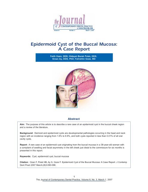
Epidermoid Cyst of the Buccal Mucosa: A Case Report
Epidermoid Cyst of the Buccal Mucosa: A Case Report
Epidermoid Cyst of the Buccal Mucosa: A Case Report

Create successful ePaper yourself
Turn your PDF publications into a flip-book with our unique Google optimized e-Paper software.
<strong>Epidermoid</strong> <strong>Cyst</strong> <strong>of</strong> <strong>the</strong> <strong>Buccal</strong> <strong>Mucosa</strong>:<br />
A <strong>Case</strong> <strong>Report</strong><br />
Aim: The purpose <strong>of</strong> this article is to describe a rare case <strong>of</strong> an epidermoid cyst in <strong>the</strong> buccal cheek region<br />
and a review <strong>of</strong> <strong>the</strong> literature.<br />
Background: Dermoid and epidermoid cysts are developmental pathologies occurring in <strong>the</strong> head and neck<br />
region with an incidence ranging from 1.6% to 6.9%, and both cysts reported in less than 0.01% <strong>of</strong> all oral<br />
cavity cysts.<br />
<strong>Report</strong>: A rare case <strong>of</strong> an epidermoid cyst originating from <strong>the</strong> buccal mucosa in a 38-year-old woman with<br />
a complaint <strong>of</strong> swelling and facial asymmetry in <strong>the</strong> left cheek just distal to <strong>the</strong> commissure for six months is<br />
presented in this report.<br />
Keywords: <strong>Cyst</strong>, epidermoid cyst, buccal mucosa<br />
Abstract<br />
Citation: Ozan F, Polat HB, Ay S, Goze F. <strong>Epidermoid</strong> <strong>Cyst</strong> <strong>of</strong> <strong>the</strong> <strong>Buccal</strong> <strong>Mucosa</strong>: A <strong>Case</strong> <strong>Report</strong>. J Contemp<br />
Dent Pract 2007 March;(8)3:090-096.<br />
1<br />
The Journal <strong>of</strong> Contemporary Dental Practice, Volume 8, No. 3, March 1, 2007
Introduction<br />
Dermoid cysts can be found anywhere in <strong>the</strong> body,<br />
particularly in areas where embryonic elements<br />
fuse toge<strong>the</strong>r. Most cases have been reported in<br />
<strong>the</strong> ovaries, <strong>the</strong> testicles, as well as <strong>the</strong> hands and<br />
feet. Dermoid and epidermoid cysts in <strong>the</strong> mouth<br />
are uncommon and account for less than 0.01%<br />
<strong>of</strong> all oral cysts. 1-2 The great majority arise in <strong>the</strong><br />
floor <strong>of</strong> <strong>the</strong> mouth, but <strong>the</strong>re are rare and usually<br />
individual case reports <strong>of</strong> examples in o<strong>the</strong>r sites.<br />
<strong>Epidermoid</strong>, dermoid, and teratoid cysts are<br />
nonodontogenic cystic lesions. They are rare<br />
lesions derived from germinal epi<strong>the</strong>lium. While<br />
a dermoid cyst has an epidermal lining with skin<br />
adnexa such as hair follicles and sebaceous<br />
glands, <strong>the</strong> epidermoid cyst contains no such<br />
adnexa. Teratoid cysts have been rarely described<br />
in <strong>the</strong> floor <strong>of</strong> <strong>the</strong> mouth. These cysts contain<br />
respiratory, gastrointestinal, and connective tissues<br />
such as bundles <strong>of</strong> striated muscle and distinct<br />
areas <strong>of</strong> fat. 1-4<br />
The purpose <strong>of</strong> this article is to describe a rare<br />
case <strong>of</strong> an epidermoid cyst in <strong>the</strong> buccal cheek<br />
region <strong>of</strong> a 38-year-old woman and a review <strong>of</strong> <strong>the</strong><br />
literature.<br />
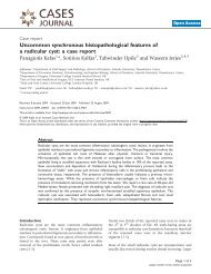
<strong>Case</strong> <strong>Report</strong><br />
A 38-year-old woman had a conspicuous swelling<br />
<strong>of</strong> <strong>the</strong> left cheek, just behind <strong>the</strong> commissure<br />
(Figure 1).<br />
The patient reported <strong>the</strong> swelling had been present<br />
for about six months. She had been prescribed<br />
an antibiotic by a general dental practitioner but<br />
stated <strong>the</strong> antibiotic had not helped. She had no<br />
history <strong>of</strong> surgery and/or trauma related to <strong>the</strong><br />
lesion location.<br />
Intraoral examination revealed a 2 x 3 x 4 cm<br />
swelling extending from <strong>the</strong> left commissure to <strong>the</strong><br />
first molar region. The swelling was not tender to<br />
palpation and formed a doughy mass freely mobile<br />
between <strong>the</strong> buccal mucosa and <strong>the</strong> buccinator<br />
muscle. The overlying mucosa was normal in color<br />
and texture with no apparent abnormality <strong>of</strong> <strong>the</strong><br />
facial skin in <strong>the</strong> area <strong>of</strong> <strong>the</strong> swelling. Submental<br />
or submandibular lymph nodes were not palpable.<br />
Under local anes<strong>the</strong>sia, <strong>the</strong> cyst was enucleated<br />
using a horizontal incision in <strong>the</strong> buccal mucosa<br />
(Figure 2).<br />
Figure 1. Extraoral appearance <strong>of</strong> <strong>the</strong><br />
patient.<br />
Figure 2. Intraoperativeappearance <strong>of</strong><br />
<strong>the</strong> cyst.<br />
The wound was closed with 3.0 silk sutures. The<br />
postoperative period was uneventful and healing<br />
was good.<br />
Histopathological examination <strong>of</strong> <strong>the</strong> surgical<br />
specimen revealed a keratinized squamous<br />
epi<strong>the</strong>lial lining with <strong>the</strong> inner surface lined with<br />
keratin lamellas and <strong>the</strong> outer surface lined<br />
with gingival connective tissue components<br />
(Figure 3, HE X10). In a detailed examination<br />
keratin lamellas, epi<strong>the</strong>lium, epi<strong>the</strong>lial lining,<br />
and fibrovascular gingival tissue were observed<br />
(Figure 4, HE, X25).<br />
The patient has shown no clinical or radiographic<br />
evidence <strong>of</strong> recurrence during 18 months <strong>of</strong><br />
follow-up evaluation.<br />
Discussion<br />
Dermoid cysts are classified into <strong>the</strong> following<br />
three categories 3-6<br />
1. <strong>Epidermoid</strong> cysts - <strong>the</strong> cystic cavity is lined<br />
with epi<strong>the</strong>lium without skin appendages;<br />
2. Dermoid cysts - <strong>the</strong> cystic cavity includes<br />
skin appendages such as hair, hair follicles,<br />
sebaceous, and sweat glands;<br />
2<br />
The Journal <strong>of</strong> Contemporary Dental Practice, Volume 8, No. 3, March 1, 2007
Figure 3. Microscopic appearance <strong>of</strong>cyst (HE X10).<br />
3. Teratoid cysts - in addition to skin<br />
appendages <strong>the</strong> cystic cavity elements <strong>of</strong><br />
<strong>the</strong> mesoderm can be found such as bone<br />
and muscle as well as gastrointestinal and<br />
respiratory tissue.<br />
Dermoid cysts <strong>of</strong> <strong>the</strong> floor <strong>of</strong> <strong>the</strong> mouth are<br />
classified topographically as sublingual and<br />
submental in relation to <strong>the</strong> mylohyoid muscle.<br />
Dermoid and epidermoid cysts are uncommon<br />
in <strong>the</strong> mouth. The incidence in <strong>the</strong> head and<br />
neck has been reported to range from 1.6% to<br />
6.9%. 1,2,4,5,7 Most reported cases have involved<br />
<strong>the</strong> floor <strong>of</strong> <strong>the</strong> mouth (sublingual dermoids),<br />
usually in <strong>the</strong> midline. 2-8 Rare cases have<br />
been reported in <strong>the</strong> tongue, 1 lips, 1 uvula, 9<br />
temporomandibular joint dermal graft, 10<br />
intradiploic, 11 intracranial, 12 and intraosseously<br />
within <strong>the</strong> mandible and maxilla; 1 only two case<br />
reports were presented 1,13 associated with an<br />
epidermoid cyst <strong>of</strong> <strong>the</strong> buccal mucosa.<br />
The etiology <strong>of</strong> <strong>the</strong>se cysts is essentially<br />
unknown. The most popular <strong>the</strong>ory is epi<strong>the</strong>lium<br />
being sequestered in lines <strong>of</strong> fusion <strong>of</strong><br />
embryonic processes 1,2,8 as a result <strong>of</strong> epi<strong>the</strong>lial<br />
tissue being traumatically implanted in utero 1<br />
or to <strong>the</strong> traumatic implantation <strong>of</strong> a piece <strong>of</strong><br />
epidermis. 6,14-16 Implantation cysts were first<br />
recognized by Werhner 17 in 1855, originally<br />
referred to by Sutton 18 in 1895, and believed<br />
to originate through implantation <strong>of</strong> epi<strong>the</strong>lium<br />
by ei<strong>the</strong>r surgical or accidental trauma into<br />
Figure 4. Increased magnification <strong>of</strong> an area <strong>of</strong><br />
Figure 3 (HE X25).<br />
deeper mesenchymal tissues. Since trauma is<br />
said to always precipitate in <strong>the</strong> formation <strong>of</strong> <strong>the</strong><br />
implantation-type epidermoid cyst, King 19 preferred<br />
<strong>the</strong> term ‘post traumatic cyst.’ Similar observations<br />
<strong>of</strong> cyst formation have been made in humans<br />
following <strong>the</strong> inclusion <strong>of</strong> epi<strong>the</strong>lium in surgical<br />
wound sites. 1,14,16<br />
However, this traditional view <strong>of</strong> processes fusing<br />
with <strong>the</strong> breakdown <strong>of</strong> <strong>the</strong> intervening epi<strong>the</strong>lium<br />
has been challenged for this <strong>the</strong>ory does not<br />
explain <strong>the</strong> presence <strong>of</strong> adnexa in dermoid cysts<br />
or <strong>the</strong> absence <strong>of</strong> cysts <strong>of</strong> this type in zones <strong>of</strong><br />
known fusion such as <strong>the</strong> palate. Shafer and<br />
co-authors 20 postulate cysts <strong>of</strong> this type in <strong>the</strong> floor<br />
<strong>of</strong> <strong>the</strong> mouth arise from totipotent blastomeres<br />
trapped during closure <strong>of</strong> <strong>the</strong> mandibular and<br />
hyoid branchial arches. However, none <strong>of</strong> <strong>the</strong><br />
traditional explanations account for <strong>the</strong> present<br />
case.<br />
Although <strong>the</strong> epidermoid cyst rarely discloses<br />
malignancy, isolated cases <strong>of</strong> premalignant and<br />
malignant conditions (Bowen’s disease, Paget’s<br />
disease, and squamous cell carcinoma) have<br />
been found in <strong>the</strong>ir walls. Dini et al. 21 described<br />
a patient with basal cell carcinoma arising in <strong>the</strong><br />
wall <strong>of</strong> an epidermoid cyst. Ikeda et al. 21 presented<br />
a case report stating basal cell carcinoma<br />
originated from an epidermoid cyst in which <strong>the</strong>y<br />
found nests <strong>of</strong> basal cell carcinoma connected<br />
with <strong>the</strong> epidermoid cyst and partially replacing<br />
<strong>the</strong> cyst wall. Lopez-Rios et al. 22 described a<br />
patient with squamous cell carcinoma arising in<br />
3<br />
The Journal <strong>of</strong> Contemporary Dental Practice, Volume 8, No. 3, March 1, 2007
<strong>the</strong> wall <strong>of</strong> an o<strong>the</strong>rwise conventional epidermoid<br />
cyst. An incorrect diagnosis could result in<br />
inappropriate <strong>the</strong>rapy. If <strong>the</strong> lesion is completely<br />
excised, <strong>the</strong> treatment is definitive.<br />
In making a differential diagnosis <strong>the</strong> clinician<br />
should first entertain a broad variety <strong>of</strong> conditions.<br />
Conditions resembling this clinical presentation<br />
are swelling <strong>of</strong> <strong>the</strong> face as odontogenic infection,<br />
buccal space infection, masseteric space<br />
infection, and dermoid cysts. These conditions<br />
can be classified as developmental, neoplastic,<br />
and infectious processes. Infectious processes,<br />
as odontogenic infection, buccal space infection,<br />
and masseteric space infection, are unlikely in<br />
this case because <strong>the</strong> lesion/mass has attained<br />
considerable size without constitutional symptoms<br />
such as malaise or fever and <strong>the</strong>re was no<br />
response to antibiotic treatment. A salivary<br />
obstruction with a mucocele or ranula formation<br />
is a possibility. A bluish-gray hue is characteristic<br />
<strong>of</strong> a ranula, but <strong>the</strong> lesion in <strong>the</strong> present case had<br />
an overlying mucosa which was normal in color.<br />
A primary malignant process is very doubtful<br />
owing to <strong>the</strong> lesion’s size, normal overlying<br />
mucosa, cystic homogeneity, and lack <strong>of</strong> nodal<br />
involvement. Benign neoplastic processes in this<br />
region may include lipoid, salivary, and vascular<br />
lesions. A lipoma would tend to be yellowish and<br />
nodular. A vascular lesion such as a hemangioma<br />
or lymphangioma is also unlikely for <strong>the</strong>se are<br />
usually lobulated and have an irregular mucosal<br />
surface. One would expect an obvious reddishpurple<br />
color (hemangioma) or almost clear<br />
translucent color (lymphangioma) with lesions <strong>of</strong><br />
this magnitude.<br />
Most salivary gland tumors probably cause an<br />
obstructive phenomena when <strong>the</strong>y reach this<br />
size. If this were a cystic hygroma, one would<br />
expect it to be present at birth (50%) or develop<br />
by age two (90%). <strong>Cyst</strong>ic hygromas are more<br />
common in <strong>the</strong> posterior triangle <strong>of</strong> <strong>the</strong> neck and<br />
are s<strong>of</strong>t, fluctuant masses. The most frequent oral<br />
site is in <strong>the</strong> anterior two thirds <strong>of</strong> <strong>the</strong> tongue.<br />
A lesion to consider in <strong>the</strong> developmental category<br />
is <strong>the</strong> oral lymphoepi<strong>the</strong>lial cyst. This cyst is an<br />
uncommon lesion developing within oral lymphoid<br />
tissue. It can occur anywhere within or adjacent<br />
to Waldeyer’s ring. The oral lymphoepi<strong>the</strong>lial cyst<br />
presents as a small (small than 1 cm), smooth,<br />
whitish-yellow, firm, painless mass. It <strong>of</strong>ten<br />
contains cheesy keratinaceous material in <strong>the</strong><br />
lumen. Because <strong>of</strong> <strong>the</strong> size, localization, and hue,<br />
<strong>the</strong> lesion presented this diagnosis was unlikely.<br />
Summary<br />
The differential diagnosis for an epidermoid cyst<br />
should include developmental, neoplastic, and<br />
infectious lesions. The developmental lesion was<br />
<strong>the</strong> most compatible category for this patient who<br />
presented with a large, slowly enlarging, smooth,<br />
doughy, painless lesion.<br />
The diagnosis between an epidermoid and<br />
dermoid cyst was initially considered and later<br />
confirmed through histopathological examination<br />
<strong>of</strong> <strong>the</strong> surgical fragment. The cyst’s features<br />
characterized by stratified squamous epi<strong>the</strong>lium<br />
with laminas <strong>of</strong> keratinization on <strong>the</strong> surface and<br />
lumen <strong>of</strong> <strong>the</strong> cyst cavity determine <strong>the</strong> histological<br />
classification. The lack <strong>of</strong> epi<strong>the</strong>lial adnexa<br />
excludes <strong>the</strong> diagnosis <strong>of</strong> dermoid cyst.<br />
The proposed treatment <strong>of</strong> epidermoid cysts is<br />
surgical removal. 3,8,23 The cyst removal procedure<br />
was simple and effective, and its success was<br />
confirmed by <strong>the</strong> lack <strong>of</strong> postsurgical alterations<br />
and no recurrence <strong>of</strong> <strong>the</strong> lesion.<br />
4<br />
The Journal <strong>of</strong> Contemporary Dental Practice, Volume 8, No. 3, March 1, 2007
References<br />
1. Rajayogeswaran V, Eveson JW. <strong>Epidermoid</strong> cyst <strong>of</strong> <strong>the</strong> buccal mucosa. Oral Surg Oral Med Oral<br />
Pathol 1989;67:181-4.<br />
2. Worley CM, Laskin DM. Coincidental sublingual and submental epidermoid cysts. J Oral Maxill<strong>of</strong>ac<br />
Surg 1993;51:787-90.<br />
3. Zachariades N, Skoura-Kafoussia C. A life-threatening epidermoid cyst <strong>of</strong> <strong>the</strong> floor <strong>of</strong> <strong>the</strong> mouth:<br />
report <strong>of</strong> a <strong>Case</strong>. J Oral Maxill<strong>of</strong>ac Surg 1990;48:400-3.<br />
4. Calderon S, Kaplan I. Concomitant sublingual and submental epidermoid cyst: a case report. J Oral<br />
Maxill<strong>of</strong>ac Surg 1993;51:790-2.<br />
5. De Ponte FS, Brunelli A, Marchetti E, Bottini DJ. Sublingual epidermoid cyst. J Crani<strong>of</strong>ac Surg<br />
2002;13:308-10.<br />
6. Walstad WR, Solomon JM, Schow SR, Ochs MW. Midline cystic lesion <strong>of</strong> <strong>the</strong> floor <strong>of</strong> <strong>the</strong> mouth.<br />
J Oral Maxill<strong>of</strong>ac Surg 1998;56:70-4.<br />
7. Cortezzi W, De Albuquerque EB. Secondarily infected epidermoid cyst in <strong>the</strong> floor <strong>of</strong> <strong>the</strong> mouth<br />
causing a life-threatening situation: report <strong>of</strong> a case. J Oral Maxill<strong>of</strong>ac Surg 1994;52:762-4.<br />
8. Correa MS, Fon<strong>of</strong>f Rde N, Ruschel HC, Parizotto SP, Correa FN. Lingual epidermoid cyst: case report<br />
in an infant. Pediatr Dent 2003;25:591-3.<br />
9. Yoshinari M, Nagayama M. <strong>Epidermoid</strong> cyst <strong>of</strong> <strong>the</strong> uvula: report <strong>of</strong> a case. J Oral Maxill<strong>of</strong>ac Surg<br />
1986;44:828-9.<br />
10. Weinberg S, Kryshtalskyj B. <strong>Epidermoid</strong> cyst in a temporomandibular joint dermal graft: report <strong>of</strong> a<br />
case and review <strong>of</strong> <strong>the</strong> literature. J Oral Maxill<strong>of</strong>ac Surg 1995;53:330-2.<br />
11. Sudhakar N, Stephenson GC. Swelling on <strong>the</strong> head- a forgotten lesson: a case report <strong>of</strong> an<br />
intradiploic epidermal cyst with an iatrogenic complication. Br J Oral Maxill<strong>of</strong>ac Surg 2004;42:155-7.<br />
12. Howng SL, Kwan AL, Hwang SL, Chien TC. Intracranial epidermoid cysts. Gaoxiong Yi Xue Ke Xue<br />
Za Zhi. 1990;6:81-7.<br />
13. Schneider LC, Mesa ML. <strong>Epidermoid</strong> cysts <strong>of</strong> <strong>the</strong> buccal mucosa. Q Natl Dent Assoc 1978;36:39-42.<br />
14. N<strong>of</strong>fke CE. Implantation-type epidermoid cyst <strong>of</strong> <strong>the</strong> mandible. Dentomaxill<strong>of</strong>ac Radiol 1999;28:383-5.<br />
15. Dohvoma CN. <strong>Epidermoid</strong> cyst: an unusual cause <strong>of</strong> obstructive sialadenitis. Br J Oral Maxill<strong>of</strong>ac<br />
Surg. 1992;30:125-7.<br />
16. Abrams MB, Andrews JE, Laskin DM. <strong>Epidermoid</strong> (implantation) cyst after temporomandibular joint<br />
surgery. J Oral Surg 1977;35:587-9.<br />
17. Werhner A. Das A<strong>the</strong>ron, ein eingebalgtes epi<strong>the</strong>lium. Virchows Arch Path Anat 1855;8:221.<br />
18. Sutton JB. A clinical lecture on some unusual tumours. Br Med J 1895;1:461-4.<br />
19. King ESJ. Post-traumatic epidermoid cysts <strong>of</strong> hands and fingers. Br J Surg 1933;21:29-43.<br />
20. Shafer WG, Hine MK, Levy BM. A textbook <strong>of</strong> oral pathology. 4th ed. Philadelphia: WB Saunders:<br />
1983. p 78-9.<br />
21. Ikeda I, Ono T. Basal cell carcinoma originating from an epidermoid cyst. J Dermatol 1990;17:643-6.<br />
22. Lopez-Rios F, Rodriguez-Peralto JL, Castano E, Benito A. Squamous cell carcinoma arising in a<br />
cutaneous epidermal cyst: case report and literature review. Am J Dermatopathol 1999;21:174-7.<br />
23. Dini M, Innocenti A, Romano GF. Basal cell carcinoma arising from epidermoid cyst: a case report.<br />
Dermatol Surg 2001;27:585-6.<br />
5<br />
The Journal <strong>of</strong> Contemporary Dental Practice, Volume 8, No. 3, March 1, 2007
About <strong>the</strong> Authors<br />
6<br />
The Journal <strong>of</strong> Contemporary Dental Practice, Volume 8, No. 3, March 1, 2007