Distinguishing the species - CIMMYT
Distinguishing the species - CIMMYT
Distinguishing the species - CIMMYT

You also want an ePaper? Increase the reach of your titles
YUMPU automatically turns print PDFs into web optimized ePapers that Google loves.
Partial funding for this publication was provided<br />
on a special project basis by <strong>the</strong> United<br />
Nations Development Programme (UNDP), Rockefeller<br />
Foundation (R F), and <strong>the</strong> Australian Development<br />
Assistance Bureau (ADAB).<br />
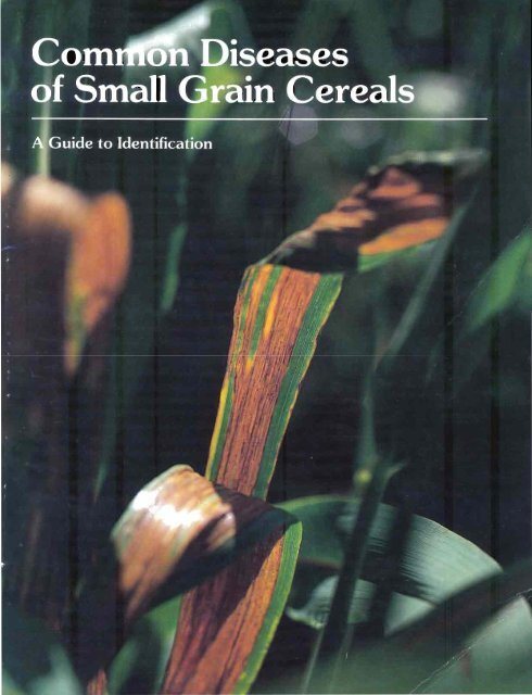
Cover: Bacterial stripe, caused by Xanthomonas<br />
translucens, on barley (photo: S. Fuentes)<br />
Cover design: An ita AI bert
Acknowledgements<br />
As with nearly all major publications, this book<br />
is <strong>the</strong> result of <strong>the</strong> interest and efforts of many<br />
people. For <strong>the</strong>ir help I am indebted and greatly<br />
appreciative; <strong>the</strong>y are mentioned here in no particular<br />
order.<br />
I would first like to acknowledge <strong>the</strong> many<br />
helpful comments and <strong>the</strong> information provided by<br />
<strong>the</strong> reviewers of this document. Thanks go to: Dr.<br />
R. J. Metzger, Oregon State University (USA), <strong>the</strong><br />
major source of information on loose, covered and<br />
flag smuts, common and dwarf bunts, and o<strong>the</strong>r<br />
diseases of winter cereals; Dr. M. B. Moore, Professor<br />
Emeritus, University of Minnesota (USA), for<br />
his constructive review and contribution of photographs;<br />
Dr. H. J. Dubin, <strong>CIMMYT</strong>, who provided a<br />
great deal of general information on disease<br />
organisms, especially those prevalent in <strong>the</strong> Andean<br />
region of South America; Dr. P. A. Burnett,<br />
<strong>CIMMYT</strong>, for his review of <strong>the</strong> section on viral and<br />
mycoplasmal diseases and his assistance in obtaining<br />
photographs; Dr. M. V. Wiese, University of Idaho<br />
(USA) and author of <strong>the</strong> Compendium of Wheat<br />
Diseases (APS, 1977), for his carefu I review of <strong>the</strong><br />
draft manuscript and kind permission to freely<br />
utilize <strong>the</strong> Compendium as a source of information;<br />
Dr. E. T. Torres, <strong>CIMMYT</strong>, for his review and<br />
editing of <strong>the</strong> Spanish translation; Dr. Real-L.<br />
Pelletier, McGill University (Canada), for his<br />
thorough review and for <strong>the</strong> translation and<br />
editing of <strong>the</strong> French version of this document.<br />
Special thanks go to <strong>the</strong> following people:<br />
Dr. J. M. Walker, CIVIl (Kew, England), for information<br />
and references and for his willingness to<br />
work with me in East Africa and Mexico in<br />
identifying <strong>the</strong> diseases afflicting wheat, triticale<br />
and barley; Lucie Gilchrist, Pathologist, Temuco,<br />
Chile, for making available to me her research<br />
information pertaining to fusarium diseases,<br />
yellow leaf blotch (tan spot) of wheat, and Free<br />
State stripe virus; Senora Haydee Barreiro de<br />
Villalpando, for her tireless efforts in preparing and<br />
maintaining <strong>the</strong> records of disease observations and<br />
photographs.<br />
This book could not have been developed<br />
without <strong>the</strong> financial support of <strong>the</strong> United Nations<br />
Development Programme (UNDP), <strong>the</strong> Rockefeller<br />
Foundation (R F), and <strong>the</strong> Australian Development<br />
Assistance Bureau (ADAB). Their continuing<br />
enthusiasm and support for this project is much<br />
appreciated. In this regard, I would also like to<br />
acknowledge and thank Dr. Clive James, Deputy<br />
Director General for Research, <strong>CIMMYT</strong>, for his<br />
efforts in assuring adequate funding, as well as for<br />
his ideas and assistance in <strong>the</strong> conceptualization of<br />
this document.<br />
As a group, <strong>CIMMYT</strong>'s Information Services<br />
Unit, under <strong>the</strong> leadership of Mr. Christopher<br />
Dowswell, has been extremely helpful in <strong>the</strong> preparation<br />
of this book. Special thanks go to: Mr. Tiffin<br />
Harris, for his editorial and design support; Senor<br />
Fernando Rulfo, for <strong>the</strong> Spanish translation; Maricela<br />
A. de Ramos and Silvia Bistrain R., for <strong>the</strong>ir<br />
patience and help in typesetting various drafts;<br />
Anita Albert, special consultant to <strong>CIMMYT</strong> in<br />
publication design from Iowa State University, for<br />
some last minute design suggestions that improved<br />
<strong>the</strong> book's utility and appearance; Miguel Mellado,<br />
for his exceptional job of preparing <strong>the</strong> guidebook<br />
for <strong>the</strong> printer. Thanks should also go to Mr.<br />
Armon Roschen of Viking Press, Inc., for his help<br />
in carrying this project to completion.<br />
Finally, a special note of appreciation to my<br />
wife, Hilda, whose support and faith over <strong>the</strong> years<br />
have enabled me to pursue my career wherever it<br />
led me. It is to her that I dedicate this book.<br />
Dr. Frank Zillinsky
Preface<br />
Dr. Frank Zillinsky came to <strong>CIMMYT</strong> in<br />
December, 1967, from <strong>the</strong> Ottawa Agricultural<br />
Research Station, where he was employed for some<br />
17 years by <strong>the</strong> Canada Department of Agriculture<br />
as a Cereal Crop Specialist. He was appointed as <strong>the</strong><br />
leader of CI MMYT's triticale improvement program,<br />
and held that position until his retirement from<br />
CIMIVIYT in July, 1982. Under Frank's guidance,<br />
triticale germplasm underwent very rapid improvement,<br />
from not much more than a biological<br />
curiosity to <strong>the</strong> threshold of widespread commercial<br />
cultivation. That alone is an accomplishment which<br />
would satisfy most agricultural researchers, yet<br />
Frank offered more.<br />
Though not formally trained as a cereal<br />
pathologist, Frank was often confronted with <strong>the</strong><br />
need to identify diseases afflicting various small<br />
grain cereals. Characteristically, Frank studied in<br />
his spare time to develop this necessary expertise.<br />
To his disappointment, he found no publications<br />
designed specifically to help a person in his situation:<br />
a plant breeder who needed to identify <strong>the</strong><br />
diseases affecting h is crops.<br />
For many years, Frank worked closely with<br />
cereal pathologists from allover <strong>the</strong> world, studying,<br />
learning, and accumulating thousands of<br />
photographs and disease observations for his own<br />
edification. About nine months before Frank's<br />
scheduled retirement from <strong>CIMMYT</strong>, Norman<br />
Borlaug suggested that hundreds of agricultural<br />
researchers in developing countries and elsewhere<br />
could benefit from <strong>the</strong> kind of book Frank had<br />
sought but not found. He also suggested that<br />
Frank was <strong>the</strong> person best qualified to write such a<br />
book. Hence, this guidebook was prepared, drawing<br />
heavily on Frank's own collection of photographs<br />
and knowledge of small grain diseases, but with<br />
significant input from friends and collegues <strong>the</strong><br />
world over.<br />
Plant diseases are probably <strong>the</strong> greatest constraint<br />
to increasing global small grains production<br />
in <strong>the</strong> 1980s and beyond. Accordingly, <strong>CIMMYT</strong><br />
is placing a very high priority on breeding wheat,<br />
triticale and barley for enhanced resistance to<br />
various diseases, and we encourage national programs<br />
in developing countries to do <strong>the</strong> same. In<br />
fact, <strong>the</strong> essence of CIIVIMYT's wheat improvement<br />
strategy lies in multilocation testing and a heavy reliance<br />
on national program staff for performance<br />
evaluations of germplasm. Moreover, many developing<br />
countries now have sophisticated plant breeding<br />
programs capable of making valuable contributions<br />
to improving disease resistance.<br />
Success in breeding for enhanced disease resistance<br />
will be <strong>the</strong> result of a cooperative effort among<br />
many national programs and CI MMYT, and will<br />
require <strong>the</strong> free exchange of germplasm and performance<br />
data. To develop useful breeding objectives<br />
and effective crossing plans, accurate information<br />
as to which diseases are affecting which cultivars<br />
is needed, as well as data on <strong>the</strong> severity of<br />
affliction. This guidebook addresses <strong>the</strong> problem of<br />
accurate disease identification. We strongly believe<br />
it is a necessary addition to <strong>the</strong> literature and that<br />
it will prove useful to thousands of agricultural<br />
researchers allover <strong>the</strong> world. It is a significant<br />
ach ievement, a fitting capstone to Fran k 's distinguished<br />
career, and perhaps <strong>the</strong> most important<br />
publication to be recently forthcoming from <strong>the</strong><br />
CINlMYT Wheat Improvement Program.<br />
Dr. Frank Zillinsky Byrd C. Curtis<br />
Director, <strong>CIMMYT</strong> Wheat<br />
Facing page: Triticale seed (wheat x rye) Improvement Program
Contents<br />
Introduction<br />
1 Objectives and scope of this guidebook<br />
2 Field and laboratory techniques and equipment<br />
2 Collecting and preserving samples<br />
6 Preparing samples for <strong>the</strong> laboratory<br />
8 Disease groups and <strong>the</strong>ir general symptoms<br />
11 The rusts<br />
13 Stem rust (Puccinia graminis)<br />
15 Leaf rust of wheat (P. recondita)<br />
16 Leaf rust of barley (P. hordei)<br />
17 Stripe rust (P. striiformis)<br />
18 Crown rust of oats (P. coronata)<br />
19 Hyperparasite of rust (Darluca filum)<br />
21 Helminthosporium diseases<br />
24 Spot blotch (Helminthosporium sativum - Perfect stage:<br />
Cochliobolus sativus)<br />
26 Yellow leaf blotch, or tan spot (H. tritici-repentis - Perfect stage:<br />
Pyrenophora tritici-repentis)<br />
28 Leaf blotch of oats (H. avenae - Perfect stage: Pyrenophora avenae)<br />
29 Net blotch (H. teres - Perfect stage: Pyrenophora teres)<br />
30 Barley stripe (H. gramineum - Perfect stage: Pyrenophora graminea)<br />
32 Zonate eyespot (H. giganteum, or Drechslera gigantea)<br />
34 Cereal leaf blotch and foot root (H. spiciferum - Perfect stage:<br />
Cochliobolus spicifer)<br />
35 Septoria complex and septoria-like diseases<br />
39 Speckled leaf blotch (Septoria tritici Perfect stage:<br />
Mycosphaerella graminicola)<br />
41 Glume blotch (S. nodorum - Perfect stage: Phaeosphaeria nodorum)<br />
43 Septoria leaf blotch of oats, wheat and triticale (s. avenae -<br />
Perfect stage: Phaeosphaeria avenaria)<br />
45 Septoria leaf blotch of barley (S. passerinii)<br />
46 Ascochyta leaf spot (Ascochyta graminicola)<br />
47 Phaeoseptoria leaf blotch (Phaeoseptoria vermiformis)
49 Smuts and bunts<br />
51 Loose and covered smuts (Vstilago <strong>species</strong>)<br />
51 Loose smut of barley and wheat [V. nuda (V. tritici)]<br />
53 Black loose smut of barley and oats [V. nigra (V. avenae)]<br />
54 Covered smut of barley and oats [V. hordei (u. kolleri)]<br />
55 Common and dwarf bunts, or stinking smut (Til/etia <strong>species</strong>)<br />
57 Karnal bunt (Til/etia indica, formerly Neovossia indica)<br />
58 Flag smut (Vrocystis agropyri)<br />
59 Fusarium diseases<br />
63 Scab and root rot (Fusarium <strong>species</strong>)<br />
63 Fusarium graminearum - Perfect stage: Gibberella zeae<br />
64 Fusarium culmorum<br />
65 Fusarium avenaceum - Perfect stage: Gibberella avenacea<br />
66 Fusarium equiseti Perfect stage: Gibberella intricans<br />
67 Fusarium leaf blotch and snow mold (F. nivale - Perfect stage:<br />
Calonectria nivalis)<br />
71 Miscellaneous root and crown diseases<br />
74 Cephalosporium stripe (Cephalosporium gramineum)<br />
75 Take-all (Ophiobolus graminis - renamed: Gaeumannomyces graminis)<br />
77 Eyespot (Pseudocercosporella herpotrichoides)<br />
78 Sclerotium wilt, or sou<strong>the</strong>rn blight (Sclerotium rolfsii)<br />
80 Anthracnose (Colletotrichum graminicola)<br />
81 Downy mildew (Sclerophthora macrospora)<br />
83 Miscellaneous leaf and head diseases<br />
85 Powdery mildew (Erysiphe graminis)<br />
87 Scald (Rhynchosporium secalis)<br />
88 Cercospora spot (Cercospora apii)<br />
89 Ergot (Claviceps purpurea)
91 Saprophytic and weakly pathogenic fungi<br />
91 Alternaria <strong>species</strong><br />
93 Stemphylium botryosum<br />
94 Cladosporium <strong>species</strong><br />
95 Pleospora <strong>species</strong><br />
96 Torula <strong>species</strong><br />
96 Phoma <strong>species</strong><br />
98 Epicoccum nigrum<br />
99 Cercosporidium graminis<br />
101 Bacterial diseases<br />
102 Bacterial stripe and black chaff (Xanthomonas transluscens)<br />
104 Basal glume rot (Pseudomonas atrofaciens)<br />
105 Halo blight of oats (P. coronafaciens)<br />
106 Bacterial spike blight (Corynebacterium tritici)<br />
107 Bacterial leaf blight (P. syringae)<br />
109 Viral and mycoplasmal diseases<br />
110 Aphid-transmitted<br />
110 Barley yellow dwarf (BYD)<br />
111 Free state strea k<br />
112 O<strong>the</strong>rs<br />
113 Leafhopper-transmitted<br />
113 American wheat striate<br />
113 Chloris striate mosaic virus (Australian wheat striate mosaic)<br />
113 Russian wheat mosaic<br />
114 Enanismo de Narino<br />
114 Aster yell ows<br />
116 Planthopper-transmitted<br />
116 Hoja blanca of rice<br />
116 African cereal streak<br />
117 Eu ropean wheat striate mosaic<br />
117 O<strong>the</strong>rs
118 Mite-transmitted<br />
118 Wheat streak mosaic<br />
119 Agropyron mosaic and rye grass mosaic<br />
119 Wheat spot mosaic<br />
120 Soil-borne<br />
120 Wheat soil-borne mosaic<br />
120 Wheat spindle streak mosaic<br />
121 Seed-borne<br />
121 Barley stripe mosaic<br />
121 Barley mosaic<br />
123 Injury from nematodes and o<strong>the</strong>r pests<br />
123 Nematodes (eel worms)<br />
125 Insects<br />
129 Birds and mammals<br />
131 Physiological and genetic disorders<br />
134 Nutrient deficiencies and environmental stresses<br />
135 Soil pH<br />
136 Soil toxicity<br />
136 Drought<br />
137 Excessive moisture<br />
137 Chemical injury and soil residues<br />
138 Frost and hail damage<br />
139 Glossary and selected references
Introduction<br />
The main purpose of this guidebook is to<br />
assist in <strong>the</strong> identification of diseases and of pathogens<br />
causing diseases in cereal crops. It is not<br />
intended as an authoritative text on diseases, nor<br />
does it provide any new information on diseases or<br />
diagnostic techniques. Its primary audience includes<br />
breeders, agronomists, and young scientists-intraining,<br />
especially in developing nations, who are<br />
responsible for identifying and recording trial data,<br />
screening segregating populations for disease resistance,<br />
and verifying <strong>the</strong> disease reactions of various<br />
types of plant material in <strong>the</strong>ir programs.<br />
At many of <strong>the</strong> research stations in developing<br />
countries, <strong>the</strong> responsibility for recording<br />
disease reactions and making selections belongs to<br />
breeders, production agronom ists, and <strong>the</strong>ir assistants.<br />
Most of <strong>the</strong>m are familiar with conspicuous<br />
diseases, such as <strong>the</strong> rusts, smuts and mildews, and<br />
feel confident that <strong>the</strong> data <strong>the</strong>y report are rei iable.<br />
Their confidence tends to fade, however, when<br />
faced with <strong>the</strong> need to identify foot rots, head<br />
blights and leaf blights. This guidebook is intended<br />
to help cereal crops researchers improve <strong>the</strong>ir<br />
knowledge and increase <strong>the</strong>ir confidence in dealing<br />
with <strong>the</strong> plant diseases <strong>the</strong>y encounter.<br />
The diseases affecting bread wheat, durum<br />
wheat, barley, oats and triticale provide <strong>the</strong> main<br />
focus of this guidebook. Triticale is now emerging<br />
as a commercial food and feed crop in some<br />
countries and production appears destined to<br />
increase. Because it is a relatively new crop, published<br />
information on <strong>the</strong> reaction of triticale<br />
to specific diseases is meager atthistime. Important<br />
observations by <strong>CIMMYT</strong> personnel and national<br />
program cooperators regarding diseases associated<br />
with triticale are included here.<br />
Figure 1. This guidebook is particularly useful to nonpatho·<br />
logists charged with <strong>the</strong> responsibility of identifying less<br />
than conspicuous diseases (photo: C. Dowswell).<br />
The scope of this guidebook is limited to <strong>the</strong><br />
use of simple techniques helpful in identifying<br />
some of <strong>the</strong> most common causal organisms of<br />
cereal crop diseases and associated saprophytes.<br />
It is recognized that many pathogens, such as those<br />
causing foot rots and viral diseases, require more<br />
elaborate techniques for accurate diagnosis. There<br />
are many excellent books available to those who<br />
wish to delve deeper into <strong>the</strong> study of plant<br />
diseases. As a start, <strong>the</strong> Compendium of Wheat<br />
Diseases (1977) and <strong>the</strong> Compendium of Barley<br />
Diseases (1982), pu bl ished by <strong>the</strong> American<br />
Phytopathological Society are especially recommended.<br />
In an effort to minimize <strong>the</strong> confusion that<br />
currently exists in <strong>the</strong> taxonomy of fungi, bacteria<br />
and viruses, <strong>the</strong> most commonly used generic and<br />
<strong>species</strong> names are used in this publication. The<br />
perfect or sexual stage is footnoted and discussed<br />
in those instances where it serves as an important<br />
stage in <strong>the</strong> initiation, recognition or spread of <strong>the</strong><br />
disease.<br />
Considerable variability exists in nature among<br />
microorganisms, just as among higher plants. In <strong>the</strong><br />
past, <strong>the</strong>re has been a tendency among mycologists<br />
to give <strong>species</strong> status to minor morphological<br />
variants or forms that are pathogenic on different<br />
host <strong>species</strong>. Greater awareness among scientists of<br />
<strong>the</strong> natural variation that exists among all life<br />
forms has led to <strong>the</strong> establishment of fewer <strong>species</strong><br />
groups. This trend is appreciated by many biological<br />
scientists outside <strong>the</strong> realm of taxonomy.<br />
1
Fungi that are pathogenic on cereal crops<br />
will generally sporulate within 48 hours on fresh or<br />
dried samples (Figure 6d). The main exceptions to<br />
this are fungi that develop sexual fruiting structures,<br />
and thus require more time to induce maturation<br />
of sexual spores. One to two weeks under semi -dry<br />
conditions, or alternate periods of drying and<br />
moistening, is all that is required for some ascomycetes.<br />
O<strong>the</strong>rs require extended periods of outdoor<br />
wea<strong>the</strong>ring.<br />
The incubated material should be examined<br />
within 48 hours; o<strong>the</strong>rwise, saprophytic contaminants<br />
tend to obscure <strong>the</strong> main pathogens. The<br />
fruiting structures can be observed and, with<br />
practice, identified under <strong>the</strong> dissecting scope at<br />
low magnification (5X to 50X). For observation at<br />
higher magnifications, spores or fruiting structures<br />
can be picked out or scraped from <strong>the</strong> surface of<br />
<strong>the</strong> plant material and placed in a drop of water on<br />
a slide. A cover slip should be applied gently to<br />
avoid air bubbles, and <strong>the</strong> slide examined under<br />
higher magnification. Using an eyepiece with a<br />
Figure 6(al. Diseased leaf material is first cut to fi t in a<br />
previously prepared petri dish ...<br />
calibrated scale will help to establish <strong>the</strong> size and<br />
identify o<strong>the</strong>r features of <strong>the</strong> spores being examined.<br />
However, with experience <strong>the</strong> relative size of<br />
spores can be established without using a scale, and<br />
disease causing organisms can be identified at lower<br />
magnification.<br />
PLEASE NOTE: Most of <strong>the</strong> photographs of spore<br />
forms included in this guidebook were taken at an<br />
original magnification of 10 x 40 (400 times <strong>the</strong><br />
actual size). These photographs were <strong>the</strong>n fur<strong>the</strong>r<br />
enlarged 250 percent for printing purposes. Thus,<br />
<strong>the</strong> final magnification of a 10 x 40 photomicrograph<br />
equals 10 x 40 x 2.5 (1000 times <strong>the</strong> actual<br />
size). In <strong>the</strong> case of close-up photographs of<br />
conidiophores, etc., which were generally taken at<br />
50 x, <strong>the</strong> final magnification equals 50 x 2.5 (125<br />
times <strong>the</strong> actual size). The original magnification of<br />
each photograph taken with <strong>the</strong> aid of a microscope<br />
is provided in each respective caption. Simply<br />
multiply that magnification by 2.5 to arrive at <strong>the</strong><br />
final magnification.<br />
.. ,(e) <strong>the</strong> samples are incubated for 24 to 48 hours at room<br />
temperature in a plastic bag that acts as a moisture<br />
chamber...<br />
.,. (b) <strong>the</strong> filter paper is <strong>the</strong>n moistened and excess moisture ... (d) and usually within 48 hours, pathogenic fungi will<br />
is poured off .. , sporulate, Shown here is sporulating Helminthosporium<br />
sativum,<br />
7
Disease Group<br />
Miscellaneous leaf<br />
and head diseases<br />
Saprophytic and<br />
weakly pathogenic<br />
fungi<br />
Bacterial diseases<br />
Viral and mycoplasmal<br />
diseases<br />
Injury from<br />
nematodes and<br />
o<strong>the</strong>r pests<br />
Physiological, nutritional<br />
and environmental<br />
stresses<br />
Symptoms<br />
The fungal pathogens included in this group have little in common<br />
except <strong>the</strong>ir tendency to invade <strong>the</strong> upper parts of <strong>the</strong> plant. Each<br />
fungus produces ra<strong>the</strong>r distinct disease symptoms, and <strong>the</strong>y usually<br />
develop <strong>the</strong>ir fruiting structures on <strong>the</strong> surface from submerged<br />
mycelia. Claviceps purpurea, <strong>the</strong> cause of ergot, differs in its development<br />
of fruiting structures; large dark sclerotia replace <strong>the</strong> kernels<br />
in infected florets.<br />
Several common saprophytes and facultative parasites are included<br />
in this group. Normally <strong>the</strong>se fungi are aggressive spore producers,<br />
producing dark, th ick-walled spores on dead or dying tissue. The<br />
affected plant parts take on a dull greyish appearance. Under conditions<br />
favorable to <strong>the</strong> fungi <strong>the</strong>y may invade living plant tissues or<br />
developing grain, usually during <strong>the</strong> maturation stage.<br />
Water-soaked streaks, spots, and blotches appear on leaves, stems or<br />
glumes. When leaves are moist with dew, droplets of bacterial exudate<br />
may develop on lesions and newly infected areas. When dry, <strong>the</strong> bacterial<br />
exudate may form crystals or a thin shiny film that flakes when<br />
scraped. Streaks may appear dark by reflected light, but often are<br />
translucent by transmitted light.<br />
Viruses produce no visible fruiting structures. They develop a broad<br />
range of symptoms on cereal crop plants, including stunting, spotting,<br />
striping, streaking, yellowing or general chlorosis. Many viral diseases<br />
are transmitted by insects or small animals (such as aphids and nematodes);<br />
o<strong>the</strong>rs are transmitted mechanically or by <strong>the</strong> fungus Polymyxa<br />
graminis. The identity of <strong>the</strong> vector is important and often essential<br />
to <strong>the</strong> identification of <strong>the</strong> disease. There may be a tendency<br />
for <strong>the</strong> onset of necrosis to be delayed longer in viral diseases than in<br />
fungal diseases.<br />
Injury can occur to below- or above-ground 'portions of <strong>the</strong> plant.<br />
Damage to roots by cutting, chewing or boring indicates worms,<br />
larvae or o<strong>the</strong>r insects. Abnormal branching and deformation of <strong>the</strong><br />
roots in <strong>the</strong> form of knots, swellings or galls indicates a nematode infestation.<br />
Above ground, mechanical damage such as chewing, clipping,<br />
boring or stripping of leaves, stems or heads indicates insects or<br />
mammals. Nematodes may form seed galls, replacing grain in <strong>the</strong><br />
heads. O<strong>the</strong>r nematodes may infest <strong>the</strong> nodes, causing swelling and<br />
deformation.<br />
Abnormal plant development caused by mechanical damage, chemical<br />
injury, environmental and physiological stresses, or genetic abnormalities;<br />
not caused by pathogens.<br />
Page<br />
82-89<br />
90-99<br />
100-107<br />
108-121<br />
122-129<br />
130-138<br />
9
The rusts<br />
Puccinia <strong>species</strong><br />
The rusts are perhaps <strong>the</strong> most destructive<br />
and also <strong>the</strong> most widely recognized of cereal crop<br />
diseases. They occur in almost all areas of <strong>the</strong><br />
world where cereal crops are grown. Any of <strong>the</strong><br />
above-ground portions of <strong>the</strong> plant may become<br />
infected, from seedling stage to maturity. The<br />
fungi, Puccinia <strong>species</strong>, are mostly obligate parasites<br />
with highly complex life cycles. Three spore forms<br />
develop on small grains and grasses, and two<br />
additional spore forms are produced on <strong>the</strong> alternate<br />
host (where it exists).<br />
Of <strong>the</strong>se spore forms, urediospores (summer<br />
spores) are most important, because <strong>the</strong>y enable<br />
repeated cycles of <strong>the</strong> disease to spread from field<br />
to field and survive from year to year. These spores<br />
vary in color from yellow-orange to reddish brown.<br />
They are produced in pustules (uredia), which<br />
erupt through <strong>the</strong> epidermis exposing <strong>the</strong> powdery<br />
spore masses (Figure 7).<br />
Teliospores (winter spores) develop as <strong>the</strong><br />
crop approaches maturity, and <strong>the</strong>y usually remain<br />
covered by <strong>the</strong> epidermis of <strong>the</strong> host plant. Teliospores<br />
differ from <strong>the</strong> urediospores in color, form<br />
F igu re 7. Uredia of stem rust (Puccinia graminis f .sp. tritici)<br />
on wheat (facing page). Note <strong>the</strong> loose epidermal tissue<br />
along <strong>the</strong> margins of <strong>the</strong> sori.<br />
<strong>Distinguishing</strong> <strong>the</strong> <strong>species</strong><br />
Urediospores of P. graminis f.spo tritici (10 x 40)<br />
and function. They are dark brown, two-celled,<br />
have thick walls, and function as <strong>the</strong> first stage in<br />
<strong>the</strong> sexual cycle of <strong>the</strong> fungus. After wea<strong>the</strong>ring, or<br />
overwintering, <strong>the</strong> teliospores germinate and produce<br />
basidiospores that are able to infect <strong>the</strong><br />
alternate host (but not cereals or grasses). The<br />
sexual cycle on alternate host plants creates diversity<br />
through genetic recombination, and establishes<br />
primary inoculum for <strong>the</strong> new crop cycle. The<br />
fungi, however, usually persist from cycle to cycle<br />
as urediospores, or as mycelium established in <strong>the</strong><br />
host tissue.<br />
At least five Puccinia <strong>species</strong> attack cereal<br />
crops; forms within <strong>species</strong> vary in <strong>the</strong>ir capacity<br />
to parasitize crops and grasses. The different forms<br />
are given form <strong>species</strong> (f.sp.) names. For example,<br />
stem rust is caused by Puccinia graminis. The form<br />
<strong>species</strong> that infects oats but not wheat or barley is<br />
identified as P. graminis f. sp. avenae. Within forms<br />
<strong>the</strong>re are physiologic races that are distinguished<br />
by <strong>the</strong>ir virulence on specific host genotypes. The<br />
occurrence of new physiologic races of rust fungi<br />
creates difficulties in developing and maintaining<br />
resistant plant varieties.<br />
Rust diseases are most effectively controlled<br />
by <strong>the</strong> use of resistant varieties and by <strong>the</strong> eradication<br />
of <strong>the</strong> alternate host. Fungicides applied as<br />
foliar sprays can be used effectively to protect<br />
crops threatened by pathogenic races, but are<br />
usually uneconomical as control systems.<br />
The rust <strong>species</strong> described here usually can be<br />
distinguished by <strong>the</strong> characteristics of <strong>the</strong> uredia,<br />
color of <strong>the</strong> urediospores, and <strong>the</strong> host <strong>species</strong> <strong>the</strong>y<br />
infect.<br />
Puccinia graminis (stem rust)<br />
Large elongate spore masses are reddish brown<br />
with loose epidermal tissue conspicuous at <strong>the</strong><br />
margins of <strong>the</strong> pustules. Uredia may develop on<br />
both <strong>the</strong> upper and lower surfaces of leaves,<br />
and on heads and stems. Urediospores are dark red,<br />
elliptical, echinulate, and measure 24-32 ,um x<br />
18-22,um.<br />
11
Puccinia recondita (leaf rust of wheat)<br />
Uredia are red, oval shaped, and scattered,<br />
without conspicuous loose epidermal tissue at <strong>the</strong><br />
margins. They are usually produced on <strong>the</strong> upper<br />
side of leaf blades. Urediospores are orange-red to<br />
dark red, spherical, and measure 20-28 .um in<br />
diameter.<br />
Puccinia hordei (leaf rust of barley)<br />
Uredia are yellow-orange, oval shaped, scattered,<br />
and occur generally on <strong>the</strong> upper side of <strong>the</strong><br />
leaf blades. Urediospores are yellow-orange, relatively<br />
large, egg shaped to spherical, and measure<br />
28-36 .um x 24-28 .um. This fungus is pathogenic<br />
on lyon barley and its close relatives.<br />
Puccinia striiformis (stripe rust)<br />
Uredia occur in conspicuous narrow yellow<br />
stripes on leaf blades and sheaths. Urediospores are<br />
yellow, more or less spherical, and large, measuring<br />
28-34 .um in diameter. Glumes and lemmas often<br />
become infected. Narrow black stripes are formed<br />
on leaves during telial development.<br />
Puccinia coronata (crown rust of oats)<br />
This fungus is pathogenic on oats, but not on<br />
o<strong>the</strong>r cereal crops. Uredia are oval to elongate<br />
blisters with bright yellow-orange urediospores. The<br />
urediospores are spherical to egg shaped, and 20-32<br />
.um in diameter. The teliospores are broad and have<br />
projections on <strong>the</strong> upper cells.<br />
12<br />
Urediospores of P. recondita (10 x 40)<br />
Urediospores of P. hordei (10 x 40)<br />
Urediospores of P. striiformis (10 x 40)<br />
Urediospores of P. coronata (10 x 40)
Stent rust<br />
Pucc;n;a gram;n;s<br />
Stem rust is a well recognized disease of cereal<br />
crops, occuring almost everywhere <strong>the</strong>se crops are<br />
grown. ,Pucc;n;a gram;n;s trit;c; infects wheat,<br />
barley and triticale. Pucc;n;a gram;n;s avenae<br />
infects oats, and P. gram;n;s seca/;s infects rye.<br />
Disease symptoms most commonly appear on <strong>the</strong><br />
stems and leaf sheaths, but leaf blades and spikes<br />
may also become infected (Figure 8) . Three spore<br />
forms are produced on <strong>the</strong> cereal hosts. Urediospores<br />
develop in pustules (uredia) that rupture <strong>the</strong><br />
epidermis and expose masses of reddish brown<br />
spores (Figure 9). The pustules are largerthanthose<br />
of leaf and stripe rust, oval shaped or elongated,<br />
Figure 8. Heavy infection of stem rust on triticale. Note<br />
that leaf blades and sheaths are also affected.<br />
with loose or torn epidermal tissue along <strong>the</strong><br />
margins. They may appear on both <strong>the</strong> upper and<br />
lower surfaces of <strong>the</strong> leaf. The urediospores of P.<br />
gram;n;s are reddish brown, elliptical to egg shaped,<br />
echinulate structures measuring 24-32 J.Lm x 18-22<br />
J.Lm (Figure 10). They continue to be produced<br />
until <strong>the</strong> plants approach maturity.<br />
Figure 9. Uredia of P. graminis. Note <strong>the</strong> loose epidermal Figure 10. Urediospores of P. graminis are oval to egg shaped<br />
tissue. (10 x 40) .<br />
13
As <strong>the</strong> plants approach maturity, urediospore<br />
production ceases and teliospores develop, ei<strong>the</strong>r in<br />
<strong>the</strong> same uredia or in different fruiting structures<br />
called telia. The tel iospores are dark brown, two<br />
celled, and somewhat wedge shaped. They retain a<br />
portion of <strong>the</strong> pedicel or stalk, have thick walls,<br />
and measure 40-60.um x 18-26 .um. The apical cell<br />
is rounded or slightly pointed (Figure 11). The<br />
teliospores frequently erupt through <strong>the</strong> epidermis,<br />
persist in <strong>the</strong> plant tissue during <strong>the</strong> winter, and<br />
may serve as <strong>the</strong> first stage in <strong>the</strong> sexual cycle by<br />
producing basidiospores. Common barberry, Berberis<br />
vulgaris, and Mahonia <strong>species</strong> are alternate<br />
hosts for P. gram in is (Figure 12).<br />
The urediospores of stem rust are carried over<br />
long distances by <strong>the</strong> wind. During <strong>the</strong> active growth<br />
period of host crops, repeated cycles of uredia are<br />
produced every 14 to 21 days, which accounts for<br />
<strong>the</strong> rapid spread of <strong>the</strong> disease over wide areas.<br />
This disease, along with leaf rust, has received<br />
lT1uch attention from pathologists, plant breeders,<br />
and o<strong>the</strong>r scientists. In <strong>the</strong> past, <strong>the</strong> creation of<br />
new physiologic races via <strong>the</strong> sexual cycle on <strong>the</strong><br />
alternate host, barberry, resu Ited in serious crop<br />
losses and reduced <strong>the</strong> effectiveness of resistant<br />
varieties. In North America, <strong>the</strong> eradication<br />
of barberry, and deliberate breeding of resistant<br />
varieties, have been important factors in improving<br />
<strong>the</strong> stability of resistance to stem rust in varieties<br />
and reducing <strong>the</strong> incidence of epiphytotics.<br />
Figure 11. Tel iospores of P. graminis (1 0 x 40). Figure 12. Aecia developing on an alternate host of P.<br />
graminis. common barberry (Berberis vulgaris).<br />
14
Leaf rust of wheat<br />
Puccinia recondita<br />
Leaf rust is a serious disease of wheat, rye, triticale<br />
and many grasses. The pathogen is mildly<br />
parasitic on barley, but does not infect oats.<br />
Ano<strong>the</strong>r closely related <strong>species</strong>, P. hordei, is a<br />
common pathogen of barley (page 16).<br />
Leaf rust appears as small, oval-shaped, dark<br />
red pustules scattered on leaf sheaths and <strong>the</strong> upper<br />
surfaces of leaf blades. The pustules (uredia) break<br />
through <strong>the</strong> epidermis, but do not cause loose epidermal<br />
tissue at <strong>the</strong> margins as is characteristic of<br />
stem rust uredia (Figure 13). The urediospores of<br />
P. recondita are orange red to dark red, echinulate,<br />
Figure 13. Uredia of P. recondita on a wheat leaf.<br />
spherical, and usually measure 20-28 J-Lm in diameter<br />
(Figure 14). The teliospores are dark brown,<br />
two celled with thick walls, and are rounded or<br />
flattened at <strong>the</strong> apex (Figure 15), The telia develop<br />
during <strong>the</strong> later stages of plant development in sori<br />
on leaf sheaths and on both surfaces of <strong>the</strong> leaf<br />
blades. The teliospores remain in <strong>the</strong> leaf tissues<br />
and are covered by <strong>the</strong> epidermis.<br />
Thalictrum and Anchusa <strong>species</strong>, <strong>the</strong> alternate<br />
hosts of P. recondita, occur in some areas of<br />
Europe, but are unimportant as a source of new<br />
races of this fungus. In temperate climates, <strong>the</strong><br />
pathogen survives from yeC1r to year as mycelium<br />
or urediospores on winter cereals and grasses. In<br />
<strong>the</strong> tropics, it oversummers at higher elevations<br />
and in cooler areas.<br />
Leaf rust is best controlled by <strong>the</strong> use of resistant<br />
cereal crop varieties or multilines. Fungicides,<br />
such as Triadimefon (bayleton) and Butrizol<br />
(indar), are effective in controlling leaf rust and<br />
can be used economically in epidemic situations.<br />
F igu re 14. U red iospores of P. recondita t end to be more Figure 15. Teliospores of P. recondita (10 x 40).<br />
spherical than those of P. gram in is (lO x 40).<br />
15
Leaf rust of barley<br />
Puccinia hordei<br />
Leaf rust occurs extensively in many barley<br />
growing areas of <strong>the</strong> world, but <strong>the</strong> crop losses<br />
it causes are usually light. The pathogenicity of<br />
this fungus is restricted to cultivated barley and its<br />
close relatives. Barley is also affected by some races<br />
of P. recondita, but to a lesser extent.<br />
The uredia of P. hordei are small, oval to<br />
round, and yellow-orange in color. They develop<br />
almost exclusively on <strong>the</strong> leaf blades and sheaths<br />
(Figure 16). The urediospores are egg shaped to<br />
spherical, yellow, and relatively large, measuring<br />
28-36 11m x 24-28 11m (Figure 17). As <strong>the</strong> crop<br />
matures, dark brown telia form in <strong>the</strong> leaf tissues<br />
and initially remain covered by <strong>the</strong> epidermis. The<br />
alternate host, Ornithogalum umbellatum (<strong>the</strong> star<br />
of Bethlehem), can be a source of aeciospores in<br />
some areas of Europe, but does not appear to<br />
become infected in North America; it is unlikely<br />
that it plays an important role in <strong>the</strong> perpetuation<br />
or dissemination of new, diverse forms of <strong>the</strong><br />
pathogen.<br />
The development and spread of leaf rust of<br />
barley is favored by warm humid wea<strong>the</strong>r. The<br />
uredial stage overwinters on fall-sown barley in<br />
areas having mild winters. In subtropical areas <strong>the</strong><br />
fungus survives summer temperatures in <strong>the</strong> cooler<br />
highlands. The use of resistant varieties is <strong>the</strong> most<br />
economical means of control.<br />
Figure 16. Uredia of P. hordei on a barley leaf. Figure 17. Urediospores of P. hordei vary from spherical to<br />
egg shaped (10 x 40).<br />
16
Stripe rust<br />
Puccinia striiformis<br />
Stripe rust is a serious disease of wheat and<br />
barley, and moderately affects triticale, rye and<br />
some grasses. Oats appear to be immune. No<br />
alternate host is known, and <strong>the</strong> disease is confined<br />
by environmental factors to a greater extent than<br />
ei<strong>the</strong>r stem rust or leaf rust. Disease development<br />
occurs at lower temperatures than are optimal for<br />
leaf and stem rust.<br />
Figure 18. Ured ia of P. striiformis on a wheat leaf. Note<br />
that <strong>the</strong>y form narrow I inear stripes.<br />
Uredia develop in narrow, yellow, linear stripes<br />
mainly on leaves and spikelets (Figure 18). When<br />
<strong>the</strong> heads are infected, <strong>the</strong> pustules appear on <strong>the</strong><br />
inner surfaces of glumes and lemmas, occasionally<br />
invading <strong>the</strong> developing kernels (Figure 19), The<br />
urediospores are yellow to orange in color, more or<br />
less spherical, echinulate, and 28-34 Jim in diameter<br />
(Figure 20). The teliospores apparently serve no<br />
function. They are dark brown, two celled, and<br />
similar in size and shape to those of P. recondita.<br />
The telia develop on leaf blades and sheaths as dark<br />
brown to black stripes and remain covered by <strong>the</strong><br />
epidermis.<br />
The urediosporesand mycelium persist through<br />
<strong>the</strong> winter on fall-sown cereals and grasses in areas<br />
where temperatures are not extreme. Aggressive<br />
secondary spread occurs during <strong>the</strong> cool temperatures<br />
of spring. New, highly virulent races of stripe<br />
rust have recently appeared in Europe, Australia,<br />
and <strong>the</strong> Andean region of South America.<br />
Figure 19. Uredia of P. striiformis on wheat florets (glumes Figure 20. Urediospores of P. striiformis (10 x 40).<br />
and lemmas) .<br />
17
Crown rust of oats<br />
Puccinia coronata<br />
Crown rust is a significant disease of oats and<br />
a few grasses, but does not affect <strong>the</strong> o<strong>the</strong>r cereal<br />
crops. The disease is prevalent wherever oats are<br />
grown. The diversity in physiologic races is very<br />
great, due at least in part to <strong>the</strong> wide distribution<br />
of <strong>the</strong> alternate host, Rhamnus cathartica<br />
(buckthorn), and related <strong>species</strong>.<br />
The uredial pustules develop mainly on <strong>the</strong><br />
leaf blades, but may also appear on leaf sheaths<br />
and spikelets. They occur as small, scattered, ovalshaped<br />
blisters with exposed urediospores that are<br />
bright orange in color (Figure 21). The epidermis is<br />
slightly lifted along <strong>the</strong> margins of <strong>the</strong> pustules.<br />
The urediospores are spherical to oval in shape,<br />
echinulate, orange-yellow in color, and measure<br />
20-32 /.lm in diameter (Figure 22). Telia develop<br />
on affected leaves as <strong>the</strong> plants approach maturity.<br />
The tel ial sori may develop as conspicuous black<br />
borders around <strong>the</strong> uredia, but <strong>the</strong>y remain covered<br />
by <strong>the</strong> epidermis. The teliospores are distinctive in<br />
shape from those of o<strong>the</strong>r <strong>species</strong>. The apical cells<br />
of <strong>the</strong> teliospores are broad and tipped with several<br />
projections, from which <strong>the</strong> name "crown" rust<br />
was derived (Figure 23).<br />
The fungus persists from season to season in<br />
<strong>the</strong> warmer climates by continuous production of<br />
urediospores. In areas with lower temperatures,<br />
<strong>the</strong> disease is initiated by spores carried by wind<br />
currents from warmer areas, or from aeciospores<br />
developing on buckthorn (Rhamnus <strong>species</strong>).<br />
In many oat producing areas, such as Africa,<br />
South America and Mexico, breeding for resistance<br />
has not kept pace with <strong>the</strong> occurrence of new<br />
physiologic races of <strong>the</strong> pathogen. Oat production<br />
in <strong>the</strong>se areas has declined because of losses from<br />
crown rust and stem rust, as well as from barley<br />
yellow dwarf virus.<br />
Figure 22. Urediospores of P. coronata (lO x 40).<br />
Figure 21 . Ured ia of P. coronata on an oat leaf. Figure 23. Teliospores of P. coronata have short projections<br />
on upper cells, which gave rise to <strong>the</strong> name "crown rust"<br />
(10x40).<br />
18
Hyperparasite of rust<br />
Oarluca filum<br />
Oarluca filum is an obligate parasite of <strong>the</strong><br />
rust fungi, occurring most commonly in warmer<br />
climates. The fungus develops dark fruiting bodies<br />
(pycnidia) inside <strong>the</strong> rust pustules. Its role in<br />
controlling disease spread is probably insignificant,<br />
but <strong>the</strong> fungus is included here because of <strong>the</strong><br />
frequency with which it is observed on cereal crops<br />
affected by rust diseases.<br />
Figure 24. Black pycnidia of D. filum forming in <strong>the</strong><br />
pustules of P. recondita (leaf rust of wheat).<br />
The fruiting structures appear in clusters and<br />
consist of several black spherical pycnidia within<br />
<strong>the</strong> rust sori (Figure 24). Conidia are released<br />
through <strong>the</strong> ostiole of <strong>the</strong> pycnidiurr, as a whitish<br />
or light grey mucous-like mass (Figure 25). The<br />
conidia are colorless, two celled, ellipsoidal to<br />
oblong in shape, and measure 14-16 /Jm x 3-4 /Jm<br />
with a few short bristles projecting from each end<br />
(Figure 26). Oarluca filum progresses too slowly<br />
during <strong>the</strong> rust cycle to serve as an effective<br />
biological control of <strong>the</strong> rusts.<br />
Figure 25. Whitish to light grey cirrhi (containing conidial Figure 26. Conidia of D. filum (10 x 40).<br />
are exuded from <strong>the</strong> pycnidia of D. filum.<br />
19
Helminthosporium<br />
diseases<br />
Five He/minthosporium <strong>species</strong> are Injurious<br />
pathogens of cereal crops and are widely distributed<br />
around <strong>the</strong> world. Two less common <strong>species</strong><br />
that have unique characteristics are also included<br />
here. The He/minthosporium <strong>species</strong> are treated as<br />
a group in this guidebook, not because of similarities<br />
in <strong>the</strong> disease symptoms produced, but because of<br />
simi larities (1) in <strong>the</strong> types of fruiting structures<br />
developed during sporulation, and (2) in <strong>the</strong> general<br />
shape of <strong>the</strong> conidia. As a group, <strong>the</strong>y are probably<br />
second in importance to <strong>the</strong> rusts as destructive<br />
pathogens of cereal crops <strong>the</strong> world over.<br />
Various types of foliar lesions, such as blotches,<br />
stripes, spots and discolorations are caused by most<br />
of <strong>the</strong> <strong>species</strong> (Figure 27). Some <strong>species</strong> also cause<br />
seed ling bl ights, foot rots, node and stem decay,<br />
and head blights.<br />
Figure 27. Early leaf-spotting symptoms (facing page)<br />
caused by Helminthosporium tritici-repentis (yellow leaf<br />
blotch or tan spot).<br />
<strong>Distinguishing</strong> <strong>the</strong> <strong>species</strong><br />
Conidia of H. sativum (10 x 40)<br />
The asexual fruiting structures develop on <strong>the</strong><br />
surface of plant tissues from mycelia within <strong>the</strong><br />
tissue. The spore-bearing stal ks (conidiophores) are<br />
as long as, or longer than, <strong>the</strong> conidia, develop<br />
singly or in clusters, are erect, septate, light brown<br />
to dark brown in color, and are mostly unbranched.<br />
Each conidiophore produces one to several conidia,<br />
which are cylindrical to slightly barrel shaped<br />
(ellipsoid), may be straight or slightly curved, vary<br />
in color from tinted (subhyaline) to dark brown or<br />
black, and have several cells with very conspicuous<br />
septa.<br />
The <strong>species</strong> within this group can be distinguished<br />
mainly by <strong>the</strong> characteristics of <strong>the</strong> conidia,<br />
<strong>the</strong> crops <strong>the</strong>y infect and <strong>the</strong> disease symptoms<br />
produced.<br />
He/minthosporium sativum (spot blotch)<br />
Conidia appear black and shiny under low<br />
magnification, but under higher magnification <strong>the</strong>y<br />
are a dark olive brown. The conidia are thick<br />
walled, typically have five to nine cells, are elliptical,<br />
may be straight or slightly curved, and measure<br />
60-120 JIm x 12-20 JIm. This pathogen can affect<br />
all small grain cereals, but is less common on oats.<br />
21
Heiminthosporium tritici-repentis (yellow leaf<br />
blotch or tan spot)<br />
The early leaf spots caused by this disease<br />
may have distinct yellow borders around straw to<br />
grey-brown, oval-shaped lesions. Young conidia are<br />
subhyaline, becoming yellow-grey with age. They<br />
are cylindrical, thin walled, taper slightly toward<br />
<strong>the</strong> top, and measure 80-170 .um x 12-24.um with<br />
five to ten septa. The basal end cell is a rounded<br />
cone suggestive of a "snake's head." This fungus is<br />
pathogenic on wheat and triticale, and less frequently<br />
affects rye and barley.<br />
Heiminthosporium avenae (leaf blotch of oats)<br />
This disease is restricted to oats. The conidia<br />
are light greyish brown, cylindrical, straight to<br />
slightly curved, four to six septate, and measure<br />
80-120 .um x 12-18 .um. They are very similar in<br />
size and shape to <strong>the</strong> conidia of H. teres.<br />
Heiminthosporium teres (net blotch)<br />
The conidia may be easily confused with<br />
those of H. gramineum, but <strong>the</strong> disease symptoms<br />
are quite distinct. Net blotch lesions start as<br />
small brown spots or short streaks and expand to<br />
irregularly shaped blotches with internal brown<br />
pigmentation. The conidia are straight, cylindrical,<br />
four to six septate and have rounded end cells.<br />
They are subhyaline at first, darken to yellowbrown<br />
with age, and measure 60-120 .um x 16-24<br />
.um. This disease is most often found on barley, but<br />
occasionally affects wheat and triticale.<br />
Heiminthosporium gramineum (barley stripe)<br />
This disease is systemic and is restricted to<br />
barley. As <strong>the</strong> plant develops, each succeeding<br />
leaf shows stripes that are yellow at first and <strong>the</strong>n<br />
turn brown or dark grey. The leaves tend to<br />
split and fray. The conidia are usually light brown<br />
in color and seldom exceed 100 .um in length.<br />
Conidiophores develop in clusters of two to six,<br />
without <strong>the</strong> pronounced swollen basal cells that<br />
occur in H. teres.<br />
22<br />
Conidia of H. tritici-repentis (10 x 40)<br />
Conidia of H. avenae (10 x 40)<br />
Conidia and dark conidiophores of H. teres (10 x 40)<br />
Conidia of H. gramineum (10 x 40)
Conidia of H. giganteum-<strong>the</strong> long, cylindrical. light<br />
colored structures (10 x 10, top; 10 x 40 bottom)<br />
Conidia of H. spiciferum (10 x 40)<br />
Helminthosporium giganteum (zonate eye spot)<br />
The lesions of this disease are distinctive,<br />
appearing as small yellow spots surrounded by dark<br />
brown borders. The conidia are straight, subhyaline,<br />
cylindrical, measure 350-400 /-Lm x 15-20<br />
/-Lm and have few septa. The disease can affect all<br />
small grain cereals, and is restricted to humid<br />
subtropical regions.<br />
Helminthosporium spiciferum (cereal leaf blotch<br />
and foot rot)<br />
The leaf blotches vary in color from light<br />
brown to grey-brown, are irregularly shaped, and<br />
usually spread across <strong>the</strong> leaves. The conidia are<br />
<strong>the</strong> smallest among <strong>the</strong> helminthosporium cereal<br />
crop pathogens. They measu re 25-40 /-Lm x 8-12<br />
/-Lm, and have three and sometimes four septa.<br />
This pathogen attacks wheat, barley and related<br />
grasses.<br />
23
Spot blotch<br />
He/minthosporium sativum *<br />
A voracious pathogen that attacks wheat,<br />
triticale, barley and many grasses, H. sativum<br />
causes spot blotch, root and crown rots, node<br />
cankers (black joint) and head and seedling blight<br />
(Figure 28). Rye is somewhat more tolerant, while<br />
oats are seldom infected. The fungus also is fully<br />
capable of surviving as a saprophyte on dead plant<br />
tissue.<br />
Infection by H. sativum may occur at any<br />
stage of plant development, but symptoms are<br />
usually more pronounced after heading. Infected<br />
seedlings develop dark brown necrotic lesions on<br />
roots, crowns and lower leaf sheaths (Figure 29).<br />
Lesions on leaf sheaths may extend to <strong>the</strong> blades.<br />
Root and crown infections occurring before or<br />
during <strong>the</strong> flowering period usually kill plants<br />
before seeds are formed. This often occurs in<br />
association with o<strong>the</strong>r root-rotting pathogens, such<br />
as Ophiobo/us graminis and Fusarium <strong>species</strong>. Node<br />
can kers appear as brownish black discolorations at<br />
<strong>the</strong> lower nodes. The diseased nodes may be<br />
partially or completely invaded, thus restricting<br />
24<br />
<strong>the</strong> flow of plant nutrients. Diseased nodes take on<br />
a greyish black, velvety appearance due to dense<br />
sporulation (Figure 30).<br />
Leaf blade and sheath infections (<strong>the</strong> spot<br />
blotch stage) develop as distinct oval to elongated<br />
blotches, light brown to dark brown in color, that<br />
contrast sharply with <strong>the</strong> normal green of <strong>the</strong> leaf<br />
(Figures 31, 32). The dead areas of leaf blades<br />
and sheaths eventually take on a dull greyish appearance.<br />
Under conditions favorable for <strong>the</strong><br />
development of <strong>the</strong> disease, spikelets may be<br />
affected, causing shriveling of <strong>the</strong> grain. Black<br />
point, which develops as a dark staining of <strong>the</strong><br />
* Perfect stage: Cochliobolus sativus<br />
Figure 29. Barley seedlings infected by H. sativum . Note<br />
<strong>the</strong> dark hrown necrotic lesions.<br />
Figure 28. Typical leaf-spotting symp Figure 30. Sporulation of H. sativum on a diseased wheat<br />
toms on barley, caused by H. sativum stem node. Note <strong>the</strong> black, velvety appearance.<br />
(photo: S. Fuentes).
embryo end of <strong>the</strong> seed, occurs under conditions<br />
favorable to <strong>the</strong> disease (warm temperatures and<br />
high humidity). The disease develops faster at<br />
temperatures above 200 C (680 F).<br />
The fruiting structures, which develop readily<br />
on moistened diseased plant tissue, serve as <strong>the</strong><br />
most reliable diagnostic feature of this pathogen.<br />
The conidiophores develop singly or in small<br />
clusters, are erect, unbranched, 100-150 11m x 6-8<br />
11m, and have several septa (Figure 33). Conidia<br />
develop laterally from pores just below <strong>the</strong><br />
conidiophore septa. The conidia are olive brown,<br />
oblong, tapered toward <strong>the</strong> end, slightly curved<br />
with smooth walls, and have a prominent basal<br />
scar. They measure 60-120 11m x 12-20 11m in size<br />
Figure 31. Spot blotch lesions on a durum wheat leaf,<br />
caused by H. sativum.<br />
and have three to nine septa (Figure 34). Isolates<br />
vary considerably in virulence. The perfect stage<br />
rarely occurs in nature.<br />
Helminthosporium sativum is among <strong>the</strong> most<br />
widely distributed of <strong>the</strong> cereal crop pathogens. The<br />
diseases caused by this fungus probably reduce <strong>the</strong><br />
yield of wheat and barley in subtropical areas more<br />
than any o<strong>the</strong>r pathogen. Its ability to survive in<br />
soil debris and on many grass <strong>species</strong> reduces <strong>the</strong><br />
effectiveness of crop rotation as a control measure.<br />
Seed treatment can be beneficial in areas where<br />
head blight results in <strong>the</strong> production of infected<br />
seed. Breeding for resistance is a slow and elusive<br />
process, but appears to offer <strong>the</strong> best probability<br />
of success in controlling this pathogen.<br />
Figure 33. Conidiophores of H. sativum on wheat (50 x).<br />
See also Figure 6d, page 7.<br />
Figure 32. Typical spot blotch lesions caused by H. sativum Figure 34. Conidia of H. sativum are dark, thick walled, and<br />
on barley (photo: S. Fuentes)' Compare <strong>the</strong>se lesions with have conspicuous septa. Note that <strong>the</strong> conidia shown here<br />
those produced on wheat (Figure 31). are slightly larger than most encountered (10 x 40).<br />
25
Yellow leaf blotch (tan spot)<br />
He/minthosporium trit,"ci-repentis'"<br />
Yellow leaf blotch is chiefly a foliar disease of<br />
wheat, triticale and several grasses. Rye and barley<br />
are less frequently affected and oats appear to be<br />
immune. The first symptoms appear as small<br />
yellow-brown spots that develop into oval-shaped,<br />
light brown blotches, usually with yellow borders<br />
(Figure 35). As <strong>the</strong> lesions merge, large areas of<br />
leaves turn yellow and die (Figure 36). Necrosis<br />
often begins near <strong>the</strong> tip and progresses toward <strong>the</strong><br />
base of <strong>the</strong> leaf. Heavy sporu lation causes <strong>the</strong><br />
centers of lesions to darken.<br />
* Perfect stage : Pyrenophora tritici-repentis<br />
26<br />
Sporulation occurs readily in moisture. The<br />
conidiophores are dark brown, septate, and measure<br />
100-400 11m x 6-8 11m . The conidia are pale yellowgrey,<br />
cylindrical, 80-170 11m x 12-24 11m in size<br />
and have four to ten septa. The basal cell tapers to<br />
a rounded cone resembling a "snake's head" (Figure<br />
37).<br />
Figure 36. More advanced leaf spotting on wheat, caused by<br />
H. tritici-repen tis.<br />
Figure 35. Typical yellow leaf blotch Figure 37. Conidia of H. tritici-repentis. Note <strong>the</strong> tapered<br />
lesions on wheat, caused by H. triticibasal cells (10 x 40).<br />
repentis.
The perfect stage, Pvrenophora tritici-repentis,<br />
develops on wea<strong>the</strong>red wheat stubble and straw.<br />
The peri<strong>the</strong>cia are black, erumpent, with dark<br />
spines surrounding <strong>the</strong> short beaks (Figures 38, 39).<br />
The mature ascospores are yellow-brown, oval<br />
shaped, have three transverse septa and one or two<br />
longitudinal septa. They measure 40-60 /-Lm long by<br />
18-25 /-Lm wide (Figure 40). Conidiophores and<br />
conidia frequently develop on <strong>the</strong> peri<strong>the</strong>cia.<br />
Ascospores and conidia may both serve as primary<br />
inoculum.<br />
Figure 38. Peri<strong>the</strong>cia of Pyrenophora tritici·repentis (<strong>the</strong><br />
perfect stage of H. tritici-repentis) developing on wea<strong>the</strong>red<br />
wheat straw.<br />
The disease caused by H. tritici-repentis occurs<br />
in most of <strong>the</strong> wheat-growing regions of <strong>the</strong> world.<br />
The pathogen appears to tolerate cool temperatures<br />
more readily than H. sativum. Losses from yellow<br />
leaf blotch in wheat crops occur regularly in <strong>the</strong><br />
Andean region of South America, <strong>the</strong> highlands of<br />
East and North Africa, <strong>the</strong> High Plateau of Mexico,<br />
and <strong>the</strong> Middle East. Clean cultivation and fungicides<br />
are suggested as possible control measures;<br />
however, resistant varieties offer a more real istic<br />
means of keeping losses to a minimum.<br />
Figure 39. A closer view of a peri<strong>the</strong>cium of P. tritici Figure 40. Asci containing ascospores of P. tritici-repentis.<br />
rep en tis. Note <strong>the</strong> dark spines surrounding <strong>the</strong> short beak Note that <strong>the</strong> ascospores have transverse and longitudinal<br />
(10 x 40). septa (10 x 40).<br />
27
Leaf blotch of oats<br />
He/minthosporium avenae *<br />
Leaf blotch, caused by H. avenae, is a common<br />
disease of oats. Apparently restricted to this crop,<br />
<strong>the</strong> fungus may be carried over from one oat<br />
crop to <strong>the</strong> next on <strong>the</strong> grain. The disease starts<br />
with <strong>the</strong> coleoptiles becoming infected. Subsequent<br />
infections develop from conidia produced on<br />
lesions. Diseased leaves develop oblong to elongated<br />
reddish brown spots (Figure 41). The leaf symptoms<br />
at <strong>the</strong> early stages of disease development are<br />
good diagnostic features. As <strong>the</strong> disease progresses,<br />
<strong>the</strong> brown merges into reddish shades with indefinite<br />
margins. The lesions gradually spread across<br />
most of <strong>the</strong> leaf blade, and eventually <strong>the</strong> affected<br />
leaves die and dry out as <strong>the</strong> disease advances.<br />
Under severe conditions, panicles can be infected,<br />
and <strong>the</strong> fungus becomes established in <strong>the</strong> hulls<br />
and kernels where it remains until <strong>the</strong> next crop<br />
cycle.<br />
The fruiting structures of this fungus are<br />
similar in shape and color to those of He/minthosporium<br />
teres (net blotch) on barley (page 29).<br />
However, since H. avenae is restricted to oat<br />
<strong>species</strong> and H. teres does not infect oats, <strong>the</strong>re<br />
should be no confusion as to <strong>the</strong> identity of <strong>the</strong><br />
fungus. The conidiophores are scattered, develop<br />
singly or in clusters of two or three, and are<br />
thickened, measuring 8-12 Mm thick and up to 200<br />
Mm long (Figure 42). The conidia are light yellow-<br />
grey when young and turn to a darker brown with<br />
age. They are straight or slightly curved, cylindrical<br />
with rounded ends, have four to six septa and<br />
measure 80-110 Mm x 12-18 Mm . The basal cell<br />
has a prominent scar (Figure 43). The perfect stage,<br />
Pyrenophora avenae, occurs infrequently in nature,<br />
and its role in disease development has not been<br />
established with certainty.<br />
He/minthosporium avenae is widespread in<br />
most oat-growing regions of <strong>the</strong> world, but crop<br />
losses are generally light. Crop rotation and seed<br />
treatment with chemical fungicides reduce <strong>the</strong><br />
severity and incidence of <strong>the</strong> disease.<br />
* Perfect stage : Pyrenophora avenae<br />
Figure 42. Sporulation of H. avenae on a diseased oat leaf<br />
(50 xl.<br />
Figure 41. Leaf blotch of oats, caused by H. avenae. Note Figure 43. Conidia of H. avenae. Note <strong>the</strong> prominent scar<br />
<strong>the</strong> reddish brown color. on <strong>the</strong> basal cell (10 x 40).<br />
28
Net blotch<br />
He/minthosporium teres *<br />
Net blotch is primarily a leaf disease of<br />
barley. It occasionally affects wheat, triticale and<br />
o<strong>the</strong>r grasses. Disease symptoms appear first as<br />
small brown spots, usually near <strong>the</strong> tip of <strong>the</strong><br />
blade (Figure 44). These spots soon elongate, becoming<br />
short stripes or blotches with internal brown<br />
pigmentation that develop in a transverse and<br />
longitudinal netlike pattern (Figure 45). Spikes<br />
may become infected as plants approach maturity.<br />
Conidial development is somewhat unpredictable.<br />
Rapid sporulation is usually observed on<br />
fresh leaves after flowering, but often fails to<br />
develop on dry leaves after a few months in<br />
storage. Conidiophores develop singly or in groups<br />
Figure 44. Early symptoms of net blotch on barley, caused<br />
by H. teres.<br />
of two or three, are light to medium brown, and<br />
are up to 200 11m long and 7-11 11m thick. The<br />
basal cell is usually enlarged. The conidia are<br />
straight, cylindrical, and light grey when young,<br />
turning greenish brown with age. Most of <strong>the</strong><br />
conidia measure 60-120 11m x 16-23 11m and have<br />
four to six septa (Figure 46) . They are similar in<br />
size and shape to H. avenae conidia (page 28).<br />
The perfect stage, Pyrenophora teres, develops<br />
on overwintered straw and stubble. The peri<strong>the</strong>cia<br />
are black, with setae (bristles) around <strong>the</strong> short<br />
beak. Mature ascospores are yellow-brown with<br />
three transverse and one longitudinal septa. The<br />
spores are constricted at <strong>the</strong> septa. The ascospores<br />
are important as inoculum in some areas where<br />
barley is produced.<br />
Net blotch may develop from infected seed,<br />
or from ascospores released from peri<strong>the</strong>cia on<br />
wea<strong>the</strong>red straw and stubble. The disease is common<br />
in all cool and humid barley-growing regions<br />
of <strong>the</strong> world. A spot blotch phase of H. teres occurs<br />
on barley in some highland areas, particularly <strong>the</strong><br />
Andes of South America. The disease can be distinguished<br />
from spot blotch, caused by H. sativum<br />
(page 24), only by observing <strong>the</strong> spores. Seed<br />
treatment with an appropriate fungicide controls<br />
<strong>the</strong> seed-borne pathogen . Resistance to H. teres has<br />
been derived from old varieties of barley originating<br />
in China, Turkey and Ethiopia.<br />
* Perfect stage : Pvrenophora teres<br />
Figure 45. Advanced symptoms of net blotch on barley. Figure 46. Conidia and dark conidiophores of H. teres<br />
(10 x 40).<br />
29
Barley Stripe<br />
Helminthosporium gramineum *<br />
Barley stripe is a systemic disease that affects<br />
barley, but no o<strong>the</strong>r cereal crops. The fungus is<br />
seed-borne, and becomes active during seed germination,<br />
invading <strong>the</strong> coleoptile and infecting each<br />
of <strong>the</strong> leaves as <strong>the</strong>y develop. Barley stripe symptoms<br />
provide excellent diagnostic features, and<br />
characteristic symptoms appear at <strong>the</strong> tillering<br />
stage. Narrow yellow stripes develop <strong>the</strong> full length<br />
of <strong>the</strong> leaf (Figure 47). These soon turn reddish<br />
brown and <strong>the</strong>n dark brown. The leaf tissues die<br />
and dry out, causing <strong>the</strong> leaves to split along <strong>the</strong><br />
stripes (Figure 48). Plants are dwarfed; <strong>the</strong> leaves<br />
turn dark, twist and fray; <strong>the</strong> spikes usually fail to<br />
* Perfect stage: Pyrenophora graminea<br />
30<br />
emerge. Although disease symptoms appear on leaf<br />
blades and sheaths during <strong>the</strong> seedling stage,<br />
sporulation is delayed until <strong>the</strong> flowering period of<br />
<strong>the</strong> barley crop. Floral infection of healthy plants<br />
occurs from air-borne conidia. The fungus becomes<br />
dormant after invading <strong>the</strong> hulls of developing<br />
seed.<br />
Figure 47. Early symptoms of barley stripe, caused by H.<br />
gramineum. Note that <strong>the</strong> narrow yellow stripes develop<br />
<strong>the</strong> full length of <strong>the</strong> leaf blade (photo: S. Fuentes).<br />
Figure 48. Advanced symptoms of barley stripe. Note <strong>the</strong><br />
dark stripes and splitting of <strong>the</strong> leaf blades along <strong>the</strong> stripes.
The conidiophores appear in clusters of two<br />
to six and are grey to dark brown in color (Figure<br />
49). The basal cell of <strong>the</strong> conidiophore is enlarged.<br />
The young conidia are light grey, turning dark<br />
brown with age. They are straight, cylindrical,<br />
slightly tapered, have rounded ends, and typically<br />
Figure 49. Sporulation of H. gramineum on a barley leaf<br />
(50 x).<br />
Figure 50. Conidia of H. gramineum (10 x 40).<br />
measure 60-90 f.1m x 15-18 f.1m with three to six<br />
septa (Figure 50). An unusual feature of this<br />
<strong>species</strong> during sporulation is <strong>the</strong> frequent development<br />
of secondary conidia from <strong>the</strong> apices of <strong>the</strong><br />
primary ones. The perfect stage, Pyrenophora<br />
graminea, seldom occurs in nature and is of no<br />
importance in <strong>the</strong> disease cycle, but is included<br />
here because <strong>the</strong> name is commonly used by<br />
mycologists to identify <strong>the</strong> fungus.<br />
The pathogen can be kept under control with<br />
fungicide seed treatment and resistant varieties.<br />
Its importance in Europe and North America has<br />
diminished considerably in recent years, though<br />
<strong>the</strong> disease is still common in some areas of South<br />
America, Africa and Asia.<br />
31
Zonate Eyespot<br />
He/minthosporium giganteum *<br />
Zonate eyespot frequently occurs on wheat,<br />
triticale, rye and barley in <strong>the</strong> coastal areas adjacent<br />
to <strong>the</strong> Gulf of Mexico in <strong>the</strong> USA and Mexico,<br />
extending south into <strong>the</strong> higher elevations of Central<br />
America. It is a common disease on tropical<br />
grasses. Early symptoms appear as numerous small,<br />
grey-brown, oval to circular spots on green leaves.<br />
The centers of <strong>the</strong> spots soon fade, becoming light<br />
grey to straw colored with distinct dark brown<br />
margins (Figure 51). The spots tend to remain<br />
small, but <strong>the</strong> tissues between <strong>the</strong> spots develop an<br />
* Also known as: Drechslera gigantea<br />
32<br />
unusual bleached appearance (Figure 52). Eventually<br />
<strong>the</strong> whole leaf turns yellow and dies. The<br />
fungus attacks plants that are growing rapidly,<br />
from <strong>the</strong> seedling stage through heading. Sporulation<br />
diminishes quickly after severe necrosis develops,<br />
and o<strong>the</strong>r pathogenic and saprophytic<br />
organisms invade <strong>the</strong> affected plant tissues.<br />
Figure 51. Distinctive zonate eyespot lesion on a wheat<br />
leaf, caused by H. giganteum. Note <strong>the</strong> definite dark brown<br />
margin.<br />
Figure 52. Advanced symptoms of zonate eyespot. Note<br />
<strong>the</strong> unusual bleached appearance of <strong>the</strong> tissue between <strong>the</strong><br />
spots.
The disease is easily recogn ized by <strong>the</strong> "zonate<br />
eyespots" and <strong>the</strong> fungus by its large fruiting<br />
structures. The conidiophores are dark brown,<br />
ra<strong>the</strong>r th in, but may reach 400,um in length (Figure<br />
53). The conidia are almost colorless cylindrical<br />
tu bes with rou nded ends, have very thin walls, two<br />
to five septa, and measure 350-400 ,um long by<br />
15-20 ,um wide (Figure 54). The conidia are<br />
sensitive to desiccation and lose <strong>the</strong>ir ability to<br />
germinate after a week or two of drying. The<br />
fungus often fails to sporulate after a secondary<br />
infection by bacteria.<br />
Figure 53. Sporulation of H. giganteum on wheat. Note<br />
how large <strong>the</strong>se conidiophores are relative to those<br />
produced by o<strong>the</strong>r He/minthosporium <strong>species</strong> (50 x).<br />
Although <strong>the</strong> disease does not usually occur<br />
in <strong>the</strong> temperate zones, it may restrict commercial<br />
production of cereal crops in <strong>the</strong> more humid<br />
sUbtropics. Native tropical grasses apparently<br />
provide <strong>the</strong> major source of primary inoculum. As<br />
yet, very little research has been done to identify<br />
resistance or tolerance to this disease. Some strains<br />
of triticale appeared to be more tolerant than<br />
wheat or barley under a heavy natural infection<br />
of H. giganteum in <strong>the</strong> Alajuela Nursery, Costa<br />
Rica, in 1978.<br />
Figure 54. The nearly colorless conidia and dark conidiophores of H. giganteum (left photo-magnification 10 x 10); a<br />
comparison of relative sizes of H. sativum conidium (small, dark brown) and larger conidium of H. giganteum, (only part<br />
of which is visible at 10 x 40 magnification in <strong>the</strong> right photo).<br />
33
Cereal leaf blotch and foot rot<br />
Helminthosporium spiciferum *<br />
This fungus occurs ocassionally on wheat,<br />
rice, and o<strong>the</strong>r cereals, and has been reported as a<br />
serious pathogen on some grasses. It is usually<br />
considered as a weak foot rot pathogen of wheat,<br />
but has been observed to cause leaf blotch symptoms<br />
on bread wheat and durums (in nor<strong>the</strong>rn<br />
India and Pakistan) and on barley (in Mexico).<br />
Foliar symptoms appear as dull greyish brown,<br />
irregularly shaped blotches extending <strong>the</strong> full<br />
length and width of <strong>the</strong> leaves (Figure 55).<br />
The fungus is easily identified by <strong>the</strong> branched<br />
superficial mycelia and distinctive conidia. Very<br />
heavy sporulation develops on diseased leaves during<br />
maturation of <strong>the</strong> host plant. Conidiophores arise<br />
from mycelia developing on <strong>the</strong> surface of plant<br />
tissues. Mycelia and conidiophores areyellow-brown<br />
to dark brown (Figure 56). The conidiophores have<br />
a knobby appearance from <strong>the</strong> scars of previous<br />
conidia. Conidia are short, oval shaped with rounded<br />
ends, straw colored to brown, normally measure<br />
25-40 Mm x 8-12 Mm, ancttypically are three septate<br />
(Figure 57).<br />
* Perfect stage: Cochliobolus spicifer<br />
The perfect stage, Cochliobolus spicifer, has<br />
been reported, but its role in disease development<br />
has not been established. Although <strong>the</strong> disease has<br />
been reported to occur in North America, Europe,<br />
Asia and Australia, its influence in cereal crop<br />
production is negligible.<br />
Figure 56. Sporulation of H. spiciferum on a diseased barley<br />
leaf.<br />
Figure 55. Typical symptoms of cereal leaf blotch on barley Figure 57. Conidia of H. spiciferum (10 x 40) .<br />
leaves, caused by H. spiciferum.<br />
34
Septoria complex<br />
and septoria-like diseases<br />
Four Septaria <strong>species</strong> are important pathogens<br />
of small grain cereals. They are <strong>the</strong> primary causes<br />
of various types of leaf spotting, blotching and<br />
necrosis. Depending upon <strong>the</strong> growth stage of <strong>the</strong><br />
host and environmental conditions, any of <strong>the</strong><br />
above-ground portions of <strong>the</strong> plant may be affected<br />
(Figure 58-next page).<br />
Septaria <strong>species</strong> possess several characteristics<br />
that distinguish <strong>the</strong>m from o<strong>the</strong>r genera of pathogenic<br />
fungi parasitizing <strong>the</strong> same crops:<br />
• The pycnidiospores (conidia) develop inside an<br />
enclosed, more or less spherical, fruiting<br />
structure called a pycnidium.<br />
• The pycnidia develop vVithin <strong>the</strong> plant tissue<br />
(embedded) and, as <strong>the</strong>y mature, break<br />
through <strong>the</strong> epidermis. The conidia are<br />
extruded in a spore mass through a pore or<br />
ostiole. The spore mass, or cirrhus, is usually<br />
slightly pink to yellowish white in color.<br />
• The conidia are threadlike or cylindrical<br />
with various lengths and widths depending<br />
upon <strong>the</strong> <strong>species</strong>. They have rounded ends,<br />
may be curved or straight, are hyaline, and<br />
usually have two to four cross walls or septa.<br />
<strong>Distinguishing</strong> <strong>the</strong> <strong>species</strong><br />
Conidia of S. tritici (10 x 40)<br />
The size and length-width ratios of <strong>the</strong><br />
conidia, <strong>the</strong> texture and color of <strong>the</strong> pycnidia<br />
or fru iting bodies, and <strong>the</strong> crop <strong>species</strong> <strong>the</strong>y infect<br />
are important diagnostic features used to identify<br />
Septaria <strong>species</strong>.<br />
Septoria diseases occur in all cereal growing<br />
regions of <strong>the</strong> world. The fungi overwinter on<br />
stubble and plant debris. Thus, clean cultivation<br />
and crop rotation are recommended as control<br />
measures in locations where severe septoria infections<br />
are frequent. Varieties that are resistant<br />
to one <strong>species</strong> of <strong>the</strong> pathogen are not necessarily<br />
resistant to o<strong>the</strong>rs.<br />
Septaria tritici (speckled leaf blotch)<br />
Pycnidia are very dark, and appear as tiny<br />
black specks in <strong>the</strong> lesions on plant tissues. Conidia<br />
are long, narrow, curved, threadlike rods, which<br />
usually range from 40-80 /-Lm x 1.7-3.0 /-Lm when<br />
mature. This <strong>species</strong> is parasitic mainly on wheat,<br />
but occurs on triticale, rye, and infrequently on<br />
some oat <strong>species</strong>.<br />
35
Conidia of S. nodorum (10 x 40)<br />
Conidia of S. avenae (10 x 40)<br />
Conidia of S. passerinii (10 x 40)<br />
Figure 58. Speckled leaf blotch on wheat (facing page).<br />
caused by Septoria tritici. Note <strong>the</strong> tiny black specks. which<br />
are <strong>the</strong> pycnidia (photo: S. Fuentes).<br />
Septoria nodorum (glume blotch)<br />
Pycnidia are jellylike and straw colored in<br />
actively developing lesions, becoming darker and<br />
tougher with age. Conidia are shorter, thicker,<br />
and straighter than any of <strong>the</strong> o<strong>the</strong>r Septoria<br />
<strong>species</strong>, and have one to three distinct septa when<br />
mature. Mature conidia are 15-241im long and 2.5-4<br />
lim wide. This <strong>species</strong> is parasitic on wheat, triticale,<br />
rye and barley.<br />
Septoria avenae (septoria leaf blotch of oats, wheat<br />
and triticale)<br />
Pycnidia are soft and slightly darker than those<br />
of S. nodorum. The cirrhi (exuded spore masses)<br />
are distinctly pinkish in color. The conidia are<br />
intermediate in length when compared to S.<br />
nodorum and S. tritici, usually 20-45 lim x 2.5-4.0<br />
lim. Forms parasitic on wheat and triticale are<br />
classified as S. avenae triticea, on oats as S. avenae<br />
avenae, and on rye as S. seca/is.<br />
Septoriapasserinii (septoria leaf blotch of barley)<br />
This <strong>species</strong> is apparently restricted to barley.<br />
The conidia resemble S. avenae in shape but are<br />
narrower, measuring 26-42 lim x 1.5-2.0 lim. The<br />
mature conidia have two or three septa, and are<br />
mostly curved.<br />
37
O<strong>the</strong>r similar fungi<br />
Several <strong>species</strong> of fungi that resemble Septoria<br />
<strong>species</strong> in fruiting structures and in <strong>the</strong> disease<br />
symptoms <strong>the</strong>y induce occur on cereals. Some are<br />
facultative parasites occurring regularly as leafspotting<br />
fungi in widely separated cereal crop<br />
areas. Two <strong>species</strong> that parasitize cereal crops<br />
and produce dark colored pycnidia are included<br />
here. They can be distinguished from <strong>the</strong> Septoria<br />
<strong>species</strong> by <strong>the</strong> shape and size of <strong>the</strong> conidia and <strong>the</strong><br />
number of septa.<br />
Ascochyta graminico/a (ascochyta leaf spot)<br />
Grey-brown, spherical pycnidia are embedded<br />
in <strong>the</strong> leaf blades and sheaths. The conidia are<br />
hyaline, short, cylindrical to oval with rounded<br />
ends, have a single septum, and measure 14-22 tLm<br />
x 4-5 tLm.<br />
Phaeoseptoria vermiformis (phaeoseptoria leaf<br />
blotch)<br />
Pycnidia are very dark, large, quite tough, and<br />
have short beaks. The conidia are light grey, long,<br />
narrow and taper toward <strong>the</strong> ends. They measure<br />
70-120 tLm x 3-4.5 tLm, have numerous septa, and<br />
resemble nematodes in shape.<br />
38<br />
Conidia of Ascochyta graminico/a (10 x 40)<br />
Conidia of Phaeoseptoria vermiformis (10 x 40)
Speckled leaf blotch<br />
Septaria tritic;*<br />
Speckled leaf blotch results in serious crop<br />
losses in many wheat-growing regions of <strong>the</strong> world.<br />
The disease is caused by <strong>the</strong> fungus S. tritici, which<br />
primarily infects wheat, though under favorable<br />
conditions it also attacks triticale and rye. It is<br />
occasionally found on barley and certain oat<br />
<strong>species</strong>.<br />
The symptoms appear first as small, irregular,<br />
reddish brown leaf spots that, being restricted<br />
by <strong>the</strong> veins of <strong>the</strong> leaf, tend to develop longitudinally<br />
(Figure 59). As <strong>the</strong> disease develops, <strong>the</strong><br />
centers of <strong>the</strong> lesions become ash colored. These<br />
lesions extend and merge, eventually expanding<br />
across <strong>the</strong> leaf, often resu Iting in complete necrosis.<br />
As <strong>the</strong> lesions enlarge, <strong>the</strong>y lose <strong>the</strong>ir dark borders<br />
and tend to turn a light greyish color. It is <strong>the</strong>n<br />
that <strong>the</strong> tiny dark specks (pycnidia) appear, hence<br />
<strong>the</strong> name "speckled" leaf blotch (Figure 60),<br />
* Perfect stage: Mvcosphaerella graminico/a<br />
Figure 59. Typical symptoms of speckled leaf blotch on Figure 60. Advanced symptoms of speckled leaf blotch on<br />
durum wheat, caused by S. tritici. bread wheat. Note <strong>the</strong> different coloration of <strong>the</strong> lesions<br />
compared to durum wheat (Figure 59).<br />
39
This disease usually appears on <strong>the</strong> lower<br />
leaves first. The rate of spread to upper leaves<br />
depends upon environmental conditions and <strong>the</strong><br />
reaction of <strong>the</strong> variety to <strong>the</strong> disease. The disease<br />
becomes less aggressive as <strong>the</strong> crop begins to<br />
mature. While developing, <strong>the</strong> fruiting structures<br />
(pycnidia) are embedded in <strong>the</strong> leaf tissue (Figure<br />
61). As <strong>the</strong>y mature, <strong>the</strong>y break through <strong>the</strong><br />
epidermis and exude white to buff-colored spore<br />
masses (cirrhi) (Figure 62). The conidia are hyaline,<br />
threadlike (filiform) rods, usually curved, and have<br />
rounded ends with three to seven inconspicuous<br />
septa. The conidia range between 40-80 J,Lm x<br />
1.7-3.0 J,Lm in size (Figure 63). Sometimes, small<br />
spores (microspores) are also produced.<br />
Speckled leaf blotch occurs in <strong>the</strong> wheatproducing<br />
areas of all continents. The crop losses<br />
in some areas, such as North Africa and sou<strong>the</strong>rn<br />
Brazil, can be devastating. The breeding of varieties<br />
resistant to S. tritici is under way in Mexico, Brazil,<br />
<strong>the</strong> USA, East and North Africa, in some countries<br />
of <strong>the</strong> Middle East, and in <strong>the</strong> Mediterranean region<br />
of Europe.<br />
Figure 62. Under moist conditions, white to buff-colored<br />
cirrhi (containing conidia) are exuded from <strong>the</strong> pycnidia of<br />
S. tritici.<br />
Figure 61. It is <strong>the</strong> dark pycnidia of S. tritici that gives Figure 63. Conidia of S. tritici (10 x 40) .<br />
lesions a speckled appearance.<br />
40
Glunte blotch<br />
Septoria nodorum *<br />
Glume blotch can be a serious disease on<br />
wheat, triticale, rye and barley. The blotches<br />
caused by S. nodorum occur not only on <strong>the</strong><br />
glumes, but just as frequently on leaf blades,<br />
leaf sheaths and nodes. Early leaf symptoms are<br />
yellowish to tan-brown, oval or lens-shaped spots<br />
with somewhat darker borders (Figure 64). The<br />
young lesions are less restricted by <strong>the</strong> leaf veins<br />
than those produced by S. tritici. The spots enlarge<br />
and merge, and as <strong>the</strong> tissues die, <strong>the</strong> necrotic areas<br />
become light grey in color. Light brown pycnidia<br />
develop randomly in scattered colonies and are less<br />
conspicuous than those of S. tritici.<br />
Figure 64. Typical leaf symptoms of glume blotch on wheat,<br />
caused by S. nodorum.<br />
Figure 65. Symptoms of glume blotch on a bread wheat<br />
glume. Note <strong>the</strong> grey·brown discoloration developing from<br />
<strong>the</strong> tip downward, and <strong>the</strong> brown pycnidia embedded in<br />
<strong>the</strong> tissue.<br />
The pathogen becomes more aggressive as <strong>the</strong><br />
crop approaches maturity, at which stage <strong>the</strong> nodes<br />
and glumes are primarily affected. The glume blotch<br />
stage usually starts at <strong>the</strong> tip of <strong>the</strong> glumes and<br />
lemmas as greyish discolorations across <strong>the</strong> glume<br />
with a brownish lower border. The disease development<br />
is downward and <strong>the</strong> brown border is eventually<br />
replaced by greyish discoloration (Figure 65).<br />
Pycnidia usually develop as small, dark grey or<br />
brown pimples before <strong>the</strong> blotch covers <strong>the</strong> top<br />
1/3 of <strong>the</strong> glume.<br />
At <strong>the</strong> glume blotch stage, <strong>the</strong> disease can<br />
be readily distinguished from black chaff, a bacterial<br />
disease caused by Xanthomonas transluscens (page<br />
102). Black chaff starts developing in narrow, dark<br />
brown to black, water-soaked streaks at <strong>the</strong> base of<br />
<strong>the</strong> awn and tip of <strong>the</strong> glume (Figure 66). Usually,<br />
two or three short streaks develop at first, and<br />
globules of yellowish brown bacterial exudate are<br />
produced on both sides of <strong>the</strong> infected lemmas and<br />
glumes after incubation on moist filter paper. The<br />
lesions caused by S. nodorum are dry, turn <strong>the</strong><br />
affected areas grey, and release white to pinkishcolored<br />
spore masses from <strong>the</strong> protruding pycnidia<br />
after 24-48 hours on moist filter paper.<br />
Pycnidia developing on leaf blades and sheaths<br />
are honey brown at first, later turning dark grey<br />
and thus becoming more difficult to distinguish<br />
from S. tritici. Microscopic examinations should be<br />
* Perfect stage: Phaeosphaeria nodorum<br />
Figure 66. Compare <strong>the</strong> typical symptoms of black chaff<br />
(shown here), caused by <strong>the</strong> bacteria Xanthomonas<br />
translucens, with those of glume blotch. Note <strong>the</strong> narrow,<br />
dark brown to black, water·soaked streaks beginning at <strong>the</strong><br />
base of <strong>the</strong> awn.<br />
41
made within 48 hours after diseased plant material<br />
is placed on moist filter paper, as <strong>the</strong> pink spore<br />
masses (cirrhil are being exuded most actively at<br />
this stage and <strong>the</strong> development of saprophytes is<br />
limited (Figure 67). Conidia of S. nodorum are<br />
hyaline, straight or slightly curved with rounded<br />
ends, and usually have three conspicuous septa.<br />
The mature conidia are 15-24 11m long and 2.5-4.0<br />
11m wide (Figure 68).<br />
The perfect stage, Phaeosphaeria nodorum,<br />
develops in older lesions, particularly on diseased<br />
glumes. These fruiting structures appear black, and<br />
<strong>the</strong> beaks of <strong>the</strong> peri<strong>the</strong>cic: protrude through <strong>the</strong><br />
epidermis (F igu re 69). Ascospores are sl ightly<br />
curved, light grey in color and have four cells. The<br />
Figure 67. Pinkish cirrhi of S. nodorum being exuded from<br />
pvcnidia that are less conspicuous than those of S. tritici.<br />
Figure 68. Conidia of S. nodorum (10 x 40).<br />
42<br />
two end cells taper to rounded tips (Figure 70).<br />
They are indistinguishable from <strong>the</strong> ascospores of<br />
P. avenaria, <strong>the</strong> perfect stage of S. avenae. The<br />
frequency with which <strong>the</strong>se occur in nature, and<br />
<strong>the</strong>ir ability to cause infection, suggest that <strong>the</strong>y<br />
have some importance in initiating <strong>the</strong> disease.<br />
The distribution of this fungus is worldwide<br />
and at least as broad as that of S. tritici. Its prevalence<br />
is less erratic and crop losses are probably<br />
greater than those caused by S. tritici. Varieties<br />
resistant to one pathogen are not necessarily<br />
resistant to <strong>the</strong> o<strong>the</strong>r. Triticales generally restrict<br />
<strong>the</strong> development of S. tritici fru iti ng structu res,<br />
but are more susceptible to S. nodorum, which<br />
occurs qu ite frequently on <strong>the</strong> nodes.<br />
Figure 69. Peri<strong>the</strong>cia of Phaeosphaeria nodorum (<strong>the</strong> perfect<br />
stage of S. nodorum) developing in an old glume blotch<br />
lesion.<br />
Figure 70. Ascospores of P. nodorum (10 x 40).<br />
•<br />
• 1#
Septoria leaf blotch<br />
of oats, wheat and triticale<br />
Septaria avenae *<br />
At least three forms of this pathogen parasitize<br />
cereal crops. The variants that are pathogenic<br />
on wheat, triticale and barley are classified as S.<br />
avenae triticea, on rye as S. seca/is, and on oats as<br />
S. avenae avenae. The pathogen produces similar<br />
pycnidial and peri<strong>the</strong>cial forms on all of <strong>the</strong>se<br />
crops. The disease caused by this pathogen on oats<br />
is generally referred to as septoria black stem<br />
(Figure 71). It is one of <strong>the</strong> most destructive<br />
diseases of oats in North America and elsewhere.<br />
Figure 71. Septaria black stem on oats, caused by S. avenae<br />
avenae.<br />
Lesions appear first on <strong>the</strong> leaves as small<br />
chocolate brown spots, becoming lens shaped as<br />
<strong>the</strong>y expand and merge (Figure 72l. The lesions<br />
soon turn to a light greyish brown, becoming<br />
difficult to distinguish from those caused by o<strong>the</strong>r<br />
Septaria <strong>species</strong> (Figure 73). This <strong>species</strong> continues<br />
to develop fruiting bodies during <strong>the</strong> ripening stage<br />
well after active' plant growth has ceased. It is<br />
found also on tissues that have been infected by<br />
o<strong>the</strong>r pathogens.<br />
* Perfect stage: Phaeosphaeria avenaria; until recently,<br />
Leptosphaeria avenaria<br />
Figure 72. Early symptoms of septaria leaf blotch on oat Figure 73. Advanced symptoms of septaria leaf blotch on<br />
leaves, caused by S. avenae avenae. oats. At this stage, <strong>the</strong> disease symptoms become more<br />
difficult to distinguish from those caused by o<strong>the</strong>r Septoria<br />
<strong>species</strong>.<br />
43
The pycnidia release pinkish-colored spore<br />
masses in profusion (more so than S. tritici - see<br />
Figure 74). The conidia are hyaline, straight or<br />
slightly curved, cylindrical with rounded ends, have<br />
three to four septa, and typically measure 20-45<br />
J..Lm x 2.5-4.0 J..Lm in size (Figure 75). They are<br />
intermediate in length between S. tritici and S.<br />
nodorum.<br />
The perfect stage, Phaeosphaeria avenaria, develops<br />
readily on diseased straw and stubble that<br />
has wea<strong>the</strong>red for some time. Ascospores developing<br />
on overwintered stubble and debris provide <strong>the</strong> initial<br />
inoculum for this disease on oats in nor<strong>the</strong>rn<br />
areas of North America (Figure 76). The ascospores<br />
are similar in size, shape and color to those<br />
produced by P. nodorum, <strong>the</strong> perfect stage of S.<br />
nodorum.<br />
The disease has been observed on wheat in<br />
Europe, Asia, North Africa, South America, <strong>the</strong><br />
Central Plains of North America and Mexico.<br />
Although occurring in <strong>the</strong> same general areas as<br />
S. nodorum, losses caused by S. avenae on crops<br />
o<strong>the</strong>r than oats are relatively minor.<br />
Figure 75. Conidia of S. avenae (10 x 40).<br />
Figure 74. A single pycnidium releasing conidia of S. avenae Figure 76. Asci and ascospores of Phaeosphaeria avenaria,<br />
(lOx 10). <strong>the</strong> perfect stage of S. avenae (lOx 40).<br />
44
Septoria leaf blotch of barley<br />
Septaria passerinii<br />
Leaf blotch of barley, caused by S. passerinii,<br />
appears to be restricted to cultivated barley and<br />
some of its wild relatives. The disease is common in<br />
<strong>the</strong> nor<strong>the</strong>rn area of North America, Eu rope and<br />
Asia, but it is <strong>the</strong> least damaging of <strong>the</strong> septoria<br />
complex.<br />
The leaf blotch symptoms are similar to those<br />
produced by S. avenae, including <strong>the</strong> dark specks<br />
(embedded pycnidia - see Figure 77). Septaria<br />
<strong>species</strong> that are pathogenic on barley produce dark<br />
pycnidia in <strong>the</strong> lesions, thus microscopic examination<br />
is necessary for accurate identification.<br />
Figure 77. Typical leaf blotch symptoms on barley, caused<br />
by S. passerinii.<br />
Colonies of dark brown pycnidia develop in<br />
<strong>the</strong> elongated lesions on leaf blades, leaf sheaths,<br />
and stems (Figure 78). The conidia are hyaline, and<br />
may be straight or curved, with one end tapering<br />
slightly more than <strong>the</strong> o<strong>the</strong>r. They measure 26-42<br />
11m x 1.5-2.0 11m and usually have three septa<br />
(Figure 79). The fungus has been observed in <strong>the</strong><br />
leaf and stem lesions of mature barley grown on<br />
<strong>the</strong> High Plateau of Mexico. These conidia are<br />
sometimes curved, and may exceed 2.0 11m in width<br />
(Figure 80).<br />
The fungus persists in plant debris, particularly<br />
on <strong>the</strong> dead leaves of Hordeum jubatum or o<strong>the</strong>r<br />
wild barleys that are common in areas where this<br />
disease occurs. Little attention has been given to<br />
<strong>the</strong> development of resistant varieties and o<strong>the</strong>r<br />
disease control measures.<br />
Figure 79. Typical conidia of S. passerinii (10 x 40).<br />
Figure 78. A close look at S. passerinii pycnidia on diseased Figure 80. Somewhat different conid ia of S. passerinii taken<br />
barley. from diseased barley on <strong>the</strong> High Plateau of Mexico (10 x 40).<br />
45
Ascochytaleafspot<br />
Ascachyta graminica/a Ascochyta leaf spot is common in areas having<br />
mild, temperate climates. The fungus persists on <strong>the</strong><br />
Ascochyta leaf spot is a minor, but widely dead leaves of native grasses. High humidity in <strong>the</strong><br />
distributed disease, usually producing leaf spots on early spring favors sporulation and disease develop<strong>the</strong><br />
lower leaves of wheat, oats, triticale, barley ment. Cereal crops are probably infected by spores<br />
and numerous grasses. Lesions occur on leaf released from plant debris, or by secondary inoculum<br />
blades and sheaths, often in association with o<strong>the</strong>r from diseased grasses.<br />
leaf-spotting fungi. They develop as circular or ovalshaped<br />
chlorotic spots, gradually merging to form<br />
light brown to grey-brown diffuse blotches (Figure<br />
81 ). The disease is often confused with leaf spots<br />
caused by Septaria <strong>species</strong>.<br />
The fruiting structures occur as colonies of<br />
dark pycnidia in <strong>the</strong> tissue within <strong>the</strong> lesion. The<br />
conidia are two celled, hyaline, short, more or less<br />
straight, cylindrical to slightly oval shaped with<br />
rounded ends; <strong>the</strong>y typically measure 14-22 J-Lm x<br />
4-5 J-Lm. A wide range in conidial sizes has been<br />
observed. For example, variants with small conidia<br />
(measuring 10 J-Lm x 2.5 J-Lm) develop on oats in<br />
Kenya (Figure 82), while large-spored variants<br />
(measuring 25 J-Lm x 7 J-Lm) have been observed on<br />
triticale on <strong>the</strong> High Plateau of Mexico (Figure 83).<br />
Figure 82. Variants of Ascochyta graminico/a with small<br />
conidia (shown here) are found on oats in Kenya (10 x 40).<br />
Figure 81. Symptoms of ascochyta leaf spot on diseased Figure 83. Large-spored variants of Ascochyta graminico/a<br />
wheat leaves, caused by Ascochyta graminico/a. have been observed on triticale grown on <strong>the</strong> High Plateau<br />
of Mexico (10 x 40).<br />
46
Phaeoseptoria leaf blotch<br />
Phaeoseptoria vermiformis<br />
Leaf blotch caused by this fungus occurs in<br />
subtropical highlands on wheat, triticale and oats.<br />
Although <strong>the</strong> disease caused by P. vermiformis has<br />
been observed frequently on <strong>the</strong> High Plateau of<br />
Mexico and in East Africa, its importance as a<br />
cereal crop disease is negligi,ble. The fungus was<br />
described by Punithalingam and Waller at <strong>the</strong> Commonwealth<br />
Mycological Institute, Kew, England, in<br />
1980.<br />
Figure 84. Phaeoseptoria leaf blotch symptoms on triticale<br />
leaves, caused by Phaeoseptoria vermiformis.<br />
Figure 85. Relatively large, black pycnidia of P. vermiformis.<br />
Note <strong>the</strong> beaks of mature pycnidia breaking through <strong>the</strong><br />
epidermis.<br />
Leaf lesions caused by P. vermiformis are similar<br />
to those produced by Septoria nodorum, and<br />
<strong>the</strong>y often occur toge<strong>the</strong>r. The blotches are light<br />
brown to medium brown with diffuse pale yellow<br />
to straw-colored areas extending lengthwise. The<br />
lesions frequently occur at <strong>the</strong> edges of <strong>the</strong> leaves<br />
(Figure 84). The pycnidia are dark brown to black,<br />
are embedded in leaf tissue, and have short beaks<br />
that rupture <strong>the</strong> epidermis (Figure 85). Light grey<br />
conidial masses are exuded after 24-48 hours in a<br />
moisture chamber. The conidia are pale grey, long,<br />
narrow, conspicuously septate, curved, and tapered<br />
toward <strong>the</strong> ends. They typically measure 70-120<br />
11m x 3.0-4.5 11m with 8 to 14 septa (Figure 86),<br />
They resemble long narrow nematodes with dark<br />
septa. The life cycle of this fungus has not been<br />
studied. It is probable that <strong>the</strong> fungus persists on<br />
native grasses in its natural habitat.<br />
47
Smuts and bunts<br />
Four genera of Basidiomycetes are included<br />
under <strong>the</strong> general heading of smuts and bunts.<br />
They are pathogenic on small grain cereals and<br />
many grasses, and produce black spore masses<br />
(teliospores*) that partially or completely replace<br />
heads, spikelets or kernels (Figure 87). They all<br />
tend to be seed- and/or soil-borne pathogens. Smut<br />
diseases caused by Usti/ago <strong>species</strong> are usually<br />
initiated via floral infection. They develop systemically<br />
in <strong>the</strong> host plant during <strong>the</strong> generation<br />
following infection, and smut spore masses are<br />
produced instead of grain. In contrast, bunt<br />
diseases caused by Ti//etia <strong>species</strong> initiate infection<br />
at <strong>the</strong> seedling growth stage of <strong>the</strong> host plant,<br />
develop systemically, and produce bunt balls<br />
instead of grain in <strong>the</strong> same generation that infection<br />
occurred. Karnal bunt, caused by Neovossia<br />
indica, is initiated exclusively via floral infection,<br />
does not develop systemically, and produces spores<br />
in <strong>the</strong> infected kernels. Flag smut, caused by<br />
Urocystis <strong>species</strong>, produces black spore masses in<br />
<strong>the</strong> leaves and stems of diseased plants, and infection<br />
is initiated at <strong>the</strong> seedling growth stage of <strong>the</strong><br />
host plant.<br />
Figure 87. A barley spike (facing page) displaying <strong>the</strong><br />
b lack spore masses typical of loose smut, caused by Ustilago<br />
nuda (photo: S. Fuentes).<br />
<strong>Distinguishing</strong> <strong>the</strong> genera<br />
Teliospores typical of Ustilago <strong>species</strong> (lO x 40)<br />
Features common to <strong>the</strong> smuts and bunts<br />
include:<br />
• They are caused by obligate parasites.<br />
• They produce dark spore masses that usually<br />
appear at <strong>the</strong> heading growth stage<br />
of <strong>the</strong> host plant.<br />
• Most of <strong>the</strong> genera have developed distinctive<br />
physiological races.<br />
The smuts and bunts are injurious diseases on<br />
cereal crops in <strong>the</strong> Eastern Hemisphere. They are<br />
particularly abundant in <strong>the</strong> Near and Middle East<br />
and <strong>the</strong> Indian Subcontinent. Numerous wild<br />
relatives of cereal crop <strong>species</strong> are indigenous to<br />
<strong>the</strong>se areas, and serve as alternative hosts for <strong>the</strong><br />
smut- and bunt-causing pathogens. The use of<br />
disease-free seed, fungicide seed dressing, crop<br />
rotation and resistant varieties (when available) are<br />
effective as control measu res.<br />
* Teliospores are thick-walled resting spores produced by<br />
rust and smut fungi. In this section, <strong>the</strong> words "spore"<br />
and "teliospore" are used interchangeably.<br />
Usti/ago <strong>species</strong> (loose and covered smuts)<br />
Dark, loose spore masses, enclosed in a fine<br />
membrane, completely replace <strong>the</strong> florets. Teliospores<br />
are very small, measuring 5-10 Ilm in diameter.<br />
49
Til/etia <strong>species</strong> (common and dwarf bunts)<br />
Greyish brown, tough-coated bunt balls containing<br />
black spore masses replace <strong>the</strong> kernels. The<br />
bunt balls emit a strong fishy odor when crushed.<br />
The teliospores are intermediate in size, measuring<br />
15-23 J-Lm in diameter.<br />
Til/etia indica* (karnal bunt)<br />
Kernels are partially or completely replaced<br />
by black spore masses. Floral infection occurs from<br />
sporidia produced by teliospores germinating near<br />
<strong>the</strong> soil surface. Teliospores are large, measuring<br />
25-30 J-Lm in diameter, and also emit a fishy odor.<br />
* Formerly Neovossia indica<br />
Urocystis <strong>species</strong> (flag smut)<br />
Black spore masses develop in <strong>the</strong> leaf blades<br />
and sheaths. Tel iospores are released in clusters of<br />
two to four viable teliospores surrounded by a<br />
layer of sterile cells. The viable teliospores measure<br />
10-20 J-Lm in diameter.<br />
50<br />
Teliospores typical of Ti/letia <strong>species</strong> (10 x 40)<br />
Teliospores of Ti/letia indica (10 x 40)<br />
Teliospores typical of Urocvstis <strong>species</strong> (10 x 40)
Loose and covered Sntuts<br />
Usti/ago <strong>species</strong><br />
Loose smuts are among <strong>the</strong> most easi Iy recognized<br />
diseases affecting cereal crops. The spikelets<br />
of diseased plants are transformed into dark olive<br />
brown to black spore masses. The disease becomes<br />
obvious only after heading. As <strong>the</strong> heads emerge,<br />
<strong>the</strong> spore masses are enclosed in delicate greyish<br />
white membranes that rupture prior to or during<br />
harvest, releasing powdery spore masses. Disease<br />
spreads ei<strong>the</strong>r by floral infection (U nuda), or by<br />
embryo infection from spores that may be carried<br />
on <strong>the</strong> seed or that are in <strong>the</strong> soi I (U nigra, U<br />
hordei). The fungus mycelium invades <strong>the</strong> growing<br />
point of <strong>the</strong> embryo and grows internally with <strong>the</strong><br />
plant until <strong>the</strong> floral structures are replaced by<br />
spore masses at <strong>the</strong> heading stage.<br />
Loose smut of barley and wheat<br />
Three closely related forms or <strong>species</strong> are recognized<br />
on <strong>the</strong> basis of morphological characteristics<br />
of <strong>the</strong> fungi, disease symptoms, and life cycle:<br />
• Loose smut of barley and wheat (U nuda).<br />
• Black loose smut of barley and oats (U<br />
nigra).<br />
• Covered smut of barley and oats (Uhordei).<br />
Physiological specializations based on <strong>the</strong><br />
reaction of crops and varieties have been established<br />
within each of <strong>the</strong>se <strong>species</strong>. Hybridization has<br />
been reported between some forms within <strong>the</strong><br />
three groups, which suggests that <strong>the</strong> forms may be<br />
more correctly regarded as variants ra<strong>the</strong>r than<br />
distinct <strong>species</strong>.<br />
Barley, oats, wheat and many grasses are<br />
attacked by smuts of <strong>the</strong> Usti/ago <strong>species</strong>, while<br />
triticale and rye are rarely infected. The incidence<br />
of loose and covered smuts has decreased substantially<br />
in North America and Europe as a result<br />
of <strong>the</strong> use of more effective seed-treatment fungicides,<br />
and more resistant varieties.<br />
Usti/ago nuda (U tritici)<br />
The disease caused by this <strong>species</strong> occurs via<br />
floral infection, and only barley and wheat are susceptible.<br />
One form of U nuda affects barley but<br />
not wheat (Figure 88); ano<strong>the</strong>r form, previously<br />
identified as U trifici, affects wheat but not barley.<br />
O<strong>the</strong>rwise, <strong>the</strong>se two forms are identical. The heads<br />
of diseased plants tend to emerge slightly earlier<br />
Figure 88. A barley spike with loose smut, caused by U.<br />
nuda. The spike has recently emerged and still retains <strong>the</strong><br />
delicate, silvery grey membrane that initially encases <strong>the</strong><br />
spores (photo: S. Fuentes).<br />
51
than those of healthy plants. The membranes<br />
enclosing <strong>the</strong> spore masses are very delicate,<br />
rupturing soon after <strong>the</strong> head emerges (Figure 89).<br />
The olive brown teliospores are dispersed by wind<br />
currents. Floral infection is initiated by teliospores<br />
landing on <strong>the</strong> flowers. Teliospores <strong>the</strong>n germinate<br />
and produce infectious mycelia, which invade <strong>the</strong><br />
ovaries and eventually <strong>the</strong> embryos of developing<br />
seed. The fungus remains dormant until <strong>the</strong> seed<br />
germinates. The mycelium grows systemically with<br />
<strong>the</strong> seedling and, as <strong>the</strong> plant approaches heading,<br />
<strong>the</strong> mycelium penetrates <strong>the</strong> head tissues and<br />
converts <strong>the</strong>m to masses of teliospores.<br />
Teliospores are spherical, finely echinulate<br />
(possessing short spines on <strong>the</strong>i r su rfacesl, are<br />
of lighter color on one side, and measure 5-10,um<br />
in diameter (Figure 90). Germinating teliospores<br />
produce a four-celled promycelium, but no sporidia<br />
(Figure 91).<br />
In barley, loose smut caused by U. nuda is<br />
difficult to distinguish from black loose smut<br />
caused by U. nigra. Usually, <strong>the</strong>re is more awn development<br />
on plants infected by U. nigra. However,<br />
<strong>the</strong> variation in awn development among varieties of<br />
barley and wheat limits <strong>the</strong> usefulness of this<br />
52<br />
characteristic as a diagnostic feature. Differences in<br />
spore size and degree of echinu lation are insufficient<br />
to distinguish <strong>the</strong> <strong>species</strong> with certainty. Differences<br />
in spore color, olive brown in U. nuda and dark<br />
chocolate brown in U. nigra, can be useful if<br />
comparative samples are available. A more reliable<br />
means of distinguishing <strong>the</strong> two <strong>species</strong> is to observe<br />
<strong>the</strong> germination of <strong>the</strong> teliospores on water agar.<br />
Usti/ago nigra produces sporidia on its germinating<br />
promycelia, while U. nuda does not.<br />
Usti/ago nuda survives between crop cycles as<br />
dormant mycelium in <strong>the</strong> embryo and endosperm<br />
of diseased seed. Fungicide seed treatments used to<br />
control seed-borne fungi are ineffective. Hot water<br />
treatments that were used to disinfect seed have<br />
now been superseded by systemic fungicides, such<br />
as Carboxin. Resistant varieties and good cultural<br />
practices have reduced <strong>the</strong> incidence of th is disease.<br />
Figure 90. Teliospores of U. nuda (10 x 40).<br />
Figure 89. What remains of a barley Figure 91. Teliospores of U. nuda germinating on water<br />
spike infected by U. nuda. The mem agar. Note <strong>the</strong> development of branched promycel ia, but<br />
brane has broken and most of <strong>the</strong> spores that no sporidia are produced (10 x 40).<br />
are dispersed (see Figures 87 and 88).
Black loose smut of barley and oats<br />
Usti/ago nigra (u. avenae)<br />
Barley and oats are susceptible to forms of'<br />
th is <strong>species</strong>. One form attacks on Iy barley; ano<strong>the</strong>r<br />
attacks only oats (Figure 92). The disease symptoms<br />
are quite similar to those of loose smut. The<br />
awns and glumes tend to develop to some extent,<br />
particularly in barley (Figure 93), but this characteristic<br />
varies with variety and environment. The<br />
spore masses are dark chocolate brown to black.<br />
The membranes enclosing <strong>the</strong> spore masses are<br />
somewhat tougher than those produced by U.<br />
nuda, and <strong>the</strong> dispersal of spores is delayed depending<br />
upon <strong>the</strong> durability of <strong>the</strong> membranes.<br />
Figure 92. Oat spikes with two kinds of Figure 93. A barley spike with black<br />
smut: black loose smut on <strong>the</strong> left loose smut, caused by U. nigra. Note<br />
(caused by U. nigra), and covered smut <strong>the</strong> awn development.<br />
on <strong>the</strong> right (caused by U. hordei).<br />
Teliospores lodge in <strong>the</strong> floral bracts, or on <strong>the</strong><br />
hulls and seed of neighboring plants where <strong>the</strong>y<br />
remain dormant until <strong>the</strong> seed is planted. Seedling<br />
infection occurs via direct penetration of <strong>the</strong><br />
coleoptile by <strong>the</strong> sporidia of germinating teliospores<br />
that are ei<strong>the</strong>r on <strong>the</strong> seed or in <strong>the</strong> soil close to<br />
<strong>the</strong> germinating embryo.<br />
The teliospores are dark brown to black, more<br />
or less spherical, 6-7.5 .um in diameter, and vary in<br />
<strong>the</strong> degree of echinulation. They are practically indistinguishable<br />
from <strong>the</strong> teliospores of U. nuda (see<br />
Figure 90). The germinating teliospores produce a<br />
promycelium and oblong sporidia. Secondary<br />
sporidia are produced by yeastlike budding. The<br />
formation of <strong>the</strong>se sporidia is a useful diagnostic<br />
feature.<br />
Seedling infection can be prevented by appropriate<br />
fungicide seed treatments. The use of varieties<br />
resistant to <strong>the</strong> local races, as well as seed<br />
treatments, usually keeps <strong>the</strong> disease under control.<br />
53
CODlDlon and dwarf bunts<br />
(stinking SDlut)<br />
Ti//etia <strong>species</strong><br />
Common and dwarf bunts are injurious pathogens<br />
of wheat, and can also affect barley and certain<br />
grass <strong>species</strong>. Bunts seldom occur on <strong>the</strong> o<strong>the</strong>r<br />
small grain cereals. The life cycle of <strong>the</strong> bunts is<br />
similar to that of covered smuts. Diseased plants<br />
are usually reduced in height, and often display increased<br />
tillering (Figure 97). The degree of dwarfing<br />
varies with <strong>the</strong> race of <strong>the</strong> bunt fungus and <strong>the</strong><br />
plant variety. The disease becomes more evident<br />
after heading. Diseased spikes are bluish green, and<br />
<strong>the</strong> glumes tend to spread apart slightly to accommodate<br />
<strong>the</strong> bunt balls that have replaced <strong>the</strong><br />
normal kernels (Figure 98). As <strong>the</strong> crop matures,<br />
<strong>the</strong> diseased spikes can be distinguished by <strong>the</strong>ir<br />
darker color, <strong>the</strong> expanded glumes with <strong>the</strong> tips of<br />
<strong>the</strong> bunt balls protruding, <strong>the</strong> variation in height,<br />
and <strong>the</strong> abnormalities in spike shape and size.<br />
Diseased club wheat spikes are distinctly more lax<br />
and longer than healthy ones.<br />
Bunt balls are greyish brown, similar to normal<br />
seed in shape, but usually more spherical<br />
(Figure 99). When <strong>the</strong>y are crushed, black powdery<br />
spores having a strong fishy odor are released. The<br />
spores from diseased plants are dispersed during<br />
harvesting operations, contaminating <strong>the</strong> grain and<br />
soil. The teliospores persist on seed or in dry soil<br />
until moisture becomes available. Germinating<br />
tel iospores form a promycel ium and 8 to 16<br />
basidiospores, wh ich fuse in pairs forming H-shaped<br />
structures, and eventually develop infectious<br />
secondary sporidia. The sporidia infect <strong>the</strong> coleoptiles<br />
of young seedlings, or <strong>the</strong> growing points of<br />
young tillers, often before <strong>the</strong>y emerge from <strong>the</strong><br />
soil. The mycelium of <strong>the</strong> pathogen grows within<br />
<strong>the</strong> seedling, advancing as <strong>the</strong> plant develops and<br />
eventually invading <strong>the</strong> meristematic tissues. The<br />
pathogen is particularly well suited to developing<br />
on winter wheat in temperate climates.<br />
Figure 97. Dwarf bunt, caused by Figure 98. Wheat spikes with bunt. Figure 99. Two wheat spikes with<br />
Til/etia controversa, results in severe Note how <strong>the</strong> glumes are spread apart common bunt (stinking smut). On <strong>the</strong><br />
dwarfing and excessive tillering. by <strong>the</strong> larger size of <strong>the</strong> bunt balls that left, bunt balls produced by a spike<br />
replace <strong>the</strong> grain. infected with T. caries; on <strong>the</strong> right.<br />
bunt balls produced by a spike infected<br />
with T. foetida.<br />
55
Three closely related fungi are involved.<br />
Tilletia caries and T. foetida cause common bunt<br />
(stinking smut) and T. controversa causes dwarf<br />
bunt. Tilletia caries and T. foetida are very similar<br />
in morphological features, life cycles, disease development,<br />
and in <strong>the</strong> occurrence of physiologic<br />
races. They could be considered variants of one<br />
<strong>species</strong>. They differ slightly in <strong>the</strong> shape of <strong>the</strong><br />
teliospores and in <strong>the</strong> texture of <strong>the</strong> spore walls.<br />
Tilletia caries produces dull grey-brown, elongated<br />
or kernel-shaped bunt balls, while T. foetida<br />
produces elongated to round bunt balls (Figure<br />
100). The teliospores of T. caries are globose,<br />
brownish black in mass, and measure 15 to 23 11m<br />
in diameter with reticulate walls (Figure 101). The<br />
teliospores of T. foetida are globose to elongated<br />
with smooth walls, measuring 17-2211m in diameter<br />
(Figure 102).<br />
Tilletia controversa, <strong>the</strong> cause of dwarf bunt,<br />
is very difficult to distinguish from T. caries in its<br />
morphological features, but differs markedly in<br />
<strong>the</strong> symptoms it causes. A more pronounced stunting<br />
occurs in plants with dwarf bunt than with<br />
common bunt (see Figure 97). The disease is also<br />
restricted to areas where snow cover remains over<br />
<strong>the</strong> soil for extended periods; <strong>the</strong> spores of dwarf<br />
bunt require longer to germinate than those of<br />
common bunt. As a consequence of this delayed<br />
germination, infection by T. controversa usually<br />
occurs at <strong>the</strong> two to three leaf stage of host plant<br />
development.<br />
The most effective control of common and<br />
dwarf bunts is obtained from a combination of<br />
seed treatment and resistant varieties. Among <strong>the</strong><br />
fungicides that are effective in <strong>the</strong> control of<br />
both seed-borne and soil-borne spores are hexachlorobenzene,<br />
pentachloronitrobenzene, Carboxin<br />
and Thiabendazole.<br />
Figure 101. Teliospores of T. caries (10 x 40).<br />
Figure 100. A comparison: normal wheat kernels (top, Figure 102. Teliospores of T. foetida (10 x 40).<br />
center); bunt balls caused by T. foetida (bottom, left);<br />
bunt balls caused by T. caries (bottom, center); seed galls<br />
or cockles caused by <strong>the</strong> nematode, Anguina tritici (bottom,<br />
rightl.<br />
56
Karnal bunt<br />
Tilletia indica"<br />
Karnal bunt is primarily a disease of bread<br />
wheat, but durum wheats and triticale may be<br />
affected (F igu re 103). The disease is usually noticed<br />
first when broken and partially smutted kernels are<br />
seen in <strong>the</strong> threshed grain. Unless <strong>the</strong> disease is<br />
severe, only a few florets per spike are affected,<br />
and <strong>the</strong>se are easily overlooked. While diseased<br />
Figure 103. Wheat spike with karnal<br />
bunt, caused by Tilletia indica.<br />
seeds usually retain a partial seed coat, <strong>the</strong> embryo<br />
and part of <strong>the</strong> endosperm can be converted to<br />
masses of dry black spores (Figure 104). They emit<br />
a fishy odor similar to that of common bunt. The<br />
teliospores of karnal bunt are large (25-30 J.Lm in<br />
diameter), more or less spherical, black and have a<br />
slightlY rough surface (Figure 105). Germinating<br />
teliospores form a promycelium from which<br />
numerous sporidia are released.<br />
The main source of inoculum is diseased seed,<br />
or soil contaminated with spores from <strong>the</strong> previous<br />
crop. The teliospores germinate, producing numerous<br />
sporidia at <strong>the</strong> soil surface, which are <strong>the</strong>n<br />
dispersed by wind and insects. Sporidia infect <strong>the</strong><br />
ovaries during <strong>the</strong> flowering stage of <strong>the</strong> host plant<br />
and diseased kernels may be partially or completely<br />
displaced by masses of teliospores.<br />
More research on <strong>the</strong> reaction of varieties and<br />
<strong>the</strong> effectiveness of fungicides is needed before<br />
control measures can be recommended. However,<br />
<strong>the</strong> use of disease-free seed is essential. The evidence<br />
ga<strong>the</strong>red to date in Mexico on chemical seed<br />
treatment for karnal bunt indicates that peNB<br />
(pentach loronitrobenzene), at 2 kg/ton of seed, provides<br />
satisfactory control of seed-borne inoculum.<br />
Although <strong>the</strong> disease is indigenous to <strong>the</strong> Punjab<br />
area of <strong>the</strong> Indian Subcontinent, it is now reported<br />
to be in Syria, Lebanon, Iraq and Turkey. Its<br />
survival in o<strong>the</strong>r parts of <strong>the</strong> world appears to be<br />
restricted by environmental factors.<br />
* Formerly Neovossia indica<br />
Figure 104. Wheat kernels with karnal bunt. Note <strong>the</strong> Figure 105. Distinctive black teliospores of Tilletia indica<br />
retention of part of <strong>the</strong> seed coat (photo: S. Fuentes). (10 x 40).<br />
57
Flag SDlut<br />
Urocystis agropyri<br />
Flag smut is primarily a disease of winter<br />
wheat in areas with mild winters. It also occurs on<br />
spring wheats grown during <strong>the</strong> winter months in<br />
some areas of <strong>the</strong> Indian Subcontinent. Stalk smut<br />
on rye is caused by an organism closely related to<br />
Urocystis agropyri. Flag smut is easy to identify,<br />
but is often overlooked because of <strong>the</strong> extreme<br />
dwarfing of host plants.<br />
Grey to black stripes develop on leaf blades<br />
and sheaths. The plants are usually dwarfed, and<br />
<strong>the</strong> leaves twist and split lengthwise after <strong>the</strong><br />
epidermis breaks open to release <strong>the</strong> spores (Figure<br />
106). Diseased plants usually fail to head (Figure<br />
107). Spores from diseased plants contaminate <strong>the</strong><br />
soil and seed. Seedlings of <strong>the</strong> new crop become<br />
infected by sporidia produced by germinating teliospores,<br />
ei<strong>the</strong>r in <strong>the</strong> soil or on <strong>the</strong> seed, and <strong>the</strong><br />
mycelium develops systemically through <strong>the</strong> diseased<br />
plant.<br />
Flag smut sori are filled with masses of spore<br />
balls covered with a layer of sterile cells. Each<br />
spore ball contains one to four teliospores that are<br />
angular to spherical, dark brown, smooth walled,<br />
and are 10-20 J.Lm in diameter (Figure 108). Germinating<br />
teliospores produce a promycelium with<br />
three to four sporidia, which fuse to form <strong>the</strong><br />
infectious mycelium.<br />
Crop rotation, combined with disease-free seed,<br />
can be used effectively to control <strong>the</strong> disease. Flag<br />
smut has been nearly eliminated in Australia<br />
through <strong>the</strong> use of crop rotations and resistant<br />
varieties. Systemic fungicides, such as Carboxin<br />
and Benomy/, are effective as seed treatments.<br />
Figure 107. Wheat plants affected by<br />
flag smut are distorted, dwarfed, and<br />
produce few tillers (photo: R.<br />
Metzger).<br />
Figure 106. Wheat leaves with flag smut, caused by Figure 108. Teliospores of Urocystis agropyri. Note <strong>the</strong><br />
Urocystis agropyri. Note <strong>the</strong> greyish black stripes. layer of sterile cell s su rrou nd ing <strong>the</strong> viable tel iospores<br />
(10x40).<br />
58
Fusarium diseases<br />
The two most common cereal crop diseases<br />
caused by Fusarium <strong>species</strong> are scab and root rot.<br />
Most of <strong>the</strong>se Fusarium fungi are soil-inhabiting<br />
and may be aggressive facultative saprophytes.<br />
The root pathogens are difficult to identify without<br />
special laboratory techniques and are <strong>the</strong>refore<br />
considered beyond <strong>the</strong> scope of this guidebook.<br />
Five Fusarium <strong>species</strong> that regularly occur<br />
as pathogens on <strong>the</strong> above-ground parts of small<br />
grains are discussed here. * These <strong>species</strong> develop<br />
conidial accumulations on <strong>the</strong> surface of <strong>the</strong> affected<br />
areas. These appear in various colors, including<br />
white, pink, peach, orange, red and tan.<br />
Scab is readily recognized by <strong>the</strong> appearance<br />
of one or more prematurely blighted spikelets after<br />
flowering. The infected spikelets develop a bleached<br />
straw color, in contrast to <strong>the</strong> normal green of<br />
healthy spikelets (Figure 109-next page). As <strong>the</strong><br />
crop matures, light pink to orange-colored conidial<br />
masses accumulate at <strong>the</strong> base of <strong>the</strong> spikelets or<br />
along <strong>the</strong> edges of <strong>the</strong> glumes and lemmas. The<br />
kernels on infected spikelets are usually badly<br />
shriveled and nonviable. Diseased grain may contain<br />
mycotoxins that are poisonous to humans and<br />
animals. The same fungi that cause scab may also<br />
cause seedling blight, crown rot, stem and node<br />
blight, and grain rot on all small grain cereals.<br />
* Problems in <strong>species</strong> identification have occurred as a<br />
result of <strong>the</strong> confused state of nomenclature, and from<br />
natural variations in morphological development in response<br />
to changes in environment and nutrition. In an effort to<br />
bring a measure of order to <strong>the</strong> classification of Fusarium,<br />
Snyder and Hansen (1945) proposed that <strong>the</strong> number of<br />
<strong>species</strong> be reduced to nine. There has been some agreement<br />
among mycologists that this was a move in <strong>the</strong> right direction.<br />
However, <strong>the</strong> concept has created some soul searching<br />
among taxonomists regarding <strong>the</strong> basis for classification of<br />
o<strong>the</strong>r fungi. For example, differences in morphological<br />
features among varieties within <strong>the</strong> <strong>species</strong> Fusarium roseum<br />
are at least as great as <strong>the</strong> differences that separate <strong>species</strong><br />
in such genera as Ustilago and Ti/letia.<br />
Dr. C. Booth, at <strong>the</strong> Commonwealth Mycological<br />
Institute, Kew, England, has prepared an excellent review<br />
entitled "Genus Fusarium" (1971), which should help<br />
researchers who spend considerable time on fusarium<br />
diseases. However, few agricultural scientists working with<br />
cereal crops have <strong>the</strong> time or <strong>the</strong> laboratory equipment to<br />
use <strong>the</strong> techniques suggested. A less complicated system of<br />
identification is needed, at least among <strong>the</strong> Fusarium <strong>species</strong><br />
that infect <strong>the</strong> above-ground parts of cereal crop plants.<br />
In early spring, snow mold caused by F. nivale<br />
invades leaf and crown tissues of fall-sown cereals.<br />
The disease can be recognized by <strong>the</strong> production of<br />
white superficial mycelia on <strong>the</strong> affected areas.<br />
Fusarium nivale is most destructive at temperatures<br />
just a few degrees above freezing. The same <strong>species</strong><br />
also causes leaf blotch in some subtropical regions.<br />
Fusarium <strong>species</strong> persist on plant debris in <strong>the</strong><br />
soil or on untreated seed. Fungicides may protect<br />
seedlings against seed-borne infection, but not<br />
from inoculum present in <strong>the</strong> soil or plant refuse.<br />
Early infections develop as root rots or seedling<br />
blights. Later infections develop as crown rots,<br />
often in association with o<strong>the</strong>r pathogens, such as<br />
Ophiobolus graminis and Helminthosporium sativum.<br />
The occurrence of "white heads" prior to crop<br />
maturation may be due to crown rot caused<br />
by Fusarium <strong>species</strong>. Spikelet infections result in<br />
scab and occur via air-borne conidia during <strong>the</strong><br />
flowering or grain-filling period.<br />
59
<strong>Distinguishing</strong> <strong>the</strong> <strong>species</strong><br />
Two types of conidia that differ considerably<br />
in size are produced by Fusarium <strong>species</strong>. Small<br />
spores, called microconidia, are produced by some<br />
<strong>species</strong> but not o<strong>the</strong>rs. Microconidia are extremely<br />
small and lack features useful in diagnosis per se,<br />
yet <strong>the</strong> presence or absence of microconidia can be<br />
helpful in <strong>species</strong> identification. Larger spores,<br />
called macroconidia, are produced by all of <strong>the</strong><br />
<strong>species</strong> and have morphological features that are<br />
usefu I in <strong>species</strong> identification.<br />
Five Fusarium <strong>species</strong> regularly occur as<br />
pathogens on <strong>the</strong> above-ground parts of small<br />
grain plants. These have distinctive macroconidia<br />
and, with practice, it shou Id be possible to identify<br />
<strong>the</strong> <strong>species</strong> by microscopic examination of fresh<br />
macroconidia sporulating on host plant tissue.<br />
Infected spikelets, nodes, and leaves usually<br />
sporulate adequately on slightly moistened filter<br />
paper in petri dishes in 24-48 hours at room<br />
temperature.<br />
Figure 109. Fusarium head scab is easily recognized by <strong>the</strong><br />
distinctive symptoms that appear after flowering (facing<br />
page) . Diseased spikelets develop a bleached straw color, in<br />
contrast to <strong>the</strong> normal green of healthy spikelets.<br />
Conidia of F. graminearum (10 x 40)<br />
Distortion of spores occurs with aging, drying,<br />
spore germination, or from contamination. Such<br />
samples should not be used for identification.<br />
When isolates are developing on nutrient media,<br />
o<strong>the</strong>r diagnostic features, such as growth rate,<br />
pigmentation, formation of microconidia and<br />
chlamydospores, and <strong>the</strong> development of peri<strong>the</strong>cia<br />
are useful in <strong>species</strong> identification. The<br />
occurrence of <strong>the</strong>se features on host tissue in nature<br />
is too erratic for <strong>the</strong>m to serve as rei iable guides,<br />
but <strong>the</strong>y should not be ignored completely.<br />
Fusarium graminearum<br />
Macroconidia usually measure between 25-50<br />
J..Lm x 2.5-5.0 J..Lm and have three to seven septa.<br />
The main body of <strong>the</strong> spore is usually straight or<br />
slightly curved, and <strong>the</strong> apical cell curves and<br />
narrows sharply to a point. The basal cells are<br />
elongated, tapered, and curve slightly. Superficial<br />
peri<strong>the</strong>cia often develop on diseased spikes and<br />
straw under prolonged periods of warm wet<br />
wea<strong>the</strong>r during harvest.<br />
61
Fusarium culmorum<br />
Macroconidia usually range from 30-60 ,um<br />
x 4-7 ,um in size. They are thick, slightly curved,<br />
have three to five distinct septa, pointed apical<br />
cells, and well-defined foot cells. Peri<strong>the</strong>cia have<br />
not been reported.<br />
Fusarium avenaceum<br />
Macroconidia are long, slender, and curve<br />
more or less uniformly throughout <strong>the</strong>ir length.<br />
They measure 50-80 ,um x 2.5-4 ,um and have<br />
four to seven septa. Peri<strong>the</strong>cia rarely occur in<br />
nature.<br />
Fusarium equiseti<br />
Macroconidia are more or less intermediate<br />
in length and width between F. culmorum and F.<br />
avenaceum. The attenuated apical cell is more<br />
strongly curved. The conidia measure 22-60 ,um x<br />
3.5-6.0 ,um, and have four to seven distinct septa<br />
and well-defined foot cells.<br />
Fusarium nivale<br />
This is <strong>the</strong> most readily identified of <strong>the</strong> Fusarium<br />
<strong>species</strong> that parasitize cereal crops. Conidia<br />
are short, curved, taper toward <strong>the</strong> ends, and foot<br />
cells are not well marked. Mature conidia measure<br />
20-28 ,um x 2.5- 5,um and usually have three septa.<br />
Peri<strong>the</strong>cia develop readily on diseased leaves and<br />
leaf sheaths during or after maturity.<br />
62<br />
Conidia of F. culmorum (10 x 40)<br />
Conidia of F. avenaceum (10 x 40)<br />
Conidia of F. equiseti (10 x 40)<br />
Conidia of F. nivale (10 x 40)
Scab and root rot<br />
Figure 110. Fruiting structures (conidial masses and aerial<br />
mycelium) of head scab on a triticale spikelet, caused by<br />
F. graminearum.<br />
Figure 111. Conidia of F. graminearum (10 x 40).<br />
Fusarium graminearum *<br />
Scab, root rot and crown rot diseases are<br />
caused by this <strong>species</strong> world wide. The diseases are<br />
especially serious on wheat in China, sou<strong>the</strong>rn<br />
Brazil, Argentina and western Europe. Fusarium<br />
graminearum apparently favors areas having mild<br />
winters and warm humid summers.<br />
Macroconidia are produced in orange to peach<br />
colored conidial masses at <strong>the</strong> base of spikelets, or<br />
at <strong>the</strong> edges of glumes or lemmas (Figure 110). The<br />
macroconidia are hyaline, may be straight or<br />
slightly curved, and often have a slightly elongated<br />
apical cell that curves more abruptly near <strong>the</strong> tip.<br />
They typically measure 25-50 pm x 2.5-5.0 pm,<br />
although some collections exceed this range (Figure<br />
111). Microconidia are not produced.<br />
The perfect stage (Gibbere//a zeae) occu rs<br />
frequently on diseased plants, and probably plays a<br />
role in perpetuating <strong>the</strong> disease from one year to<br />
<strong>the</strong> next. It is <strong>the</strong> only<strong>species</strong> of <strong>the</strong> F. graminearum<br />
group that produces peri<strong>the</strong>cia regularly in nature.<br />
The peri<strong>the</strong>cia develop superficially in clusters on<br />
diseased heads of cereal crops (Figure 112). The<br />
ascospores are subhyaline to light yellow-brown,<br />
fusoid and slightly curved with rounded ends<br />
(Figure 113). They resemble <strong>the</strong> ascospores of<br />
Phaeosphaeria avenaria (see page 44), but without<br />
<strong>the</strong> enlargement of one of <strong>the</strong> mid-cells. When<br />
mature, <strong>the</strong> ascospores are three septate and<br />
measure 20-25 pm x 34 pm.<br />
* Perfect stage: Gibbere/la zeae<br />
Figure 112. Peri<strong>the</strong>cia of Gibbere/la Figure 113. Ascospores of Gibberellazeae (10 x 40).<br />
zeae (<strong>the</strong> perfect stage of Fusarium<br />
graminearum) on a wheat spikelet.<br />
63
Figure 118. Rye spikes infected by F.<br />
avenaceum .<br />
Fusarium avenaceum *<br />
This <strong>species</strong> also commonly causes scab, crown<br />
rot, and foot rot diseases. It is widely distributed<br />
over a range oftemperature environments. Fusarium<br />
avenaceum is readily identified by its very long and<br />
very narrow, bow-shaped macroconidia. It is a<br />
frequent scab pathogen on rye (Figure 118) and<br />
triticale on <strong>the</strong> High Plateau of Mexico. Usually,<br />
small, light pink spore masses accumulate on <strong>the</strong><br />
surface of <strong>the</strong> glumes (Figure 119). The macroconidia<br />
are hyaline, uniformly curved, taper<br />
gradually at <strong>the</strong> ends, and typically measure 50-80<br />
/-Lm x 2.5-4.0 /-Lm (Figure 120). The form and size<br />
of macroconidia appear to vary considerably.<br />
Peri<strong>the</strong>cia are seldom encountered on diseased<br />
host plants.<br />
* Perfect stage: Gibberel/a avenacea<br />
Figure 119. Conidial development of Figure 120. Conidia of F. avenaceum (10 x 40).<br />
F. avenaceum on a rye glume.<br />
65
Fusarium equiseti*<br />
This <strong>species</strong> occurs most often as a soil fungus,<br />
forming a high proportion of <strong>the</strong> soil microflora in<br />
some areas . However, <strong>the</strong> <strong>species</strong> is not considered<br />
a serious cereal crop pathogen. Pink spore masses<br />
of F. equiseti occur frequently on spikes of cereal<br />
crops on <strong>the</strong> High Plateau of Mexico (Figures 121,<br />
122) . In this region, F. equiseti has also been<br />
observed to sporulate on <strong>the</strong> immature leaves of<br />
cereal crop plants. The macroconidia are hyaline,<br />
curved, and have elongated apical cells that bend<br />
sharply inward. The mature conidia measure 22-60<br />
j.1m x 3.5-6.0 j.1m (Figure 123). Many strains have<br />
dark conspicuous septa. Microconidia are not<br />
produced. The peri<strong>the</strong>cial state has been reported,<br />
but it seldom occurs on cereal crops or in culture.<br />
* Perfect stage : Gibberella intricans<br />
Figure 122. A closer view of a fruiting structure of F.<br />
equiseti on a durum wheat spikelet.<br />
Figure 121. Conidial development of F. equiseti on an Figure 123. Conidia of F. equiseti (10 x 40).<br />
oat leaf.<br />
66
FusariuDl leaf blotch and snow Dlold<br />
Fusarium niva/e *<br />
Fusarium niva/e is an important pathogen in<br />
cool temperate zones and causes snow mold of<br />
winter cereals. It also causes seedling infection and<br />
foot rot during cool temperatures. Much of <strong>the</strong><br />
winter killing of winter cereals can be attributed to<br />
infection by F. niva/e.<br />
In <strong>the</strong> cooler areas of <strong>the</strong> subtropics, this<br />
organism causes leaf blotches on wheat, triticale<br />
and rye (Figure 124). The leaf blotching caused by<br />
F. niva/e is prevalent on <strong>the</strong> High Plateau of<br />
Mexico, in <strong>the</strong> high valleys of East Africa, and in<br />
<strong>the</strong> Andean region of South America. Under severe<br />
attack, comp lete defoliation may occur. Head<br />
Figure 124. Early symptoms of fusarium leaf blotch on<br />
wheat, caused by F. niva/e.<br />
blight diseases do occur, but <strong>the</strong> major crop losses<br />
result from poor grain development following <strong>the</strong><br />
leaf blotch disease. Durum wheat and triticale are<br />
generally more severely affected by <strong>the</strong> disease<br />
than wheat or rye. Oats and barley appear to be<br />
immune.<br />
Leaf blotch symptoms usually appear at about<br />
heading time. Young lesions occur as oval or<br />
irregularly shaped, greyish green mottled areas. The<br />
lesions enlarge rapidly, develop into greyish brown,<br />
oval-shaped blotches with grey centers (Figure<br />
125). The leaf tissue in <strong>the</strong> older lesions becomes<br />
brittle and tends to split or fold.<br />
Fruiting structures of <strong>the</strong> fungus develop<br />
rapidly in young lesions. White to light pink<br />
sporodochia develop from <strong>the</strong> stomata. Under<br />
low magnification <strong>the</strong>y appear as rows of light<br />
colored or pinkish stitches parallel to <strong>the</strong> veins of<br />
<strong>the</strong> leaf (Figure 126). As <strong>the</strong> lesion becomes older,<br />
<strong>the</strong> sporodochia turn yellowish. Numerous hyaline,<br />
sickle-shaped conidia are produced on <strong>the</strong> sporodochia<br />
(Figure 127- next page). Mature conidia<br />
are uniform in size, measuring 20-28 11m x 2.5-5<br />
11m, with three septa. The apical cell tapers to a<br />
sharper point than does <strong>the</strong> foot cell (Figure 128).<br />
* Perfect stage: Ca/onectria niva/is<br />
Figure 125. The lesions of fusarium leaf blotch cause leaves Figure 126. Pinkish-colored sporodochia of F. niva/e<br />
to become d istorted or frayed. develop in rows parallel to <strong>the</strong> veins of leaves.<br />
67
The perfect stage (Ca/onectria niva/is) develops pycnidia of <strong>the</strong> septoria complex, but can be distinreadily<br />
in <strong>the</strong> lesions of diseased leaves when plants guished by <strong>the</strong> asci and ascospores <strong>the</strong>y contain,<br />
are maturing, but mature ascospores are not ra<strong>the</strong>r than conidia. The ascospores are small,<br />
released until some wea<strong>the</strong>ring has occurred (Figure hyaline, elliptical, irregularly curved, have one to<br />
129). The peri<strong>the</strong>cia are embedded in <strong>the</strong> leaf three septa, and measure 10-18 Mm x 3.5 Mm<br />
tissue. A pore through which ascospores are (Figure 131).<br />
released protrudes through <strong>the</strong> epidermis (Figure<br />
130). At first glance, <strong>the</strong> peri<strong>the</strong>cia look like<br />
Figure 128. Conidia of F. nivale (10 x 40).<br />
Figure 127. A closer look at a single sporodocium of F. Figure 129. Peri<strong>the</strong>cia of Calonectria nivalis (<strong>the</strong> perfect<br />
nivale. Note <strong>the</strong> production of conidia (10 x 40). stage of F. nivale) developing on a triticale leaf.<br />
68
Snow mold is <strong>the</strong> more common disease<br />
caused by F. niva/e, and occurs on winter cereals<br />
in <strong>the</strong> winter wheat areas of <strong>the</strong> USA and Europe.<br />
Plants affected by snow mold become chlorotic,<br />
with pinkish-colored mycelium, sporodochia, and<br />
visible spore accumu lations on <strong>the</strong> necrotic leaves.<br />
Crops are most likely to be damaged under prolonged<br />
snow cover (Figure 132). Snow mold has<br />
not been observed on winter cereals on <strong>the</strong> High<br />
Plateau of Mexico, although night temperatures<br />
below OOC occur regularly from November to<br />
February. The leaf blotching form of <strong>the</strong> disease<br />
develops only in midsummer.<br />
Figure 130. A closer view of <strong>the</strong> peri<strong>the</strong>cia of C. nivalis.<br />
l\lote that <strong>the</strong>y are embedded in <strong>the</strong> leaf tissue.<br />
Researchers are generally more familiar with<br />
snow mold than with <strong>the</strong> leaf blotching caused<br />
by F. niva/e. Some success in <strong>the</strong> control of snow<br />
mold has been achieved by using crop rotations<br />
that include more resistant varieties. Little research<br />
has been done on <strong>the</strong> control of <strong>the</strong> leaf blotching<br />
form. <strong>CIMMYT</strong> researchers have found that<br />
8enomv/, used as a foliar spray at two week<br />
intervals starting at <strong>the</strong> heading stage, kept <strong>the</strong><br />
disease in check on cereals in <strong>the</strong> Toluca nursery in<br />
Mexico. Some progress has been achieved in<br />
developing strains of wheat, triticale and durums<br />
with resistance to fusarium leaf blotch.<br />
Figure 131. Asci containing ascospores of C. nivalis, being Figure 132. Winter wheat suffering from snow mold caused<br />
released from a peri<strong>the</strong>cium (10 x 40). by F. nivale (photo: G.W. Bruehl).<br />
69
Miscellaneous root<br />
and crown diseases<br />
The pathogens described in this section are all<br />
soil-borne fungi. Initial infection usually occurs on<br />
<strong>the</strong> roots or at <strong>the</strong> base of <strong>the</strong> plant. Some of <strong>the</strong><br />
fungi, such as Ophiobolus graminis (Figure 133),<br />
Pseudocercosporella herpotrichoides and Sclerotium<br />
roifsii, produce lesions at or near <strong>the</strong> soil surface.<br />
Diseased plants usually wilt or develop general<br />
necrosis of <strong>the</strong> basal portions without <strong>the</strong> secondary<br />
spread of disease to o<strong>the</strong>r parts of <strong>the</strong> plant.<br />
Colletotricum graminicola usually causes early<br />
lesions near <strong>the</strong> plant base, and often secondary<br />
lesions on leaf blades and sheaths. Cephalosporium<br />
gramineum and Sclerophthora macrospora invade<br />
<strong>the</strong> plant through roots or crowns and subsequently<br />
become systemic. The symptoms become conspicuous<br />
on leaf blades and sheaths, usually accompanied<br />
by dwarfing or distortion. All of <strong>the</strong><br />
fungi in this group persist on debris of <strong>the</strong> host<br />
plants.<br />
Figure 133. Foot rot symptoms of take·all on wheat<br />
(facing page), caused by Ophiobolus graminis.<br />
<strong>Distinguishing</strong> <strong>the</strong> fungi<br />
Conidia are less useful as diagnostic features<br />
to identify <strong>the</strong> fungi in this group. However,<br />
when possible <strong>the</strong>y are included with <strong>the</strong> description<br />
of o<strong>the</strong>r symptoms.<br />
Sporodochia of Cephalosporium gramineum<br />
Cephalosporium gramineum (cephalosporium<br />
stripe)<br />
This is a disease of winter cereals in temperate<br />
climates. One or two pale green to white stripes develop<br />
<strong>the</strong> fu II length of leaf blades and sheaths. Their<br />
color darkens to reddish brown as <strong>the</strong> stripes age.<br />
Unicellular slime spores are produced on sporodochia<br />
on <strong>the</strong> overwintered straw and stubble of<br />
diseased plants. Conidia are hyaline, oval shaped,<br />
nonseptate, and small, measuring 4-7 ,um x 2-3 ,um.<br />
71
Ophiobofus graminis (take-all)<br />
Dark brown to black dry rot occurs on roots<br />
and <strong>the</strong> bases of stems. Mats of mycelial strands<br />
develop on <strong>the</strong> surface of stems and leaf sheaths<br />
near <strong>the</strong> base of <strong>the</strong> plant. Diseased plants may<br />
develop "white heads" prior to maturity. lVIature<br />
peri<strong>the</strong>cia release long, narrow, hyaline and septate<br />
ascospores, measuring 70-90 11m x 2.5-3.5 11m.<br />
Pseudocercosporella herpotrichoides (eyespot)<br />
This disease usually causes lodging and premature<br />
senescence. Elliptic eyespot-type lesions<br />
develop on stems close to <strong>the</strong> soil surface. The<br />
lesions are a conspicuous light grey to tan with<br />
brown margins. Sporulation occurs on diseased<br />
stubble in <strong>the</strong> early spring. The conidia are hyaline,<br />
curved, several septate, and measure 30-60 11m x<br />
1.5-3.5 11m.<br />
Sclerotium roffsii (sclerotium wilt or sou<strong>the</strong>rn<br />
blight)<br />
Masses of white mycelia develop on wilting or<br />
dead plants just below <strong>the</strong> soil line. Spherical<br />
sclerotia, white at first and gradually turning with<br />
age to a dark greyish brown, form on <strong>the</strong> roots and<br />
stem bases.<br />
72<br />
Dark mycelial strands and peri<strong>the</strong>cial initials of Ophiobolus<br />
graminis<br />
Eyespot lesion typical of those produced by Pseudocer·<br />
cosporella herpotrichoides (photo: B. Fitt)<br />
Spherical sclerotia and white mycelia typical of Sclerotium<br />
rolfsii
Fruiting structures typical of those produced by Col/eta·<br />
trichum graminico/a<br />
Symptoms typical of those caused by<br />
Sclerophthora macrospora<br />
Colletotrichum graminicola (anthracnose)<br />
Fruiting structures provide <strong>the</strong> most reliable<br />
diagnostic feature of this fungus. Small, black,<br />
oval to elongated fruiting structures (acervuli)<br />
develop on <strong>the</strong> stems and leaf sheaths, usually near<br />
<strong>the</strong> soil line. The fruiting structures are raised<br />
above <strong>the</strong> epidermis, and have numerous black<br />
setae (spines) protruding above <strong>the</strong> conidial layer.<br />
The conidia are sickle shaped, single celled, hyaline<br />
to light grey, and measure 20·30 Mm x 4·6 Mm.<br />
Sclerophthora macrospora (downy mildew)<br />
Diseased plants are stunted, yellowed and have<br />
thickened, distorted leaves. If heading occurs, <strong>the</strong><br />
flora I bracts are fleshy, elongated, and leafl ike.<br />
No viable grain is produced. Oospores can be<br />
found in <strong>the</strong> dead tissue of lower leaves. They are<br />
spherical, smooth walled and 36-60 Mm in diameter.<br />
The disease is most prevalent in water-logged areas.<br />
73
Cephalosporiunt stripe<br />
Cephalosporium gramineum<br />
This is a common disease of fall-sown cereals.<br />
Wheat is <strong>the</strong> most common host, although oats,<br />
barley, triticale and rye are susceptible. Cephalosporium'<br />
stripe occurs in <strong>the</strong> midwestern and<br />
northwestern USA, in western Europe and eastern<br />
Asia. The disease is rarely serious on spring-sown<br />
cerea I crops.<br />
Diseased plants can occur singly or in patches.<br />
The symptoms may appear at any growth stage,<br />
but are more conspicuous as <strong>the</strong> plants approach<br />
heading. One or more white to pale green stripes<br />
with diffuse edges develop along <strong>the</strong> full length of<br />
<strong>the</strong> leaf blades and sheaths. The stripes darken and<br />
turn reddish brown (Figure 134). The remainder of<br />
<strong>the</strong> leaves soon turn chlorotic or yellow. As plants<br />
approach maturity, <strong>the</strong> stem areas below <strong>the</strong> nodes<br />
become dark. Plants that are infected early are<br />
dwarfed and sterile.<br />
The fungus C. gramineum is a soil-inhabiting<br />
microorganism. Sporu lation occurs on <strong>the</strong> senescent<br />
stems and leaves of diseased plants. Unicellular<br />
conidia develop in yellow to straw colored slime<br />
masses from sporodochia produced on moist straw<br />
(Figure 135). These conidia are hyaline, oval<br />
shaped, nonseptate and quite small, measuring 4-7<br />
,um x 2-3,um (Figure 136).<br />
The fungus persists on plant debris. Inoculum<br />
tends to build up in soils that are continuously<br />
planted to cereal crops and is becoming more<br />
prevalent where minimum tillage is practiced.<br />
Native grasses help perpetuate <strong>the</strong> fungus. The<br />
inoculum may be disseminated on seed. Following<br />
initial root infection, <strong>the</strong> disease develops systemically.<br />
Some wheat varieties display a considerable<br />
degree of tolerance to <strong>the</strong> disease. Rotations with<br />
crops o<strong>the</strong>r than cereals, as well as deep plowing<br />
of diseased stubble, are <strong>the</strong> recommended cultural<br />
practices to reduce <strong>the</strong> incidence of <strong>the</strong> disease.<br />
Figure 135. Sporodoch ia of C. gram;neum on wea<strong>the</strong>red<br />
wheat straw (photo: W.W. Bockus) .<br />
Figure 134. Leaf-striping symptoms of cephalosporium Figure 136. Conidia of C. gram;neum (10 x 40) .<br />
stripe on winter wheat, caused by C. gram;neum (photo:<br />
W.W. Bockus).<br />
74
Take-all<br />
Ophiobo/us graminis *<br />
Take-all is a root and foot rot disease of<br />
cereals and grasses. The disease is usually more<br />
destructive on winter crops, but spring-sown crops<br />
may be heavily damaged. Although infection can<br />
occur from <strong>the</strong> seedling stage onward, <strong>the</strong> disease is<br />
most often observed soon after heading. The foliage<br />
turns to pale green, and <strong>the</strong> heads become<br />
bleached and ripen prematurely (Figure 137). The<br />
heads are sterile or develop shriveled seed (Figure<br />
138). When soil moisture is limited, <strong>the</strong> plants are<br />
more severely stunted and tillering is reduced.<br />
Figure 137. A patch of spring wheat displaying <strong>the</strong><br />
symptoms of take-all, caused by O. graminis. Note <strong>the</strong><br />
bleached, prematurely ripe heads.<br />
Diseased plants have dark brown to black dry rot<br />
or decay on roots and <strong>the</strong> basal portions of stems<br />
(Figure 139). Mats of dark mycelial strands develop<br />
superficially under <strong>the</strong> lowest leaf sheath. Stems<br />
are weakened, plants are prone to lodging, and<br />
<strong>the</strong>y break easily at or near <strong>the</strong> crowns when<br />
pulled.<br />
* Renamed : Gaeumannomyces graminis<br />
Figure 138. Wheat spikes and grain from plants infected Figure 139. Lower culms and roots<br />
by O. graminis at different growth stages. A disease-free decayed by O. gram in is.<br />
spike on <strong>the</strong> right.<br />
75
In recent years, several closely related fungi<br />
have been implicated as causes of take-all. Although<br />
<strong>the</strong> fungi have remained <strong>the</strong> same, <strong>the</strong>ir names have<br />
been changed, resulting in some confusion. The<br />
take-all pathogen,originally identified as Ophiobo/us<br />
graminis, develops peri<strong>the</strong>cial initials among <strong>the</strong><br />
mats of dark mycelia usually found at <strong>the</strong> base<br />
of <strong>the</strong> stem under <strong>the</strong> leaf sheath (Figure 140).<br />
Under favorable conditions, <strong>the</strong> peri<strong>the</strong>cia develop<br />
and produce mature ascospores (Figure 141). The<br />
peri<strong>the</strong>cia are spherical to oblong, black in color,<br />
and frequently develop a beak. The ascospores are<br />
narrow, threadlike, hyaline, septate, measure about<br />
70-90 .um x 2.5-3.5 .um, and taper toward <strong>the</strong> ends<br />
(Figure 142). The peri<strong>the</strong>cia and ascospores are<br />
excellent diagnostic features but, because <strong>the</strong>y<br />
often fail to develop, <strong>the</strong> mats of dark mycelial<br />
strands that develop superficially on diseased stems<br />
serve as <strong>the</strong> more consistent distinguishing feature<br />
of <strong>the</strong> fungus.<br />
The fungus survives as mycelia and peri<strong>the</strong>cia<br />
on plant debris. Infection occurs by direct<br />
mycelial penetration of root and stem tissue. The<br />
density of inoculum in <strong>the</strong> soil is influenced by <strong>the</strong><br />
soil environment and <strong>the</strong> presence of antagonistic<br />
microorganisms. Crop rotations that include<br />
legumes or o<strong>the</strong>r resistant crops reduce <strong>the</strong> amount<br />
of inoculum in <strong>the</strong> soil.<br />
Figure 141. A mature peri<strong>the</strong>ciu m of O. graminis rei easi ng<br />
ascospores (10 x 4).<br />
Figure 140. Dark mycelial strands and peri<strong>the</strong>cial initials Figure 142. Ascospores of O. gram inis (10 x 40).<br />
of O. graminis, developing on <strong>the</strong> basal internode of a<br />
wheat stem.<br />
76<br />
Eyespot<br />
Pseudocercospore//a herpotrichoides<br />
Eyespot is generally assumed to be a disease<br />
of winter wheat, but spring cereals in many regions<br />
of <strong>the</strong> world can be severely damaged. Usually<br />
restricted to <strong>the</strong> basal portions of <strong>the</strong> plant,<br />
symptoms seldom appear on roots or extend<br />
more than six to eight inches above <strong>the</strong> soil line<br />
(Figure 143). All of <strong>the</strong> cereals and many grasses<br />
are hosts of this fungus. Cool wet wea<strong>the</strong>r favors<br />
disease development.<br />
The disease symptoms become more obvious<br />
as <strong>the</strong> crop approaches maturity. Elliptical or eyespot<br />
lesions develop on <strong>the</strong> lowest leaf sheaths<br />
and adjacent internodes just above <strong>the</strong> soil line<br />
(Figure 144), Young lesions have brown elongated<br />
borders with straw-colored centers. Eventually <strong>the</strong><br />
whole stem base may be encircled. The center area<br />
of lesions becomes darker with age (Figure 145).<br />
Severe attacks are usually accompanied by lodging.<br />
Sporulation occurs most frequently during<br />
<strong>the</strong> cool moist wea<strong>the</strong>r of early spring, and it may<br />
cease entirely during <strong>the</strong> summer. Spore production<br />
is usually difficult to induce in mature stems of<br />
diseased plants. Unbranched, short, erect conidiophores<br />
develop from <strong>the</strong> dark stromalike mycelium.<br />
The conidia taper toward <strong>the</strong> apex, are hyaline,<br />
straight or slightly curved, measure 30-60 11m<br />
x 1.5-3.5 11m and have three to seven septa.<br />
The fungus persists as mycelia on crop residue.<br />
Infection may occur from conidia produced<br />
on diseased straw (or from active mycelia) invading<br />
<strong>the</strong> lower leaf sheaths. The disease develops inward<br />
and laterally, eventually producing cottony mycelia<br />
in <strong>the</strong> hollow center of <strong>the</strong> stem. Conidia produced<br />
on diseased plants during <strong>the</strong> early growth stages<br />
serve as a secondary source of inoculum. Suggested<br />
control measures include delayed fall seeding and<br />
rotations with crops o<strong>the</strong>r than cereals, thus<br />
reducing <strong>the</strong> amount of inoculum in <strong>the</strong> soil.<br />
Figure 144. Eyespot lesions occur at<br />
or near <strong>the</strong> soil line on <strong>the</strong> base of<br />
stems (photo: B. F itt).<br />
Figure 143. Advanced symptoms of eyespot on triticale Figure 145. A typical eyespot lesion caused by Pseudostems.<br />
cercospore//a herpotrichoides (photo: B. Fitt).<br />
77
SclerotiuDl wilt (sou<strong>the</strong>rn blight)<br />
Sclerotium rolfs;;<br />
Sclerotium wilt is a widely distributed, warm<br />
climate disease with a very broad host range.<br />
Among its hosts are grain legumes, numerous fruit<br />
and vegetable crops, cereals, ornamentals and<br />
weeds.<br />
The first symptoms of <strong>the</strong> disease are <strong>the</strong> yellowing<br />
and wilting of <strong>the</strong> plants. This may happen<br />
to scattered individual plants or in small patches<br />
(Figure 146). Infection may occur at any stage<br />
from tillering to maturity. Most of <strong>the</strong> diagnostic<br />
symptoms occur within a few inches of <strong>the</strong> soil<br />
surface. Masses of white mycelia develop on <strong>the</strong><br />
78<br />
roots, stems and leaf sheaths at, or just below, <strong>the</strong><br />
soil line (Figure 147). Fruiting bodies (sclerotia)<br />
develop after <strong>the</strong> plants begin to wilt. The sclerotia<br />
develop as small white spherical structures on <strong>the</strong><br />
mycelia (Figure 148), As <strong>the</strong>y grow in size, <strong>the</strong><br />
color changes from white to tan, <strong>the</strong>n to light<br />
brown, and finally to medium brown (Figure 149).<br />
The fruiting bodies may develop in <strong>the</strong> soil, on <strong>the</strong><br />
roots or on <strong>the</strong> lower portion of <strong>the</strong> plant (Figure<br />
150).<br />
Figure 147. Young sclerotia and white mycelia of S. rolfsii.<br />
Figure 146. A barley plant showing Figure 148. A closer view of young sclerotia caused by<br />
symptoms of sclerotium wilt, caused S. rolfsii. Note <strong>the</strong>ir white, spherical, smooth appearance.<br />
by S. rolfsil' (photo: H. Vivar).
The fungus is identified most readily by <strong>the</strong><br />
masses of white mycelia and <strong>the</strong> white to brown<br />
sclerotia at <strong>the</strong> bases of <strong>the</strong> wi Ited plants. The<br />
basidial state, Corticum rolfsii, is occasionally<br />
observed on dead plant tissue, but is probably of<br />
minor importance in <strong>the</strong> transmission of <strong>the</strong><br />
disease and survival of <strong>the</strong> fungus.<br />
The fungus is a highly efficient facultative<br />
parasite. It may continue to grow extensively on<br />
dying plant tissue hear <strong>the</strong> soil surface. It persists<br />
as sclerotia or mycelia in plant debris in <strong>the</strong> soil.<br />
The fungus prefers acidic soils. Sclerotium ralfsii<br />
may be very difficult to eradicate after it becomes<br />
established because of its wide host range, its<br />
capability to develop saprophytically, and its<br />
longevity via sclerotia and mycelia in <strong>the</strong> soil.<br />
Control measures include removal of all plant<br />
debris, deep plowing, complete weed control<br />
and liming if soil is acidic.<br />
Figure 149. Sclerotia of S. rolfsii turn brown with age. Figure 150. Sclerotia of S. rolfsii are<br />
sometimes produced on <strong>the</strong> lower<br />
portion of <strong>the</strong> stem, near <strong>the</strong> soil line.<br />
79
Anthracnose<br />
Co//etotrichum graminico/a<br />
Anthracnose can occur on all small grains,<br />
maize, sorghum and many grasses. Symptoms usually<br />
appear on <strong>the</strong> lower portions of <strong>the</strong> plant, but<br />
may also develop on <strong>the</strong> upper leaves and heads.<br />
Although <strong>the</strong> disease is relatively common, it<br />
seldom causes serious losses in cereal crops.<br />
During <strong>the</strong> early stages, <strong>the</strong> symptoms are<br />
obscure. Diseased plants develop a bleached appearance,<br />
ripen prematurely, and are susceptible to<br />
lodging. They may be easily confused with plants<br />
having o<strong>the</strong>r diseases of <strong>the</strong> foot rot complex. The<br />
fruiting structures that develop as <strong>the</strong> plants<br />
approach maturity serve as <strong>the</strong> most reliable<br />
diagnostic feature. Small, black, oval to elongated<br />
fruiting structures (acervuli) develop on <strong>the</strong> surface<br />
of stems and leaf sheaths (Figure 151). These<br />
acervuli have a raised appearance due to <strong>the</strong><br />
presence of numerous black setae (spines) that<br />
protrude above <strong>the</strong> surface of <strong>the</strong> plant tissue<br />
(Figures 152, 153). The conidia that develop<br />
within <strong>the</strong> acervulus among <strong>the</strong> setae are sickle<br />
shaped, single celled, hyaline to light grey, and<br />
measure 20-30 11m x 4-6 11m.<br />
The fungus persists on host residue as conidia<br />
or mycelium. Infection usually occurs at <strong>the</strong> base<br />
of <strong>the</strong> plant or on <strong>the</strong> roots from soil -borne inoculum.<br />
Fruiting structures that appear on <strong>the</strong><br />
upper leaves and heads :levelop from <strong>the</strong> secondary<br />
spread of conidia. Wild grasses may serve as sources<br />
of primary and secondary inoculum.<br />
Relatively little research has been devoted to<br />
this disease, probably because it never occurs on<br />
an epidemic scale. However, it occurs over a broad<br />
range of environments. The disease may be more<br />
prevalent in soils low in phosphorus and potash.<br />
Crop rotations that include legume crops may be<br />
usefu I as a control measure in areas where <strong>the</strong><br />
disease occurs regularly.<br />
Figure 152. A closer view of <strong>the</strong> acervul i of C. graminico/a.<br />
Note <strong>the</strong> raised appearance, which is due to <strong>the</strong> presence<br />
of spines (satae) that protrude above <strong>the</strong> surface of <strong>the</strong><br />
plant tissue.<br />
Figure 151. Black fruiting structures (acervuli) on <strong>the</strong> Figure 153. A closer view of <strong>the</strong> long, dark spines that<br />
surface of stems and leaf sheaths are symptomatic of surround <strong>the</strong> acervuli of C. graminico/a (10 x 10).<br />
anthracnose, caused by Colletotrichum graminico/a.<br />
80
Downy ntildew<br />
Sclerophthora macrospora<br />
Downy mildew of cereals is usually associated<br />
with water-logged or irrigated soils. The fungus,<br />
Sclerophthora macrospora, has a broad host range,<br />
including small grain cereals, maize, sorghum and<br />
many grasses. Disease symptoms most frequently<br />
appear during <strong>the</strong> early growth stages of host<br />
plants as leaf yellowing (Figure 154), severe stunting<br />
and excessive tillering (Figure 155). Many<br />
severely diseased plants die during <strong>the</strong> tillering to<br />
early elongation growth stages. Diseased plants may<br />
develop 'thickened and twisted leaves, and usually<br />
fail to head. Where heading occurs, <strong>the</strong> floral bracts<br />
are fleshy, distorted and elongated (Figure 156),<br />
These plants rarely produce viable grain.<br />
The downy appearance, due to <strong>the</strong> development<br />
of conidiophores and conidia, occurs on<br />
Figure 154. Downy mildew developing on spring wheat<br />
being grown on poorly drained soil.<br />
many crops, but is seldom observed on small<br />
grains. Oospores can be found readily in <strong>the</strong><br />
mesophyll tissues of lower leaves and leaf sheaths<br />
of diseased plants. The oospores are spherical,<br />
smooth walled and measure 36-60 11m in diameter<br />
(Figure 157). The oospores persist in diseased plant<br />
residue in dry soil. These oospores germinate in<br />
water or wet soil to produce sporangia, from which<br />
zoospores are released. Infection takes place by<br />
water-borne zoospores, and seedlings appear to be<br />
more susceptible than adult plants.<br />
Figure 156. Triticale infected by<br />
Sclerophthora macrospora exhibits<br />
deformed stems, leaves and spikes.<br />
Figure 155. Wheat plants with downy mildew are dwarfed Figure 157. Oospores of Sclerophthora macrospora develoand<br />
chlorotic (center, right); a healthy plant is on <strong>the</strong> ping in diseased le3f sheath tissue (10 x 40).<br />
left.<br />
81
Miscellaneous leaf<br />
and head diseases<br />
The leaf pathogens in this group cause conspicuous<br />
disease symptoms. They produce superficial<br />
fruiting structures on leaves, and occasionally<br />
on <strong>the</strong> heads. Their reproduction activity is generally<br />
restricted to living plants, although <strong>the</strong> sexual<br />
cycle may occur on dead plant tissue. C/aviceps<br />
purpurea, which causes ergot, is strictly a head<br />
pathogen (Figure 158).<br />
Figure 158. Symptoms of ergot : honeydew and sclerotia<br />
formation on a barley spike (facing page) infected by<br />
Claviceps purpurea (photo: V . Pederson).<br />
<strong>Distinguishing</strong> <strong>the</strong> fungi<br />
Hyphae of Erysiphe graminis on rye<br />
Erysiphe gram inis (powdery mildew)<br />
Fruiting structures develop as white to<br />
light grey patches of superficial hyphae and conidia<br />
on <strong>the</strong> upper surface of leaves. The undersides of<br />
<strong>the</strong>se leaves turn a discolored pale green to yellow<br />
below <strong>the</strong> affected areas. The conidia are oval,<br />
hyaline, single celled, and about 20-35 11m x 8-10<br />
11m in size. The mycelium can be scraped off <strong>the</strong><br />
leaf surface. As <strong>the</strong> fruiting structures age, <strong>the</strong>y<br />
darken to a yellowish grey color. The older leaves<br />
develop black spherical fruiting structures (cleisto<strong>the</strong>cia)<br />
within <strong>the</strong> mycelial mats.<br />
83
Rhynchosporium seca/is (scald)<br />
Early lesions are a pale blu ish grey, and with<br />
time en large to irregu larly shaped blotches with<br />
dark brown borders. Conidia are produced in a<br />
thin moist layer on <strong>the</strong> surface of fertile stroma.<br />
The conidia are hyaline, single septate, vary in<br />
shape, usually have a hook or beak, and measure<br />
16-20 /-lm x 3-5 /-lm.<br />
Cercospora apii (cercospora spot)<br />
Lesions appear as dark brown, rectangular<br />
to elongated spots on leaf blades and sheaths.<br />
Clusters of conspicuous, dark brown, septate<br />
conidiophores produce thin, hyaline, tapered and<br />
septate conidia. The conidia range from 50-200/-lm<br />
in length, and taper from 3-5 /-lm wide at <strong>the</strong> base<br />
to 1 /-lm at <strong>the</strong> tip.<br />
C/aviceps purpurea (ergot)<br />
This fungus produces dark purplish grey,<br />
hornlike sclerotia that replace <strong>the</strong> seed in diseased<br />
florets. Infection occurs at flowering time, and <strong>the</strong><br />
infected ovaries exude droplets of sticky slime<br />
(honeydew) prior to sclerotia formation. Conidia<br />
are nonseptate, cylindrical with rounded ends,<br />
hyaline, and measure 6-9 /-lm x 5-6 /-lm.<br />
84<br />
Older lesions produced by Rhynchosporium secalis on a<br />
barley leaf<br />
A lesion typical of those produced by Cercospora apii<br />
A wheat head with <strong>the</strong> sclerotia<br />
produced by Claviceps purpurea
Powdery ntildew<br />
Erysiphe gram inis<br />
Powdery mildew is a common disease of<br />
cereals and some grasses, particularly in humid<br />
areas. Economic losses are greater on barley than<br />
o<strong>the</strong>r cereals. Erysiphe graminis is an obligate<br />
parasite.<br />
Figure 159. Powdery mildew on wheat, caused by Erysiphe<br />
graminis. Note <strong>the</strong> white superficial hyphae (photo: C.<br />
Dowswell).<br />
Early symptoms appear as colonies of fluffy<br />
superficial mycelium, white to light grey in color,<br />
on <strong>the</strong> upper surfaces of <strong>the</strong> leaf blades (Figure<br />
159). Under favorable conditions, <strong>the</strong> leaf sheaths<br />
and spikes may be affected. The mycelial"fruiting<br />
bodies" darken to a yellowish grey with age (Figure<br />
160). The undersides of affected leaves have<br />
yellowish necrotic spots at <strong>the</strong> infection sites. Dry<br />
powdery conidia are produced in abundance from<br />
conidiophores among <strong>the</strong> mycelium. Late in <strong>the</strong><br />
season, black spherical fruiting structures develop<br />
in <strong>the</strong> mycelial mats.<br />
The mycelium develops superficially, except<br />
for haustoria that form in <strong>the</strong> epidermal cells.<br />
The conidia are ovoid, hyaline, single celled and<br />
measure 20-35 11m x 8-10 11m (Figure 161).<br />
Figure 160. The superficial mycelium and fruiting structures Figure 161. Conidia of E. graminis (10 x 40).<br />
of powdery mildew turn yellowish grey with age.<br />
85
The sexual fruiting structures (cleisto<strong>the</strong>cia)<br />
are black, spherical, measure about 150-300 11m in<br />
diameter and have a very tough outer coating<br />
(Figure 162). They usually require considerable<br />
wea<strong>the</strong>ring or overwintering to produce mature<br />
ascospores. Cleisto<strong>the</strong>cia that develop on growing<br />
plants usually contain immature asci (Figure 163).<br />
Erysiphe graminis has acquired a high degree<br />
of specialization. Each cereal crop apparently has<br />
its own special form. Triticale appears to be highly<br />
resistant to <strong>the</strong> wheat and rye forms of <strong>the</strong> fungus.<br />
Fur<strong>the</strong>r specialization exists in <strong>the</strong> form of physiologic<br />
races. Reaction to <strong>the</strong> fungus varies with<br />
varieties: chlorosis and browning, accompanied by<br />
dense mycelial formation and sporulation, occurs<br />
on <strong>the</strong> more susceptible varieties, while light<br />
flecking without mycelial development occurs on<br />
<strong>the</strong> more resistant varieties.<br />
Cleisto<strong>the</strong>cia develop readily on diseased plant<br />
tissue, but mature ascospores are rarely observed.<br />
The importance of ascospores as primary innoculum<br />
is uncertain. Mycelia that survive on plant debris<br />
and on overwintering plants are probably most<br />
important in <strong>the</strong> perpetuation of <strong>the</strong> disease.<br />
The use of resistant varieties is <strong>the</strong> most<br />
effective means of controlling <strong>the</strong> disease. However,<br />
fungicides are used as foliar sprays in some parts of<br />
Europe.<br />
Figure 162. Cleisto<strong>the</strong>cia of E. graminis appear as black Figure 163. Immature asci are released from <strong>the</strong> cleisto<strong>the</strong>cia<br />
dots in <strong>the</strong> mycelium on this barley leaf (photo: M. Moore). of E. graminis.<br />
86
Scald<br />
RhVnchosporium seca/is<br />
Scald is primarily a foliar disease of barley<br />
and rye, though it also affects several common<br />
grasses, which may serve as reservoirs for primary<br />
inoculum. The disease is reported to be most<br />
serious on barley in <strong>the</strong> cool moist areas of <strong>the</strong><br />
temperate zone, but it is also destructive in <strong>the</strong><br />
highland tropics. Scald is usually restricted to <strong>the</strong><br />
leaves, but under severe conditions may also affect<br />
<strong>the</strong> glumes and awns of barley.<br />
Scald is readily identified from its leaf symptoms.<br />
Early symptoms appear as oval, elongated or<br />
elliptical lesions, having bluish grey centers with<br />
dark brown to reddish brown borders (Figure 164).<br />
With time, <strong>the</strong> lesions enlarge and merge, producing<br />
patterns of various shapes and sizes. The centers of<br />
<strong>the</strong> lesions progress through several color changes,<br />
from bluish grey (Figure 165) through light grey to<br />
straw brown (Figure 166).<br />
Figure 164. Early leaf symptoms of scald on barley, caused<br />
by Rhyncosporium secalis.<br />
Sporulation is favored by cool moist wea<strong>the</strong>r.<br />
Spore production diminishes, or may cease entirely,<br />
during hot dry wea<strong>the</strong>r, or as <strong>the</strong> plants approach<br />
maturity. Scald lesions provide suitable sites for<br />
facultative or saprophytic organisms to develop.<br />
The conidia develop in a thin slime layer on <strong>the</strong><br />
surface of <strong>the</strong> lesion from a mat of spore-producing<br />
mycelia. They appear as a wet, compact layer of<br />
colorless yeast cells. The conidia are hyaline with a<br />
single septum, and vary considerably in shape and<br />
size. Most spores have a hook or beak, and measure<br />
16-20 J.Lm x 3-5 J.Lm (Figure 167).<br />
The fungus survives as mycelia on plant<br />
debris and on diseased cereals and grasses. Spore<br />
production begins early in <strong>the</strong> season during cool<br />
wet wea<strong>the</strong>r. Primary and secondary infections<br />
occur from conidia. Control is achieved with<br />
resistant varieties and by rotations involving crops<br />
o<strong>the</strong>r than barley or rye.<br />
Figure 166. Scald lesions merge and become more brown<br />
with age.<br />
Figure 165. An early scald lesion (photo: S. Fuentes)' Figure 167. Conidia of Rhyncosporium secalis (10 x 40).<br />
87
Cercospora spot<br />
Cercospora apii<br />
Leaf spots caused by Cercospora apii are<br />
common on a wide range of herbaceous plants, but<br />
seldom cause serious economic losses on cereal<br />
crops. The fungus usually behaves as a weak<br />
parasite, but occasionally causes leaf spot disease<br />
on small grain cereals when conditions are favorable.<br />
Cercospora spot is more prevalent on wheat and<br />
triticale under warm humid conditions.<br />
The disease develops as small brown rectangu<br />
lar to elongated necrotic spots with darkened<br />
areas in <strong>the</strong> center of <strong>the</strong> lesions (Figure 168). The<br />
fruiting structures develop superficially as clusters<br />
of dark conidiophores, usually emerging through<br />
Figure 168. A dark elongated cercospora spot lesion on<br />
wheat, caused by Cercospora apii.<br />
stomata from mycelia embedded in <strong>the</strong> leaf tissue<br />
(Figure 169). The conidiophores are usually unbranched<br />
and slightly swollen at <strong>the</strong> base. The<br />
basal portions of conidiophores are darker than <strong>the</strong><br />
upper portions. They measure about 30-70 J.Lm<br />
long, 3-4 J.Lm thick at <strong>the</strong> apex, and 5-9 J.Lm at <strong>the</strong><br />
base (Figure 170). The conidia are clavate, straight<br />
or slightly curved, 3-5 J.Lm thick near <strong>the</strong> base,<br />
and gradually taper to about 1 J.Lm at <strong>the</strong> apex.<br />
They are smooth, hyal ine, 9-17 septate, and vary<br />
from 50 J.Lm to 200 J.Lm in length (Figure 171).<br />
Research on varieties resistant to Cercospora spot<br />
has not been reported.<br />
Figure 170. A closer view of a fruiting structure of<br />
Cercospora apii (10 x 10).<br />
Figure 169. Clusters of dark conidiophores give <strong>the</strong> lesions Figure 171. A conidium of Cercospora apii.<br />
produced by Cercospora apii a dark, bushy appearance<br />
(50 x),<br />
88
Ergot<br />
Claviceps purpurea<br />
Ergot is usually considered a disease of rye,<br />
but it also occurs on wheat, triticale, barley, oats,<br />
millet and many grasses. The fungus attacks only<br />
<strong>the</strong> seed-producing organs, and usually <strong>the</strong> yield<br />
losses caused by <strong>the</strong> disease are less serious than <strong>the</strong><br />
losses from discounted grain quality. The ergot<br />
bodies that replace <strong>the</strong> kernels are toxic to humans<br />
and animals, and <strong>the</strong>y are difficult to remove from<br />
harvested grain without special equipment.<br />
Ergot is readily identifiable. The first symptoms<br />
appear during flowering as yellowish droplets<br />
of sticky exudate on <strong>the</strong> infected florets (<strong>the</strong><br />
honeydew stage-Figure 172). As plants approach<br />
maturity, some of <strong>the</strong> kernels are replaced by<br />
purplish black sclerotia that protrude from <strong>the</strong><br />
spikelets (Figure 173). The dark ergot sclerotia can<br />
be detected in <strong>the</strong> threshed grain as well.<br />
The fungus overwinters on <strong>the</strong> soil surface or<br />
as sclerotia in plant debris. In <strong>the</strong> spring and early<br />
summer, small stalklike stromata grow out of <strong>the</strong><br />
sclerotia (Figure 174). Peri<strong>the</strong>cia buried in <strong>the</strong><br />
mushroom-shaped heads of <strong>the</strong> stromata release<br />
ascospores that may infect <strong>the</strong> stigmas and ovaries<br />
of small grains and grasses. Within a few days,<br />
honeydew that contains conidia is exuded from<br />
infected ovaries. The honeydew is disseminated<br />
among <strong>the</strong> flowers by insects, and <strong>the</strong> conidia<br />
cause floret infection similar to <strong>the</strong> ascospores.<br />
Mature conidia are nonseptate, hyaline, cylindrical<br />
with rounded ends, and measure 6-9 11m x 5-6 11m.<br />
As <strong>the</strong> flowering stage passes, <strong>the</strong> production of<br />
honeydew ceases and infected ovaries form fungal<br />
sclerotia instead of grain. By <strong>the</strong> time <strong>the</strong> plant has<br />
matu red, <strong>the</strong> sclerotia have developed into conspicuous<br />
dark, hornlike structures, two or three<br />
times as large as normal kernels.<br />
Ergot is more prevalent in cool, temperate'<br />
climates. The prevalence of <strong>the</strong> disease may be<br />
related to <strong>the</strong> susceptibility of native grasses in<br />
<strong>the</strong>se areas. The level of incidence among varieties<br />
usually depends more on flowering habits and<br />
floral structures than on genetic resistance to <strong>the</strong><br />
fungus. Crops in which <strong>the</strong> florets remain open for<br />
extended periods during <strong>the</strong> flowering stage are<br />
more likely to become infected. The incidence of<br />
disease may be reduced by clipping <strong>the</strong> grasses<br />
along <strong>the</strong> roadways and borders of <strong>the</strong> fields.<br />
Figure 172. Honeydew being exuded Figure 173. Ergot bodies (sclerotia) of Figure 174. Stromata developing from<br />
from <strong>the</strong> florets of a barley spike Claviceps purpurea. (photo: V. an ergot sclerotium after wea<strong>the</strong>ring<br />
infected with Claviceps purpurea Pederson). (photo: M. Moore).<br />
(photo: V. Pederson).<br />
89
Saprophytic and<br />
weakly pathogenic fungi<br />
Often referred to as field fungi, <strong>the</strong>se organisms<br />
are aggressive spore producers. They are<br />
widely distributed, reproducing on diseased, dead,<br />
or ripe plant material, especially during periods of<br />
high humidity. Field fungi often occur on plants<br />
affected by o<strong>the</strong>r diseases or stresses, such as root<br />
rots, leaf blights, lodging, aphid attacks, and bacterial<br />
or viral diseases. The failure of field fungi to<br />
appear on individual plants in a plot or nursery<br />
where a particular disease is prevalent indicates that<br />
ei<strong>the</strong>r <strong>the</strong>se plants escaped <strong>the</strong> primary pathogen<br />
or were resistant.<br />
The more common genera, such as Alternaria,<br />
Stemphvlium, Epicoccum, Cladosporium and Torula<br />
invade leaves, stems and heads of ripening or<br />
severely stressed cereal crop plants. The grey or<br />
black discoloration on heads and seed due to such<br />
fungal infections is referred to as sooty molds,<br />
black point, or smudge (Figure 175). The development<br />
of mycelia and discoloration of grain (black<br />
point) may reduce yield and grain quality.<br />
Some weak pathogens are prevalent on grasses<br />
and weeds, but may infect cereal crops if conditions<br />
are suitable. Among this group are Ph oma, Pleospora<br />
Figure 175. Typical sooty mold on wheat spikes (facing<br />
page), caused by Torula <strong>species</strong>.<br />
Alternaria <strong>species</strong><br />
Figure 176. Leaf blight on wheat, caused by Alternaria<br />
triticina.<br />
and Cercosporidium <strong>species</strong>. Their occurrence on<br />
cereals is usually as a secondary infection but <strong>the</strong>y<br />
may also behave as primary pathogens by producing<br />
spots or blotches on leaves and floral bracts.<br />
Economic losses due to <strong>the</strong>se fungi are usually<br />
insignificant, but <strong>the</strong>y complicate disease identification<br />
by occurring with <strong>the</strong> primary pathogens in <strong>the</strong><br />
same lesions.<br />
Saprophytic fungi may alter <strong>the</strong> appearance<br />
of invaded tissue, but seldom produce distinctive<br />
symptoms. They are identified by <strong>the</strong> morphology<br />
of <strong>the</strong>ir fruiting structures, particularly <strong>the</strong> spores.<br />
A large number of Alternaria <strong>species</strong> have<br />
become established throughout <strong>the</strong> world. Most<br />
<strong>species</strong> exist as saprophytes or as pathogens of<br />
crops o<strong>the</strong>r than cereals. Some <strong>species</strong> of Alternaria,<br />
such as A. triticina, cause severe blights on <strong>the</strong><br />
leaves (Figure 176) and spikes of wheat and<br />
triticale, but <strong>the</strong>y do not infect barley or oats.<br />
These diseases were first reported in India, and<br />
have recently occurred on wheat in western Asia<br />
and North Africa.<br />
91
The fungal colonies are clark grey to black<br />
in color, and develop from immersed or partially<br />
superficial mycelia. The conidiophores are dark<br />
to olivaceous brown, arise singly or occasionally<br />
in small groups, may be simple or branched and<br />
are usually 3-6 J.Lm thick (Figure 177). The conidia<br />
of Alternaria <strong>species</strong> have unique morphological<br />
characteristics by which this genus can be readily<br />
identified. However, similarities among <strong>species</strong><br />
within <strong>the</strong> genus make <strong>species</strong> identification very<br />
difficult. The conidia of most of <strong>the</strong> saprophytic<br />
Alternaria <strong>species</strong> that occur on cereals develop<br />
in chains, are ovoid or ellipsoidal, and often taper<br />
to a beak at one end. They are medium to dark<br />
brown in color. They have smooth or slightly<br />
roughened walls, several transverse and longitudinal<br />
or oblique septa, and measure 20-90 J.Lm x 8-20<br />
J.Lm (Figure 178).<br />
The <strong>species</strong> that cause leaf spots develop<br />
singly or in chains, have somewhat larger conidia,<br />
up to 100 J.Lm long and 30 J.Lm wide, but are very<br />
simi lar in o<strong>the</strong>r characterisitcs (F igure 179). They<br />
can be distingu ished from <strong>the</strong> o<strong>the</strong>r saprophytic<br />
forms by <strong>the</strong>ir ability to parasitize wheat.<br />
Figure 178. Conidia of (wild) saprophytic Alternaria<br />
<strong>species</strong> (10 x 40).<br />
Figure 177. Conidiophores of Alternaria <strong>species</strong> (50x). Figure 179. Larger conidia of Alternaria triticina (10 x 40).<br />
Compare with Figure 178.<br />
92
Stemphylium botryosum<br />
This <strong>species</strong> occurs everywhere as a common<br />
saprophyte on dead plant material. Some <strong>species</strong><br />
occur as parasites on legume and vegetable crops.<br />
Fruiting structures are scattered in colonies, and<br />
short conidiophores arise singly from submerged<br />
mycelia or stroma, each producing a single conidium<br />
(Figures 180,181). The conidiophores develop dark<br />
terminal swellings one after ano<strong>the</strong>r, successive<br />
swellings appearing as each new conidium is<br />
produced. The conidia vary in shape, from globoseovoid<br />
to cylindrical with rounded ends. They have<br />
several transverse and longitudinal oblique septa,<br />
but lack <strong>the</strong> prominent beak that characterizes<br />
Alternaria <strong>species</strong>. Conidia are dark brown to<br />
black in color, have a slightly rough surface and<br />
measure 25-40 11m x 20-30 11m (Figure 182).<br />
Figure 180. Lesion on a wheat leaf in which <strong>the</strong> fruiting<br />
structures of Stemphylium <strong>species</strong> are developing.<br />
Figure 181. A dense, sporulating colony of Stemphylium Figure 182. Conidia of Stemphylium botryosum (10 x 40).<br />
botryosum on a wheat leaf (50x).<br />
93
Cladosporium <strong>species</strong><br />
This genus includes a large number of forms<br />
that display a considerable variation in morphological<br />
characteristics. Two of <strong>the</strong> most common<br />
<strong>species</strong> especially abundant on dead plant material<br />
are Cladosporium herbarum and C. macrosporum.<br />
94<br />
Figure 183. Fruiting structures of<br />
Cladosporium <strong>species</strong> (50x). Note<br />
<strong>the</strong> dark, bUshy appearance.<br />
Figure 184. A cluster of conidiophores<br />
of Cladosporium <strong>species</strong> developing<br />
from submerged mycelia.<br />
The fruiting structures develop as bushy, grey<br />
to olive green to dark brown clusters (Figure 183).<br />
Conidiophores are usually branched, particularly at<br />
<strong>the</strong> upper end (Figures 184, 185). The conidia are<br />
ovoid, cylindrical or oblong with rounded ends,<br />
medium brown to olivaceous brown, have smooth<br />
to rough walls, and are zero to three septate.<br />
Conidia size varies considerably within and between<br />
<strong>species</strong> (Figure 186).<br />
Figure 185. Two conidiophores of<br />
Cladosporium <strong>species</strong> showing conidial<br />
development at <strong>the</strong> upper ends .<br />
•<br />
Figure 186. Conidia of Cladosporium <strong>species</strong> (10 x 40).<br />
Note <strong>the</strong> variation in size and shape.<br />
•
Pleospora <strong>species</strong><br />
Several leaf-spotting Pleospora <strong>species</strong> develop<br />
peri<strong>the</strong>cia on cereals and grasses (Figure 187). These<br />
fungi occur as parasites or saprophytes, and ei<strong>the</strong>r<br />
produce leaf spots or inhabit lesions produced by<br />
o<strong>the</strong>r parasites. The fruiting structures (peri<strong>the</strong>cia)<br />
occur in small scattered colonies in necrotic<br />
areas (Figure 188). The ascospores vary in size,<br />
shape and color, but all have transverse and longitudinal<br />
septa (Figures 189, 190). Some of <strong>the</strong><br />
<strong>species</strong> inhabiting cereal crops have yellow to<br />
brown ascospores. Pleospora herbarum is <strong>the</strong><br />
sexual stage of Stemphyl;um botryosum.<br />
Figure 187. A colony of peri<strong>the</strong>cia of Pleospora <strong>species</strong><br />
in oats (50x).<br />
Figure 189. Two peri<strong>the</strong>cia and <strong>the</strong> asci (containing<br />
ascospores) of Pleospora <strong>species</strong> (10 x 10).<br />
Figure 188. Peri<strong>the</strong>cia of Pleospora <strong>species</strong> developing Figure 190. Ascospores of Pleospora <strong>species</strong> typically<br />
in <strong>the</strong> green leaf tissue of wheat (50x). develop transverse and longitudinal septa (10 x 40).<br />
95
Torula <strong>species</strong><br />
Torula <strong>species</strong> are typical sooty mold fungi.<br />
They invade heads, upper stems and leaf sheaths<br />
of cereal crop plants, which become enveloped in a<br />
velvety, dark grey to brownish black fungal growth<br />
(Figure 191). Surface development of mycelia is<br />
restricted, and conidiophores are inconspicuous.<br />
Conidia develop in long chains, and appear to arise<br />
Figure 191. Stems, spike and grain of wheat affected by<br />
Torula <strong>species</strong> (left). Healthy wheat on <strong>the</strong> right.<br />
Phoma <strong>species</strong><br />
Phoma <strong>species</strong> are widely distributed and<br />
usually occur as pathogens on many herbaceous<br />
and woody plants. Their pathogenic activity is<br />
considerably restricted on cereal crops and grasses.<br />
Phoma glomerata has been observed to cause leaf<br />
diseases on wheat and triticale in Mexico and in<br />
Central and South America. Its appearance as a<br />
pathogen usually occurs after prolonged periods of<br />
humid wea<strong>the</strong>r. Phoma <strong>species</strong> are frequently<br />
observed as secondary invaders of <strong>the</strong> leaves (F igure<br />
193) and floral bracts (Figure 194) of cereals<br />
during maturation.<br />
96<br />
from segments of superficial mycelia. When mature,<br />
<strong>the</strong> conidial chains break into segments of from<br />
one to many cells. The single-celled conidia are<br />
spherical, about 6 .um in diameter, minutely<br />
echinulate and dark brown to black in color<br />
(Figure 192). The fungal growth frequently develops<br />
on cereals in <strong>the</strong> High Plateau of Mexico<br />
during wet harvests.<br />
Figure 192. Dark, thick-walled conidia of Torula <strong>species</strong><br />
form in chains and are covered with an oily film when<br />
dry (photo: K.A. Mujeeb-magnification 10 x 16 x 1.25).<br />
Figure 193. Phoma <strong>species</strong> developing on leaves already<br />
under stress.
The fruiting structures are dark, spherical<br />
pycnidia, which ei<strong>the</strong>r develop superficially or<br />
are embedded in <strong>the</strong> host tissue. A conspicuous<br />
beak is present on <strong>the</strong> pycnidia of most <strong>species</strong><br />
that occur on cereals (Figures 195, 196). The<br />
conidia are ovoid to elongate, unicellular and<br />
subhyaline. Unicellular conidia distinguish Phoma<br />
<strong>species</strong> from <strong>the</strong> pycnidial fungi of <strong>the</strong> septoria<br />
complex. The conidia of P. g/omerata are subhyaline<br />
at first, become light greyish brown when <strong>the</strong>y<br />
mature and measure 4-8 J.1m x 3 J.1m (Figures 197,<br />
198).<br />
Figure 194. Wheat spikelets covered<br />
with <strong>the</strong> pycnidia of Phoma glomerata.<br />
Figure 196. The pycnidia of Phoma <strong>species</strong> often develop<br />
in compact colonies and produce spores in profusion<br />
(10 x 10).<br />
Figure 197. Two pycnidia of Phoma <strong>species</strong> releasing<br />
conidia. Note <strong>the</strong> prominent beak on <strong>the</strong> right (10 x 10).<br />
Figure 195. The pycnidia of Phoma <strong>species</strong> are partially Figure 198. Unicellular conidia of Phoma glomerata<br />
embedded in host plant tissue. Note <strong>the</strong> spherical shape (10 x 40).<br />
and pronounced beaks (50x) .<br />
97
Epicoccum nigrum<br />
Th is fungus occu rs everywhere as a saprophyte<br />
on dead plant material and frequently occurs<br />
on leaf spot lesions caused by o<strong>the</strong>r fungi or<br />
bacteria. The fruiting structures appear as small<br />
hemispherical clusters of black spores developing<br />
from sporodochia (Figures 199, 200). The mycelia<br />
are embedded in plant tissue. Conidiophores are<br />
compact, very short, smooth and produce a single<br />
terminal spore. Mature conidia are dark brown to<br />
black, globose or somewhat angu lar and irregu larly<br />
septate (Figure 201). The septa are often obscured<br />
by <strong>the</strong> thick rough spore wall. O<strong>the</strong>r than variation<br />
in spore size, <strong>the</strong>re is a considerable degree of<br />
uniformity in morphological characteristics among<br />
Epicoccum <strong>species</strong> throughout <strong>the</strong> world. The<br />
black sporodochia may be confused under field<br />
observations with <strong>the</strong> pycnidia of Septoria <strong>species</strong>.<br />
However, <strong>the</strong> sporodochia of Epicoccum nigrum<br />
are easily rubbed off with a finger, while septoria<br />
pycnidia remain embedded in plant tissues.<br />
Figure 200. A closer view of a sporodochium releasing <strong>the</strong><br />
conidia of Epicoccum nigrum (10 x 40) .<br />
Figure 199. Sporodochia of Epicoccum nigrum developing Figure 201. Conidia of Epicoccum nigrum (10 x 40).<br />
on triticale straw (50x).<br />
98
Cercosporidium graminis<br />
Cercosporidium graminis usually occurs as a<br />
leaf pathogen on grasses and occassionally infects<br />
rye, triticale and oats. Lesions develop in narrow<br />
elongated spots or streaks. The spots appear<br />
bleached, yellowijreen to light grey, with numerous<br />
black specks scattered throughout <strong>the</strong> necrotic<br />
areas. Lesions on oat leaves have a yellowish red<br />
tinge (Figure 202).<br />
Figure 202. Yellowish red lesion on an oat leaf, caused by<br />
Cercosporidium graminis (50x). Note <strong>the</strong> scattered clusters<br />
of fruiting structures (see Figure 203).<br />
The fruiting structures appear as tiny black<br />
tufts raised above <strong>the</strong> leaf surface. The conidia are<br />
formed singly on <strong>the</strong> apices of compact clusters<br />
of conidiophores protruding through <strong>the</strong> epidermis<br />
(Figure 203). The conidia are smooth, pale<br />
grey to dark brown, usually have one or two septa,<br />
taper gradually to a rounded apex, and are 8-12<br />
11m wide near <strong>the</strong> base and up to 60 11m long<br />
(Figure 204).<br />
Figure 203. Dense clusters of <strong>the</strong> fruiting structures of Figure 204. Greyish conidia of C. graminis usually have one<br />
C. graminis erupt through <strong>the</strong> epidermis of leaves (10 x 10). to several septa (10 x 40).<br />
99
Bacterial diseases<br />
Bacterial plant pathogens are small unicellular<br />
rods from 1 to 3 11m in length. They do not have a<br />
well-defined nucleus, nor a nuclear membrane.<br />
Bacteria are spread by insects, air currents,<br />
splashing rain, and by mechanical means. Free<br />
moisture is usually necessary for infection, and<br />
penetration of host tissue occurs through wounds or<br />
stomatal openings. These pathogens invade <strong>the</strong><br />
vascular system or intercellular spaces in host<br />
tissue, and necrosis resu Its from toxins produced or<br />
enzymatic activity of <strong>the</strong> bacteria (Figure 205).<br />
Symptoms on cereal crops appear as watersoaked<br />
streaks, spots or rots. Disease identification<br />
in this guidebook is limited to <strong>the</strong> symptoms<br />
produced and crop <strong>species</strong> attacked. Some bacterial<br />
pathogens form droplets of bacterial ooze<br />
on <strong>the</strong> surface beyond <strong>the</strong> area of <strong>the</strong> lesion under<br />
humid conditions (Figure 206). These bacteria are<br />
capable of movement between <strong>the</strong> cells in a gelatinous<br />
matrix. Some o<strong>the</strong>r <strong>species</strong> of bacteria do not<br />
form exudate on <strong>the</strong> surface, and <strong>the</strong> spread of<br />
disease is thus more or less restricted to <strong>the</strong> lesions<br />
surrounding <strong>the</strong> initial infection.<br />
Figure 205. Bacterial stripe on barley leaves (facing page),<br />
caused by Xanthomonas translucens {photo: S. Fuentes}.<br />
Figure 206. Droplets of bacterial exudate can develop<br />
under conditions of high humidity on <strong>the</strong> surface of diseased<br />
leaves. Some bacterial pathogens produce a clear yellow<br />
exudate; o<strong>the</strong>rs, such as X. translucens (shown here)<br />
produce a cloudy yellow exudate.<br />
Pathogenic bacteria are most commonly<br />
spread by air currents, water and insects. They also<br />
persist on seed, plant debris and in <strong>the</strong> soil. An<br />
important control measure is to use disease-free<br />
seed or seed that has been disinfected. Diseaseresistant<br />
varieties are available for some bacterial<br />
diseases. Bactericide sprays are used effectively on<br />
vegetable and ornamental crops, but are usually<br />
uneconomical on cereals.<br />
101
Bacterial stripe and black chaff<br />
Xanthomonas trans/ucens<br />
Bacterial stripe and black chaff, caused by<br />
X. trans/ucens, are <strong>the</strong> most common bacterial<br />
diseases of cereal crops and occur in all of <strong>the</strong><br />
cereal-growing regions of <strong>the</strong> world. The pathogen<br />
attacks wheat, barley, rye, triticale and many<br />
grasses. All of <strong>the</strong> above-ground parts of <strong>the</strong> plant<br />
may be affected, but <strong>the</strong> disease occurs most<br />
commonly on <strong>the</strong> leaves and glumes.<br />
When it attacks leaves and stems, <strong>the</strong> disease<br />
is referred to as bacterial stripe. The early symptoms<br />
appear as small, linear, light brown, water-<br />
102<br />
soaked spots or streaks. The lesions tend to develop<br />
longitudinally between <strong>the</strong> veins during <strong>the</strong> early<br />
stages (Figure 207), but eventually expand and<br />
merge, producing irregularly shaped grey-brown<br />
blotches. Under high humidity, droplets of yellow<br />
bacterial exudate form along <strong>the</strong> lesions (Figure<br />
208). Small yellowish granules or thin shiny scales<br />
form on <strong>the</strong> surfaces of leaf blades when <strong>the</strong><br />
exudate dries. The disease progresses to <strong>the</strong> leaf<br />
sheaths and adjacent culms, causing dark staining<br />
and weakening of <strong>the</strong> stems.<br />
Figure 207. Xanthomonas translucens on a barley leaf.<br />
Note how <strong>the</strong> early lesions tend to develop longitudinally<br />
between <strong>the</strong> veins.<br />
Figure 208. Cloudy, yellowish bacterial exudate, typical of<br />
X. translucens (50x).
The disease is called black chaff when <strong>the</strong><br />
heads and neck are affected. Black chaff is easily<br />
recognized by <strong>the</strong> dark, linear, water-soaked streaks<br />
on <strong>the</strong> glumes and lemmas (Figure 209). Usually<br />
<strong>the</strong> symptoms appear first on <strong>the</strong> upper parts of<br />
<strong>the</strong> glumes. As <strong>the</strong> disease develops, <strong>the</strong> lesions<br />
merge producing a dark staining of <strong>the</strong> glumes,<br />
lemmas and peduncles. Under severe conditions <strong>the</strong><br />
kernels may become stained and shriveled.<br />
Figure 209. Symptoms of black chaff on a wheat glume,<br />
caused by X. translucens. Note <strong>the</strong> narrow dark stripes<br />
beginning at <strong>the</strong> base of <strong>the</strong> awn and <strong>the</strong> slimy appearance,<br />
which is due to bacterial exudate.<br />
Figure 210. Bacterial stripe blight lesions on oat leaves,<br />
caused by Pseudomonas striafaciens. Note how <strong>the</strong> lesions<br />
are found at or near <strong>the</strong> edges of <strong>the</strong> leaves (photo: M.<br />
Moore).<br />
This disease is often confused with brown<br />
necrosis (page 132) and glume blotch (page 41).<br />
Bacterial stripe of oats, reported to be caused by<br />
Pseudomonas striafaciens, develops symptoms very<br />
similar to those caused by X. trans/ucens (Figure 210).<br />
103
Basal gluDle rot<br />
Pseudomonas atrofaciens<br />
Pseudomonas atrofaciens occurs on wheat,<br />
barley, rye and triticale. It is widespread, but<br />
usually of minor economic importance. The<br />
disease affects <strong>the</strong> floral bracts and grain, and can<br />
be recognized by light grey-brown discolored areas<br />
on <strong>the</strong> lower part of <strong>the</strong> glumes (Figure 211). The<br />
dark staining is more pronounced on <strong>the</strong> inner sides<br />
of glumes and lemmas, and <strong>the</strong> staining may<br />
extend to <strong>the</strong> rachis and kernels (Figure 212).<br />
During extended wet periods, a whitish grey<br />
bacterial exudate may form on <strong>the</strong> discolored<br />
lesions. Early infections of basal glume rot cause<br />
shriveling and discoloration of <strong>the</strong> kernels.<br />
104<br />
Figure 211. Symptoms typical of basal<br />
glume rot, caused by Pseudomonas<br />
atrofaciens. Note <strong>the</strong> grey-brown discoloration<br />
of <strong>the</strong> lower part of <strong>the</strong><br />
glumes.<br />
Figure 212. The staining caused by P.<br />
atrofaciens is more pronou nced on <strong>the</strong><br />
inside of <strong>the</strong> glumes. At right is a seed<br />
totally destroyed by <strong>the</strong> bacteria.
Halo blight of oats<br />
Pseudomonas coronafaciens<br />
Halo blight occurs in most oat-growing<br />
regions of <strong>the</strong> world. Cultivated <strong>species</strong> of oats<br />
and several grasses are susceptible. The disease<br />
occurs most frequently on leaf blades, but <strong>the</strong> leaf<br />
sheaths and panicles may also be affected. The<br />
early lesions are small, oval-shaped, water-soaked<br />
spots, straw to light brown in color (F igure 213).<br />
Figure 213. Typical early lesion of halo blight of oats,<br />
caused by Pseudomonas coronafaciens. Note <strong>the</strong> watersoaked<br />
appearance.<br />
The borders surrounding <strong>the</strong> spots gradually turn<br />
light yellow and become water soaked (Figure 214).<br />
Under moist conditions, bacterial exudate may<br />
form in <strong>the</strong> lesions, but not to <strong>the</strong> same extent as<br />
on plants infected by Xanthomonas trans/ucens or<br />
Pseudomonas striafaciens. The disease lesion tends<br />
to be restricted to <strong>the</strong> brown area surrounding <strong>the</strong><br />
initial point of infection (Figure 215).<br />
Aphids and o<strong>the</strong>r insects carry <strong>the</strong> bacteria<br />
from plant to plant and, through <strong>the</strong>ir feeding,<br />
provide <strong>the</strong> means by which <strong>the</strong> bacteria may enter<br />
<strong>the</strong> host tissue. The disease may be seed borne, but<br />
because <strong>the</strong> bacteria survive on plant debris and<br />
o<strong>the</strong>r grass hosts, seed treatment does not ensure<br />
freedom from <strong>the</strong> disease.<br />
Figure 214. As <strong>the</strong> lesions of halo blight of oats age, <strong>the</strong> Figure 215. The lesions caused by P. coronafaciens tend<br />
borders surrounding <strong>the</strong> lesions turn a yellowish green to be restricted to <strong>the</strong> brown areas surrounding <strong>the</strong> infection<br />
(photo: M. Moore). sites.<br />
105
Bacterial spike blight<br />
Corynebacterium tritici<br />
Spike blight is a disease of wheat, and is also<br />
referred to as "tundu" and yellow ear rot. The<br />
disease occurs in North Africa, Asia and Australia,<br />
and is usually associated with a gall-forming<br />
nematode, Anguina tritici. The gall nematode has<br />
been observed in some areas of North America, but<br />
without <strong>the</strong> spike blight bacterial disease.<br />
The disease is generally restricted to <strong>the</strong><br />
heads, and appears as a sticky, yellow slime on<br />
wheat spikes. Spikes emerge from <strong>the</strong> sheath<br />
covered with a mass of sticky bacterial exudate.<br />
The leaves and spikes are distorted and produce<br />
little or no viable seed (Figure 216). If <strong>the</strong> gall<br />
nematode is associated with <strong>the</strong> disease, <strong>the</strong> kernels<br />
are replaced by seed galls (Figure 217). The disease<br />
is dispersed through contaminated seed, in soil or<br />
in association with seed galls. Larvae of Anguina<br />
tritici are released from <strong>the</strong> gall in <strong>the</strong> soil and may<br />
serve as vectors for <strong>the</strong> bacteria with which <strong>the</strong>y<br />
have become contaminated (Figure 218).<br />
106<br />
Figure 217. A wheat spike, normal seed and <strong>the</strong> seed galls<br />
(cockles) caused by an infestation of <strong>the</strong> nematode, Anguina<br />
tritici. These nematodes can carry <strong>the</strong> inoculum of C.<br />
tritici.<br />
Figure 216. Distorted flag leaves and <strong>the</strong> Figure 218. A closer view of a seed gall (cockle) from a<br />
formation of sticky exudate on <strong>the</strong> wheat spike. The gall has been cut open to reveal <strong>the</strong><br />
spikes are symptoms typical of mass of dormant nematodes.<br />
bacterial spike blight, caused by<br />
Corynebacterium tritici (photo: E.E.<br />
Saari).
Bacterial leaf blight<br />
Pseudomonas syringae<br />
This <strong>species</strong> is a common pathogen on maize,<br />
sorghum and beans in many areas of <strong>the</strong> world,<br />
but only recently has it been recognized as a<br />
serious pathogen of small grain cereals. The disease<br />
is prevalent and destructive on bread wheat,<br />
durums and triticale in <strong>the</strong> Great Plains of <strong>the</strong><br />
USA, and probably o<strong>the</strong>r cereal growing areas as<br />
well.<br />
The early lesions appear as small, bleached<br />
green, water-soaked spots that expand rapidly under<br />
favorable conditions to produce irregular<br />
yellow-brown blotches (Figure 219). The disease<br />
develops most rapidly under warm, humid conditions,<br />
and disease lesions produce bacterial ooze in<br />
moisture. Pseudomonas syringae does not produce<br />
<strong>the</strong> leaf stripe or black chaff symptoms that characterize<br />
X. translucens.<br />
Little is known regarding <strong>the</strong> etiology of <strong>the</strong><br />
disease or its control. Some wheat varieties have<br />
been reported as being resistant to <strong>the</strong> bacteria.<br />
Figure 219. Lesions typical of those produced on wheat<br />
leaves by Pseudomonas syringae.<br />
107
Viral and<br />
mycoplasmal diseases<br />
Viruses are very small life forms capable of<br />
infecting living cells and causing injury to <strong>the</strong>ir<br />
hosts. lVlycoplasmas, since <strong>the</strong>y cause viruslike<br />
diseases, were at one time considered to be viruses,<br />
but more recently have been given a category of<br />
<strong>the</strong>ir own between bacteria and viruses. Mycoplasmas<br />
somewhat resemble bacteria, but are<br />
smaller and do not have a rigid cell wall. They have<br />
a very fine outer membrane, which allows greater<br />
flexibility than bacteria.<br />
Viruses and mycoplasmas that cause diseases<br />
in cereal crops may be transmitted by aphids, leafhoppers,<br />
planthoppers, mites, fungi, seed or<br />
mechanically. They are identified by <strong>the</strong> symptoms<br />
produced, <strong>the</strong> host range, vectors, and by more<br />
complicated laboratory procedures, including electron<br />
microscopy and serological techniques.<br />
Viral diseases generally cause symptoms that<br />
distingu ish <strong>the</strong>m from o<strong>the</strong>r infectious diseases.<br />
The most common symptoms are dwarfing, excessive<br />
tillering, and various forms of leaf streaking,<br />
spotting, chlorosis and necrosis (Figure 220).<br />
Viruses do not produce visible fruiting structures<br />
or ooze. Many viral diseases produce somewhat<br />
similar disease symptoms and are difficult to<br />
identify with certainty. Determining <strong>the</strong> mode of<br />
transmission helps considerably in reducing <strong>the</strong><br />
number of possibilities. Beyond this, <strong>the</strong> plant<br />
scientist may determine <strong>the</strong> crop specificity and<br />
consider <strong>the</strong> geographic distribution of a particular<br />
virus or vector. To assist in identification, <strong>the</strong> viral<br />
diseases discussed here are grouped by vector.<br />
Figure 220. Streaking, general chlorosis and yellowing of<br />
leaves, stunting, and sterile spikes (see Figure 236) are<br />
symptomatic of many viral diseases. Here, <strong>the</strong> symptoms of<br />
aster yellows, a leafhopper-transmitted viral disease (photo:<br />
E. Banttari).<br />
Successful control of viral diseases depends<br />
upon <strong>the</strong> vectors involved and <strong>the</strong>ir relationships<br />
with <strong>the</strong> hosts in transmitting disease. Seed-borne<br />
viruses can be controlled by <strong>the</strong> use of virus-free<br />
seed. Soil areas contaminated by fungal vectors<br />
should be sown to resistant crops in long-cycle<br />
rotations. I nsect vectors, particu larly aph ids, may<br />
transmit a virus during very brief feeding periods.<br />
Insecticides are not very effective against aphids,<br />
but can be successfully used against leafhoppers,<br />
which require longer feeding periods to become<br />
viruliferous and to transmit <strong>the</strong> virus. Systemic<br />
insecticides are more effective against those insect<br />
vectors that cause leaves to curl, thus forming a<br />
protective covering against predators and contact<br />
insecticide sprays. Genetic resistance against <strong>the</strong><br />
vector or <strong>the</strong> pathogen is <strong>the</strong> most effective means<br />
of control, but is not always available.<br />
109
Aphid-transntitted viral diseases<br />
Aphids are among <strong>the</strong> most common and<br />
efficient vectors of viral diseases. Disease development<br />
on cereal crops requires infestations of<br />
viruliferous aphids. Normally, small populations<br />
of aphids migrate from neighboring crops or<br />
grasses. Diseased plants occur singly or in small<br />
patches, usually starting at <strong>the</strong> borders of <strong>the</strong><br />
field. Under certain conditions, <strong>the</strong> aphid population<br />
builds up to <strong>the</strong> point where extensive windborne<br />
migrations may infest large areas almost<br />
simu Itaneously.<br />
General chlorosis, yellowing and dwarfing<br />
have been considered <strong>the</strong> typical disease symptoms<br />
of aphid transmitted viruses on cereal crops. I n recent<br />
years, however, cereal crop viral diseases with<br />
mosaic and striping symptoms have been reported<br />
as having aphid vectors.<br />
Barley yellow dwarf virus (BYDV)<br />
This disease is among <strong>the</strong> most widely distributed<br />
viral diseases on cereals and can cause serious<br />
economic losses. Wheat, barley, oats, rye, triticale<br />
and grasses are hosts of BYDV. Symptom expression<br />
on barley and oats is quite conspicuous-bright<br />
yellowing (on wheat and barley-Figure 221) or<br />
reddening (on oats-Figure 222) of <strong>the</strong> leaves<br />
starting from <strong>the</strong> tip and developing toward<br />
<strong>the</strong> base, stunting, excessive tillering, <strong>the</strong> development<br />
of white sterile spikes, and <strong>the</strong> presence of<br />
aph ids are common diagnostic characteristics.<br />
Many <strong>species</strong> of aphids are vectors for barley<br />
yellow dwarf, but among <strong>the</strong> most common are<br />
Rhopa/osiphum, Metop%phium and Macrosiphum<br />
<strong>species</strong> (Figure 223). Schizaphis graminis, <strong>the</strong><br />
greenbug, may also transmit <strong>the</strong> virus (though<br />
somewhat less efficiently) and can also produce<br />
Figure 222. Severe dwarfing, sterility<br />
and a reddish yellow discoloration of<br />
leaves in oats may be due to BYDV.<br />
Figure 221. A wheat leaf showing symptoms of barley yellow Figure 223. Several aphid <strong>species</strong> serve as common vectors<br />
dwarf virus (BYDV). Note<strong>the</strong>yellow,bleached appearanceof of BYDV, including Rhopa/osiphum maidis (shown here).<br />
<strong>the</strong> leaf. Symptoms are similar on barley (photo S. Fuentes). The main vector by far is R. padi, wh ich is similar in appear·<br />
ance to R. maidis but with reddish hindquarters.<br />
110
spots or blotches that develop on <strong>the</strong> leaves due to<br />
toxins released during feeding (Figure 224l.<br />
Considerable research has been focused on <strong>the</strong><br />
development of varieties resistant to barley yellow<br />
dwarf virus.<br />
Figure 224. Greenbugs (Schizaphis<br />
graminis) act a s vectors of BYDV, and<br />
also cause damage when feeding.<br />
Free State strea k<br />
Free State streak is an aphid-transmitted disease<br />
that occurs in Mexico and in sou<strong>the</strong>rn Africa.<br />
It is found on wheat, barley, rye, triticale and<br />
some annual brome <strong>species</strong>, but does not produce<br />
symptoms on oats. Diseased plants develop one to<br />
three pale green stripes on <strong>the</strong> leaves (Figure 225).<br />
The upper leaves tend to roll lengthwise, developing<br />
a rigid lion ion leaf" appearance. A pale reddish or<br />
purple color occasionally develops among <strong>the</strong><br />
stripes on wheat and triticale. Spikes are usually<br />
fleshy and distorted, and rarely produce viable<br />
grain (Figure 226). The aphid vectors, known as<br />
Diuraphis noxia in Africa (Figures 227, 228-next<br />
page) and D. mexicana in Mexico, are found inside<br />
<strong>the</strong> rolled upper leaves. Young barley plants tend<br />
to recover when <strong>the</strong> aphids are removed or killed.<br />
Figure 225. Aphid vectors of Free State streak are found in· Figure 226. Distorted, chlorotic barley<br />
side <strong>the</strong> rolled upper leaves of affected plants. Note <strong>the</strong> pale spikes are symptomatic of Free State<br />
green stripes caused by <strong>the</strong> virus. streak.<br />
111
O<strong>the</strong>r aphid-transmitted viral diseases<br />
Several o<strong>the</strong>r aphid-transmitted viral diseases<br />
occur in central and eastern Asia_ Wheat yellow<br />
leaf occurs on wheat and barley in Japan. The<br />
symptoms are somewhat similar to barley yellow<br />
dwarf virus. The corn leaf aphirl, Rhopa/osiphum<br />
maidis, is <strong>the</strong> vector. Barley mosaic and Cardamon<br />
streak mosaic are viral diseases that commonly<br />
occur in India. Rhopa/osiphum maidis and o<strong>the</strong>r<br />
aphid <strong>species</strong> serve as vectors. Wheat, oats, barley<br />
and some grasses are susceptible.<br />
Figure 227. A winged adult Russian grain aphid (Diuraphis Figure 228. Young Russian grain aphids (0. noxia).<br />
noxia), a vector of Free State streak.<br />
112
Leafhoppertransntitted<br />
viral diseases<br />
Leafhoppers serve as vectors for several cereal<br />
crop viruses and at least one mycoplasma. Several<br />
wheat striate mosaic diseases produce ra<strong>the</strong>r similar<br />
symptoms, making disease identification based on<br />
symptoms very difficult. The symptoms occur as<br />
fine discontinuous chlorotic streaks along <strong>the</strong> veins<br />
of leaves (Figure 229). Later, <strong>the</strong> leaves develop<br />
necrotic spots (Figure 230). The plants may<br />
become stunted, with considerable head sterility.<br />
Wheat, oats, barley, rye and various grasses are<br />
hosts to most of <strong>the</strong>se viruses. Winter crops often<br />
are more seriously damaged than spring crops, but<br />
delaying fall seeding by several weeks usually<br />
reduces leafhopper infestation. Disease tolerant<br />
varieties also help to reduce losses.<br />
American wheat striate mosaic<br />
This disease occurs in <strong>the</strong> central and nor<strong>the</strong>rn<br />
USA and in Canada. The leafhopper vectors are<br />
Endria inimica (Figure 231) and E/ymana virescens.<br />
Winter wheat is <strong>the</strong> most commonly affected crop,<br />
but o<strong>the</strong>r small grains are also susceptible.<br />
Chlorois striate mosaic virus (Australian wheat<br />
striate mosaic)<br />
The leafhopper Nesoc/utha obscura is <strong>the</strong><br />
vector for this virus. The vector overwinters on<br />
corn and grasses, which also serve as hosts for <strong>the</strong><br />
virus. The disease is relatively unimportant on<br />
small grain crops.<br />
Russian wheat mosaic<br />
This disease can be serious on both spring and<br />
winter wheats in eastern Europe and Russia. It also<br />
occurs on oats, barley and rye. The symptoms<br />
are typical of those reported for <strong>the</strong> American<br />
wheat striate and chlorois striate mosaic viruses.<br />
Severe stunting occurs following seedling infection.<br />
Two leafhoppers, Psammotettix striatus and<br />
Macroste/es /aevis, are vectors.<br />
Figure 230. Later symptoms of wheat striate mosaic diseases<br />
on barley (photo: J. Slykhius).<br />
Figure 229. Typical leaf symptoms of wheat striate mosaic Figure 231. The leafhopper, Endria inimica, a vector of<br />
diseases on wheat. Note <strong>the</strong> fine, discontinuous chlorotic American wheat striate mosaic.<br />
streaks or striations.<br />
113
Enanismo de Narino<br />
This is <strong>the</strong> name given to a cereal dwarfing<br />
virus that occurs in highland areas of Colombia and<br />
Ecuador. The disease is associated with <strong>the</strong> leafhopper<br />
Cicadu/ina pastusae (Figure 232). Serious<br />
losses have occurred in wheat, oats and barley in<br />
some areas. Diseased seedlings are severely stunted.<br />
Plants infected after elongation develop leaf<br />
enations or galls on <strong>the</strong> new leaves (Figure 233). It<br />
is suspected that a toxin produced by <strong>the</strong> leafhopper<br />
C. pastusae is responsible for <strong>the</strong> gall<br />
formation .<br />
Aster yellows<br />
Aster yellows is a minor disease of wheat,<br />
barley and some o<strong>the</strong>r Gramineae, but may cause<br />
serious economic losses to vegetable and ornamental<br />
crops. It occurs in many areas of North America,<br />
Europe and Asia. Leafhoppers of <strong>the</strong> Macroste/es<br />
(Figure 234) and Endria genera are <strong>the</strong> most<br />
common vectors. The pathogen is now classified as<br />
a mycoplasma, although <strong>the</strong> disease symptoms and<br />
transmission are similar to those associated with<br />
some viral diseases. Diseased plants develop general<br />
chlorosis and yellowing of <strong>the</strong> leaves, stunting and<br />
sterile spikes (Figures 235, 236). The symptoms on<br />
cereal crops are qu ite similar to those caused by<br />
BYDV.<br />
Figure 233. Leaf enations and d istortions symptomatic of<br />
enanismo de narino<br />
Figure 232. A leafhopper nymph of <strong>the</strong> genus Cicadulina Figure 234. The leafhopper, Macrosteles fascifrons, a<br />
(50 x). common vector of aster yellows. (photo : E. Banttari).<br />
114
Figure 235. General chlorosis and<br />
yellowing are symptoms typical of<br />
aster yellows (photo: E. Banttari).<br />
Figure 236. Barley spikes affected by aster yellows (center<br />
and right). Note <strong>the</strong> steril ity and <strong>the</strong> chlorotic, distorted<br />
appearance (photo: Dr. Banttari).<br />
115
Planthoppertransntitted<br />
viral diseases<br />
Planthopper-transmitted viral diseases occur<br />
on wheat, barley, triticale, oats, rye and various<br />
grasses. Disease symptoms are similar to those of<br />
o<strong>the</strong>r viral diseases. White to yellow streaks on<br />
leaves, stunting, excessive tillering and sterility are<br />
fairly common. Viral diseases transmitted by<br />
planthoppers appear most frequently in Eu rope<br />
and East Asia. The most efficient vectors appear to<br />
be Laodelphax and Javese//a <strong>species</strong>.<br />
Hoja blanca of rice<br />
Hoja blanca, or white leaf, is primarily a<br />
disease of rice, but may affect all of <strong>the</strong> small grain<br />
cereals. Its distribution is restricted to <strong>the</strong> presence<br />
of <strong>the</strong> vectors, Sogatodes <strong>species</strong>, occurring on<br />
cultivated rice. The "white leaf" characteristic is<br />
more pronounced in <strong>the</strong> upper leaves and spikes.<br />
The lower leaves may display mottling and striping.<br />
This disease occurs in <strong>the</strong> rice-growing areas of <strong>the</strong><br />
sou<strong>the</strong>rn USA, <strong>the</strong> Caribbean, and Central and<br />
South America.<br />
116<br />
African cereal streak<br />
Th is disease is common on wheat in Kenya<br />
and Ethiopia. It may occur on all small grain<br />
cereals and local grasses. The disease usually<br />
occurs on scattered plants or in small patches, and<br />
is more prevalent on crops grown during <strong>the</strong> dry<br />
season. The virus is transmitted by Toya catilina.<br />
Early symptoms appear as thin pale streaks on<br />
leaves. These tend to expand, particularly on<br />
upper leaves, producing pale yellow stripes or<br />
general leaf chlorosis (Figures 237, 238, 239). Crop<br />
losses are of little economic importance.<br />
Figure 238. Dwarfing that is characteristic of African cereal<br />
streak (photo : D. Harder),<br />
Figure 237. Leaves and spike of wheat Figure 239. A closer view of <strong>the</strong><br />
with African cereal streak. Note <strong>the</strong> symptoms typical of African cereal<br />
pale yellow stripes and general chlorosis. streak.
Mite-transOlitted viral diseases<br />
At least three mite-transmitted viral diseases<br />
of cereals occur in North America and o<strong>the</strong>r<br />
cereal-growing areas of <strong>the</strong> world (Figure 243).<br />
Two <strong>species</strong> of mites, Aceria tu/ipae (Figure 244)<br />
and Abacarus hystrix, are common vectors of<br />
<strong>the</strong> viruses. The diseases are more serious on wheat<br />
than on barley and rye.<br />
Wheat streak mosaic<br />
Wheat streak mosaic is a serious disease of<br />
winter wheat in <strong>the</strong> winter wheat belt of <strong>the</strong><br />
central USA. The disease is widely distributed in<br />
North America, Europe and <strong>the</strong> Near East. The<br />
wheat curl mite, Aceria tu/ipae, is <strong>the</strong> common<br />
vector. Diseased plants develop mottled chlorotic<br />
streaks (Figure 245). Stunting is more pronounced<br />
on plants infected during <strong>the</strong> seedling stage.<br />
Figure 244. Wheat curl mites (Aceria tulipae) are common<br />
vectors of several viruses (photo: T. Harvey).<br />
Figure 243. Leaf symptoms of three different mite· Figure 245. Leaf symptomsof wheat streak mosaic on wheat<br />
transmitted viral diseases: wheat streak mosaic (top), (photo: J. Slykhius).<br />
agropyron mosaic (center). and wheat spot mosa ic (bottom).<br />
118
Soil-borne viral diseases<br />
The occurrence of soil-borne viral diseases is<br />
worldwide. Those found on cereal crops in North<br />
America are transmitted primarily by <strong>the</strong> primitive<br />
soil-inhabiting fungus Po/ymyxa graminis (Figure<br />
249). Nematodes and certain o<strong>the</strong>r soil-inhabiting<br />
organisms serve as vectors for some viruses. Wheat,<br />
barley and rye are common hosts to soil-borne<br />
viruses, but oats are ei<strong>the</strong>r resistant or 00 not produce<br />
symptoms. The diseases tend to appear in <strong>the</strong><br />
same area each year and usually spread slowly. The<br />
fungal vector is soil inhabiting and is difficult to<br />
control. Soil fumigation is effective, but not<br />
economical on cereal crops.<br />
Wheat soil-borne mosaic<br />
This disease causes serious losses to winter<br />
wheat crops in North America. Barley and rye<br />
may also be affected. The disease is transmitted by<br />
<strong>the</strong> fungus Po/ymyxa graminis, and appears to be<br />
more common in wet-soil areas. Diseased plants are<br />
stu nted and develop leaf strea ks or various ch lorotic<br />
patterns (Figures 250, 251). When infected in <strong>the</strong><br />
seedling growth stage, susceptible varieties develop<br />
a "mosaic-rosette" and do not produce heads.<br />
Resistant varieties of winter wheat are available.<br />
Wheat spindle streak mosaic<br />
Wheat spindle streak is ano<strong>the</strong>r viral disease<br />
transmitted by <strong>the</strong> fungus P. graminis. The disease<br />
occurs in <strong>the</strong> Great Lakes and o<strong>the</strong>r regions of <strong>the</strong><br />
USA and Canada. Diseased plants develop more<br />
distinct symptoms during cool wea<strong>the</strong>r (Figure<br />
252). Diseased plants tend to recover during warm<br />
wea<strong>the</strong>r.<br />
Figure 250. Wheat soil·borne mosaic<br />
on wheat (photo: S. Fuentes).<br />
Figure 251. Fleshy, distorted and<br />
sterile wheat spikes are symptomatic<br />
of wheat soil-borne mosaic virus.<br />
Figure 249. The soil-inhabiting fungus, Polymyxa graminis, Figure 252. Leaf symptoms of wheat spindle streak mosaic,<br />
in wheat root tissue. Th is fungus can serve as a vector of transmitted by P. graminis (photo: J. Slykhius).<br />
certain soil-borne viral diseases (photo: D.J.S. Barr).<br />
120
Seed-borne viral diseases<br />
Barley stripe mosaic<br />
Barley stripe mosaic is one of <strong>the</strong> few cereal<br />
crop viral diseases known to be seed transmitted.<br />
The disease occurs principally in barley, but may<br />
also affect wheat, oats, maize and several grasses.<br />
The disease is distributed worldwide. The virus can<br />
be transmitted mechanically by plants brushing toge<strong>the</strong>r<br />
in <strong>the</strong> field, or may be pollen transmitted.<br />
Disease symptoms from seed-borne viruses<br />
tend to appear early in <strong>the</strong> season. Diseased barley<br />
plants develop chlorotic streaking or striping of <strong>the</strong><br />
leaves (Figure 253). Severe stunting and excessive<br />
tillering occur on <strong>the</strong> more susceptible varieties<br />
(Figure 254). The stripe symptoms are somewhat<br />
similar to <strong>the</strong> early symptoms of barley stripe<br />
caused by Heiminthosporium gramineum (page<br />
30). Symptoms on wheat are usually moresubdued,<br />
although susceptible varieties develop typical<br />
mosaic symptoms. The use of disease-free seed and<br />
<strong>the</strong> limiting of barley volunteers from previous<br />
crops usually provide adequate control.<br />
Barley mosaic<br />
Barley mosaic, an uncommon viral disease<br />
reported in India, is usually transmitted by <strong>the</strong><br />
aphid Rhopaiosiphum maidis, but may also be<br />
transmitted through seed and mechanically to<br />
barley, wheat and oats. The disease has not been<br />
reported outside of India. Diseased plants are<br />
stunted, <strong>the</strong>ir leaves displaying mosaic symptoms<br />
late in <strong>the</strong> growth cycle.<br />
Figure 253. Leaf symptoms typical of those produced by Figure 254. Stunting and excessive<br />
barley stripe mosaic virus. Note how <strong>the</strong> necrosis tends to tillering are symptomatic of barley<br />
scribe a "V" or "w" across <strong>the</strong> leaf. stripe mosaic virus.<br />
121
.. ..
Injury from nematodes<br />
and o<strong>the</strong>r pests<br />
Injury to cereal crop plants from nematodes<br />
and o<strong>the</strong>r pests is usually associated with feeding<br />
and mechanical damage. However, such parasites as<br />
nematodes and insects may transmit disease<br />
pathogens. The damage caused by pests can be as<br />
serious as from disease organisms. Pests vary<br />
greatly in size, form and activity. With larger<br />
animal pests, <strong>the</strong> nature of <strong>the</strong> injury inflicted and<br />
<strong>the</strong> presence of <strong>the</strong> pest or its debris provide clues<br />
as to <strong>the</strong> injurious agent involved. With smaller<br />
forms, such as nematodes and small insects, it is<br />
frequently necessary to examine symptoms or signs<br />
under magnification to identify <strong>the</strong> cause. Only a<br />
very brief introduction to some of <strong>the</strong> most<br />
important groups of cereal crop parasites and pests<br />
is included in this section.<br />
Nematodes (eelworms)<br />
Nematodes have been recognized as cereal<br />
crop pests for over 300 years, but <strong>the</strong>ir ability to<br />
transmit viral diseases was discovered only recently.<br />
Nematodes occur in large numbers, and most live<br />
in soil, water or on plant and animal debris close<br />
to <strong>the</strong> soil surface. They are small, transparent<br />
worms that can be observed under low magnification<br />
(Figure 255). The <strong>species</strong> parasitizing cereal<br />
crop plants cause mechanical injury, rotting, root<br />
knots, galls and <strong>the</strong> distortion of plant parts<br />
(Figure 256). Nematodes affect <strong>the</strong> yield and<br />
quality of <strong>the</strong> crops <strong>the</strong>y infest. They may act as<br />
vectors for some viruses and provide bacteria and<br />
fungi with a means of entry through mechanical<br />
injury to <strong>the</strong> plant.<br />
Identification of <strong>the</strong> injurious agent can be<br />
made from symptoms on <strong>the</strong> roots or aboveground<br />
parts of affected plants, and by <strong>the</strong><br />
presence of galls, cysts and nodules in which <strong>the</strong><br />
eggs are found.<br />
Among <strong>the</strong> more important parasitic forms<br />
infesting cereal crops are <strong>the</strong> seed gall nematode<br />
(Anguina tritici), <strong>the</strong> oat cyst nematode (Heterodera<br />
avenae), and root knot nematodes (Meloidogyne<br />
<strong>species</strong>).<br />
Figure 255. Nematodes are small transparent worms that<br />
are visible under low magnification (10 x 10). This one<br />
(faCing page) is typical of those taken from infested wheat<br />
nodes on <strong>the</strong> High Plateau of Mex ico (see Figure 256). Figure 256. Distorted wheat nodes infested with nematodes.<br />
123
Seed gall nematodes (Anguina tritici) were<br />
among <strong>the</strong> first to be recognized as a cause of<br />
disease in wheat. This parasite also infests rye,<br />
triticale and related <strong>species</strong>. Seed gall occurs on<br />
wheat chiefly in <strong>the</strong> nor<strong>the</strong>rn part of <strong>the</strong> Eastern<br />
Hemisphere, particularly throughout <strong>the</strong> Near and<br />
Middle East. The symptoms appear before heading<br />
as distorted leaves and lower stems. As <strong>the</strong> plants<br />
approach matu rity, ga lis are formed in <strong>the</strong> florets<br />
and replace <strong>the</strong> seed. The galls are similar in size to<br />
<strong>the</strong> seed <strong>the</strong>y replace, dark brown in color, and<br />
Figure 257. A comparison of seed galls (cockles), caused by<br />
<strong>the</strong> nematode Anguina tritici (left)' with normal wheat seed<br />
(right).<br />
Figure 258. Seed galls are filled with dormant nematodes<br />
(A. tritie;), which become active in moisture.<br />
124<br />
more difficult to crush than bunt balls (Figure<br />
257). Large numbers of motile nematode larvae<br />
are contained in <strong>the</strong> seed galls (Figure 258) and<br />
become active after <strong>the</strong> galls have been moistened.<br />
The seed galls, when mixed with seed to be planted,<br />
provide a means of propagation.<br />
Oat cyst nematodes (Heterodera avenae)<br />
are widespread in <strong>the</strong> cereal-growing areas of <strong>the</strong><br />
world. Wheat, oats and rye are susceptible. The<br />
roots of infested plants develop frequent branches<br />
and swellings (knots). Cysts can be observed<br />
clinging to <strong>the</strong> roots (Figure 259). The cysts are<br />
light grey when young and dark brown when old.<br />
The female of H. avenae has a characteristic saclike<br />
body containing large numbers of eggs that remain<br />
in <strong>the</strong> sac even after her death.<br />
\<br />
Figure 259. Wheat roots infested with<br />
oat cyst nematodes. Note <strong>the</strong> cysts<br />
clinging to <strong>the</strong> roots.
Insects<br />
Figure 260. Symptoms of a root knot<br />
nematode infestation. Note <strong>the</strong> excessive<br />
branch ing of <strong>the</strong> roots and <strong>the</strong><br />
stunted, yellow appearance of <strong>the</strong><br />
above-ground parts (photo: S. Fuentes).<br />
Figure 261. Greenbugs on a triticale leaf. Note <strong>the</strong> discolored<br />
blotch caused by toxins emitted by <strong>the</strong> greenbugs as <strong>the</strong>y<br />
feed.<br />
Root knot nematodes (Meloidogyne <strong>species</strong>)<br />
have a wide range of hosts, including all of <strong>the</strong><br />
cereal crops. Meloidogyne naasi is <strong>the</strong> most common<br />
<strong>species</strong> attacking cereal crops and some grasses.<br />
Root knot nematode infestations are recognized<br />
by small knots or galls, usually curved or twisted,<br />
near <strong>the</strong> tips of <strong>the</strong> roots. Above ground, <strong>the</strong><br />
infested plants are stunted and yellow (Figure 260).<br />
Excessive branching of infested roots sometimes<br />
occurs. Nematodes usually invade plants in <strong>the</strong><br />
spring or early summer. Each knot envelops one to<br />
several females within <strong>the</strong> root tissue. The females<br />
produce large egg masses within <strong>the</strong>ir saclike<br />
bodies.<br />
Nematicides and soil fumigants are effective<br />
in controlling most nematodes, but are seldom economical<br />
on cereal crops. The most effective control<br />
is through crop rotations that include resistant<br />
crop <strong>species</strong>. Anguina tritici larvae, which persist<br />
in seed galls, are released when contaminated seed<br />
is planted in wet soil. Thus, use of clean seed is one<br />
important precaution.<br />
The number of insect <strong>species</strong> that feed on<br />
cereal crops is very large. Only a few of <strong>the</strong> most<br />
common pests are mentioned here. Insects cause<br />
injury by feeding, and predispose weakened plants<br />
to disease. They produce wounds that serve as<br />
infection sites, and often carry pathogens with<br />
<strong>the</strong>m from plant to plant. Insects are by far <strong>the</strong><br />
most numerous and most efficient vectors of<br />
viruses and mycoplasmas. Some insects, such as<br />
green bugs, produce toxic substances or byproducts<br />
that cause fur<strong>the</strong>r injury to plant tissues (Figure<br />
261 ).<br />
125
Careful observation of <strong>the</strong> type of injury, and<br />
<strong>the</strong> presence of <strong>the</strong> insects or <strong>the</strong>ir droppings help<br />
to identify <strong>the</strong> injurious agent. Insects that have<br />
chewi ng mouth parts remove tissue or cut off<br />
young plants. Wire worms, cut worms and June<br />
bug (May beetle) larvae ei<strong>the</strong>r feed under <strong>the</strong> soil<br />
surface or return below <strong>the</strong> surface after feeding.<br />
The worms can usually be found below <strong>the</strong> soil<br />
surface close to <strong>the</strong> damaged plants.<br />
The larvae of Hessian flies and shoot flies are<br />
very destructive to cereal crops in many areas of<br />
<strong>the</strong> world. Individual tillers of young plants are<br />
killed, and <strong>the</strong> remaining tillers of infested plants<br />
often develop root rot (Figure 262). The larvae and<br />
pupae live and feed inside <strong>the</strong> succulent stems of<br />
young tillers (Figure 263). Sawfly larvae (Figure<br />
264) and wheat stem maggots live inside <strong>the</strong> hollow<br />
stems, causing head sterility and lodging. The type<br />
of damage, and <strong>the</strong> presence of <strong>the</strong> larvae, pupae<br />
and debris help to identify <strong>the</strong> source of <strong>the</strong><br />
problem.<br />
Figure 263. Pupae of Hessian flies in wheat (photo: S.<br />
Fuentes!'<br />
Figure 262. Damage caused by Hessian fly larvae. Figure 264. Sawfly larva in a wheat stem (photo: S. Fuentes).<br />
126
Insects that feed on <strong>the</strong> surface of leaves,<br />
spikes or stems of cereal crops are usually more<br />
easily identified. Grasshoppers (Figure 265), army<br />
worms, aphids (Figure 266) and leafhoppers may<br />
infest crops in large numbers, or <strong>the</strong>ir numbers<br />
may build up rapidly. Smaller, less conspicuous<br />
<strong>species</strong>, such as thrips (Figure 267), mites and<br />
cereal leaf beetles (Figures 268, 269, 270 - next<br />
page), are often overlooked, particularly when<br />
populations are low.<br />
Figure 265. Grasshopper feeding on a<br />
durum wheat spike (photo: S. Fuentes).<br />
Grain-destroying insects do <strong>the</strong>ir damage<br />
during grain filling and at maturity. In some areas<br />
of Africa and Asia, grain losses are caused by ants<br />
and termites. Ants usually climb <strong>the</strong> stems and remove<br />
grain from <strong>the</strong> florets, allowing it to drop<br />
to <strong>the</strong> ground where o<strong>the</strong>r ants carry it away. Termites<br />
burrow into <strong>the</strong> crown and cause <strong>the</strong> plants<br />
to lodge. Clay tunnels are <strong>the</strong>n built along <strong>the</strong><br />
stems to underground storage areas (Figure 271).<br />
Weevils and grain borers are particularly destructive<br />
to stored grain in areas where dry, tight storage<br />
facilities are unavailable (Figures 272, 273). These<br />
insects are less of a problem in areas having low<br />
winter temperatures.<br />
Figure 266. Corn leaf aphids on a barley leaf (photo: S. Figure 267. Thrips on barley (photo:<br />
Fuentes). S. Fuentes)'<br />
127
128<br />
Figure 268. Cereal leaf beatle larva<br />
feeding on a wheat leaf (photo: C.<br />
Dowswell).<br />
Figure 270. Typical damage (feeding<br />
pattern) of cereal leaf beatle larvae<br />
(photo: S. Fuentes)'<br />
Figure 269. Cereal leaf beatle larvae Figure 271.Clay tunnels built by termites along lodged<br />
spray <strong>the</strong>ir backs with fecal material wheat stems.<br />
(photo: S. Fuentes)'
Figure 272. Grain weevils taken from infested triticale seed.<br />
Birds and mammals<br />
Birds are attracted to cereal crops from <strong>the</strong><br />
milk stage to maturity. Grain is removed from <strong>the</strong><br />
spikelets, leaving <strong>the</strong> heads damaged and <strong>the</strong><br />
glumes and lemmas scattered on <strong>the</strong> ground. Stems<br />
are broken from <strong>the</strong> weight of <strong>the</strong> feeding birds<br />
(Figure 274). In Asia and Africa, parrots and<br />
similar birds clip off <strong>the</strong> heads completely, leaving<br />
only barren stems. Bird damage is usually most<br />
severe along <strong>the</strong> edges of <strong>the</strong> fields.<br />
Figure 273. A sample of damaged triticale grain, infested<br />
with grain weevils. Note <strong>the</strong> holes in <strong>the</strong> grain and loose<br />
"flour dust."<br />
Rodents cause serious economic losses to<br />
cereal crops, both in <strong>the</strong> field (Figure 275) and in<br />
storage. Gophers, groundhogs and some <strong>species</strong> of<br />
rats dig burrows in <strong>the</strong> field. In some countries <strong>the</strong><br />
losses due to burrowing rodents are serious enough<br />
to preclude cereal crop production. Larger animals<br />
clip <strong>the</strong> plants while grazing. Tracks, droppings<br />
and <strong>the</strong> type of damage are means by which <strong>the</strong><br />
culprits may be identified.<br />
Figure 274. Damage typical of that caused by birds. Note Figure 275. Durum wheat nursery plots damaged by rodents.<br />
<strong>the</strong> broken stems.<br />
129
Brown necrosis occurs on some wheat varieties<br />
that have derived <strong>the</strong>ir resistance to stem<br />
rust from such progenitors as Hope and H44. Under<br />
certain environmental conditions, a dark brown<br />
pigmentation develops on <strong>the</strong> glumes and peduncles<br />
Figure 279. Brown necrosis on wheat<br />
spikelets. Compare <strong>the</strong>se symptoms<br />
with those shown in Figures 280 and<br />
281 (photo: S. Fuentes).<br />
(Figure 279). Th is condition may be confused with<br />
black chaff, caused by <strong>the</strong> bacteria Xanthomonas<br />
trans/ucens, or glume blotch, caused by Septoria<br />
nodorum (Figures 280, 281-also, see pages 102<br />
and41).<br />
Brittle rachis and preharvest sprouting are<br />
genetic problems associated with some strains of<br />
triticale. Certain lines tend to have a much higher<br />
number of plants with brittle rachis (Figure 282).<br />
Delaying selection until 10 days to 2 weeks after<br />
<strong>the</strong> plants have ripened is very effective in eliminating<br />
<strong>the</strong> problem.<br />
Figure 281. Symptoms of glume blotch, caused by Septoria<br />
nodorum. Note <strong>the</strong> brownish grey discoloration that<br />
progresses from <strong>the</strong> base of <strong>the</strong> awns downward, <strong>the</strong> distinct<br />
brownish border, and <strong>the</strong> embedded pycnidia.<br />
Figure 280. Symptoms of black chaff, caused by Figure 282. Brittle rachis, resulting in shattering of <strong>the</strong><br />
Xanthomonas translucens. Note <strong>the</strong> distinct brownish spikelets, in a triticale line having Secale montonum as<br />
streaks that begin at <strong>the</strong> base of <strong>the</strong> awn. a proginator.<br />
132
The tendency to preharvest sprouting varies<br />
considerably; some strains even tend to sprout on<br />
<strong>the</strong> spike as soon as <strong>the</strong>y mature (Figure 283).<br />
Screening for nonsprouting lines among segregating<br />
populations (especially during extended, moist<br />
harvest periods) has increased <strong>the</strong> degree of<br />
dormancy.<br />
Blast is a common problem in oats, and results<br />
in <strong>the</strong> production of empty spikelets under some<br />
environmental conditions (Figure 284). Unbalanced<br />
nitrogen-phosphorous ratios, unfavorable temperatures<br />
and high soil moisture are among <strong>the</strong> factors<br />
that have been implicated as causes of blast.<br />
Varieties differ considerably in <strong>the</strong> degree of<br />
blasting that occurs.<br />
Figure 283. Preharvest sprouting in triticale spikes. Figure 284. Blast in oats (right)<br />
compared with a healthy plant<br />
(photo: M. Moore),<br />
133
Nutrient deficiencies<br />
and environmental stresses<br />
Nutrient deficiencies in plants may occur<br />
ei<strong>the</strong>r as a result of low levels of nutrients in <strong>the</strong><br />
soil, or because nutrients are present in a form that<br />
makes <strong>the</strong>m unavailable to <strong>the</strong> plants. This latter<br />
condition may be due to <strong>the</strong> soil pH, or o<strong>the</strong>r abnormal<br />
chemical or physical properties of <strong>the</strong> soil.<br />
Some of <strong>the</strong>se can be corrected by <strong>the</strong> application<br />
of commercial fertilizer or lime, while o<strong>the</strong>rs<br />
requ ire foliar sprays or seed treatments with specific<br />
chemical compounds.<br />
Nitrogen, phosphorous, and potassium are <strong>the</strong><br />
three most common nutrients depleted by growing<br />
crops. Deficiencies may be determined from soil<br />
analysis or from agronomy trials. Nitrogen-deficient<br />
crops appear pale green, chlorotic or yellow during<br />
early growth stages (F igu re 285). As plants develop,<br />
<strong>the</strong>ir lower leaves die prematurely. Phosphorous<br />
deficiency is usually expressed as red, purple, or<br />
brownish discolorations on leaves; necrosis starts at<br />
<strong>the</strong> tips of <strong>the</strong> leaves. Also symptomatic of phosphorous<br />
deficiency are less developed root systems<br />
(Figure 286) . Potassium deficiency symptoms '<br />
appear somewhat similar to those caused by<br />
nitrogen deficiency. The yellowing occurs on <strong>the</strong><br />
tips of older leaves first, and develops into streaks or<br />
general necrosis.<br />
Minor element deficiencies are usually more<br />
difficult to identify than 1\], P, or K deficiencies.<br />
Several minor element deficiencies occur that may<br />
substantially reduce crop yields. Manganese, copper,<br />
and zinc are <strong>the</strong> most common minor element<br />
deficiencies in cereal crops.<br />
Manganese deficiency is relatively easily detected<br />
in such crops as oats or peas. Symptoms<br />
are most apparent at <strong>the</strong> seed ling stage, especia Ily<br />
when growing during cool wea<strong>the</strong>r. Irregular<br />
spots, light green to grey in color, occur on <strong>the</strong><br />
leaves. These enlarge, merge and turn in color to<br />
straw to light brown (Figure 287). The dying leaves<br />
tend to twist or become distorted. This condition<br />
is referred to as grey speck, and rapid recovery may<br />
be obtained from foliar sprays with manganese<br />
sulfate.<br />
Figure 286. Test plots in phosphorous deficient soil. The<br />
effects of applied phosphorous on <strong>the</strong> right (photo:<br />
P. Wall).<br />
Figure 285. Symptoms of nitrogen deficiency in wheat. Figure 287. Symptoms of manganese deficiency on oat<br />
Note <strong>the</strong> general yellowing. leaves (photo: V . Cooke).<br />
134
Copper deficiencies are common in areas of<br />
Australia, Europe, Asia and East Africa. The deficient<br />
seedlings develop a characteristic necrosis<br />
and curling at <strong>the</strong> tips of leaves (Figure 288).<br />
Plants become stunted, pale green and produce<br />
bleached spikes. Seed treatment with copper<br />
compounds are quite effective in controlling <strong>the</strong><br />
problem.<br />
Zinc deficiency often occurs on alkaline soils,<br />
but is not restricted to <strong>the</strong>m. The problem tends to<br />
increase under continuous high phosphate fertilization.<br />
Zinc-deficient plants are stunted, chlorotic<br />
and have characteristic white stripes on <strong>the</strong> midribs<br />
Figure 288. Symptoms of copper deficiency in wheat. Note<br />
<strong>the</strong> curling of <strong>the</strong> leaf tips.<br />
of new leaves. A reddish brown speckled discoloration<br />
occurs on <strong>the</strong> leaves of some crops. Foliar<br />
sprays or seed dressings with zinc sulphate are effective<br />
corrective measures.<br />
Soil pH<br />
Many soil related problems result from abnormal<br />
pH levels. Cereal crops generally are sensitive<br />
to soil acidity, although some varieties of rye tolerate<br />
moderate acidity levels (pH 6.0-4.5) quite well.<br />
Acid soils tend to reduce <strong>the</strong> amount of available<br />
phosphorous and increase <strong>the</strong> levels of free aluminum<br />
and manganese. Toxic levels of both elements<br />
may occur in acid soils (Figure 289),<br />
Soil acidity may be corrected to some extent<br />
by <strong>the</strong> application of agricultural lime. Alkaline<br />
and calcareous soils usually have pH levels above<br />
7.5. Though barley is probably <strong>the</strong> most tolerant,<br />
cereal crops as a group generally are not tolerant to<br />
alkaline soils (Figure 290). The salt concentration<br />
of soil can usually be reduced by providing drainage<br />
and leaching.<br />
Figure 289. Acid soils can drastically retard <strong>the</strong> growth of Figure 290. Poor drainage can result in<br />
certain small grain crops. a build-up of salts, which inhibit <strong>the</strong><br />
growth of most small grains (photo:<br />
T. Harris).<br />
135
Soil toxicity<br />
Several mineral elements may occur in soils<br />
at levels that are detrimental to plant growth.<br />
Aluminum, selenium, copper and boron are among<br />
<strong>the</strong> more common elements to occur in toxic<br />
concentrations. Aluminum toxicity usually occurs<br />
in acidic soils. Many of <strong>the</strong> wea<strong>the</strong>red laterite soils<br />
have a high aluminum content. Cereal crop varieties<br />
differ in <strong>the</strong>ir reaction to aluminum (Figures 291,<br />
292). Rye and triticale are usually more tolerant<br />
than o<strong>the</strong>r cereals. lime and phosphorous help to<br />
reduce <strong>the</strong> effect of aluminum toxicity. The<br />
uptake of copper can be reduced through application<br />
of calcium phosphate, and liming reduces <strong>the</strong><br />
availability of boron.<br />
Figure 291. Differing reactions of wheat to a high level of<br />
aluminum in <strong>the</strong> soil.<br />
Drought<br />
Moisture stress is probably <strong>the</strong> single most<br />
limi'ting crop production factor (Figures 293, 294).<br />
Very little genetic improvement in <strong>the</strong> drought<br />
resistance of cereal crops has occurred as a result of<br />
plant breeding. Water conservation by improved<br />
tillage practices, and <strong>the</strong> use of drought-escaping<br />
techniques (such as early maturing varieties and <strong>the</strong><br />
careful timing of seeding) have been effective in<br />
increasing yields.<br />
Figure 293. Wheat spikes stressed by severe drought early in<br />
<strong>the</strong> growing season.<br />
Figure 292. A wheat test plot showing susceptibility to Figure 294. Leaf necrosis caused by drought, high tempera·<br />
aluminum toxicity (photo: S. Fuentes)' tures and wind.<br />
136
Excessive moisture<br />
Water logging, resulting from poor drainage,<br />
reduces <strong>the</strong> productivity of cereal crop plants in<br />
several ways (Figure 295). Reduced oxygen availability,<br />
reduced nutrient uptake, low soil temperature<br />
and high incidence of soil-borne diseases are<br />
some of <strong>the</strong> main factors associated with high<br />
levels of soil moisture. Wet soils also tend to build<br />
up high levels of soil salts. Improved drainage is <strong>the</strong><br />
best means of controlling water logging.<br />
Chemical injury and soil residues<br />
Pesticides used to protect crops against weeds,<br />
insects and diseases occasionally produce side effects<br />
that are injurious to crop plants (Figure 296,<br />
297). The damage usually results when <strong>the</strong> chemi-<br />
Figure 295. Early symptoms of water logging in irrigated<br />
wheat. Note <strong>the</strong> yellowing of <strong>the</strong> leaves.<br />
cals are applied in excessive amounts, or to susceptible<br />
crops, or at improper stages of plant development.<br />
Fertilizers applied too closely to <strong>the</strong> seed at<br />
planting or directly on <strong>the</strong> foliage causes injury to<br />
seedlings of <strong>the</strong> growing crop. Atrazine is an<br />
effective herbicide in maize production, but leaves<br />
residues in <strong>the</strong> soil that are detrimental to small<br />
grain cereals (Figure 298).<br />
Figure 297. Spikelet deformation in wheat due to excessive<br />
application of 2,4-0 herbicide (photo: S. Fuentes).<br />
Figure 296. Grey blotch on wheat, caused by nonselective Figure 298. Yellowing and leaf necrosis in wheat, caused by<br />
herbicide spray. residual herbicide (atrazine).<br />
137
Frost and hail damage<br />
Several factors influence <strong>the</strong> degree of damage<br />
caused by frost (Figures 299, 300). They include<br />
<strong>the</strong> physiological condition of <strong>the</strong> plants when <strong>the</strong><br />
frost occurs, <strong>the</strong> frost tolerance of <strong>the</strong> plants and<br />
<strong>the</strong> stage of plant development. Fall -sown crops<br />
require hardening at temperatures close to freezing<br />
before being able to withstand severe frosts. The<br />
inherent frost tolerance of winter cereals is greater<br />
in rye and wheat than in barley or oats. Small grain<br />
crops are most sensitive to frost damage after<br />
heading and during <strong>the</strong> soft dough stage, and are<br />
least sensitive during <strong>the</strong> tillering stage.<br />
Hail is a hazard in most small grain production<br />
areas. Cereal crops tend to recover from hail<br />
damage incurred during early growth stages, but<br />
<strong>the</strong> capacity for recovery diminishes after stem<br />
elongation. Little regrowth can be expected if<br />
hail damage occurs after flowering. Hail damage<br />
appears as randomly broken stems, bleached spots<br />
on stems and leaf sheaths, and bruised or distorted<br />
leaf blades and spikes (Figure 301). Hail damage<br />
predisposes cereal crops to outbreaks of various<br />
leaf, stem and head blights, especially those caused<br />
by bacteria and Septaria <strong>species</strong>.<br />
Figure 300. Sterility of <strong>the</strong> upper<br />
portions of wheat spikes, caused by<br />
freezing temperatures during <strong>the</strong> early<br />
growth stages (photo: S. Fuentes).<br />
Figure 299. Frost damage on wheat leaves {photo: S. Figure 301. Damaged spikes, broken<br />
Fuentes}. stems and spl it leaves are typical of <strong>the</strong><br />
damage caused by hail {photo:<br />
S. Fuentes}.<br />
138
Glossary and<br />
selected references<br />
acervulus (pI. acervuli}-saucer-shaped, exposed<br />
fruiting structure that produces conidia from a<br />
compact layer of conidiophores, often surrounded<br />
by spines<br />
aeciospores-spores in a chainlike series within an<br />
aecium<br />
aecium-<strong>the</strong> fruiting body of rust fungi<br />
alternate host-a second host <strong>species</strong> required by<br />
some rusts and o<strong>the</strong>r organisms to complete<br />
<strong>the</strong>ir life cycle<br />
apical cell-<strong>the</strong> cell located at <strong>the</strong> tip or apex<br />
apices-plural of apex<br />
ascocarp-a mature fungal fruiting body containing<br />
asci and ascospores<br />
Ascomycete-a class of fungi that produces ascospores<br />
within a saclike ascus<br />
ascospore-a spore produced within a saclike<br />
structu re (<strong>the</strong> ascus)<br />
ascus (pI. asci}-an oval or tubular spore sac containing<br />
ascospores<br />
aseptate-without cross walls (septa)<br />
asexual spore-a spore produced by cell division,<br />
which is capable of developing without conjugation<br />
into a new individual<br />
avirulent-nonpathogenic<br />
basal cell (foot cell}-<strong>the</strong> cell at <strong>the</strong> foot or base of<br />
a conidium or conidiophore<br />
basidial state-<strong>the</strong> part of <strong>the</strong> life cycle of Basidiomycetes<br />
during which haploid spores are formed<br />
Basidiomycete-a class of fungi that produce sexual<br />
spores on a basidium<br />
basidiospore-a haploid sexual spore produced on a<br />
basidium<br />
basidium-a short threadlike structure, produced<br />
by germinating teliospores, which gives rise to<br />
basidiospores or sporidia; also called pro mycelium<br />
in <strong>the</strong> smut fungi<br />
chlamydospore-a thick-walled, resting spore<br />
chlorosis -yellowing or whitening of <strong>the</strong> normally<br />
green tissue of plants<br />
cirrhus (pI. cirrhi}-a ribbonlike column of spores<br />
extruded from <strong>the</strong> ostioles of pycnidia or<br />
peri<strong>the</strong>cia<br />
clavate-club shaped<br />
cleisto<strong>the</strong>cium-a closed fruiting structure containing<br />
asci<br />
conidiophore-a threadlike stalk upon which conidia<br />
are produced<br />
conidium (pI. conidial-any asexual spore formed<br />
on a conidiophore<br />
diploid-having two sets of chromosomes<br />
echinulate-having short projections on <strong>the</strong> surface<br />
of spores<br />
ellipsoid-shaped like an ellipse or oval<br />
enation-a disease condition in which deformed<br />
tissue or galls develop on plant leaves or stems<br />
epiphytotic-<strong>the</strong> sporadic recurrence of a disease,<br />
usually over a wide area and affecting large<br />
numbers of susceptible plants<br />
erumpent-<strong>the</strong> ability of some fungi to grow<br />
vigorously from beneath <strong>the</strong> leaf surface,<br />
bursting through and rising above <strong>the</strong> epidemis<br />
etiology-all <strong>the</strong> factors contributing to <strong>the</strong> cause<br />
of a disease<br />
extrude-to release or discharge spores<br />
exudate-accumulation of spores or bacterial ooze<br />
facultative parasite-a saprophytic organism capable<br />
of behaving as a pathogen<br />
filiform-threadlike<br />
filamentous-threadlike<br />
flexuous-having bends or waves<br />
foot rot-disease symptoms, such as discoloration,<br />
necrosis and decay, affecting <strong>the</strong> roots and<br />
basal portions of <strong>the</strong> plant<br />
fruiting body-<strong>the</strong> structure from which spores are<br />
produced<br />
fungicide-a chemical or toxin that kills or inhibits<br />
fungi<br />
fusoid (fusiform) -tapering toward <strong>the</strong> ends<br />
gall-an abnormal growth or swelling, usually<br />
caused by pathogenic organisms, nematodes, or<br />
insects<br />
germ tube-<strong>the</strong> hypha of a germinating spore<br />
globose-more or less spherical in shape<br />
haploid-having one set of chromosomes<br />
haustorium (pI. haustoria}-a specialized structure<br />
for extracting nutrients that is formed on some<br />
fungal hyphae following plant cell penetration<br />
herbicide-a chemical that is toxic to some herbacious<br />
plants<br />
honeydew-a sticky exudate (containing conidia)<br />
produced during one stage of <strong>the</strong> life cycle of<br />
Claviceps purpurea<br />
hyaline-transparent or colorless<br />
hymenium-<strong>the</strong> layer of a fruiting body that<br />
produces spores<br />
139
hypha (pI. hyphae)-a tubular threadlike filament<br />
of fungal mycelium<br />
immune-not affected by pathogens<br />
imperfect state-<strong>the</strong> asexual period during <strong>the</strong> life<br />
cycle of a fu ngus<br />
inoculum-spores or o<strong>the</strong>r disease material that<br />
may cause infection<br />
isolate-a culture of an organism<br />
latent-<strong>the</strong> lack of visible symptoms following<br />
infection<br />
lesion-a visable area of diseased tissue on an<br />
infected plant<br />
macroconidia-<strong>the</strong> taryer, and usually more common,<br />
of <strong>the</strong> conidia produced by fungi<br />
microconidia-<strong>the</strong> smaller of <strong>the</strong> conidia produced<br />
by fungi<br />
morphology-referring to <strong>the</strong> form or structure of<br />
an organism<br />
mosaic-a pattern of disease symptoms displaying<br />
mixed green and white patches<br />
motile-capable of movement<br />
multiseptate-having several septa (crosswalls)<br />
mycelium (pI. mycelia)-a mass of hyphae that<br />
form <strong>the</strong> body of a fungus<br />
mycoplasma-minute unicellular organisms that are<br />
smaller than bacteria, variable in shape, and<br />
lack rigid cell walls<br />
necrosis-death of plant tissue, usually accompanied<br />
by discoloration<br />
obligate parasite-an organism that develops only<br />
on living host tissue<br />
oospore-thick-walled, resting spore of Phycomycetes<br />
ostiole-a pore or opening on pycnidia or peri<strong>the</strong>cia<br />
through which spores are released<br />
ovoid-egg shaped<br />
pathogen-a microorganism that causes disease<br />
pathogenic-capable of causing disease<br />
pathogenicity-capacity for causing disease<br />
perfect stage-sexual stage of reproduction<br />
peri<strong>the</strong>cium (pI. peri<strong>the</strong>cia)-a closed ascocarp<br />
having an ostiole or opening<br />
persistant-pertaining to viruses that remain<br />
infectious in insect vectors for tong periods<br />
Phycomycetes-a large class of fungi that range in<br />
form from an undifferentiated mass to a<br />
well-developed mycelium with many branches<br />
140<br />
physiological forms-groups within <strong>species</strong> that are<br />
morphologically similar, but differ in pathogenicity<br />
or o<strong>the</strong>r characteristics<br />
primary inoculum-spores or fragments of mycelium<br />
capable of initiating a disease<br />
promycelium-hypha of a germinating teliospore<br />
on which basidiospores are produced<br />
pustule-a spore mass developing below <strong>the</strong> epidermis,<br />
usually breaking through at maturity<br />
pycnidium (pI. pycnidia) -a flask-shaped fruiting<br />
body producing asexual spores<br />
race-a group of organisms within a <strong>species</strong> that is<br />
distinguished by its pathogenicity<br />
resistance-inherent capacity of a host plant to<br />
prevent or retard <strong>the</strong> development of disease<br />
resting spore-a spore that remains dormant for a<br />
period of time before germination<br />
reticulate walls-spore walls having a pattern of<br />
superficial lines or ridges<br />
rosette-a very short, bunchy growth<br />
saprophyte-an organism that uses dead organic<br />
matter as food<br />
sclerotium (pI. sclerotia) -a dense, compact mycelial<br />
mass capable of remaining dormant for<br />
extended periods<br />
senescence-<strong>the</strong> phase of plant growth that extends<br />
from full maturity to death<br />
septum-a cross wall or partition<br />
serology-a technique for identifying antigens and<br />
antibodies<br />
seta (pI. setae)-short hairlike tufts<br />
sexual spore-a spore produced during <strong>the</strong> sexual<br />
stage of <strong>the</strong> fungal life cycle<br />
sign-fruiting structures or o<strong>the</strong>r features of <strong>the</strong><br />
pathogen that are visible on <strong>the</strong> host plant<br />
sorus (pl . sori) -a spore mass erupting through, or<br />
replacing, host tissue<br />
sporangium (pI. sporangia) -a compact fungal mass<br />
within which spores that are usually asexual are<br />
produced<br />
spore-a minute reproductive unit in fungi and<br />
lower plant forms<br />
sporidium (pl. sporidia) -<strong>the</strong> haploid sexual spore<br />
developing from a basidium; a basidiospore<br />
sporodochium (pl. sporodochia) -a cushionlike<br />
fruiting body producing conidiophores and<br />
conidia over its surface<br />
sporulation-active spore production<br />
striate-displaying narrow parallel streaks or bands
stroma (pI. stromata) -a mass of mycelium from<br />
which spores develop<br />
susceptible-being subject to infection or injury by<br />
a pathogen<br />
symptom-a visible response of a host plant to a<br />
pathogenic organism<br />
systemic-<strong>the</strong> capacity of pathogens or chemicals<br />
to spread throughout plants, ra<strong>the</strong>r than<br />
remaining localized<br />
telium (pI. telia) -pustule containing telisopores<br />
teliospore-a thick-walled resting spore produced<br />
by rust and smut fungi<br />
tolerant-<strong>the</strong> ability of a host plant to develop and<br />
reproduce fairly efficiently while sustaining<br />
disease<br />
Selected references<br />
Commonwealth Mycological Institute. 1968. Plant<br />
Pathologist's Pocketbook. Kew, England:<br />
CAB.<br />
Commonwealth Mycological Institute. Description<br />
of Pathogenic Fungi and Bacteria. Kew,<br />
England: CAB (Publised in series).<br />
James, W. C. 1971. Manual of Assessment Keys<br />
for Plant Diseases. Canadian Development<br />
Agency: Pub. no. 1458.<br />
Jones, D. G.; and Clifford, B. C. 1978. Cereal<br />
Diseases: Their Pathology and Control. BASF<br />
United Kingdom Limited, Agrochemical Division:<br />
Perivan Press Limited.<br />
toxin-a poison produced by an organism<br />
transmission-<strong>the</strong> spread of a virus or o<strong>the</strong>r pathogen<br />
among individual hosts<br />
urediospore-an asexual spore of <strong>the</strong> rust fungi<br />
vector-an organism capable of transmitting inoculum<br />
vermiform-worm shaped<br />
virulence-<strong>the</strong> relative ability of a microorganism<br />
to overcome <strong>the</strong> defenses of a host<br />
viruliferous-carrying a virus<br />
water soaked-appearing wet, darkened, and<br />
partially translucent<br />
zoospore-a fungal spore that is motile in water<br />
Mathre, D. E., ed. 1982. Compendium of Barley<br />
Diseases. American Phytopathological Society.<br />
Schaad, N. W., ed. 1982. Identification of Plant<br />
Pathogenic Bacteria. American Phytopathological<br />
Society.<br />
Wiese, M. V. 1977. Compendium of Wheat Diseases.<br />
American Phytopathological Society.<br />
141
Correct Citation: Centro Internacional de Mejoramiento de IVaIZ y Trigo. Cornrron Diseases of Small Grain Cereals: A Guide to<br />
Identification. Zillinsky, F.J., 1983.<br />
<strong>CIMMYT</strong>. The International lVa ize and w,eat Improvement Center (<strong>CIMMYT</strong>) receives suWQrt from government agencies of<br />
Australia, Canada, Denmark, France, Federal Republic of Germany, Japan, Mexico, The Ne<strong>the</strong>rlands, Norway, The Philippines, Spain,<br />
SNitzerland, United Kingdom, and USA; and from <strong>the</strong> Ford Foundation, Inter-American Developrrent Bank, OPEC Fund for<br />
International Developrrent, Patronato de Sonora (PI EAES) , Rockefeller Foundation, United Nations Developrrent Programre and<br />
<strong>the</strong> World Bank. Responsibility for this publication rests solely with <strong>CIMMYT</strong>.