Comparative Safety of Various Intracanal Irrigation Systems - The ...
Comparative Safety of Various Intracanal Irrigation Systems - The ...
Comparative Safety of Various Intracanal Irrigation Systems - The ...

You also want an ePaper? Increase the reach of your titles
YUMPU automatically turns print PDFs into web optimized ePapers that Google loves.
<strong>Comparative</strong> <strong>Safety</strong> <strong>of</strong> <strong>Various</strong> <strong>Intracanal</strong> <strong>Irrigation</strong> <strong>Systems</strong><br />
Pranav Desai, BDS, DDS, and Van Himel, DDS<br />
Abstract<br />
<strong>The</strong> objective <strong>of</strong> this project was to evaluate the safety<br />
<strong>of</strong> various intracanal irrigation systems by measuring the<br />
apical extrusion <strong>of</strong> irrigant. Twenty-two single canal, extracted<br />
mature teeth were instrumented and secured<br />
through the lid <strong>of</strong> a scintillation vial to collect apically<br />
extruded irrigant. A precision syringe pump delivered<br />
controlled amounts <strong>of</strong> irrigant at a constant flow. <strong>The</strong><br />
irrigation systems used were EndoVac Micro and Macro<br />
Cannula, EndoActivator, manual irrigation with Max-I-<br />
Probe needle, Ultrasonic Needle <strong>Irrigation</strong>, and Rinsendo.<br />
Results were analyzed by using one-way analysis<br />
<strong>of</strong> variance with Scheffé test (P < .05). <strong>The</strong> EndoVac<br />
Micro and Macro cannulae groups did not extrude irrigant,<br />
and there was no statistically significant difference<br />
between these 2 groups and the EndoActivator group.<br />
Within the groups that produced extrusion, EndoActivator<br />
extruded statistically significantly less irrigant than<br />
Manual, Ultrasonic, and Reinsendo groups. <strong>The</strong>re was<br />
no statistically significant difference among Manual,<br />
Ultrasonic, and Rinsendo groups. This study showed<br />
that the EndoVac did not extrude irrigant after deep intracanal<br />
delivery and suctioning the irrigant from the<br />
chamber to full working length. EndoActivator had<br />
a minimal, although statistically insignificant, amount<br />
<strong>of</strong> irrigant extruded out <strong>of</strong> the apex when delivering irrigant<br />
into the pulp chamber and placing the tip into<br />
the canal and initiating the sonic energy <strong>of</strong> the EndoActivator.<br />
Manual, Ultrasonic, and Rinsendo groups had<br />
significantly greater amount <strong>of</strong> extrusion compared with<br />
EndoVac and EndoActivator. (J Endod 2009;35:545–549)<br />
Key Words<br />
EndoActivator, EndoVac, RinsEndo, safety, ultrasonic<br />
needle<br />
From the Department <strong>of</strong> Endodontics and Operative<br />
Dentistry, University <strong>of</strong> Tennessee Health Science Center,<br />
College <strong>of</strong> Dentistry, Memphis, Tennessee.<br />
Address requests for reprints to Dr Pranav Desai, Department<br />
<strong>of</strong> Endodontics and Operative Dentistry, University <strong>of</strong><br />
Tennessee, College <strong>of</strong> Dentistry, 875 Union Ave, Memphis,<br />
TN 38163. E-mail address: pdesai@utmem.edu.<br />
0099-2399/$0 - see front matter<br />
Copyright ª 2009 American Association <strong>of</strong> Endodontists.<br />
doi:10.1016/j.joen.2009.01.011<br />
Basic Research—Technology<br />
Chemomechanical debridement is an important part <strong>of</strong> endodontic treatment. Elimination<br />
<strong>of</strong> pulpal tissue, microbiota and their by-products, and organic and inorganic<br />
debris removal by using instruments and intracanal irrigants are objectives <strong>of</strong><br />
this important phase <strong>of</strong> treatment. Sodium hypochlorite along with ethylenediaminetetraacetic<br />
acid is able to achieve the goal <strong>of</strong> chemical debridement (1, 2). Sodium hypochlorite<br />
carries risk <strong>of</strong> extrusion into periapical tissues causing inflammation,<br />
ecchymoses, hematoma, and sometimes even necrosis and paresthesia (3–5). Accordingly,<br />
any root canal irrigation delivery system that reduces the risk <strong>of</strong> sodium hypochlorite<br />
extrusion into the periapical tissues would greatly benefit patient care.<br />
In vitro studies have demonstrated that when root canals are instrumented and<br />
irrigated with patent apical terminations, extrusion <strong>of</strong> irrigants beyond the apical<br />
constriction is routine (6–9). Accordingly, the premise <strong>of</strong> this study was to create<br />
the worst case scenario for testing the potential <strong>of</strong> each device to extrude endodontic<br />
irrigants: a tooth with a patent apical foramen, not covered by either bone or membrane,<br />
and terminating in an atmospheric neutral environment.<br />
<strong>The</strong> specific aim <strong>of</strong> this in vitro study was to compare the relative safety <strong>of</strong> various<br />
intracanal irrigation systems. <strong>The</strong> volume <strong>of</strong> irrigant that extruded beyond the minor<br />
diameter <strong>of</strong> the apical foramen was measured. <strong>The</strong> device ’s safety was then directly<br />
correlated to the amount <strong>of</strong> extruded irrigant. Five irrigation delivery and/or activation<br />
systems with different irrigation principles were included in this study.<br />
<strong>The</strong> EndoVac apical negative pressure irrigation system (Discus Dental, Smart<br />
Endodontics, Culver City, CA) has 3 components: Micro cannula (MICRO) (test group<br />
1) (Fig. 1B), the Macro cannula (MACRO) (test group 2) (Fig. 1A), and the Master<br />
Delivery Tip (MDT) (Fig. 1C-3). <strong>The</strong> MDT simultaneously delivers and evacuates the<br />
irrigant (Fig. 2). <strong>The</strong> Macro cannula is used to suction irrigant from the chamber to<br />
the coronal and middle segments <strong>of</strong> the canal. <strong>The</strong> Micro cannula contains 12 microscopic<br />
holes and is capable <strong>of</strong> evacuating debris to full working length. Nielsen and<br />
Baumgartner (10) concluded that EndoVac was significantly better for root canal<br />
debridement at the apical termination than positive pressure needle irrigation.<br />
<strong>The</strong> EndoActivator) (Advanced Endodontics, Santa Barbara CA) (test group 3)<br />
(Fig. 1D-1) uses sonic energy to irrigate root canal systems. This system has 2 components,<br />
a handpiece and activator tips (Yellow 15/02, Red 25/04, Blue 35/04). <strong>The</strong><br />
battery-operated handpiece activates from 2,000–10,000 cycles/min. <strong>The</strong> manufacturer<br />
recommends using this device after completion <strong>of</strong> cleaning and shaping and irrigation<br />
<strong>of</strong> the canal with a manual syringe and an endodontic irrigation needle (11).On<br />
placing irrigant into the canal and chamber, passively fitting tips are activated at 10,000<br />
cycles/min for 30–60 seconds. It has been reported that sonic irrigation is capable <strong>of</strong><br />
producing clean canals (12, 13).<br />
Manual irrigation with a side-ported needle (Max-I-Probe; Dentsply International,<br />
York, PA) (MAX) by using positive pressure (test group 4) (Fig. 1C-2) within 2–3 mm<br />
<strong>of</strong> working length is the most commonly used endodontic irrigation system. Instances <strong>of</strong><br />
expressing irrigants into periapical tissues and causing significant tissue damage and<br />
postoperative pain have been reported with the use <strong>of</strong> positive pressure (3–5).<br />
A unique Ultrasonic Needle system (UN) capable <strong>of</strong> delivering and agitating the<br />
irrigant simultaneously was used in this study (test group 5) (Fig. 1C-1). It has been<br />
observed that the needle can produce cavitations with high ultrasonic output in shaped<br />
canals by removing pulpal tissues and debris better than hand and rotary instrumentation<br />
alone from canals and isthmi (14).<br />
Rinsendo (RE) (Air Techniques Inc, New York, NY) (test group 6) irrigates the<br />
canal by using pressure-suction technology. Its components are a handpiece, a cannula<br />
with a 7-mm-long exit aperture, and a syringe carrying irrigant (Fig. 1D-2). <strong>The</strong><br />
JOE — Volume 35, Number 4, April 2009 <strong>Comparative</strong> <strong>Safety</strong> <strong>of</strong> <strong>Various</strong> <strong>Intracanal</strong> <strong>Irrigation</strong> <strong>Systems</strong> 545
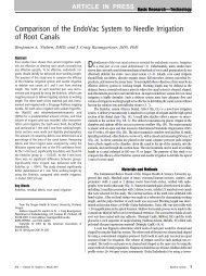
Basic Research—Technology<br />
Figure 1. (A) <strong>The</strong> EndoVac plastic Macro and (B) stainless steel Micro cannulae are shown inserted in their respective titanium components. <strong>The</strong> Micro ’s tip<br />
(enlargement) terminates with array <strong>of</strong> twelve 100-mm holes (only 6 are visible) extending between an area 0.2–0.7 mm from the spherical end <strong>of</strong> the cannula. (C)<br />
PSP at top was used to deliver irrigant through (C-1) the ultrasonic needle, (C-2) the Max-I-Probe, and (C-3) the EndoVac ’s MDT. (D1) <strong>The</strong> battery-operated<br />
EndoActivator is shown with a plastic activation tip inserted. (D2) <strong>The</strong> Rinsendo is shown fully assembled; it delivers irrigant via internal pneumatic pressure.<br />
handpiece is powered by dental air compressor and has irrigation<br />
speed <strong>of</strong> 6.2 mL/min. Research has shown promising results in cleaning<br />
the root canal system. Periapical extrusion <strong>of</strong> irrigant has also been<br />
reported (15).<br />
Materials and Methods<br />
Twenty-two single-rooted, extracted maxillary central and lateral<br />
incisors with mature apices were selected. <strong>The</strong> same 22 teeth were<br />
used in all 6 groups to avoid variables <strong>of</strong> different canal anatomy and<br />
apical diameter. A consistent and known volume <strong>of</strong> irrigant was delivered<br />
to each pulp canal, and all apical extrusion was trapped in a collection<br />
vial similar to that <strong>of</strong> Brown et al (8). <strong>The</strong> percent difference<br />
between the extruded and delivered irrigant was calculated and<br />
analyzed.<br />
Canal Preparation<br />
After conventional access preparation, canals were shaped by<br />
using a crown-down technique with Endo Sequence, rotary nickel titanium<br />
instruments (Brasseler USA Dental Instrumentation, Savannah,<br />
GA) to a master apical file (MAF) size <strong>of</strong> #50/04. MAF is defined as<br />
the largest file that binds slightly at correct working length after<br />
straight-line access. Once the teeth were shaped to MAF, a micro capillary<br />
tip (Ultradent Products Inc, South Jordan, UT) was used to deliver<br />
6.0% sodium hypochlorite through the prepared root canal space, until<br />
no visual evidence <strong>of</strong> intracanal organic tissue was found.<br />
Test Units and Irrigant Control<br />
<strong>The</strong> test units were prepared in the following manner (Fig. 3). <strong>The</strong><br />
prepared teeth were mounted through a hole in the mating lid (Fig. 3A-<br />
1) <strong>of</strong> a removable 20-mL collection vial (Research Product Interna-<br />
tional Corp, Mt Prospect, IL) (Fig. 3A-4) next to an atmospheric equalization<br />
18-gauge needle (Ultradent Products Inc) (Fig. 3A-3). Both the<br />
tooth and the 18-gauge needle were secured and sealed to the lid by<br />
using light-cure composite resins (Esthet-X, Dentsply Caulk; Dentsply<br />
International, Milford, DE) and yellow sticky wax (Kerr Lab, Sybron<br />
Dental, Orange, CA) (Fig. 3A-2). <strong>The</strong> collection vial was dried and<br />
weighed on a digital scale (Sauter; August Sauter <strong>of</strong> America, New<br />
York, NY) and then securely screwed into the tooth/needle/lid assembly<br />
(8).<br />
In all tests, irrigation was accomplished with room temperature<br />
tap water delivered to the pulp canal according to manufacturer ’s<br />
instruction. To maintain irrigation consistency, a programmable precision<br />
syringe pump (PSP) (Fig. 1C) (Alladin, AL 1000; World Precision<br />
Instruments, Inc, Sarasota, FL) was used to deliver between 3.48 and<br />
3.53 mL at the precise rate <strong>of</strong> 7.0 mL/min, except for the Rinsendo,<br />
because it contains its own pneumatic pump and irrigation syringe. A<br />
custom-made Fluid Recovery Trap (FRT) (Fig. 3A-5) collected coronally<br />
expressed irrigant in group 3 (Fig. 3C) or the irrigant flow through<br />
the Micro and Macro cannulae in groups 1 and 2 (Fig. 3A).<br />
Testing Procedure<br />
Group 1: Micro Cannula, EndoVac. <strong>The</strong> MDT was attached to the<br />
PSP to deliver irrigant into the pulp chamber (Fig. 3A-6). <strong>The</strong> micro<br />
cannula was attached to FRT (Fig. 3A-8), placed at full working length,<br />
and used according to manufacturer ’s instructions.<br />
Group 2: Macro Cannula, EndoVac. <strong>The</strong> Macro cannula was<br />
used as described in group 1. Its apical advancement ended wherever<br />
the intracanal diameter prevented its further apical extension.<br />
Group 3: EndoActivator. <strong>The</strong> PSP was attached to irrigation<br />
needle that delivered irrigant into the pulp chamber (Fig. 3C). <strong>The</strong><br />
546 Desai and Himel JOE — Volume 35, Number 4, April 2009
Figure 2. <strong>The</strong> EndoVac ’s MDT delivers irrigant from its stainless steel tip (A)<br />
into an access opening (B) and concurrently aspirates the excess (C) via its<br />
evacuation hood (D), thus ensuring a brimful access opening necessary for<br />
successful apical negative pressure irrigation. Because the MDT delivers<br />
more irrigant than is actually drawn through the Macro and Micro cannulae,<br />
it was necessary to measure the actual volume flow via an FRT (Fig. 3A). Fig. 3A<br />
shows the MDT as an abstract schematic without the detail shown here.<br />
EndoActivator tip (35/04) was placed within 2 mm <strong>of</strong> WL and activated<br />
while moving in an up and down motion for 30 seconds.<br />
Group 4: Manual Syringe and Max-I-Probe Needle. <strong>The</strong> 30gauge<br />
Max-I-Probe needle attached to the PSP was placed 2 mm short <strong>of</strong><br />
working length without binding and moved in an up and down motion<br />
during irrigation (Fig. 3B).<br />
Group 5: Ultrasonic Needle <strong>Irrigation</strong>. <strong>The</strong> Ultrasonic unit<br />
used was Spartan MTS, 115 V (Obtura Spartan USA, Fenton, MO).<br />
<strong>The</strong> 25-gauge diameter, experimental beveled ultrasonic needle (Becton<br />
Dickinson & Co, Franklin Lakes, NJ) measured 1.5 inches in length<br />
and was mounted at a 45-degree angle to the ultrasonic handpiece. <strong>The</strong><br />
PSP was attached to the ultrasonic needle, which delivered the irrigant.<br />
<strong>The</strong> ultrasonic needle was placed short <strong>of</strong> the binding point and moved<br />
in an up and down motion during irrigation (14).<br />
Group 6: Rinsendo. Its syringe was filled with 3.50 mL <strong>of</strong> irrigant<br />
and weighed before and after the experiment to confirm the volume <strong>of</strong><br />
the irrigant in the syringe. Rinsendo was operated at 45-PSI pressure.<br />
<strong>The</strong> cannula was placed into the coronal third <strong>of</strong> the canal without<br />
binding and moved up and down during irrigation (Fig. 3D).<br />
Data Collection and Analysis<br />
<strong>The</strong> volume <strong>of</strong> irrigant delivered into each pulp canal via the PSP<br />
was recorded from the pump ’s digital display. After each test, the lid<br />
assembly was separated from the collection vial, weighing the collection<br />
vial again and subtracting the pre-test tare weight to calculate the apical<br />
extrusion. Because the experiment was conducted at room temperature<br />
with water as the irrigant, no conversion between the weight and volume<br />
Basic Research—Technology<br />
was performed because the specific gravity <strong>of</strong> water at 25 C (77 F) is<br />
1.00 at the second decimal place, reflecting the limit <strong>of</strong> the PSP ’s display.<br />
<strong>The</strong> percentage <strong>of</strong> extrusion in each test was calculated (Apical irrigant<br />
extrusion/Total irrigant delivered) and recorded. Results were analyzed<br />
by using one-way analysis <strong>of</strong> variance with Scheffé test (P < .05).<br />
Results<br />
At the end <strong>of</strong> the experiment 22 teeth were left. Four teeth were<br />
eliminated because <strong>of</strong> cracked roots resulting from desiccation.<br />
<strong>The</strong> apical negative pressure group 1 (EndoVac Micro Cannula)<br />
and group 2 (EndoVac Macro Cannula) were the only ones that did<br />
not extrude irrigating solution into the collection vial (Fig. 4). <strong>The</strong>re<br />
was no statistically significant difference between groups 1, 2, and 3<br />
(EndoVac Micro, EndoVac Macro, EndoActivator). Group 3 extruded<br />
statistically significantly less irrigant compared with group 4 (Max-I-<br />
Probe Needle), group 5 (Ultrasonic needle), and group 6 (Rinsendo).<br />
<strong>The</strong>re was no statistically significant difference among groups 4, 5, and<br />
6. Group 6 extruded highest irrigant followed by groups 5, 4, and 3<br />
(Fig. 5).<br />
Discussion<br />
Results <strong>of</strong> this study broadly correlated with studies by Lambrianidis<br />
et al (6), Brown et al (8), Myers and Montgomery (9), and Roy and<br />
Laurence (16), which noted that irrigation with positive pressure resulted<br />
in periapical extrusion. This study also supports the result <strong>of</strong> Fukumoto<br />
et al (17) that negative pressure irrigation technique reduced<br />
periapical extrusion.<br />
EndoVac Micro and Macro cannulae did not extrude irrigant<br />
through the apex. Because nothing was extruded, the amount <strong>of</strong> irrigant<br />
circulating through the Macro and Micro cannulae could be questioned.<br />
To address this concern, it was decided to collect the irrigants<br />
circulating through these components by using the FRT. Data from the<br />
FRT demonstrated that 82%–99% <strong>of</strong> the irrigant circulated through the<br />
Macro cannula, whereas 51%–54% circulated through the Micro<br />
cannula. <strong>The</strong> MDT was responsible for suctioning the coronal overflow<br />
(Fig. 3A-7)(Fig. 2).<br />
Although Endoactivator extruded irrigant, the volume was very<br />
small, and the clinical significance is not known. However, the manufacturer<br />
’s instructions at the time <strong>of</strong> research did not suggest the use<br />
<strong>of</strong> manual irrigation before using Endoactivator. In a recent publication<br />
by Ruddle (11), he suggested the use <strong>of</strong> intracanal irrigation before<br />
using EndoActivator. To relate these results to the manufacturer ’s<br />
instructions, groups 3 and 4 could be added together and then<br />
compared with the other groups. This would potentially make the differences<br />
between the EndoActivator and the EndoVac even greater.<br />
<strong>The</strong> protocol for this study was designed to maximize the possibility<br />
<strong>of</strong> irrigant extrusion through an unrestricted, yet normal apex.<br />
It is understood that in clinical situations several factors might decrease<br />
the extent to which these systems extrude solutions. Periapical tissues<br />
and bone provide resistance to apical extrusion as well as non-patent<br />
canals. If quantities <strong>of</strong> periapical extrusion occurred clinically such<br />
as reported in this article, greater adverse treatment reactions associated<br />
with full-strength sodium hypochlorite would most likely occur.<br />
<strong>The</strong> model used most likely correlates, by design, to a canal that is<br />
open to atmospheric pressure, such as occurs when the apex <strong>of</strong> a tooth<br />
is extruding into the maxillary sinus with no apical covering or restriction<br />
(18, 19).<br />
Because the basic goal <strong>of</strong> successful endodontic therapy is to<br />
eradicate microorganisms and other intracanal debris from the root<br />
canal system, the clinician must be able to deliver antimicrobial and<br />
tissue solvent solutions in predictable volumes safely to full working<br />
JOE — Volume 35, Number 4, April 2009 <strong>Comparative</strong> <strong>Safety</strong> <strong>of</strong> <strong>Various</strong> <strong>Intracanal</strong> <strong>Irrigation</strong> <strong>Systems</strong> 547
Basic Research—Technology<br />
Figure 3. All tests used the same set <strong>of</strong> teeth (A-1), mounted and sealed via composite and wax (A-2) to a removable cap, perforated, and sealed with a pressure<br />
equalization cannula (A-3). This cap unit could be assembled and disassembled from apical extrusion collection vials (A-4). An FRT (A-5) was used in 2 test<br />
groups. Except for Rinsendo, all irrigant was delivered via a PSP (A-6). EndoVac ’s (A) Macro and Micro (not shown) received irrigant at the access opening<br />
via the PSP, coronal excess was evacuated into the Hi-Vac (A-7), while the irrigant flowing through the Macro/Micro cannulae was trapped (A-8). (B) <strong>The</strong><br />
Max-I-Probe and ultrasonic needles both received their irrigant from the PSP. (C) <strong>The</strong> EndoActivator received its irrigant at the access opening via the PSP,<br />
and coronal excess was trapped. (D) <strong>The</strong> Rinsendo delivered irrigant to its cannula via its internal pneumatic pump.<br />
length. This goal seems to have been accomplished by using the Endo-<br />
Vac system in terms <strong>of</strong> safety (no apical extrusion) and volume (data<br />
from the FRT). Fear <strong>of</strong> a procedural error attributed to full-strength<br />
sodium hypochlorite extrusion might cause clinicians to use an inadequate<br />
flow <strong>of</strong> sodium hypochlorite at full working length (20), thus<br />
decreasing the efficacy <strong>of</strong> full-strength sodium hypochlorite at full<br />
working length. This observation is supported by a recent study testing<br />
positive and negative postoperative cultures (21) as well as studies<br />
examining intracanal debris and smear layer in the apical region<br />
(10, 17).<br />
Figure 4. Percent apical irrigant extrusion by group. EA, EndoActivator.<br />
Figure 5. Statistical group comparison with P value. EA, EndoActivator.<br />
*Statistical significance.<br />
548 Desai and Himel JOE — Volume 35, Number 4, April 2009
This study concluded that the EndoVac did not extrude irrigant<br />
after deep intracanal delivery and suctioning the irrigant from the<br />
chamber to full working length. EndoActivator had a minimal, although<br />
statistically insignificant, amount <strong>of</strong> irrigant extruded out <strong>of</strong> the apex<br />
when delivering irrigant into the pulp chamber, placing the tip into<br />
the canal, and initiating the sonic energy <strong>of</strong> the EndoActivator. Manual,<br />
Ultrasonic, and Rinsendo groups had significantly greater amounts <strong>of</strong><br />
extrusion compared with EndoVac and EndoActivator groups.<br />
Acknowledgments<br />
We thank Discus Dental, Advanced Endodontics, and Air Techniques<br />
Inc for providing us all the necessary supplies to complete<br />
the research. We also thank Dr John Schoeffel for his assistance<br />
in this project and Dr Mark Scarbecz for his help in performing<br />
statistical analysis.<br />
References<br />
1. Clegg MS, Vertucci FJ, Walker C, Belanger M, Britto LR. <strong>The</strong> effect <strong>of</strong> exposure to<br />
irrigant solutions on apical dentin bi<strong>of</strong>ilms in vitro. J Endod 2006;32:434–7.<br />
2. Dunavant TR, Regan JD, Glickman GN, Solomon ES, Honeyman AL. <strong>Comparative</strong><br />
evaluation <strong>of</strong> endodontic irrigants against Enterococcus faecalis bi<strong>of</strong>ilms. J Endod<br />
2006;32:527–31.<br />
3. Ehrich DG, Brian JD Jr, Walker WA. Sodium hypochlorite accident: inadvertent<br />
injection into the maxillary sinus. J Endod 1993;19:180.<br />
4. Bowden JR, Ethunandan M, Brennan PA. Life-threatening airway obstruction<br />
secondary to hypochlorite extrusion during root canal treatment. Oral Surg Oral<br />
Med Oral Pathol Oral Radiol Endod 2006;101:402–4.<br />
5. Mehdipour O, Kleier DJ, Averbach RE. Anatomy <strong>of</strong> sodium hypochlorite accidents.<br />
Compend Contin Educ Dent 2007;28:544–6, 548,550.<br />
6. Lambrianidis T, Tosounidou E, Tzoanopoulou M. <strong>The</strong> effect <strong>of</strong> maintaining apical<br />
patency on periapical extrusion. J Endod 2001;27:696–8.<br />
Basic Research—Technology<br />
7. Tinaz AC, Alacam T, Uzun O, Maden M, Kayaoglu G. <strong>The</strong> effect <strong>of</strong> disruption <strong>of</strong> apical<br />
constriction on periapical extrusion. J Endod 2005;31:533–5.<br />
8. Brown DC, Moore BK, Brown CE Jr, Newton CW. An in vitro study <strong>of</strong> apical extrusion <strong>of</strong><br />
sodium hypochlorite during endodontic canal preparation. J Endod 1995;21:587–91.<br />
9. Myers GL, Montgomery S. A comparison <strong>of</strong> weights <strong>of</strong> debris extruded apically by<br />
conventional filing and Canal Master techniques. J Endod 1991;17:275–9.<br />
10. Nielsen BA, Baumgartner J. Comparison <strong>of</strong> the EndoVac system to needle irrigation<br />
<strong>of</strong> root canals. J Endod 2007;33:611–5.<br />
11. Ruddle C. Endodontic disinfection-tsunami irrigation. Endodontic Practice 2008;5:<br />
8–17.<br />
12. Sabins RA, Johnson JD, Hellstein JW. A comparison <strong>of</strong> the cleaning efficacy <strong>of</strong> shortterm<br />
sonic and ultrasonic passive irrigation after hand instrumentation in molar<br />
root canals. J Endod 2003;29:674–8.<br />
13. Jensen SA, Walker TL, Hutter JW, Nicoll BK. Comparison <strong>of</strong> the cleaning efficacy <strong>of</strong><br />
passive sonic activation and passive ultrasonic activation after hand instrumentation<br />
in molar root canals. J Endod 1999;25:735–8.<br />
14. Burleson A, Nusstein J, Reader A, Beck M. <strong>The</strong> in vivo evaluation <strong>of</strong> hand/rotary/<br />
ultrasound instrumentation in necrotic, human mandibular molars. J Endod<br />
2007;33:782–7.<br />
15. Pouch D, Bohne W, Enkel B, Pilet P, Calas P, Laboux O. Cleaning qualities <strong>of</strong> Rinsendo:<br />
an in vitro study. European Cells and Materials 2007;13:7.<br />
16. Roy G, Laurence JW. Apical extrusion <strong>of</strong> root canal irrigants when using Er:YAG and<br />
Er, Cr:YSGG lasers with optical fibers: an in vitro dye study. J Endod 2008;34:<br />
706–8.<br />
17. Fukumoto Y, Kikuchi I, Yoshioka T, Kobayashi C, Suda H. An ex vivo evaluation <strong>of</strong><br />
a new root canal irrigation technique with intracanal aspiration. Int Endod J 2006;<br />
39:93–9.<br />
18. Hauman CH, Chandler NP, Tong DC. Endodontic implications <strong>of</strong> the maxillary sinus:<br />
a review. Int Endod J 2002;35:127–41.<br />
19. Zairi A, Lambrianidis T. Accidental extrusion <strong>of</strong> sodium hypochlorite into the maxillary<br />
sinus. Quintessence Int 2008;39:745–8.<br />
20. Bradford CE, Eleazer PD, Downs KE, Scheetz JP. Apical pressures developed by needles<br />
for canal irrigation. J Endod 2002;28:333–5.<br />
21. Hockett JI, Dommisch JK, Johnson JD, Cohenca N. Antimicrobial efficacy <strong>of</strong> two irrigation<br />
techniques in tapered and nontapered canal preparations: an in vitro study. J<br />
Endod 2008;34:1374–7.<br />
JOE — Volume 35, Number 4, April 2009 <strong>Comparative</strong> <strong>Safety</strong> <strong>of</strong> <strong>Various</strong> <strong>Intracanal</strong> <strong>Irrigation</strong> <strong>Systems</strong> 549