A Comparative Study of Smear Layer Removal ... - Home page | ART
A Comparative Study of Smear Layer Removal ... - Home page | ART
A Comparative Study of Smear Layer Removal ... - Home page | ART

You also want an ePaper? Increase the reach of your titles
YUMPU automatically turns print PDFs into web optimized ePapers that Google loves.
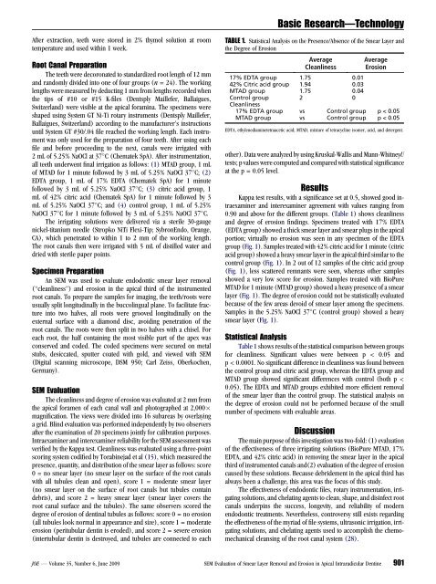
After extraction, teeth were stored in 2% thymol solution at room<br />
temperature and used within 1 week.<br />
Root Canal Preparation<br />
The teeth were decoronated to standardized root length <strong>of</strong> 12 mm<br />
and randomly divided into one <strong>of</strong> four groups (n = 24). The working<br />
lengths were measured by deducting 1 mm from lengths recorded when<br />
the tips <strong>of</strong> #10 or #15 K-files (Dentsply Maillefer, Ballaigues,<br />
Switzerland) were visible at the apical foramina. The specimens were<br />
shaped using System GT Ni-Ti rotary instruments (Dentsply Maillefer,<br />
Ballaigues, Switzerland) according to the manufacturer’s instructions<br />
until System GT #30/.04 file reached the working length. Each instrument<br />
was only used for the preparation <strong>of</strong> four teeth. After using each<br />
file and before proceeding to the next, canals were irrigated with<br />
2 mL <strong>of</strong> 5.25% NaOCl at 37 C (Chematek SpA). After instrumentation,<br />
all teeth underwent final irrigation as follows: (1) MTAD group, 1 mL<br />
<strong>of</strong> MTAD for 1 minute followed by 3 mL <strong>of</strong> 5.25% NaOCl 37 C; (2)<br />
EDTA group, 1 mL <strong>of</strong> 17% EDTA (Chematek SpA) for 1 minute<br />
followed by 3 mL <strong>of</strong> 5.25% NaOCl 37 C; (3) citric acid group, 1<br />
mL <strong>of</strong> 42% citric acid (Chematek SpA) for 1 minute followed by 3<br />
mL <strong>of</strong> 5.25% NaOCl 37 C; and (4) control group, 1 mL <strong>of</strong> 5.25%<br />
NaOCl 37 C for 1 minute followed by 3 mL <strong>of</strong> 5.25% NaOCl 37 C.<br />
The irrigating solutions were delivered via a sterile 30-gauge<br />
nickel-titanium needle (Stropko NiTi Flexi-Tip; SybronEndo, Orange,<br />
CA), which penetrated to within 1 to 2 mm <strong>of</strong> the working length.<br />
The root canals then were irrigated with 5 mL <strong>of</strong> distilled water and<br />
dried with sterile paper points.<br />
Specimen Preparation<br />
An SEM was used to evaluate endodontic smear layer removal<br />
(‘‘cleanliness’’) and erosion in the apical third <strong>of</strong> the instrumented<br />
root canals. To prepare the samples for imaging, the teeth/roots were<br />
usually split longitudinally in the buccolingual plane. To facilitate fracture<br />
into two halves, all roots were grooved longitudinally on the<br />
external surface with a diamond disc, avoiding penetration <strong>of</strong> the<br />
root canals. The roots were then split in two halves with a chisel. For<br />
each root, the half containing the most visible part <strong>of</strong> the apex was<br />
conserved and coded. The coded specimens were secured on metal<br />
stubs, desiccated, sputter coated with gold, and viewed with SEM<br />
(Digital scanning microscope, DSM 950; Carl Zeiss, Oberkochen,<br />
Germany).<br />
SEM Evaluation<br />
The cleanliness and degree <strong>of</strong> erosion was evaluated at 2 mm from<br />
the apical foramen <strong>of</strong> each canal wall and photographed at 2,000<br />
magnification. The views were divided into 16 subareas by overlaying<br />
a grid. Blind evaluation was performed independently by two observers<br />
after the examination <strong>of</strong> 20 specimens jointly for calibration purposes.<br />
Intraexaminer and interexaminer reliability for the SEM assessment was<br />
verified by the Kappa test. Cleanliness was evaluated using a three-point<br />
scoring system codified by Torabinejad et al (15), which measured the<br />
presence, quantity, and distribution <strong>of</strong> the smear layer as follows: score<br />
0 = no smear layer (no smear layer on the surface <strong>of</strong> the root canals<br />
with all tubules clean and open), score 1 = moderate smear layer<br />
(no smear layer on the surface <strong>of</strong> root canals but tubules contain<br />
debris), and score 2 = heavy smear layer (smear layer covers the<br />
root canal surface and the tubules). The same observers scored the<br />
degree <strong>of</strong> erosion <strong>of</strong> dentinal tubules as follows: score 0 = no erosion<br />
(all tubules look normal in appearance and size), score 1 = moderate<br />
erosion (peritubular dentin is eroded), and score 2 = severe erosion<br />
(intertubular dentin is destroyed, and tubules are connected to each<br />
Basic Research—Technology<br />
TABLE 1. Statistical Analysis on the Presence/Absence <strong>of</strong> the <strong>Smear</strong> <strong>Layer</strong> and<br />
the Degree <strong>of</strong> Erosion<br />
Average<br />
Cleanliness<br />
Average<br />
Erosion<br />
17% EDTA group 1.75 0.01<br />
42% Citric acid group 1.94 0.03<br />
MTAD group 1.75 0.04<br />
Control group 2 0<br />
Cleanliness<br />
17% EDTA group vs Control group p < 0.05<br />
MTAD group vs Control group p < 0.05<br />
EDTA, ethylenediaminetetraacetic acid; MTAD, mixture <strong>of</strong> tetracycline isomer, acid, and detergent.<br />
other). Data were analyzed by using Kruskal-Wallis and Mann-WhitneyU<br />
tests; p values were computed and compared with statistical significance<br />
at the p = 0.05 level.<br />
Results<br />
Kappa test results, with a significance set at 0.5, showed good intraexaminer<br />
and interexaminer agreement with values ranging from<br />
0.90 and above for the different groups. (Table 1) shows cleanliness<br />
and degree <strong>of</strong> erosion findings. Specimens treated with 17% EDTA<br />
(EDTA group) showed a thick smear layer and smear plugs in the apical<br />
portion; virtually no erosion was seen in any specimen <strong>of</strong> the EDTA<br />
group (Fig. 1). Samples treated with 42% citric acid for 1 minute (citric<br />
acid group) showed a heavy smear layer in the apical third similar to the<br />
control group (Fig. 1). In 2 out <strong>of</strong> 12 samples <strong>of</strong> the citric acid group<br />
(Fig. 1), less scattered remnants were seen, whereas other samples<br />
showed a very low score for erosion. Samples treated with BioPure<br />
MTAD for 1 minute (MTAD group) showed a heavy presence <strong>of</strong> a smear<br />
layer (Fig. 1). The degree <strong>of</strong> erosion could not be statistically evaluated<br />
because <strong>of</strong> the few areas devoid <strong>of</strong> smear layer among the specimens.<br />
Samples in the 5.25% NaOCl 37 C (control group) showed a heavy<br />
smear layer (Fig. 1).<br />
Statistical Analysis<br />
Table 1 shows results <strong>of</strong> the statistical comparison between groups<br />
for cleanliness. Significant values were between p < 0.05 and<br />
p < 0.0001. No significant difference in cleanliness was found between<br />
the control group and citric acid group, whereas the EDTA group and<br />
MTAD group showed significant differences with control (both p <<br />
0.05). The EDTA and MTAD groups exhibited more efficient removal<br />
<strong>of</strong> the smear layer than the control group. The statistical analysis on<br />
the degree <strong>of</strong> erosion could not be performed because <strong>of</strong> the small<br />
number <strong>of</strong> specimens with evaluable areas.<br />
Discussion<br />
The main purpose <strong>of</strong> this investigation was two-fold: (1) evaluation<br />
<strong>of</strong> the effectiveness <strong>of</strong> three irrigating solutions (BioPure MTAD, 17%<br />
EDTA, and 42% citric acid) in removing the smear layer in the apical<br />
third <strong>of</strong> instrumented canals and(2) evaluation <strong>of</strong> the degree <strong>of</strong> erosion<br />
caused by these solutions. Because debridement in the apical third has<br />
always been a challenge, this area was the focus <strong>of</strong> this study.<br />
The effectiveness <strong>of</strong> endodontic files, rotary instrumentation, irrigating<br />
solutions, and chelating agents to clean, shape, and disinfect root<br />
canals underpins the success, longevity, and reliability <strong>of</strong> modern<br />
endodontic treatments. Nevertheless, controversy still exists regarding<br />
the effectiveness <strong>of</strong> the myriad <strong>of</strong> file systems, ultrasonic irrigation, irrigating<br />
solutions, and chelating agents used to accomplish the chemomechanical<br />
cleansing <strong>of</strong> the root canal system (28).<br />
JOE — Volume 35, Number 6, June 2009 SEM Evaluation <strong>of</strong> <strong>Smear</strong> <strong>Layer</strong> <strong>Removal</strong> and Erosion in Apical Intraradicular Dentine 901