Determination of critical aggregation concentration and ... - YIC-IR
Determination of critical aggregation concentration and ... - YIC-IR
Determination of critical aggregation concentration and ... - YIC-IR

You also want an ePaper? Increase the reach of your titles
YUMPU automatically turns print PDFs into web optimized ePapers that Google loves.
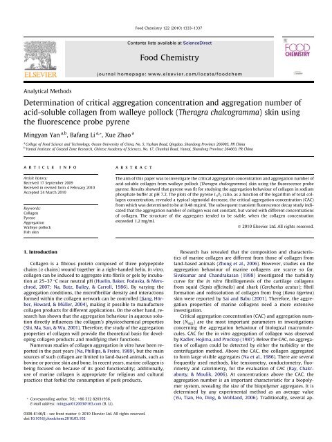
Analytical Methods<br />
<strong>Determination</strong> <strong>of</strong> <strong>critical</strong> <strong>aggregation</strong> <strong>concentration</strong> <strong>and</strong> <strong>aggregation</strong> number <strong>of</strong><br />
acid-soluble collagen from walleye pollock (Theragra chalcogramma) skin using<br />
the fluorescence probe pyrene<br />
Mingyan Yan a,b , Bafang Li a, *, Xue Zhao a<br />
a College <strong>of</strong> Food Science <strong>and</strong> Technology, Ocean University <strong>of</strong> China, No. 5, Yushan Road, Qingdao, Sh<strong>and</strong>ong Province 266003, PR China<br />
b Yantai Institute <strong>of</strong> Coastal Zone Research, Chinese Academy <strong>of</strong> Sciences, No. 17, Chunhui Road, Yantai, Sh<strong>and</strong>ong Province 264003, PR China<br />
article info<br />
Article history:<br />
Received 17 September 2009<br />
Received in revised form 4 February 2010<br />
Accepted 24 March 2010<br />
Keywords:<br />
Collagen<br />
Pyrene<br />
Aggregation<br />
Walleye pollock<br />
Fish skin<br />
1. Introduction<br />
abstract<br />
Collagen is a fibrous protein composed <strong>of</strong> three polypeptide<br />
chains (a chains) wound together in a right-h<strong>and</strong>ed helix. In vitro,<br />
collagen can be induced to aggregate into fibrils or gels by incubation<br />
at 25–37 °C near neutral pH (Huelin, Baker, Poduska, & Merschrod,<br />
2007; Na, Butz, Bailey, & Carroll, 1986). By varying the<br />
<strong>aggregation</strong> conditions, the micr<strong>of</strong>ibrillar density <strong>and</strong> interactions<br />
formed within the collagen network can be controlled (Jiang, Hörber,<br />
Howard, & Müller, 2004), making it possible to manufacture<br />
collagen products for different applications. On the other h<strong>and</strong>, research<br />
has shown that the <strong>aggregation</strong> behaviour in aqueous solution<br />
directly influences the collagen’s physicochemical properties<br />
(Shi, Ma, Sun, & Wu, 2001). Therefore, the study <strong>of</strong> the <strong>aggregation</strong><br />
properties <strong>of</strong> collagen will provide the theoretical basis for developing<br />
collagen products <strong>and</strong> modifying their functions.<br />
Numerous studies <strong>of</strong> collagen <strong>aggregation</strong> in vitro have been reported<br />
in the past years (Na, Phillips, & Freire, 1989), but the main<br />
sources <strong>of</strong> such collagen are limited to l<strong>and</strong>-based animals, such as<br />
bovine or porcine skin <strong>and</strong> bone. In recent years, marine collagen is<br />
being focused on because <strong>of</strong> its good functionality; additionally,<br />
use <strong>of</strong> marine collagen is appropriate for religious <strong>and</strong> cultural<br />
practices that forbid the consumption <strong>of</strong> pork products.<br />
* Corresponding author. Tel.: +86 532 82031936.<br />
E-mail address: mingyan012003@163.com (B. Li).<br />
0308-8146/$ - see front matter Ó 2010 Elsevier Ltd. All rights reserved.<br />
doi:10.1016/j.foodchem.2010.03.102<br />
Food Chemistry 122 (2010) 1333–1337<br />
Contents lists available at ScienceDirect<br />
Food Chemistry<br />
journal homepage: www.elsevier.com/locate/foodchem<br />
The aim <strong>of</strong> this paper was to investigate the <strong>critical</strong> <strong>aggregation</strong> <strong>concentration</strong> <strong>and</strong> <strong>aggregation</strong> number <strong>of</strong><br />
acid-soluble collagen from walleye pollock (Theragra chalcogramma) skin using the fluorescence probe<br />
pyrene. Results showed that pyrene was fit for studying the <strong>aggregation</strong> behaviour <strong>of</strong> collagen in sodium<br />
phosphate buffer at pH 7.2. The plots <strong>of</strong> the pyrene I 1/I 3 ratio, as a function <strong>of</strong> the logarithm <strong>of</strong> total collagen<br />
<strong>concentration</strong>, revealed a typical sigmoidal decrease, the <strong>critical</strong> <strong>aggregation</strong> <strong>concentration</strong> (CAC)<br />
from which was determined to be at 0.48 mg/ml. The subsequent transient fluorescence decay study indicated<br />
that the <strong>aggregation</strong> number <strong>of</strong> collagen was not constant, but varied with different <strong>concentration</strong>s<br />
<strong>of</strong> collagen. The structure <strong>of</strong> the aggregates tended to be stable, when the collagen <strong>concentration</strong><br />
exceeded 1.2 mg/ml.<br />
Ó 2010 Elsevier Ltd. All rights reserved.<br />
Research has revealed that the composition <strong>and</strong> characteristics<br />
<strong>of</strong> marine collagen are different from those <strong>of</strong> collagen from<br />
l<strong>and</strong>-based animals (Zhong et al., 2006). However, studies on the<br />
<strong>aggregation</strong> behaviour <strong>of</strong> marine collagens are scarce so far.<br />
Sivakumar <strong>and</strong> Ch<strong>and</strong>rakasan (1998) investigated the turbidity<br />
curve for the in vitro fibrillogenesis <strong>of</strong> the cartilage collagens<br />
from squid (Sepia <strong>of</strong>ficinalis) <strong>and</strong> shark (Carcharius acutus); fibril<br />
formation <strong>and</strong> redissolution <strong>of</strong> collagen from frog (Rana tigerina)<br />
skin were reported by Sai <strong>and</strong> Babu (2001). Therefore, the <strong>aggregation</strong><br />
properties <strong>of</strong> marine collagens need a more extensive<br />
investigation.<br />
Critical <strong>aggregation</strong> <strong>concentration</strong> (CAC) <strong>and</strong> <strong>aggregation</strong> number<br />
(Nagg) are the most important parameters in investigations<br />
concerning the <strong>aggregation</strong> behaviour <strong>of</strong> biological macromolecules.<br />
CAC for the in vitro <strong>aggregation</strong> <strong>of</strong> collagen was observed<br />
by Kadler, Hojima, <strong>and</strong> Prockop (1987). Below the CAC, no <strong>aggregation</strong><br />
<strong>of</strong> collagen could be detected by either the turbidity or the<br />
centrifugation method. Above the CAC, the collagen aggregated<br />
to form large visible aggregates (Na et al., 1986). There are several<br />
frequently used methods, like tensiometry, conductometry, fluorimetry<br />
<strong>and</strong> calorimetry, for the evaluation <strong>of</strong> CAC (Ray, Chakraborty,<br />
& Moulik, 2006). At <strong>concentration</strong>s above the CAC, the<br />
<strong>aggregation</strong> number is an important characteristic for a biopolymer<br />
system, revealing the size <strong>of</strong> the biopolymer aggregates. It is<br />
determined by any experimental method as an average value<br />
(Yu, Tian, Ho, Ding, & Wohl<strong>and</strong>, 2006). Traditionally, several ap-
1334 M. Yan et al. / Food Chemistry 122 (2010) 1333–1337<br />
proaches have been used to determine this parameter, including<br />
small-angle neutron scattering (SANS), fluorescence, <strong>and</strong> NMR<br />
self-diffusion coefficient measurements (Javadian et al., 2008).<br />
In this paper, we used pyrene as a fluorescence probe to explore<br />
the <strong>aggregation</strong> behaviour <strong>of</strong> collagen. Pyrene, a typical polycyclic<br />
aromatic hydrocarbon, is constituted <strong>of</strong> four fused benzene rings<br />
<strong>and</strong> contains no functional group otherwise (Siddharth, Redden,<br />
Hendricks, Fletcher, & Palmer, 2003). It has earlier been used as a<br />
fluorescence marker to study the <strong>aggregation</strong> behaviour <strong>of</strong> chitosan<br />
<strong>and</strong> derivatives (Amiji, 1995; Li et al., 2007), <strong>and</strong> to determine<br />
the CAC <strong>of</strong> some surfactants, such as sodium dodecyl sulphate,<br />
dodecyltrimethylammonium bromide, Triton X-100, octaethylene<br />
glycol monododecyl ether, etc. (Aguiar, Carpena, Molina-Bolivar,<br />
& Ruiz, 2003), <strong>and</strong> the N agg <strong>of</strong> binary mixed systems involving a sugar-based<br />
surfactant <strong>and</strong> different n-alkyltrimethylammonium<br />
bromides (Hierrezuelo, Aguiar, & Ruiz, 2006). Fujimori <strong>and</strong> Shambaugh<br />
(1983) employed pyrene to label collagens from young <strong>and</strong><br />
old rat tail tendon for studying the cross-linking.<br />
To the best <strong>of</strong> our knowledge, up to now there have been no reports<br />
about studying collagen <strong>aggregation</strong> behaviour using the<br />
fluorescence probe pyrene. The goal <strong>of</strong> the present study was to<br />
elaborate the potential <strong>of</strong> pyrene fluorescence spectra for studying<br />
the <strong>critical</strong> <strong>aggregation</strong> <strong>concentration</strong> <strong>and</strong> <strong>aggregation</strong> number <strong>of</strong><br />
acid-soluble collagen from walleye pollock skin in Na-phosphate<br />
buffer at pH 7.2.<br />
2. Materials <strong>and</strong> methods<br />
2.1. Preparation <strong>of</strong> collagen<br />
Walleye pollock (Theragra chalcogramma) skins were collected<br />
from a local fish processing factory in Qingdao. They were transported<br />
to the laboratory <strong>and</strong> stored at 20 °C until used. Collagen<br />
was prepared according to the method <strong>of</strong> Nagai <strong>and</strong> Suzuki (2000)<br />
with a slight modification. The extraction procedure <strong>and</strong> characterisation<br />
<strong>of</strong> collagen were shown in the paper <strong>of</strong> Yan et al. (2008).<br />
2.2. Preparation <strong>of</strong> pyrene solution<br />
Pyrene was purchased from Sigma Chemicals (Sigma–Aldrich,<br />
Munich, Germany) <strong>and</strong> used without further purification. A stock<br />
solution (1 mM) <strong>of</strong> pyrene was prepared in ethanol. The mixture<br />
was sonicated to dissolve completely. Pyrene, used as a fluorescence<br />
probe, was employed at a final <strong>concentration</strong> <strong>of</strong> 2 lM in<br />
the aggregate solution <strong>of</strong> collagen wherein the ethanol <strong>concentration</strong><br />
was 0.2%. Such a small <strong>concentration</strong> <strong>of</strong> ethanol was considered<br />
not to affect the spectral <strong>and</strong> <strong>aggregation</strong> behaviour <strong>of</strong><br />
collagen.<br />
2.3. Measurement <strong>of</strong> fluorescence spectroscopy<br />
Acid-soluble collagen sample was dissolved in 0.1 M acetic acid<br />
at 4 °C. Aggregation process was initiated by mixing collagen solution<br />
with cooled Na-phosphate/NaCl buffer in an ice bath. After<br />
mixing, <strong>concentration</strong>s were 5 mM Na-phosphate buffer <strong>and</strong><br />
60 mM NaCl, collagen covering a range <strong>of</strong> 0.001–3 mg/ml. The<br />
above solutions were homogenised. After checking that the pHs<br />
were 7.2, the stock solution <strong>of</strong> pyrene was introduced at a final<br />
<strong>concentration</strong> <strong>of</strong> 2 lM <strong>and</strong> the mixtures were homogenised, <strong>and</strong><br />
then the samples were immediately placed in a temperature-controlled<br />
thermostated water bath at 37 °C in total darkness. Every<br />
12 h, fluorescence measurements were taken using a Hitachi F-<br />
2500 fluorescence spectrophotometer (Hitachi, Tokyo, Japan), with<br />
a 10-mm path length quartz cuvette, according to the method <strong>of</strong> Li<br />
et al. (2007). Pyrene was excited at 343 nm, <strong>and</strong> the emission spec-<br />
trum was collected in the range <strong>of</strong> 360–500 nm at a scanning rate<br />
<strong>of</strong> 300 nm/min. The slit openings for both excitation <strong>and</strong> emission<br />
were fixed at 10 <strong>and</strong> 2.5 nm, respectively. All measurements were<br />
carried out in triplicate.<br />
2.4. Turbidity measurement<br />
The solutions (pH 7.2), containing 5 mM Na-phosphate buffer,<br />
60 mM NaCl, <strong>and</strong> collagen at <strong>concentration</strong>s <strong>of</strong> 0.5, 0.6 <strong>and</strong><br />
1.0 mg/ml, respectively, were incubated in a water bath at 37 °C.<br />
The <strong>aggregation</strong> process was examined by recording the increase<br />
in absorbance at 313 nm at various time intervals using a Shimadzu<br />
spectrophotometer UV-2550 (Shimadzu, Kyoto, Japan). The assays<br />
were conducted at least in triplicate on separate occasions.<br />
3. Results <strong>and</strong> discussion<br />
3.1. Theory <strong>of</strong> determining the <strong>aggregation</strong> number<br />
The values <strong>of</strong> <strong>aggregation</strong> number can be obtained by a transient<br />
fluorescence decay method, using pyrene as the fluorescence<br />
probe (Jiang, Ye, & Wu, 1992). Our approach was based on the<br />
assumption that pyrene molecules r<strong>and</strong>omly distribute amongst<br />
the aggregates, which is accurately described by the Poisson distribution.<br />
Nagg can be determined by measuring the total fluorescence<br />
intensity <strong>of</strong> pyrene monomer at the decay time, I t<br />
M . The fluorescence<br />
decay is described by a double exponential function:<br />
I t<br />
M<br />
¼ I0<br />
M<br />
S<br />
e hie kEt ½ ð 1Þ<br />
k1tŠ ; ð1Þ<br />
where I 0<br />
M corresponds to the fluorescence intensity <strong>of</strong> pyrene monomer<br />
at decay time zero, k1 <strong>and</strong> kE are the fluorescence decay rate<br />
constants <strong>of</strong> pyrene monomer <strong>and</strong> excimer, respectively, <strong>and</strong> hSi<br />
is defined as the average occupied degree <strong>of</strong> pyrene in the aggregates,<br />
a parameter related to the distribution <strong>of</strong> pyrene in the aggregates,<br />
which is given by<br />
hSi ¼ cPyrene<br />
; ð2Þ<br />
cAggregate<br />
where cPyrene refers to the <strong>concentration</strong> <strong>of</strong> pyrene in the whole system,<br />
<strong>and</strong> cAggregate can be written as<br />
cAggregate ¼ cCollagen CAC<br />
: ð3Þ<br />
Nagg<br />
It follows from Eqs. (2) <strong>and</strong> (3) that:<br />
Nagg ¼ hSi cCollagen CAC<br />
cPyrene<br />
: ð4Þ<br />
In the case <strong>of</strong> e k Et ! 0, or using the linear part in the double<br />
exponential decay curve, Eq. (1) can be written as<br />
I t<br />
M ¼ I0<br />
Me hSiþk ð 1tÞ<br />
; ð5Þ<br />
or<br />
ln It<br />
M<br />
I 0 ¼ hSi k1t; ð6Þ<br />
M<br />
hSi can be obtained by extending the linear part to the y-axis in the<br />
plots <strong>of</strong> ln(I t<br />
M =I0 M ) versus time, <strong>and</strong> then Nagg is calculated by fitting<br />
Eq. (4) to the data.<br />
3.2. Selection <strong>of</strong> the probe<br />
Pyrene was used as a fluorescence probe to detect the <strong>aggregation</strong><br />
behaviour <strong>of</strong> collagen in Na-phosphate buffer at pH 7.2. It has<br />
the following feature: the ratio <strong>of</strong> intensities <strong>of</strong> the first <strong>and</strong> third
Fig. 1. Fluorescence spectra <strong>of</strong> pyrene (2 lM) <strong>and</strong> acid-soluble collagen from<br />
walleye pollock skin (0.6 mg/ml) in the collagen/Na-phosphate buffer system at<br />
37 °C.<br />
vibronic peaks, I 1/I 3, is <strong>of</strong>ten considered to estimate the polarity level<br />
<strong>of</strong> the microenvironment where pyrene is located (Ray et al.,<br />
2006), <strong>and</strong>, as a result, the conformation change <strong>of</strong> collagen was<br />
deduced from the changes <strong>of</strong> polarity in the solution. The emission<br />
spectra <strong>of</strong> pyrene in the collagen/Na-phosphate buffer system at<br />
37 °C are shown in Fig. 1. It could be found that in the period <strong>of</strong> collagen<br />
<strong>aggregation</strong>, the emission spectra <strong>of</strong> pyrene all showed four<br />
peaks at 374, 380, 385 <strong>and</strong> 394.5 nm, similar to those in methanol<br />
<strong>and</strong> water in Ray’s report (Ray et al., 2006), suggesting that pyrene<br />
exhibited stable fluorescent properties in the collagen/Na-phosphate<br />
buffer system at 37 °C. However, a continuous decrease in<br />
the fluorescence intensity was observed. The reason might be that<br />
with collagen self-aggregating, pyrene molecules moved into the<br />
hydrophobic microdomains <strong>of</strong> the aggregates so that the collisions<br />
<strong>of</strong> pyrene <strong>and</strong> collagen were increased, resulting in the decline <strong>of</strong><br />
pyrene fluorescence intensity (Qiao & Jin, 2000). From Fig. 1, we<br />
could also find that pyrene I1/I3 ratio decreased with collagen<br />
<strong>aggregation</strong>, revealing a gradual reduction in the polarity <strong>of</strong> the<br />
solution.<br />
Collagen has aut<strong>of</strong>luorescence, due to the presence <strong>of</strong> some<br />
phenylalanine <strong>and</strong> tyrosine residues. To minimise the disturbance<br />
due to aut<strong>of</strong>luorescence <strong>of</strong> collagen, an appropriate excitation<br />
wavelength should be selected at the site where the chromophore<br />
<strong>of</strong> collagen has very little absorbance. Additionally, an appropriate<br />
emission wavelength should also be selected at the site where the<br />
chromophore <strong>of</strong> collagen has little emission (Shi et al., 2001). In our<br />
experiment, pyrene was excited at 343 nm, <strong>and</strong> the emission spectrum<br />
was collected over the range <strong>of</strong> 360–500 nm. Under the studied<br />
conditions, pyrene was well excited, while the chromophore <strong>of</strong><br />
collagen showed little emission, as shown in Fig. 1. Besides, the<br />
emission spectra <strong>of</strong> collagen did not change significantly in the<br />
period <strong>of</strong> <strong>aggregation</strong>, <strong>and</strong> the fluorescence intensity was near to<br />
zero. Therefore, the disturbance <strong>of</strong> collagen aut<strong>of</strong>luorescence could<br />
be avoided when the excitation wavelength <strong>of</strong> pyrene was selected<br />
at 343 nm. From the abovementioned results, it could be concluded<br />
that the fluorescence probe pyrene is suitable for studying<br />
the <strong>aggregation</strong> behaviour <strong>of</strong> acid-soluble collagen from walleye<br />
pollock skin in Na-phosphate buffer at pH 7.2.<br />
3.3. <strong>Determination</strong> <strong>of</strong> the <strong>critical</strong> <strong>aggregation</strong> <strong>concentration</strong> <strong>of</strong><br />
collagen<br />
Plots <strong>of</strong> pyrene I1/I3 ratio versus logarithm <strong>of</strong> collagen <strong>concentration</strong><br />
are shown in Fig. 2. Analysing by curve fitting, the pyrene<br />
M. Yan et al. / Food Chemistry 122 (2010) 1333–1337 1335<br />
Fig. 2. Plots <strong>of</strong> pyrene I1/I3 ratio versus the logarithm <strong>of</strong> different <strong>concentration</strong>s <strong>of</strong><br />
acid-soluble collagen from walleye pollock skin in Na-phosphate buffer at pH 7.2<br />
after incubation for 72 h at 37 °C.<br />
I1/I3 ratio data for the studied collagen are well fitted by a Boltzmann<br />
function:<br />
1:18<br />
y ¼ 0:94 þ<br />
1 þ eðxþ0:86Þ=Dx R2 ¼ 0:99956 ;<br />
where the variable y is the pyrene I1/I3 ratio, <strong>and</strong> the independent<br />
variable x is the logarithm <strong>of</strong> collagen <strong>concentration</strong>. It was shown<br />
that at lower collagen <strong>concentration</strong>s, pyrene I1/I3 ratio value corresponded<br />
to a polar environment; as the collagen <strong>concentration</strong> increased,<br />
the pyrene I1/I3 ratio decreased rapidly, indicating that<br />
pyrene was sensing a more hydrophobic environment, <strong>and</strong> then levelled<br />
<strong>of</strong>f with further increase in collagen <strong>concentration</strong>, due to the<br />
incorporation <strong>of</strong> the probe into the hydrophobic region <strong>of</strong> the <strong>aggregation</strong><br />
(Aguiar et al., 2003).<br />
We adopted the term ‘‘<strong>critical</strong> <strong>aggregation</strong> <strong>concentration</strong> (CAC)”<br />
to describe the effect <strong>of</strong> collagen <strong>concentration</strong> on the <strong>aggregation</strong><br />
behaviour. By using this term, no assumption about the nature, the<br />
size, or the shape <strong>of</strong> the aggregates are made: the aggregates can be<br />
<strong>of</strong> finite size (micellar system) or infinite (phase separation)<br />
(Gar<strong>of</strong>alakis, Murray, & Sarney, 2000). The CAC for acid-soluble collagen<br />
aggregates was graphically determined from the plots <strong>of</strong> pyrene<br />
I1/I3 ratio versus the logarithm <strong>of</strong> collagen <strong>concentration</strong> in this<br />
study, as shown in Fig. 2 (Aguiar et al., 2003). It was found that the<br />
CAC value <strong>of</strong> acid-soluble collagen from walleye pollock skin in Naphosphate<br />
buffer <strong>of</strong> pH 7.2 at 37 °C might be 0.138 mg/ml (CAC 1)<br />
or 0.48 mg/ml (CAC2). On the other h<strong>and</strong>, the <strong>aggregation</strong> kinetics<br />
<strong>of</strong> collagen could be detected by absorbance at 313 nm (Sivakumar<br />
& Ch<strong>and</strong>rakasan, 1998). The higher absorbance indicated more or<br />
larger collagen aggregates. As shown in Fig. 3, absorbance at<br />
313 nm showed a marked increase in the <strong>aggregation</strong> kinetics <strong>of</strong><br />
the collagen solutions at 0.6 <strong>and</strong> 1.0 mg/ml, while a rise in the<br />
<strong>aggregation</strong> kinetics <strong>of</strong> collagen solution with a <strong>concentration</strong> <strong>of</strong><br />
0.5 mg/ml was not found. Thus it could be concluded that the<br />
CAC <strong>of</strong> collagen was in the range <strong>of</strong> 0.5–0.6 mg/ml. The CAC determined<br />
by the plot <strong>of</strong> pyrene I1/I3 ratio versus the logarithm <strong>of</strong> different<br />
collagen <strong>concentration</strong>s was 0.48 mg/ml, a slight deviation<br />
from the range <strong>of</strong> CAC obtained by the <strong>aggregation</strong> kinetics perhaps<br />
showing the difference between theoretical <strong>and</strong> experimental<br />
value. The CAC <strong>of</strong> collagen from pollock skin was close to the<br />
threshold <strong>concentration</strong> <strong>of</strong> the collagen <strong>aggregation</strong> from porcine<br />
skin in 0.1 M acetic acid reported by Shi et al. (2001), but much<br />
higher than that <strong>of</strong> pC-collagen from the medium <strong>of</strong> cultured fibroblasts<br />
from normal human skin found by Kadler et al. (1987). The<br />
reasons were as follows. Firstly, the <strong>aggregation</strong> behaviour <strong>of</strong> col-
1336 M. Yan et al. / Food Chemistry 122 (2010) 1333–1337<br />
Fig. 3. Aggregation kinetics <strong>of</strong> collagen from walleye pollock skin at different<br />
<strong>concentration</strong>s in Na-phosphate buffer at pH 7.2 <strong>and</strong> 37 °C (closed square, 0.5 mg/<br />
ml; closed circle, 0.6 mg/ml; closed triangle, 1.0 mg/ml).<br />
lagen was closely related to its cross-linking. Collagen from walleye<br />
pollock skin shows rather less cross-linking than those from<br />
mammals (Liu, Li, & Guo, 2007; Yan et al., 2008), resulting in a<br />
much higher CAC. Secondly, the long lag time in the <strong>aggregation</strong><br />
kinetics <strong>of</strong> collagen (Fig. 3) revealed that the collagen was mainly<br />
in the monomeric form, which also led to a much higher CAC.<br />
Thirdly, ionic strength <strong>and</strong> temperature could affect the <strong>aggregation</strong><br />
<strong>of</strong> collagen, too. Generally, increasing the ionic strength in collagen<br />
solution resulted in a higher CAC for collagen, while<br />
increasing the temperature induced a lower CAC for collagen (Na<br />
et al., 1989). However, we believe that the higher CAC <strong>of</strong> collagen<br />
from walleye pollock skin was mainly ascribed to the lower<br />
cross-linking <strong>and</strong> the higher monomer content <strong>of</strong> collagen in the<br />
study.<br />
Collagen self-<strong>aggregation</strong> in aqueous solution can be explained<br />
by its molecular structure. The regular three-str<strong>and</strong> helical structure<br />
<strong>of</strong> collagen can be destroyed after extraction. In neutral solution,<br />
there was no repelling action in collagen molecular chains for<br />
the net charge near to zero. At lower collagen <strong>concentration</strong>s, few<br />
aggregates appeared in the solution, due to less combination between<br />
the carbonyl <strong>and</strong> amide groups. When collagen <strong>concentration</strong><br />
was increased to the CAC, the carbonyl groups in collagen<br />
chains, which could combine with the amide groups in neutral<br />
solution, increased. Thus the action <strong>of</strong> the intra- <strong>and</strong> intermolecular<br />
hydrogen bonds increased, which resulted in the formation <strong>of</strong><br />
irregular coil structures <strong>and</strong> hydrophobic microdomains in the<br />
solution (Shi et al., 2001). As collagen <strong>concentration</strong> was further increased,<br />
the majority <strong>of</strong> carbonyl <strong>and</strong> amide groups participated in<br />
the formation <strong>of</strong> hydrogen bonds. Simultaneously, the hydrophobic<br />
interactions <strong>of</strong> the hydrophobic amino acid residues in the polypeptide<br />
chains became stronger, <strong>and</strong> the actions <strong>of</strong> the intermolecular<br />
hydrogen bonds posed by the hydroxyl groups <strong>of</strong><br />
hydroxyproline in the polypeptide chains enhanced. Besides, the<br />
hydroxyl groups <strong>of</strong> hydroxyproline also interacted with water, to<br />
produce the hydrogen bonds. So the aggregates were formed,<br />
which induced a roughly constant value <strong>of</strong> pyrene I1/I3 ratio.<br />
3.4. <strong>Determination</strong> <strong>of</strong> the <strong>aggregation</strong> number <strong>of</strong> collagen<br />
We have used the well-established transient fluorescence decay<br />
method to determine the mean <strong>aggregation</strong> numbers <strong>of</strong> acid-soluble<br />
collagen from walleye pollock skin. In this study, pyrene was<br />
used as a fluorescence probe, <strong>and</strong> the decay experiments were analysed<br />
using Eqs. (4) <strong>and</strong> (6). We had carried out the experiments in<br />
Fig. 4. Plots <strong>of</strong> the natural logarithm <strong>of</strong> pyrene I t<br />
374 =I0 374 versus time in the acidsoluble<br />
collagen (0.6 mg/ml)/Na-phosphate buffer system at 37 °C.<br />
Fig. 5. Aggregation numbers <strong>of</strong> acid-soluble collagen from walleye pollock skin at<br />
different <strong>concentration</strong>s in Na-phosphate buffer at pH 7.2 <strong>and</strong> 37 °C.<br />
aggregate solutions with different collagen <strong>concentration</strong>s. Similar<br />
results were found in all cases that the natural logarithm <strong>of</strong> pyrene<br />
I t<br />
374 =I0 374 decreased drastically as the <strong>aggregation</strong> went on, <strong>and</strong> then<br />
decreased linearly (Fig. 4). At the same time, good linearity<br />
(r 2 > 0.9) was achieved in all cases after some <strong>aggregation</strong> time.<br />
From the interception obtained by extending the linear part <strong>of</strong><br />
the pyrene fluorescence decay curve to the y-axis, as shown in<br />
Fig. 4, <strong>and</strong> using the CAC value above, we had determined the Nagg<br />
<strong>of</strong> collagen aggregates according to Eq. (4). Fig. 5 shows that at<br />
lower collagen <strong>concentration</strong>s, Nagg rose sharply, due to increases<br />
in <strong>concentration</strong>, <strong>and</strong> when collagen <strong>concentration</strong> reached<br />
1.2 mg/ml <strong>and</strong> above, it rose slightly. The result was similar to<br />
the changes <strong>of</strong> <strong>aggregation</strong> numbers <strong>of</strong> nonaethylene glycol<br />
monododecyl ether (C12E9) with <strong>concentration</strong> increase (Yu<br />
et al., 2006), but different from those <strong>of</strong> sodium dodecyl sulphate,<br />
sodium dodecyl sulphonate (Fang et al., 2001) <strong>and</strong> quaternary trimeric<br />
surfactant (Li et al., 2005). From the results, we could conclude<br />
that Nagg <strong>of</strong> acid-soluble collagen from walleye pollock skin<br />
was not constant, but varied with different <strong>concentration</strong>s <strong>of</strong> collagen,<br />
whereas the <strong>aggregation</strong> numbers obtained for N-dodecyllactobionamide<br />
show no clear dependence on surfactant<br />
<strong>concentration</strong> (Kjellin, Reimer, & Hansson, 2003). Nagg increasing<br />
with increases in the collagen <strong>concentration</strong> showed that the<br />
aggregates might be incomplete in structure, but when the concen-
tration reached 1.2 mg/ml <strong>and</strong> above, Nagg rose slightly, indicating<br />
that the structure <strong>of</strong> aggregates tended to be stable. We presume<br />
that Nagg shows no significant dependence on collagen <strong>concentration</strong><br />
above 1.2 mg/ml, in good agreement with Kjellin’s conclusion<br />
(Kjellin et al., 2003). Nagg at the CAC could not be determined using<br />
transient pyrene fluorescence decay. Usually, it is obtained by<br />
extrapolating the Nagg versus collagen <strong>concentration</strong> experimental<br />
curve (Fang et al., 2001). In the experiment, N agg at the CAC <strong>of</strong> collagen<br />
from pollock skin was found to be about 68.<br />
The results above verified that the fluorescence probe pyrene<br />
could be applied to the study <strong>of</strong> the <strong>aggregation</strong> behaviour <strong>of</strong> acidsoluble<br />
collagen from walleye pollock skin. The advantage <strong>of</strong> using<br />
the fluorescence probe was that the <strong>concentration</strong> <strong>of</strong> the probe used<br />
could be very small, which minimised the possibility <strong>of</strong> any probe–<br />
probe interaction that might occur. The method was fast, simple<br />
<strong>and</strong> accurate, <strong>and</strong> also showed high sensitivity <strong>and</strong> good selectivity.<br />
Also, both the <strong>critical</strong> <strong>aggregation</strong> <strong>concentration</strong> <strong>and</strong> <strong>aggregation</strong><br />
number <strong>of</strong> collagen could be acquired by this method; tensiometry,<br />
conductometry <strong>and</strong> calorimetry can only evaluate the <strong>critical</strong> <strong>aggregation</strong><br />
<strong>concentration</strong> (Ray et al., 2006), while small-angle neutron<br />
scattering (SANS) <strong>and</strong> NMR self-diffusion coefficient measurements<br />
only determine the <strong>aggregation</strong> number (Javadian et al., 2008).<br />
Thirdly, the fluorescence probe pyrene was more convenient in the<br />
study on the collagen <strong>aggregation</strong> behaviour, because pyrene could<br />
be used directly in the experiment, whereas the fluorescence probe<br />
3-methoxy-4 0 -N,N-dimethylamino flavone derivative employed by<br />
Shi et al. (2001), in the study on the <strong>aggregation</strong> behaviour <strong>of</strong> collagen<br />
from pig skin must be synthesised.<br />
4. Conclusion<br />
The results presented here clearly suggested that acid-soluble<br />
collagen from walleye pollock skin could self-aggregate in Naphosphate<br />
buffer at pH 7.2 above a certain <strong>concentration</strong>. We have<br />
introduced an alternative method to determine the <strong>critical</strong> <strong>aggregation</strong><br />
<strong>concentration</strong> <strong>and</strong> <strong>aggregation</strong> number <strong>of</strong> collagen by fluorescence<br />
spectroscopy <strong>of</strong> pyrene. The CAC <strong>of</strong> collagen was found to<br />
be at 0.48 mg/ml graphically from the plots <strong>of</strong> pyrene I1/I3 ratio<br />
versus the logarithm <strong>of</strong> collagen <strong>concentration</strong>. Transient fluorescence<br />
decay study revealed that Nagg was not constant, but varied<br />
with different <strong>concentration</strong>s <strong>of</strong> collagen. The structure <strong>of</strong> aggregates<br />
tended to be stable, when the collagen <strong>concentration</strong> exceeded<br />
1.2 mg/ml.<br />
In summary, the fluorescence probe pyrene was suitable to describe<br />
the <strong>aggregation</strong> behaviour <strong>of</strong> acid-soluble collagen from<br />
walleye pollock skin. We believe that these results will aid further<br />
study on collagen <strong>aggregation</strong> behaviour.<br />
Acknowledgements<br />
This work was supported by National Natural Science Foundation<br />
<strong>of</strong> China (No. 30871943) <strong>and</strong> the High Technology Research<br />
<strong>and</strong> Development Programme <strong>of</strong> China (No. 2006AA09Z438).<br />
References<br />
Aguiar, J., Carpena, P., Molina-Bolivar, J. A., & Ruiz, C. C. (2003). On the<br />
determination <strong>of</strong> the <strong>critical</strong> micelle <strong>concentration</strong> by the pyrene 1:3 ratio<br />
method. Journal <strong>of</strong> Colloid <strong>and</strong> Interface Science, 258, 116–122.<br />
Amiji, M. M. (1995). Pyrene fluorescence study <strong>of</strong> chitosan self-association in<br />
aqueous solution. Carbohydrate Polymer, 26, 211–213.<br />
M. Yan et al. / Food Chemistry 122 (2010) 1333–1337 1337<br />
Fang, Y., Liu, X. F., Xia, Y. M., Yang, Y., Cai, K., Suh, J. M., et al. (2001). <strong>Determination</strong><br />
<strong>of</strong> <strong>critical</strong> micellar <strong>aggregation</strong> numbers by steady-state fluorescence probe<br />
method. Acta Physico–Chimica Sinica, 17(9), 828–831.<br />
Fujimori, E., & Shambaugh, N. (1983). Cross-linking <strong>and</strong> fluorescence <strong>of</strong> pyrenelabeled<br />
collagen. Biochimica et Biophysica Acta, 742(1), 155–161.<br />
Gar<strong>of</strong>alakis, G., Murray, B. S., & Sarney, D. B. (2000). Surface activity <strong>and</strong> <strong>critical</strong><br />
<strong>aggregation</strong> <strong>concentration</strong> <strong>of</strong> pure sugar esters with different sugar headgroups.<br />
Journal <strong>of</strong> Colloid <strong>and</strong> Interface Science, 229, 391–398.<br />
Hierrezuelo, J. M., Aguiar, J., & Ruiz, C. C. (2006). Interactions in binary mixed<br />
systems involving a sugar-based surfactant <strong>and</strong> different nalkyltrimethylammonium<br />
bromides. Journal <strong>of</strong> Colloid <strong>and</strong> Interface Science,<br />
294, 449–457.<br />
Huelin, S. D., Baker, H. R., Poduska, K. M., & Merschrod, S. E. F. (2007). Aggregation<br />
<strong>and</strong> adsorption <strong>of</strong> type I collagen near an electrified interface. Macromolecules,<br />
40, 8440–8444.<br />
Javadian, S., Gharibi, H., Sohrabi, B., Bijanzadeh, H., Safarpour, M. A., &<br />
Behjatmanesh-Ardakani, R. (2008). <strong>Determination</strong> <strong>of</strong> the physical–chemical<br />
parameters <strong>and</strong> <strong>aggregation</strong> number <strong>of</strong> surfactant in micelles in binary alcohol–<br />
water mixtures. Journal <strong>of</strong> Molecular Liquids, 137, 74–79.<br />
Jiang, F., Hörber, H., Howard, J., & Müller, D. J. (2004). Assembly <strong>of</strong> collagen into<br />
microribbons: Effects <strong>of</strong> pH <strong>and</strong> electrolytes. Journal <strong>of</strong> Structural Biology, 148,<br />
268–278.<br />
Jiang, Y. C., Ye, J. P., & Wu, S. K. (1992). Premicelle formation in surfactant solution<br />
<strong>and</strong> measurement <strong>of</strong> its average <strong>aggregation</strong> number. Acta Chimica Sinica, 50,<br />
1080–1084.<br />
Kadler, K. E., Hojima, Y., & Prockop, D. J. (1987). Assembly <strong>of</strong> collagen fibrils de novo<br />
by cleavage <strong>of</strong> the type I pC-collagen with procollagen C-proteinase. Assay <strong>of</strong><br />
<strong>critical</strong> <strong>concentration</strong> demonstrates that collagen self-assembly is a classical<br />
example <strong>of</strong> an entropy-driven process. Journal <strong>of</strong> Biological Chemistry, 262,<br />
15696–15701.<br />
Kjellin, U. R. M., Reimer, J., & Hansson, P. (2003). An investigation <strong>of</strong> dynamic surface<br />
tension, <strong>critical</strong> micelle <strong>concentration</strong>, <strong>and</strong> <strong>aggregation</strong> number <strong>of</strong> three<br />
nonionic surfactants using NMR, time-resolved fluorescence quenching, <strong>and</strong><br />
maximum bubble pressure tensiometry. Journal <strong>of</strong> Colloid <strong>and</strong> Interface Science,<br />
262, 506–515.<br />
Li, Y. Y., Chen, X. G., Liu, C. S., Cha, D. S., Park, H. J., & Lee, C. M. (2007). Effect <strong>of</strong> the<br />
molecular mass <strong>and</strong> degree <strong>of</strong> substitution <strong>of</strong> oleoylchitosan on the structure,<br />
rheological properties, <strong>and</strong> formation <strong>of</strong> nanoparticles. Journal <strong>of</strong> Agricultural<br />
<strong>and</strong> Food Chemistry, 55, 4842–4847.<br />
Li, X. B., Xu, L., Meng, X. W., Han, Z. H., Luo, T. L., & Liu, G. J. (2005). <strong>Determination</strong> <strong>of</strong><br />
<strong>critical</strong> micellar <strong>aggregation</strong> numbers <strong>of</strong> CTTTA by steady-state fluorescence<br />
probe method. Acta Physico–Chimica Sinica, 21(12), 1403–1406.<br />
Liu, H., Li, D., & Guo, S. (2007). Studies on collagen from the skin <strong>of</strong> channel catfish<br />
(Ictalurus punctaus). Food Chemistry, 101, 621–625.<br />
Na, G. C., Butz, L. J., Bailey, D. G., & Carroll, R. J. (1986). In vitro collagen fibril<br />
assembly in glycerol solution: Evidence for a helical cooperative mechanism<br />
involving micr<strong>of</strong>ibrils. Biochemistry, 25, 958–966.<br />
Na, G. C., Phillips, L. J., & Freire, E. I. (1989). In vitro collagen fibril assembly:<br />
Thermodynamic studies. Biochemistry, 28, 7153–7161.<br />
Nagai, T., & Suzuki, N. (2000). Isolation <strong>of</strong> collagen from fish waste-skin, bone <strong>and</strong><br />
fins. Food Chemistry, 68, 277–281.<br />
Qiao, J., & Jin, W. (2000). Study on the microenvironment properties <strong>of</strong> C60-micellar<br />
aqueous solution using pyrene as a fluorescence probe. Chemical Research <strong>and</strong><br />
Application, 12(4), 398–402.<br />
Ray, G. B., Chakraborty, I., & Moulik, S. P. (2006). Pyrene absorption can be a<br />
convenient method for probing <strong>critical</strong> micellar <strong>concentration</strong> (cmc) <strong>and</strong><br />
indexing micellar polarity. Journal <strong>of</strong> Colloid <strong>and</strong> Interface Science, 294, 248–254.<br />
Sai, K. P., & Babu, M. (2001). Studies on Rana tigerina skin collagen. Comparative<br />
Biochemistry <strong>and</strong> Physiology Part B: Biochemistry <strong>and</strong> Molecular Biology, 128,<br />
82–90.<br />
Shi, X., Ma, W., Sun, C., & Wu, S. (2001). The <strong>aggregation</strong> behavior <strong>of</strong> collagen in<br />
aqueous solution <strong>and</strong> its property <strong>of</strong> stabilizing liposomes in vitro. Biomaterials,<br />
22, 1627–1634.<br />
Siddharth, P., Redden, R. A., Hendricks, A. E., Fletcher, K. A., & Palmer, C. P. (2003).<br />
Characterization <strong>of</strong> the salvation environment provided by dilute aqueous<br />
solutions <strong>of</strong> novel siloxane polysoaps using the fluorescence probe pyrene.<br />
Journal <strong>of</strong> Colloid <strong>and</strong> Interface Science, 262, 579–587.<br />
Sivakumar, P., & Ch<strong>and</strong>rakasan, G. (1998). Occurrence <strong>of</strong> a novel collagen with three<br />
distinct chains in the cranial cartilage <strong>of</strong> the squid Sepia <strong>of</strong>ficinalis: Comparison<br />
with shark cartilage collagen. Biochimica et Biophysica Acta, 1381, 161–169.<br />
Yan, M. Y., Li, B. F., Zhao, X., Ren, G. Y., Zhuang, Y. L., Hou, H., et al. (2008).<br />
Characterization <strong>of</strong> acid-soluble collagen from the skin <strong>of</strong> walleye pollock<br />
(Theragra chalcogramma). Food Chemistry, 107, 1581–1586.<br />
Yu, L. L., Tian, M. Y., Ho, B., Ding, J. L., & Wohl<strong>and</strong>, T. (2006). <strong>Determination</strong> <strong>of</strong> <strong>critical</strong><br />
micelle <strong>concentration</strong>s <strong>and</strong> <strong>aggregation</strong> numbers by fluorescence correlation<br />
spectroscopy: Aggregation <strong>of</strong> a lipopolysaccharide. Analytica Chimica Acta, 556,<br />
216–225.<br />
Zhong, Z., Li, C., Gu, H., Dou, H., Zhang, X., & Xie, B. (2006). Study <strong>of</strong> <strong>aggregation</strong><br />
behaviors <strong>of</strong> fish scale collagen by AFM. Fine Chemicals, 23(10), 983–987.