De novo desmin-mutation N116S is associated with ... - ResearchGate
De novo desmin-mutation N116S is associated with ... - ResearchGate
De novo desmin-mutation N116S is associated with ... - ResearchGate

Create successful ePaper yourself
Turn your PDF publications into a flip-book with our unique Google optimized e-Paper software.
4600 Human Molecular Genetics, 2010, Vol. 19, No. 23<br />
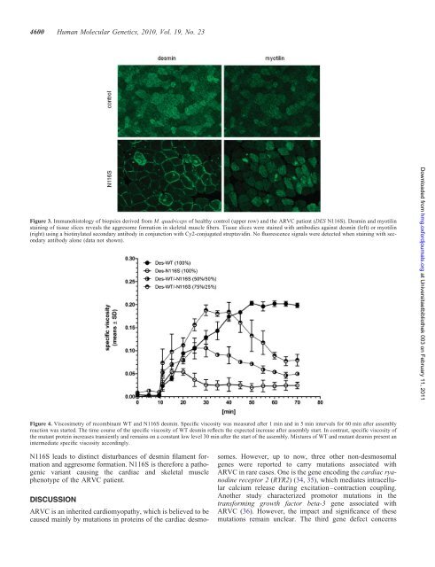
Figure 3. Immunoh<strong>is</strong>tology of biopsies derived from M. quadriceps of healthy control (upper row) and the ARVC patient (DES <strong>N116S</strong>). <strong>De</strong>smin and myotilin<br />
staining of t<strong>is</strong>sue slices reveals the aggresome formation in skeletal muscle fibers. T<strong>is</strong>sue slices were stained <strong>with</strong> antibodies against <strong>desmin</strong> (left) or myotilin<br />
(right) using a biotinylated secondary antibody in conjunction <strong>with</strong> Cy2-conjugated streptavidin. No fluorescence signals were detected when staining <strong>with</strong> secondary<br />
antibody alone (data not shown).<br />
Figure 4. V<strong>is</strong>cosimetry of recombinant WT and <strong>N116S</strong> <strong>desmin</strong>. Specific v<strong>is</strong>cosity was measured after 1 min and in 5 min intervals for 60 min after assembly<br />
reaction was started. The time course of the specific v<strong>is</strong>cosity of WT <strong>desmin</strong> reflects the expected increase after assembly start. In contrast, specific v<strong>is</strong>cosity of<br />
the mutant protein increases transiently and remains on a constant low level 30 min after the start of the assembly. Mixtures of WT and mutant <strong>desmin</strong> present an<br />
intermediate specific v<strong>is</strong>cosity accordingly.<br />
<strong>N116S</strong> leads to d<strong>is</strong>tinct d<strong>is</strong>turbances of <strong>desmin</strong> filament formation<br />
and aggresome formation. <strong>N116S</strong> <strong>is</strong> therefore a pathogenic<br />
variant causing the cardiac and skeletal muscle<br />
phenotype of the ARVC patient.<br />
DISCUSSION<br />
ARVC <strong>is</strong> an inherited cardiomyopathy, which <strong>is</strong> believed to be<br />
caused mainly by <strong>mutation</strong>s in proteins of the cardiac desmo-<br />
somes. However, up to now, three other non-desmosomal<br />
genes were reported to carry <strong>mutation</strong>s <strong>associated</strong> <strong>with</strong><br />
ARVC in rare cases. One <strong>is</strong> the gene encoding the cardiac ryanodine<br />
receptor 2 (RYR2) (34, 35), which mediates intracellular<br />
calcium release during excitation–contraction coupling.<br />
Another study characterized promotor <strong>mutation</strong>s in the<br />
transforming growth factor beta-3 gene <strong>associated</strong> <strong>with</strong><br />
ARVC (36). However, the impact and significance of these<br />
<strong>mutation</strong>s remain unclear. The third gene defect concerns<br />
Downloaded from<br />
hmg.oxfordjournals.org at Universitaetbibliothek 003 on February 11, 2011