Château-Musa - Bioversity International
Château-Musa - Bioversity International
Château-Musa - Bioversity International

Create successful ePaper yourself
Turn your PDF publications into a flip-book with our unique Google optimized e-Paper software.
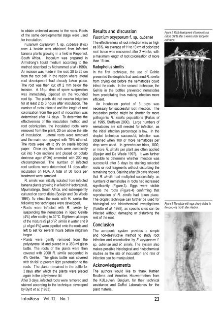
to obtain unlimited access to the roots. Roots<br />
of the same developmental stage were used<br />
for inoculation.<br />
Fusarium oxysporum f. sp. cubense (Foc)<br />
race 4 isolate was obtained from infected<br />
banana plants growing in a field in Kiepersol,<br />
South Africa. Inoculum was prepared in<br />
Armstrong’s liquid medium according to the<br />
method described by Mohammed et al. (1999).<br />
An incision was made in the root, 20 to 25 cm<br />
from the root ball, in the region where lateral<br />
root development had already taken place.<br />
The root was then cut off 2 mm below the<br />
incision. A 15-µl drop of spore suspension<br />
was immediately pipetted on the wounded<br />
root tip. The plants did not receive irrigation<br />
for at least 2 to 3 hours after inoculation. The<br />
number of roots infected and the length of root<br />
colonization from the point of inoculation was<br />
determined after 14 days. To determine the<br />
effectiveness of the inoculation method and<br />
root colonization, the inoculated roots were<br />
removed from the plant, 20 cm above the site<br />
of inoculation. Lateral roots were removed<br />
and the main root sprayed with 70% ethanol.<br />
The roots were left to dry on sterile blotting<br />
paper. Once dry, the roots were aseptically<br />
cut into 1-cm sections and placed on potato<br />
dextrose agar (PDA) amended with 200 mg<br />
chloramphenicol. The number of infected<br />
root sections were determined 14 days after<br />
incubation on PDA. A total of 50 roots per<br />
treatment were sampled.<br />
R. similis was initially isolated from infected<br />
banana plants growing in a field in Hectorspruit,<br />
Mpumalanga, South Africa, and subsequently<br />
cultured on carrot disks (Speijer and De Waele<br />
1997). To infect the roots with R. similis the<br />
following two techniques were developed.<br />
• Roots were infected with R. similis by<br />
suspending the nematodes in liquid Gelrite<br />
(4%) after cooling to 30°C. Eighteen-µl drops<br />
of the mixture (9 µl of R. similis in water and 9<br />
µl of gel 4%) were pipetted onto the roots and<br />
left to set for several hours before irrigation<br />
began.<br />
• Plants were gently removed from the<br />
polystyrene lid and placed in a 350-ml glass<br />
bottle. The roots of the plants were then<br />
covered with 2000 R. similis suspended in<br />
4% Gelrite. The glass bottle was covered<br />
with tin foil to prevent light penetration to the<br />
roots. The plants remained in the bottle for<br />
3 days after which the plants were placed<br />
again in the polystyrene lid.<br />
After 3 days, infected roots were removed and<br />
stained according to the technique developed<br />
by Byrd et al. (1983).<br />
Results and discussion<br />
Fusarium oxysporum f. sp. cubense<br />
The effectiveness of root infection was as high<br />
as 96%. An average of 11 to 13 cm of colonized<br />
root tissue was recovered after 2 weeks, with<br />
a maximum length of root colonisation of more<br />
than 15 cm.<br />
Radopholus similis<br />
In the first technique, the use of Gelrite<br />
prevented the droplets that contained R. similis<br />
from drying out before the nematodes could<br />
infect the roots. In the second technique, the<br />
Gelrite in the bottles prevented nematodes<br />
from precipitating thus making infection more<br />
efficient.<br />
An incubation period of 3 days was<br />
necessary for successful root infection. The<br />
incubation period might be shorter for more<br />
pathogenic R. similis populations (Fallas et<br />
al. 1995, Stoffelen 2000). Large numbers of<br />
nematodes are still needed for infection, as<br />
the initial infection percentage is low. In the<br />
droplet technique successful, infection was<br />
obtained when 100 or more nematodes per<br />
drop were used. In greenhouse trials, 1000,<br />
or more R. similis per plant are often applied<br />
(Speijer and De Waele 1997). It was further<br />
possible to determine whether infection was<br />
successful after 3 days by staining selected<br />
roots or root fragments without disturbing the<br />
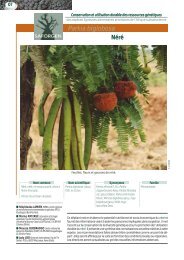
remaining roots. Staining after 28 days showed<br />
that R. similis had multiplied successfully, as<br />
numbers of nematodes in roots had increased<br />
significantly (Figure 3). Eggs were visible<br />
inside the roots (Figure 4) confirming that<br />
reproduction of R. similis had taken place.<br />
The droplet technique can further be used for<br />
histological and histochemical investigations<br />
(Valette et al. 1998), as specific sites can be<br />
infected without damaging or disturbing the<br />
rest of the root.<br />
Conclusion<br />
The aeroponic system provides a simple<br />
and non-destructive method to study root<br />
infection and colonisation by F. oxysporum f.<br />
sp. cubense and R. similis. The system also<br />
makes possible histological and histochemical<br />
studies as the site of inoculation and rate of<br />
infection can be manipulated.<br />
Acknowledgements<br />
The authors would like to thank Katrien<br />
Beullens and Annelies Hauwermeiren from<br />
the KULeuven, Belgium, for their technical<br />
assistance and DuRoi Laboratories for the<br />
plant material.<br />
Info<strong>Musa</strong> - Vol 12 - No.1 23<br />
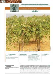
Figure 2. Root development of banana tissue<br />
culture plants after 3 weeks under aeroponic<br />
cultivation.<br />
Figure 3. Nematode with eggs clearly visible in<br />
the root, one month after infection.