Suraj Mal
Suraj Mal
Suraj Mal

You also want an ePaper? Increase the reach of your titles
YUMPU automatically turns print PDFs into web optimized ePapers that Google loves.
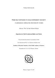
INSTITUTE OF PHYSICAL CHEMISTRY<br />
POLISH ACADEMY OF SCIENCES<br />
Kasprzaka 44/52<br />
01-224 Warsaw, Poland<br />
Synthesis and Photoluminescence of Lanthanide<br />
Ternary Complexes with Heterocyclic Ligands<br />
Ph.D. Thesis<br />
(prepared within the International Ph.D. in Chemistry Studies at Institute of Physical<br />
Chemistry of the Polish Academy of Sciences)<br />
<strong>Suraj</strong> <strong>Mal</strong><br />
Supervisor:<br />
Prof. dr hab. Marek Pietraszkiewicz<br />
Department of Photochemistry and Spectroscopy<br />
Warsaw, January 2012
This PhD work was financially supported by Project FP7 Marie<br />
Curie Initial Training Networks (MCITN), Contract PITN-GA-<br />
2008-215399 “Cavity-confined Luminophores for Advanced<br />
Photonic Materials: A Training Action for Young Researchers”<br />
(FINELUMEN).<br />
ii
Acknowledgements<br />
I am so fortunate to have the opportunity to live, experience, and study in the simply<br />
wonderful city – Warsaw, Poland. It’s amazing skyline, peaceful place for effective work and<br />
especially the polite, diligent, fortitudinous people gave me unforgettable impression<br />
throughout my life. The invaluable experience I gained throughout my studies and living here<br />
has shaped up me a lot. The confidence and courage I obtained here promote me to pursue<br />
more goals and dreams that are laid ahead. There have been many people offering me help<br />
throughout the past three years. My heartfelt thanks for everyone who shared the joys and<br />
pain in the past three years.<br />
My sincere gratitude must initially be expressed to Professor Marek Pietraszkiewicz, my<br />
research supervisor, for his guidance, encouragement, and wholehearted support throughout<br />
the study. His devoted research attitude, perseverant willpower to facing failures, and creative<br />
ideas about frontier research gave me deep impression. I would like to thank Professor Teresa<br />
Borowiak from Dept. of Chemistry, University of Adam Mickiewicz, Poznań for X-ray<br />
analysis.<br />
My sincere appreciation is expressed to Madam Oksana Pietraszkiewicz without whom, I<br />
would never have succeeded. She helped me throughout my stay here not only scientifically<br />
but also socially to run life comfortably.<br />
I would also like to thank my lab mates dr. Arkadiusz Listkowski and dr. Igor Czerski for<br />
there consistent help and valuable discussion throughout my research work.<br />
Many thanks also goes to Professor Jerzy Karpiuk, mgr Ewelina Karolak-Solarska and mgr<br />
Alina Majka for their generous help in the photoluminescence measurements.<br />
My wholehearted gratitude goes to all collaborative members of FINELUMEN Project,<br />
especially,<br />
-Professor Enrico Dalcanale and his team members, University of Parma, Parma, Italy.<br />
-Professor Kristiaan Neyts, Oksana Drobchak and his team members, University of Gent<br />
(UGENT), Gent, Belgium.<br />
-Professor Katalin Kamaras, Nitin Chelwani and all members of the group, Hungarian<br />
Academy of Sciences (RISSPO), Budapest, Hungary, for the secondements during my study.<br />
I would also like to acknowledge Professor David Parker, dr. Anurag Mishra, Durham<br />
University, Durham, UK for there consistent help and guidance during my visit to his lab.<br />
My deepest thank goes to dr. Gonzalo Angulo, IPC PAS, Warsaw for his invaluable<br />
discussions and helping in calculations during my study.<br />
iii
Special thanks also go to my research colleague, Michał Maciejczyk, Valentina<br />
Utochnikowa, and my all dear friends from the Institute of Physical Chemistry and Warsaw.<br />
I would like to express my appreciation to Miss Dorota Cegielska for her great assistance in<br />
all academic purposes.<br />
Last but not least, I would like to thank God, my family, all friends from India, and Tamanna<br />
for their long-lasting patience, support, love and care.<br />
iv
TABLE OF CONTENTS<br />
INTRODUCTION……………………………………………………………………….<br />
AIM OF THE PROJECT………………………………………………………………..<br />
GLOSSARY……………………………………………………………………………..<br />
Chapter 1<br />
1.1<br />
1.1.1.<br />
1.1.2.<br />
1.1.3.<br />
1.1.4.<br />
1.2<br />
1.3<br />
1.3.1.<br />
1.3.2.<br />
1.3.3.<br />
1.4<br />
1.5<br />
1.5.1.<br />
1.5.2.<br />
1.5.3.<br />
1.5.4.<br />
1.6<br />
1.6.1.<br />
1.6.2.<br />
1.6.3.<br />
1.7<br />
1.8<br />
1.9<br />
1.9.1.<br />
1.9.2.<br />
1.9.3.<br />
LITERATURE SURVEY………………………………………………..<br />
General introduction of lanthanides and lanthanide complexes……..<br />
Spectral features of lanthanide ions……………………………………<br />
f-electronic levels and Dieke diagram…………………………………...<br />
General mechanism of lanthanide sensitization………………………….<br />
Lanthanide emission sensitization with other metals…………………….<br />
Mathematical basics for photophysical processes - short outline……….<br />
Design of lanthanide sensitizing ligands……………………………….<br />
Brief survey on sensitizing ionizable ligands…………………………..<br />
Diketonates and their analogues…………………………………………<br />
Acyl-pyrazolones………………………………………………………….<br />
Carboxylates……………………………………………………………...<br />
NIR emitting complexes………………………………………………...<br />
Tetrazoles as sensitizers…………………………………………………<br />
General properties of tetrazoles………………………………………….<br />
Synthetic strategies for 5-pyridine-oxide tetrazoles (HPTO)…………….<br />
Lanthanide complexes based on tetrazolates…………………………….<br />
Tetrazolate transition metal complexes…………………………………..<br />
Isoxazolones as sensitizers………………………………………………<br />
General Properties of Isoxazolones………………………………………<br />
Synthetic approach for substituted isoxazolone…………………………..<br />
Lanthanide complexes based on isoxazolonates…………………………<br />
Auxiliary neutral sensitizing ligands and there role in lanthanide<br />
sensitization……………………………………………………………...<br />
Two-photon sensitization……………………………………………….<br />
Applications……………………………………………………………...<br />
Lasing systems……………………………………………………………<br />
Biomedical applications………………………………………………….<br />
Optoelectronic applications……………………………………………...<br />
1<br />
3<br />
4<br />
8<br />
8<br />
8<br />
8<br />
11<br />
17<br />
18<br />
22<br />
23<br />
23<br />
27<br />
31<br />
31<br />
34<br />
34<br />
35<br />
38<br />
41<br />
42<br />
42<br />
42<br />
44<br />
48<br />
54<br />
56<br />
56<br />
59<br />
64<br />
v
Chapter 2<br />
2.1<br />
2.2<br />
2.3<br />
2.4<br />
2.4.1.<br />
2.4.2.<br />
2.4.3.<br />
2.5<br />
2.6<br />
2.7<br />
2.7.1.<br />
2.7.2.<br />
RESULTS AND DISCUSSIONS………………………………………...<br />
Lanthanide complexes based on tetrazolates………………………….<br />
Synthesis of pyridine-oxide tetrazole…………………………………..<br />
Characterization of pyridine-oxide tetrazolate ligand………………..<br />
Synthesis and characterization of lanthanide pyridine-oxide<br />
tetrazolate complexes……………………………………………………<br />
Photophysical studies of HPTO and lanthanide pyridine-oxide<br />
tetrazolate complexes I and II………………………………………….<br />
Ligand centered luminescence HPTO……………………………………<br />
Absorption and excitation characteristics of complexes I and II………..<br />
Metal centered luminescence.....................................................................<br />
Synthesis and spectral characterization of auxiliary phosphine oxide<br />
coligands…………………………………………………………………<br />
Synthesis and characterization of ternary complexes of Eu(PTO)3<br />
with selected phosphine oxides…………………………………………<br />
Photophysical studies of ternary europium-tetrazolate complexes III<br />
to XIII……………………………………………………………………<br />
Absorption and excitation characteristics……………………………….<br />
Metal centered luminescence…………………………………………….<br />
Chapter 3 Lanthanide complexes based on isoxazolonates………………………. 105<br />
3.1<br />
3.2<br />
3.2.1.<br />
3.2.2.<br />
3.3<br />
3.3.1.<br />
3.3.2.<br />
3.4<br />
3.5<br />
3.5.1<br />
Synthesis and characterization of synthesized isoxazolonates………..<br />
Synthesis and characterization of lanthanide isoxazolonate<br />
complexes………………………………………………………………...<br />
tris-(3-phenyl-4-benzoyl-5-isoxazolone) Eu(III) and Tb(III) complexes...<br />
tris-(4-isobutyryl-3-phenyl-5-isoxazolone)Tb(III) complex.......................<br />
Photophysical studies of lanthanide tris-isoxazolonate complexes<br />
XIV, XV and XVI……………………………………………………….<br />
Absorption and excitation characteristics……………………………….<br />
Metal centered luminescence…………………………………………….<br />
Synthesis and characterization of ternary isoxazolonate complexes<br />
with neutral auxiliary sensitizing coligands…………………………..<br />
Photophysical studies……………………………………………………<br />
Spectroscopic studies of ternary Eu(III) isoxazolonate (PBI) complexes<br />
70<br />
70<br />
70<br />
70<br />
73<br />
77<br />
77<br />
78<br />
80<br />
83<br />
87<br />
96<br />
96<br />
100<br />
105<br />
107<br />
107<br />
108<br />
109<br />
109<br />
111<br />
115<br />
118<br />
vi
3.5.2.<br />
3.6<br />
3.6.1.<br />
3.6.2.<br />
Chapter 4<br />
4.1<br />
4.2<br />
4.3<br />
4.4<br />
4.5<br />
4.6<br />
4.7<br />
4.8<br />
4.9<br />
(XVII to XIX)……………………………………………………………...<br />
Metal centered luminescence…………………………………………….<br />
Spectroscopic studies of ternary Tb(III) isoxazolonate (IBPI)<br />
complexes (XX to XXII)………………………………………………...<br />
Absorption and excitation characteristics……………………………….<br />
Metal centered luminescence…………………………………………….<br />
EXPERIMENTAL………………………………………………………..<br />
Methods and procedures………………………………………………..<br />
General…………………………………………………………………...<br />
Synthesis of tetrazole 5-(2-pyridyl-1-oxide)tetrazole (HPTO)………..<br />
Ligand based on isoxazoles……………………………………………..<br />
Synthesis of phosphine oxide coligands (P1-P11)……………………..<br />
Synthesis of lanthanide complexes based on tetrazolate ligand……...<br />
Synthesis of ternary-(tetrazolate)-Eu(III) complexes (III to XIII)…...<br />
Lanthanide complexes based on isoxazolonate ligand………………..<br />
Europium ternary complexes with phosphine oxides based on 3-<br />
phenyl-4-benzoyl-5-isoxazolone (HPBI) ligand (XVII to XIX)………<br />
Terbium ternary complexes with phosphine oxides based on 4-<br />
isobutyryl-3-phenyl-5-isoxazolone (HIBPI) ligand (XX to XXII)……<br />
FINAL CONCLUSIONS AND PERSPECTIVES………………………. 141<br />
118<br />
119<br />
122<br />
122<br />
123<br />
126<br />
126<br />
126<br />
127<br />
127<br />
129<br />
131<br />
132<br />
136<br />
138<br />
139<br />
vii
FINELUMEN<br />
viii
INTRODUCTION<br />
Excerpt from the FINELUMEN project workplan<br />
FINELUMEN is a 4-year project aiming at the preparation and extensive characterization of<br />
luminescent materials in which suitably designed organic and inorganic luminophores are<br />
encapsulated within nano-containers (carbon nanotubes and coordination cages) in which<br />
they can preserve and even improve their emission output. The ultimate goal is to create a<br />
library of luminescent modules emitting throughout the VIS-NIR region for producing<br />
superior functional hybrid materials. The emission color tunability is defined by the emitting<br />
guest, while the versatility in the final application is controlled via tailored chemical<br />
functionalization of the host. The versatile properties of these materials will make them<br />
attractive in at least 3 applicative areas, i.e. bioimaging, optoelectronic devices and sensors.<br />
The participation of a big company and a high-tech SME in the consortium ensures a quick<br />
patenting and industrial scale up of the most promising luminescent materials, strengthening<br />
Europe’s competitiveness in a field of huge growth potential in the next decade. The research<br />
endeavor inside FINELUMEN calls for a multidisciplinary team in which key groups, experts<br />
in many different fields of chemistry, physics and engineering tightly interact. For this reason<br />
training and exchange of young researchers represents the core of the FINELUMEN<br />
activities. To both early-stage and experienced researchers, a multidisciplinary training in the<br />
realm of synthetic/supramolecular/physical chemistry, photosciences, and engineering as well<br />
as management, communication, and IPR is offered, preparing them for positions in<br />
academia, industry, and government labs. The local and network-wide training and transfer of<br />
knowledge activities are strengthened by 2 authoritative visiting scientists, enriched by 3<br />
FINELUMEN international summer schools and conferences, and complemented via PhD<br />
programs in co-tutoring among partners based in different countries.<br />
This PhD work was supposed to support also other FINELUMEN groups involved into<br />
physico-chemical research and supramolecular devices construction based on materials<br />
prepared within IPC. Schematic representation of scientific concept and interconnecting node<br />
within FINELUMEN partners, involving research and training is shown below:<br />
1
Preparation and<br />
functionalization of<br />
molecular containers<br />
NANOCYL, PFA,<br />
FUNDP, and UNIPR<br />
Raman and IR spectroscopy<br />
RISSPO and WMI BAdW<br />
Material Design<br />
ALL MEMBERS<br />
Theoretical modeling of<br />
structural and optical properties<br />
of supramolecular assemblies<br />
FUNDP, UGENT and PFA<br />
Photophysical and microscopic<br />
evaluation<br />
CNR/ISOF<br />
Supramolecular encapsulation<br />
FUNDP and UNIPR<br />
New material and device generation<br />
PFA, NANOCYL and UGENT<br />
Synthesis of Organic and<br />
Inorganic Luminophores<br />
CNRS and IPC<br />
Main scientific and training<br />
working interaction<br />
Characterization of new materials<br />
2
AIM OF THE WORK<br />
Luminescence of lanthanide complexes is a characteristic property which makes them<br />
attractive materials for various technologies, such as low cost full color displays, organic<br />
light-emitting devices (OLED), optical materials, biomedical imaging etc. As a result of pure<br />
colors and high emission efficiencies lanthanide complexes have attracted much research<br />
interest.<br />
As mentioned in Introduction, this PhD work is not a standard scientific venture. It is<br />
associated with FP7 ITN M. Curie Project FINELUMEN involving ten institutional partners<br />
in Europe. This project is multidisciplinary, comprising organic and inorganic synthesis of<br />
fluorophores, physicochemical aspects and theoretical modeling, serving other groups and<br />
their PhD students to investigate the properties and making photonic devices based on new<br />
phtoluminescent materials. Thus, the task of organic groups was challenging and urgent: to<br />
design and synthesize novel materials with outstanding photoluminescent properties.<br />
My task was focused on highly photoluminescent lanthanide complexes to be confined by<br />
other partners in large molecular containers by self-assembly, or in carbon nanotubes. Where<br />
they can preserve or even improve their emission output which make them useful materials<br />
for three areas, i.e. bioimaging, Optoelectronic devices and sensors.<br />
In order to achieve significant and desirable impact on cooperation within different groups,<br />
the following research strategy was undertaken: two classes of the core ionizable sensitizing<br />
ligands were selected – 1) isoxazolones, 2) tetrazoles. Both classes of ligands are planar.<br />
They are not susceptible for bleaching, air oxidation and are thermally stable. They form<br />
neutral complexes with lanthanides of 1:3 stoichiometry with 6-coordination pattern. Due to<br />
the planarity of the ligands, there is a room for sensitizing co-ligands, complementing the<br />
coordination sphere. Thus, this modular approach can serve a quick progress in achieving<br />
good photoluminescent properties of ternary complexes formed, by combinatorial<br />
methodology. Once the complexes were formed, they were fully characterized<br />
spectroscopically (UV, fluorescence, phosphorescence, PL lifetimes and quantum yields),<br />
and thermogravimetrically. The most prospective complexes were submitted to other groups<br />
for further investigations. Thus this project deserved tremendous added value.<br />
Keywords: Lanthanides, Isoxazolonate, Tetrazolate, Luminescence, Photoluminescence<br />
quantum yield, bioimaging, optoelectronic devices, OLED, phosphine oxides.<br />
3
GLOSSARY - ABBREVIATIONS USED<br />
bath 4,7-diphenyl-1,10-phenanthroline (bathophenanthroline)<br />
bipy 2,2-bipyridine<br />
But4N tetrabutylammonium<br />
dmso dimethylsulfoxide<br />
Et4N tetraethylammonium<br />
Hacac acetylacetone, 2,4-pentanedione<br />
Hbfa benzoyl-2-furanoylmethane<br />
Hbtfac Benzoyltrifluoroacetone<br />
Htfac 1,1,1-trifluoro-2,4-pentanedione; trifluoroacetylacetone<br />
Hfac hexafluoroacetylacetone, 1,1,1,5,5,5-hexafluoro-2,4-pentanedione<br />
Hthd 2,2,6,6-tetramethyl-3,5-heptanedione (Hdpm and Htmhd)<br />
Hfod 6,6,7,7,8,8,8-heptafluoro-2,2-dimethyl-3,5-octanedione<br />
Hbzac benzoylacetone, 1-phenyl-1,3-butanedione<br />
Htrimh 2,2,6-trimethyl-3,5-heptanedione<br />
Hfdh 6,6,6-trifluoro-2,2-dimethyl-3,5-hexanedione<br />
Hbtfac benzoyltrifluoroacetone<br />
Hdbm dibenzoylmethane, 1,3-diphenyl-1,3-propanedione<br />
Hbpp 1,3-bis(3-pyridyl)-1,3-propanedione<br />
Hdmbm 4,4-dimethoxydibenzoylmethane<br />
Htta 2-thenoyltrifluoroacetone, 4,4,4-trifluoro-1-(2-thienyl)-1,3-<br />
butanedione<br />
Hdnm dinaphthoylmethane<br />
Hntac 2-naphthoyltrifluoroacetone, 4,4,4-trifluoro-1-(2-naphthyl)-1,3-<br />
butanedione<br />
Hex4N tetrahexylammonium<br />
OLED organic light emitting diode<br />
phen 1,10-phenanthroline<br />
py Pyridine<br />
tppo triphenylphosphine oxide<br />
4
acpy acylprazolones<br />
PMAP 1-phenyl-3-methyl-4-acetyl-pyrazolone-5<br />
PMPP 1-phenyl-3-methyl-4-propionyl-pyrazolone-5<br />
PMIP 1-phenyl-3-methyl-4-isobutyryl-pyrazolone-5<br />
PMBP 1-phenyl-3-methyl-4-benzoyl-pyrazolone-5<br />
PhCN pyrazino(2,3-f)(1,10)-phenanthroline-2,3-dicarbonitrile<br />
trenPMBP Tris-((4-(3-methyl-1-phenyl-5-pyrazolonyl)-phenylmethylidene)- 2aminoethyl)amine<br />
dbso di-n-butylsulfoxide<br />
diglyme diethyleneglycol dimethyl ether<br />
terpy 2,2’,6’,2’’-terpyridyl<br />
tetraglyme tetraethyleneglycol dimethyl ether<br />
tbpo tri-n-butylphosphine oxide<br />
tbp tri-n-butylphosphate<br />
Hpbm 2-(2-pyridyl)benzimidazole<br />
HPBI 3-phenyl-4-benzoyl-5-isoxazolone<br />
PHA N-phenylacetamide<br />
TTA thenoyltrifluoroacetonate<br />
DPEPO bis(2-(diphenylphosphino)phenyl)ether oxides<br />
tmhd 2,2,6,6-tetramethylheptane-3,5-dione<br />
TAPO (4-diphenylamine-phenyl)-diphenylphosphine oxide<br />
TMOADPO 2-(diphenylphosphoryl)-N-(2-(diphenylphosphoryl)-4methoxyphenyl)-<br />
4-methoxy-N-(4-methoxyphenyl)aniline<br />
EtCzDPO 3,6-bis-(diphenylphosphoryl)-9-ethyl-9H-carbazole<br />
PhCzDPO 3,6-bis(diphenylphosphoryl)-9-phenyl-9H-carbazole<br />
H3pytztcn tri-aza-cyclononanepyridinetetrazole<br />
H2terpytz ter-pyridinetetrazole<br />
5
PyTzH 2-(1H-tetrazol-5-yl)pyridine<br />
PzTzH 2-(1H-tetrazol-5-yl)pyrazine<br />
BrPyTzH 5-bromo-2-(1H tetrazol-5- yl)pyridine<br />
BrPhL3 4’-(4-bromophenyl)-6,6’’-bis(1H-tetrazol-5-yl)-2,2’:6’,2’’terpyridine<br />
BrThL4 4’-(5-bromothiophen-2-yl)-6,6’’-bis(1H-tetrazol-5-yl)-2,2’:6’,2’’-<br />
terpyridine<br />
dmbpy 4,4-dimethoxy-2,2-bipyridine<br />
HPTI 3-phenyl-4-(4-toluoyl)-5-isoxazolone<br />
HPPI 3-phenyl-4-propionyl-5-isoxazolone<br />
HIBPI 4-isobutyryl-3-phenyl-5-isoxazolone<br />
PHB poly β-hydroxybutyrate<br />
btfac Benzoyltrifluoroacetylacetonate<br />
BCP 2,9-dimethyl-4,7-diphenyl-1,10-phenanthroline<br />
ITO indium tin oxide<br />
LED light emitting diode<br />
TPD N,N’-diphenyl-N,N’-(3-methyl phenyl)-1,1’-biphenyl-4,4’-diamine<br />
PDB 2-tert-butylphenyl-5-biphenyl-1,3,4-oxadiazole<br />
PMPS poly(methylphenylsilane)<br />
PVK poly(N-vinylcarbazole)<br />
PBD 2-(4-biphenylyl)-5-(4-tert-butylphenyl)-1,3,4-oxadiazole<br />
CN-PPP poly[2-(6’-cyano-6’-methyl-heptyloxy-1,4-phenylene)]<br />
dnm Dinaphthoylmethanate<br />
tmhd 2,2,6,6-tetramethyl-3,5-heptanedionate<br />
tfc 3-(trifluoromethylhydroxymethylene)-(+)-camphorate<br />
tmphen 3,4,7,8-tetramethyl-1,10-phenanthroline<br />
pms bis-perfluoro-alkyl-sulfonyl-aminato<br />
6
PyTzH 2-(1H-tetrazol-5-yl)pyridine<br />
PzTzH 2-(1H-tetrazol-5-yl)pyrazine<br />
BrPyTzH 5-bromo-2-(1H tetrazol-5- yl)pyridine<br />
7
LITERATURE SURVEY<br />
1. General introduction of lanthanides and lanthanide complexes<br />
1.1. Spectral features of lanthanide ions<br />
1.1.1. f-electronic levels and Dieke diagram<br />
Lanthanides are situated at the bottom of periodic table ranging from lanthanum (Z = 57) to<br />
lutetium (Z = 71) and commonly named as lanthanide series as shown in figure 1.1. It is<br />
difficult to separate rare earth elements among each other by means of chemical properties.<br />
With the use of ion-exchange methods the separation of an individual rare earth element can<br />
be accomplished with greater ease and precision. By following all the rules (Aufbau, Pauli’s<br />
exclusion and Hund’s rule) the general electronic configuration of lanthanides are [Xe]4f<br />
n+1 6s 2 .<br />
Figure 1.1. Periodic table of all elements showing lanthanides and actinides at the bottom (picture<br />
from http://www.bpc.edu/mathscience/chemistry/history_of_the_periodic_table.html).<br />
The principle source of rare earth elements is mineral monazite. The most stable oxidation<br />
state of lanthanides are Ln(III) which gives electronic configuration [Xe]4f n (where n = 0 to<br />
14). The outer 4f orbitals of lanthanides are greatly shielded by filled xenon (54 electrons) 5s<br />
and 5p orbitals. The interesting photophysical properties of lanthanides arise because of<br />
transitions of f-electrons which display sharp absorption and emission bands and<br />
luminescence within µs – ms range. Due to low extinction coefficient because of forbidden f-<br />
8
f transition, making direct photoexcitation of lanthanide ions is difficult. This can be<br />
overcome by using energy transfer process from organic chromophores to lanthanide ions.<br />
The extensive measurements of energy levels of 4f n configuration of lanthanide ions were<br />
carried out in the 1950’s and 1960’s. Most of the work in this field was done by Dieke and<br />
co-workers and the data summarized in his 1968 published book 1 . Most of the lanthanide ions<br />
show luminescence in the visible region of optical spectrum and the lines from the different<br />
lanthanides has been well prescribed. To visualize this knowledge one can use so-called<br />
Dieke diagram in which the allowed optical transitions are plotted as energies for different<br />
ions as shown in figure 1.2 for several lanthanide ions. These diagrams are useful because the<br />
energies of the J multiplets vary by only a small amount which allows rapid identification of<br />
the energy levels and plays an important role to design suitable materials for phosphors.<br />
Figure 1.2. Energy level diagram for the Ln(III) ions showing the main emitting levels and the<br />
transitions to the ground state levels.<br />
The f-electrons in most of lanthanide complexes are considered to have properties which are<br />
close to those of isolated ions. The energies of ground and excited state multiplets are<br />
determined by LS-coupling, inter-electronic repulsion and the ligand field. Due to inter-<br />
electronic repulsion the 4f 6 and 4f 8 configurations of Eu(III) and Tb(III) respectively splits<br />
1 G. H. Dieke, Spectra and energy levels of rare earth ions in crystals, Interscience Publishers, New<br />
York (1968).<br />
9
into various terms which are denoted as term symbol and calculated by using formulae<br />
{ (2S+1) ГJ}, where Г = S, P, D and F when the quantum number J = 0, 1, 2 or 3 as shown in<br />
figure 1.3 and 1.4 and (2S+1) represents the spin multiplicity. The different J states are well<br />
separated by a value of 10 2 cm -1 due to spin-orbit coupling and the different states arising<br />
from 4f n 5d 0 configurations are splitted by Columbic interactions 2 are generally separated by<br />
10 4 cm -1 .<br />
4f 5 5d<br />
5 L<br />
5 5<br />
D4 D3<br />
5 D2<br />
5 D1<br />
5 D0<br />
2*10 4 cm -1 Eu(III)<br />
7 FJ J = 6543210<br />
10 3 cm -1 }<br />
Figure 1.3. Schematic energy diagram for the Eu(III) ions, showing the splitting of energy levels.<br />
4f 7 5d<br />
4f 8<br />
5 L<br />
5 G<br />
5 D0<br />
5 D1<br />
5 D2<br />
5 D 5D3<br />
2*10 4 cm -1 Tb(III)<br />
7 F<br />
6*10 3 cm -1<br />
7<br />
FJ J =<br />
0<br />
1<br />
2<br />
3<br />
4<br />
56<br />
5 D4<br />
}<br />
10 3 cm -1<br />
10 2 cm -1<br />
2*10 2 cm -1<br />
Figure 1.4. Schematic energy diagram for the Tb(III) ions, showing the splitting of energy levels.<br />
2 S.F.A. Kettle, Physical Inorganic Chemistry: A Coordination Chemistry Approach, Oxford<br />
University Press, Oxford, 1996.<br />
}<br />
10
There are 295 (2S+1) ГJ spectroscopic levels for f 6 and f 8 configurations whose relative energies<br />
are predicted by Hund’s rule. The ground state term symbol for Eu(III) and Tb(III) are 7 F0<br />
and 7 F6 respectively. All trivalent lanthanide ions have unpaired electrons except lutetium.<br />
The Gd(III) ions consisting maximum number of unpaired f-electrons i.e. seven (overall spin<br />
= 7/2) make gadolinium complexes as good contrast agents for magnetic resonance imaging.<br />
1.1.2. General mechanism of lanthanide sensitization<br />
Luminescence is the characteristic and interesting feature of lanthanide trivalent ions. It can<br />
be divided into several categories depending on the source of excitation and can be defined as<br />
the emission of light from an electronically excited state to ground state. Depending on the<br />
source of light to achieve excited state, luminescence term will be converted to<br />
chemiluminescence (chemical reaction), bioluminescence (biochemical reaction),<br />
radioluminescence (radiochemical reactions), electroluminescence (electric field) or<br />
photoluminescence (light). On the basis of luminescence lifetime and spin of the initial<br />
(emitting) and final state (usually ground state), luminescence can be divided into two more<br />
categories named as fluorescence (∆S = 0, lifetime typically 10 ns) and phosphorescence (∆S<br />
≠ 0, lifetime generally milliseconds to seconds) 3 . The luminescence from the lanthanide ions<br />
is the result of competition of radiative and non-radiative pathways in the relaxation of an<br />
electronically excited species. By the selection rule ∆J = 0, ±1 (∆J = 0 is forbidden)<br />
hypothetically in lanthanide ions only magnetic dipole transitions are allowed 4 . In<br />
coordinating sphere of lanthanide, electric dipole transitions are also favored as the ligand<br />
field mixes odd parity configurations slightly into [Xe]4f n 5d 0 configuration. As coordinating<br />
chromophores absorb energy, most of the lines of absorption and emission come out due to<br />
electric dipole transition. Both magnetic dipole and electric dipole transitions of lanthanide<br />
ions are quite weak as compared to fully allowed transition in organic chromophores<br />
separately. The excited state of lanthanides is not solely relaxed by radiative process but also<br />
by non-radiative process too. The radiative and non-radiative transition nature of lanthanides<br />
is summarized in figure 1.5.<br />
3 Hemmila, I. (1991) Applications of fluorescence in immunoassays, New York, John Wiley & Sons.<br />
4 Weber, M. J., Varitimos, T. E. and Matsinger, B. H. (1973) Phys. Rev. B, 8, 47 – 53.<br />
11
Higher excited state Ln(III)**<br />
Lowest luminescent Ln(III)*<br />
state<br />
Ground state Ln(III)<br />
1 2 3 4 5 6 7<br />
Figure 1.5. Electronic transitions in lanthanide ions: 1. absorption/excitation, 2. excited state<br />
absorption, 3. direct excitation into a higher excited state, 4. emission from the lowest excited state, 5.<br />
non-radiative relaxation, 6. radiative transition between excited states, 7. emission from a higher<br />
excited state.<br />
Due to high energy vibrations in organic chromophores, the excitation energy of lanthanide<br />
complexes can be dissipated by vibrations of surrounding matrix through a process known as<br />
multi-phonon relaxation 5 . Due to low energy gap immediate decay took place from the higher<br />
excited state to low lying excited state that efficiently undergo multi-phonon relaxation. The<br />
various emissive wavelengths, emissive energy levels and transition energy levels of some<br />
lanthanide ions in solution are shown in table 1.1. A typical f-f emission diagram of various<br />
lanthanides is shown in figure 1.6 which covers the visible spectrum: (e.g. Tb(III) green,<br />
Eu(III) red, Dy(III) yellow, Sm(III) pink).<br />
Table 1.1. Emission bands of some lanthanide ions in solution with hypersensitive transition in<br />
highlighted in bold.<br />
Ions Emissive energy level Final energy level Emission wavelength (nm)<br />
Eu(III) 5 D0 7 FJ (J=0…5) 580, 590, 613, 650, 690, 710<br />
Tb(III) 5 D4 7 FJ (J=6…2) 490, 545, 590, 620, 650<br />
Nd(III) 4 F3/2 4 FJ (J=9/2,11/2, 13/2) 880, 1060, 1330<br />
Er(III) 4 I13/2 4 IJ (J = 15/2) 1550<br />
Yb(III) 2 F5/2 2 FJ (J = 7/2) 980<br />
5 Weber, M. J. (1973), Phys. Rev. B, 8, 54 – 64.<br />
12
Figure 1.6. Emission spectra of some lanthanide ions (picture from http://perso.bretagne.ens-<br />
cachan.fr/~mwerts/lanthanides/ln_shine.html).<br />
The energy migration process solely depend on the surrounding environment (inorganic<br />
matrices or organic ligand) of lanthanide ions: it may be singlet state to triplet state 6,7 , intra-<br />
ligand charge transfer states 8,9,10,11 and metal-to-ligand back energy transfer states 12,13,14,15 .<br />
The luminescent properties of Eu(III) , Tb(III), Sm(III) and Dy(III) ions are of much interest<br />
during last decades due to energy gap displayed by these lanthanides i.e. ∆E = 12300 ( 5 D0 –<br />
7 F6), 14800 ( 5 D4 – 7 F0), 7400 ( 4 G5/2 – 6 F11/2) and 7850 ( 4 F9/2 – 6 F3/2) cm -1 respectively 16 . On<br />
the basis of energy gap between lowest luminescent energy state and highest non-luminescent<br />
energy state, lanthanide ions are divided into two categories. First category consist of Pr(III),<br />
6 M. Kleinerman, J. Chem. Phys., 1969, 51, 2370.<br />
7 M. Kleinerman and Choi Sang-I1. J. Chem. Phys., 1968, 49, 3901.<br />
8 D B. Nie, Z. Q. Chen, Z. Q. Bian, J. Q. Zhou, Z. W. Liu, F. F. Chen, Y. L. Zhao and C. H.<br />
Huang, New J. Chem., 2007, 31, 1639.<br />
9 L. N. Puntus, A.-S. Chauvin, S. Varbanov and J.-C. G. Bünzli, Eur. J. Inorg. Chem., 2007, 2315.<br />
10 S. G. Roh, N. S. Baek, Y. H. Kim and H. K. Kim, Bull. Korean Chem. Soc., 2007, 28, 1249.<br />
11 A. Vogler and H. Kunkely, Inorg. Chim. Acta, 2006, 359, 4130.<br />
12 B. H. Bakker, M. Goes, N. Hoebe, H. J. van Ramesdonk, J. W. Verhoeven, M. H. V. Werts and J.<br />
W. Hofstraat, Coord. Chem. Rev., 2000, 208, 3.<br />
13 G. Blasse, Struct. Bonding, 1976, 26, 45.<br />
14 F. F. Chen, Z. Q. Bian, Z. W. Liu, D. B. Nie, Z. Q. Chen and C. H. Huang, Inorg. Chem.,<br />
2008, 47(7), 2507.<br />
15 R. Ziessel, S. Diring, P. Kadjane, L. J. Charbonnière, P. Retailleau and C. Philouze, Chem. Asian.<br />
J., 2007, 2, 975.<br />
16 Jean-Claude G. Bünzli and Claude Piguet, Chem. Soc. Rev., 2005, 34, 1048–1077.<br />
13
Nd(III), Yb(III), Ho(III), Er(III) and Tm(III) because of low energy gap between transition<br />
states of these lanthanide ions, which favors radiationless decay and giving low luminescence<br />
in visible or near infrared region. The second category of lanthanide ions Sm(III), Eu(III),<br />
Tb(III) and Dy(III) having large energy gap between lowest luminescent energy state to<br />
highest non-luminescent energy state. This gives rise to strong luminescence in visible range<br />
because of low sensitivity towards vibronic quenching by high frequency oscillators. In<br />
Gd(III) ion there is very large energy gap between ground and excited state, so transition and<br />
luminescence are rarely observed. Due to completely empty or completely filled 4f subshell<br />
in La(III) and Lu(III) ions respectively, they did not possess any luminescence property, as no<br />
intra 4f transitions are possible in these cases. The luminescence intensity and excited state<br />
lifetime is linearly proportional to the number of quenchers (especially water molecules)<br />
present inside the inner coordination sphere of lanthanide ion 17,18 , which is responsible for<br />
non-radiative decay from the excited state of lanthanide to ground state. One way to<br />
overcome the problem of quenching, is the incorporation of organic ligands (generally termed<br />
as chromophores or “antenna”). Organic ligands generally remove solvent and water<br />
molecule from the inner coordination sphere of the lanthanide ions. The overall process of<br />
energy transfer is quite complicated and followed by mainly three steps suggested by Grosby<br />
and Whan 19 : absorption of light by surrounding ligand, transfer to the lanthanide ion and<br />
emission of light. The surrounding matrix, or ligand not only provide alternate pathway for<br />
energy transfer but also enlarge the Stokes shift which allow an easy spectral separation of<br />
the remaining matrix luminescence from the metal ion emission 20 . Weissman 21 firstly noticed<br />
that the emission of lanthanide ions could be more easily observed when these ions were<br />
coordinated to aromatic carboxylic acids or 1,3-diketones and interpreted this phenomenon as<br />
an energy transfer from ligand to metal ion, in a sensitization process. The process that occurs<br />
between the absorption and emission of light is usually illustrated by Jablonski diagram 22<br />
17 Y. Haas, G. Stein, J. Phys. Chem. 75 (1971) 3677.<br />
18 W.DeW. Horrocks, D.R. Sudnick, J. Am. Chem. Soc. 101 (1979) 334.<br />
19 Bunzli, J.C.; Eliseeva, S.V., Basics of Lanthanide Photophysics. In Lanthanide Spectroscopy,<br />
Materials and Bio-Applications, Hanninen, P.; Harma, H., Eds. Springer: 2009; Vol. 7.<br />
20 Jean-Claude G. Bünzli, Steve Comby, Anne-Sophie Chauvin, Caroline D. B. Vandevyver Journal<br />
of rare earths 25 (2007) 257 – 274.<br />
21 Weissman, S.I., The Journal of Chemical Physics, 1942, 10 (4), 214-217.<br />
22 Jablonski A. 1935, Journal of physics, 94, 38-46.<br />
14
epresented in figure 1.7 and 1.8. Excitation of lanthanide complex is two-step process; firstly<br />
the chromophore is excited from the ground state to the first excited state (S1) under<br />
ultraviolet wavelength irradiation which further relaxes to lower vibrational level of excited<br />
state rapidly. From here it can relax either by non-radiatively (NR) or radiatively by releasing<br />
energy as photons through fluorescence (F) or can undergo non-radiative inter system<br />
crossing (ISC) to triplet state (T1), which can also be deactivated radiatively by spin<br />
S1 CH3 CH 3<br />
NR<br />
{Ligand} {Ln(III)}<br />
ISC<br />
ET<br />
T 1<br />
CH 3<br />
CH 3<br />
NR<br />
F P L<br />
Figure 1.7. Energy transfer diagram of luminescent lanthanide complexes (abbreviations are described<br />
BT<br />
ET<br />
in below explained phrase).<br />
IC<br />
CH 3<br />
CH 3<br />
f**<br />
f*<br />
NR<br />
Figure 1.8. Architecture and energy transfer diagram of luminescent lanthanide tris complexes.<br />
forbidden transition called as phosphorescence (P). Secondly, the energy can be transferred<br />
from triplet state of chromophore non-radiatively through an intramolecular energy-transfer<br />
(ET) process to the excited states of lanthanide ion (f*, f**,…). Both the singlet and triplet<br />
state of a ligand can transfer energy to the excited state of lanthanide ion, but due to short<br />
15
lifetime of singlet state the direct energy transfer from this state is not so significant.<br />
Thereafter, the excited state of lanthanide ion may undergo radiative transition to lower 4f<br />
state giving characteristic line like photoluminescence (L) or it can deactivate non-<br />
radiatively. To overcome the problem of back energy (BT) transfer process which is the<br />
energy transfer back from the emissive level of metal ion to the ligand, one should be careful<br />
to design “antenna” chromophore, as the photoluminescence quantum yield much depend on<br />
the energy gap between the triplet state (T1) of chromophore and the emissive level of<br />
lanthanide ion. This energy gap should be optimal for particular lanthanide cation.<br />
The emissive properties of lanthanides can be enhanced by increasing excited state<br />
population and minimizing non radiative pathways. As lanthanides generally possess nine co-<br />
ordination number, the bi-dentate antenna chromophore leads to hexa-coordinated tris-<br />
lanthanide complexes, but still the inner coordination is not fully saturated which is<br />
completed by coordination of some water or solvent molecules. To substitute these water or<br />
solvent molecules some auxiliary neutral coligands can be used which have suitable triplet<br />
energy levels corresponding to lanthanide emissive levels such as heterocyclic coligands<br />
(1,10-phenanthroline or bi-pyridine), N-oxides or phosphine oxides. Coligands can also act as<br />
sensitizers and increase the emissive population of lanthanide ions and can improve the<br />
overall photoluminescence quantum yield by alternate energy transfer process as shown in<br />
figure 1.9.<br />
{Ligand} {Ln(III)}<br />
S 1<br />
T 1<br />
CH 3<br />
CH 3<br />
ISC<br />
NR<br />
ET<br />
L<br />
5 DJ<br />
CH 3<br />
CH 3<br />
NR<br />
ET<br />
{Coligand}<br />
S1 *<br />
Figure 1.9. Architecture and energy transfer diagram of luminescent (europium, terbium) lanthanide<br />
ISC<br />
NR<br />
complexes with saturated inner coordination sphere of lanthanide ions.<br />
CH 3<br />
CH 3<br />
T 1 *<br />
16
For appended coligand with tris-lanthanide complex the intra-molecular energy transfer<br />
process is most important step influencing the photoluminescence quantum yield. It is well<br />
established that the energy gap between the triplet state (T1) of the ligand as well as coligand<br />
and emissive level of lanthanide ion plays an important role in effective energy transfer<br />
process. By extensive study on the energy gap it is experimentally proved that energy transfer<br />
process is much more effective in case if the energy gap between the triplet state (ligand, co-<br />
ligand) and emissive level of lanthanide ion (europium and terbium) is more than 2000 cm -1 ,<br />
which prevents the back-energy transfer process from the emissive level of lanthanide ions to<br />
the triplet state of ligands.<br />
1.1.3. Lanthanide emission sensitization with other metals<br />
There is another special case also reported for lanthanides sensitization by using several other<br />
metals. Lanthanide complexes can be excited with visible light using d-block elements and<br />
emission from lanthanide ion can be controlled by tuning the physiochemical properties of d-<br />
block chromophores. A number of different transition metal complexes such as Ru(II), Os(II)<br />
Fe(II), Pt(II) Au(I), Pd(II), Re(I), Cr(III), Co(III), Zn(II) and Ir(III) have been used to form d-<br />
f heteronuclear complexes. Mostly Ru(II), Os(II), Re(I) and Ir(III) complexes are successfully<br />
incorporated and reported to sensitize the photoluminescence of lanthanide ions. The energy<br />
transfer mechanism behind such incorporated complexes took place through 3 MLCT (triplet<br />
state metal to ligand charge transfer) donor level of the transition elements as shown in figure<br />
1.10, while in some cases such as Pt(II) chromophores it proceeded via broad mixed 3 MLCT<br />
and 3 MMLCT (metal to metal ligand charge transfer) emission levels 23 .<br />
Figure 1.10. Directional energy transfer mechanism diagram exhibiting Ru II - to - Ln III .<br />
23 M.D. Ward, Coord. Chem. Rev. 251 (2007) 1663.<br />
17
1.1.4. Mathematical basics for photophysical processes - short outline<br />
One of the most common form of luminescence is photoluminescence in which excitation<br />
occurs via the absorption of light by a molecule or complex. The most important feature of<br />
luminescent lanthanide complexes among lots of other photophysical parameters is the value<br />
of photoluminescence quantum yield (PLQY) and its accurate determination. Generally the<br />
quantum yield of photoexcited molecule is defined as the ratio of number of photons emitted<br />
with that of number of photons absorbed while excited at a particular wavelength. The<br />
quantum yield of lanthanide complexes arises by the balancing behavior of several<br />
phenomena such as ligand to Ln(III) energy transfer, multiphonon-relaxation, energy back<br />
transfer, crossover to charge transfer states etc. There are several competing processes such<br />
as fluorescence of the antenna (which competes with intersystem crossing), quenching of<br />
triplet state by dissolved molecular oxygen in case of NIR emitting ions (competing with the<br />
energy transfer to the lanthanide ion) and the presence of solvent or water molecules,<br />
generally lowers the value of photoluminescence quantum yield. Therefore, the overall<br />
quantum yield of sensitized emission (ΦS) is the product of the intersystem crossing quantum<br />
yield (ΦISC), the energy transfer quantum yield (ΦET) and the lanthanide luminescence<br />
quantum yield (Φlum):<br />
ΦS = ΦISC. ΦET. Φlum (1)<br />
The overall quantum yield of lanthanide containing organic ligands can be illustrated by<br />
equation 2,<br />
where Φ<br />
and Φ<br />
Φ<br />
= Φ<br />
= (<br />
) = (<br />
) (2)<br />
are the quantum yield resulting from indirect and direct excitation and<br />
represent the efficiency with which electromagnetic energy is transferred from the<br />
surrounding matrix to the metal ions, Krad is the radiative rate constant, Kobs is the sum of the<br />
rates of various deactivation processes and τrad is the radiative life time. The intrinsic<br />
quantum yield, or the photon emitted by directly excited state of lanthanide ion can be<br />
calculated by using equation 3:<br />
Φ<br />
= (3)<br />
18
Nonradiative rate constant, can be attributed by: back energy transfer to the<br />
sensitizer 24,25,26 , quenching by matrix vibrations or by electron transfer mainly for lanthanides<br />
having low reduction potential such as Eu(III) and Yb(III) ions 27,28 . To calculate the intrinsic<br />
quantum yield there are two proposed mechanisms, first by rapid diffusion enhanced<br />
resonance energy transfer in case of lanthanide complexes in solution after mixing the<br />
lanthanide complex with a known quantum yield acceptor and efficiency of energy transfer<br />
between them can be calculated from both lifetime and intensity parameters. In second<br />
method, which is only valid for europium and relies only on the fact that the intensity of<br />
purely magnetic dipolar 5 D0 – 7 F1 transition is independent of the chemical environment of<br />
the ion, and that the radiative lifetime can be calculated from its emission spectrum 27 . The<br />
measurement of absolute quantum yield is very sophisticated task; it can be calculated by<br />
determination of relative quantum yield with that of standard sample of known absolute<br />
quantum yield. The method described by Williams et al. 29 and D. F. Eaton 30 can be used to<br />
calculate the absolute quantum yield of lanthanide complexes in solution by using equation 4.<br />
Φ = Φ<br />
Where Φ represents the absolute quantum yield of sample to determine, Φ is the absolute<br />
quantum yield of reference compound , S and ST are the refractive index of solvent of<br />
sample and reference respectively and represents the fraction of light absorbed by<br />
reference and sample. For accurate determination of quantum yields, one must take into<br />
account a number of factors e.g. internal filter effects, self-quenching, reliability of the<br />
standard value etc. The emission spectra must be corrected for the spectroscopic sensitivity of<br />
the spectrophotometer because the sensitivity of the photomultiplier tube is not flat with<br />
24 M. L. Bhaumik, J. Chem. Phys., 1964, 40, 3711.<br />
25 N. Sabbatini, M. Guardigli and I. Manet, Adv. Photochem., 1997, 23, 213.<br />
26 L. Prodi, M. Maestri, V. Balzani, J. M. Lehn and C. Roth, Chem. Phys. Lett., 1991, 180, 45.<br />
27 Yuetao Yang, Junjia Li, Xiaojun Liu, Shuyi Zhang, Kris Driesen, Peter Nockemann, Koen<br />
Binnemans, ChemPhysChem, 2008, 9, 600 – 606.<br />
28 N. Sabbatini, S. Perathoner, G. Lattanzi, S. Dellonte and V. Balzani, J. Phys. Chem., 1987, 91,<br />
6136.<br />
29 A. T. R. Williams, S. A. Winfield and J. N. Miller, Analyst, 1983, 108, 1067.<br />
30 D.F. Eaton, Pure Appl. Chem. 60 (1988) 110.<br />
(4)<br />
19
wavelength and steeply decreases outside the 200-800 nm region. This can be done either by<br />
using standard compounds with known corrected emission spectra or by using a standard<br />
lamp of known color temperature. One important feature for recording quantum yield is to<br />
choose standard of similar photophysical properties with that of sample. Ruthenium<br />
tris(bipyridine) complex and qunine sulphate have been frequently used as a standard to<br />
determine the relative quantum yields for emissive metal complexes. Until now, the standard<br />
value for [Ru(bpy)3] 2+ was considered as 2.8% in non-deaerated water and 5.9% in<br />
acetonitrile 31,32 . Recently H. Ishida et al. reported the reevaluated quantum yield value of<br />
[Ru(bpy)3] 2+ in solutions based on absolute method by using an integrating sphere with a<br />
multichannel spectrometer. The author claims the revised quantum value of standard<br />
[Ru(bpy)3] 2+ were 6.3% in deaerated H2O, 4% in aerated H2O, which are significantly higher<br />
than the previously accepted quantum yield values 33 .<br />
One another important parameter so called luminescence lifetime can be defined as the time<br />
generally an electron spent in excited state after excitation 34 and can be ascribed by equation<br />
5.<br />
=<br />
Furthermore radiative and non-radiative constants (Krad and Knr respectively) can calculated<br />
by equation 6 and 7.<br />
(5)<br />
Krad = Φ/τ (6)<br />
1/τ = Krad + Knr (7)<br />
Both indirect or direct attachment of multiple chromophores increases the overall absorbance<br />
of lanthanide complex and energy transfer process from ligand to metal ion can estimated by<br />
Forster 35 and Dexter 36 mechanism for direct chromophore attachment. While in case of<br />
indirect attachment of chromophore with that of lanthanide ion, energy transfer proceeds<br />
31 K. Nakamaru, Bull. Chem. Soc. Jpn. 55 (1982) 1639.<br />
32 K. Nakamaru, Bull. Chem. Soc. Jpn. 55 (1982) 2697.<br />
33 Hitoshi Ishida, Seiji Tobita, Yasuchika Hasegawa, Ryuzi Katoh, Koichi Nozaki, Coordination<br />
Chemistry Reviews, 254 (2010) 2449–2458.<br />
34 Lakowicz, J. (1999) Principles of Fluorescence Spectroscopy, New York, Kluwer<br />
Academic/Plenum Publishers.<br />
35 Forster, T., Chem. Phys. Lett. 1971, 12, 422-424.<br />
36 Dexter, D. L., J. Chem. Phys. 1953, 21, 836-850.<br />
20
through-space Forster-type mechanism. According to Forster’s dipole - dipole mechanism the<br />
separation between two chromophores can be accessible through the determination of energy<br />
transfer efficiency ( ) by equation 8,<br />
= -<br />
=<br />
where, and are the lifetime of donor chromophore in presence and in absence of<br />
acceptor chromophore respectively, and are the decay rates of acceptor without and<br />
with donor respectively, and is the distance between donor – acceptor and critical<br />
distance respectively. The critical distance depends on quantum yield of donor without<br />
acceptor, refractive index n of the medium, the overlap integral between the emission<br />
spectrum of donor and absorption spectrum of acceptor and orientation factor κ having<br />
isotropic limit of 2/3 and can be given as equation 9.<br />
=<br />
⁄<br />
(<br />
)<br />
(8)<br />
= . - ( . . . - ) (9)<br />
Judd-Ofelt analysis is a useful technique to estimate the population of odd-parity electron<br />
transitions for Eu(III) complexes. Interaction parameters of the ligand fields are given by the<br />
Judd-Ofelt parameter; Ωλ. Particularly Ω2 is more sensitive to the symmetry and sequence<br />
fields and need to designed anti-symmetrical europium(III) complexes with larger Ω2<br />
parameter for faster radiative rates. The experimental intensity parameters (Ωλ where λ = 2<br />
and 4) can be determined from the emission spectrum of Eu(III) complex based on the<br />
5 D0→ 7 F2 and 5 D0→ 7 F4 transitions, with the 5 D0→ 7 F1 magnetic-dipole-allowed transitions as<br />
reference and estimated according to equation 10.<br />
=<br />
∑ ⟨ | |<br />
ARAD is the correspondent coefficient of spontaneous emission, e is the electronic charge, ω is<br />
the angular frequency of the transition, ћ is Planck’s constant over 2π, c is the velocity of<br />
light, and χ is the Lorentz local field correction term which is given by n(n 2 +2) 2 /9 with a<br />
refraction index n = 1.43, and ⟨ | |<br />
⟩<br />
(10)<br />
⟩ are the squared reduced matrix elements<br />
whose values are 0.0032 and 0.0023 for J = 2 and 4 respectively. The Ω6 parameter is<br />
21
difficult to determine because the 5 D0→ 7 F6 transition could not be experimentally detected in<br />
most of Eu(III) complexes.<br />
1.2. Design of lanthanide sensitizing ligands<br />
The introduction of antenna in lanthanide complexes provide an alternate pathway for energy<br />
transfer and enrich the lanthanide emitting levels which then relax to ground state by emitting<br />
light. For an effective sensitization process in sensitizer–functionalized–lanthanide<br />
complexes for various applications generally the chromophores should fulfil some<br />
requirements as mentioned below:<br />
1. The antenna chromophore should possess high molar extinction coefficient to obtain<br />
high photoluminescence quantum yield in the process of Absorption-Energy Transfer-<br />
Emission.<br />
2. The antenna chromophore should match the triplet state energy levels for effective<br />
energy transfer to the lanthanide luminescent states. If the energy transfer between<br />
donor and acceptor is too big it may lead to slower energy transfer rates, whereas a<br />
thermally activated back energy transfer can occur in small energy gap.<br />
3. The antenna chromophore should be in close proximity to lanthanide ion for effective<br />
energy transfer process.<br />
4. The intersystem crossing yield of the antenna chromophore should be high.<br />
5. To get rid-off the quenching problem by water or solvent molecules, the antenna<br />
chromophore should saturate the inner coordination sphere of lanthanide metal ion –<br />
coordination number at least 8.<br />
According to Reinhoudt’s empirical rule 37 the intersystem process will be effective when ΔE<br />
(S1-T1) is at least 5000 cm -1 for all type of ligands. At the beginning there was a controversy<br />
about the transfer of energy from chromophore to lanthanide weather it will be from singlet<br />
excited state or triplet state of ligand to lanthanide emissive levels. <strong>Mal</strong>ta et. al. has reported<br />
that transfer of energy from singlet excited state of ligand to lanthanide emissive levels are<br />
not so important by considering several examples with a theoretical model 38 . The triplet state<br />
37 F.J. Steemers, W. Verboom, D.N. Reinhoudt, E.B. Vander Tol, J.W. Verhoeven, J. Am. Chem. Soc.<br />
117 (1995) 9408.<br />
38 De Sa, G.F.; <strong>Mal</strong>ta, O.L.; Donega, C.D.; Simas, A.M.; Longo, R.L.; Santa-Cruz, P.A.; Da Silva,<br />
E.F., Coordination Chemistry Reviews 2000, 196, 165-195.<br />
22
thus play an important role in energy transfer process which is also confirmed by<br />
experimental evidences, as back–energy transfer has been reported in many cases if there is<br />
low energy gap between the lowest triplet state of chromophore and lanthanide emissive<br />
levels. Latva et al supported this phenomenon by his extensive study on terbium complexes.<br />
An empirical rule given by Latwa states that ligand–to–metal energy transfer took place only<br />
when ΔE (T1 - 5 D4) will be greater than 2000 cm -1 for Tb(III) and 2500 cm -1 in case of Eu(III)<br />
complexes, which results in higher photoluminescence quantum yield 39 . To calculate the<br />
triplet energy state of an organic antenna chromophore, phosphorescence spectra of<br />
gadolinium complex with the corresponding antenna chromophore is quite useful. As the<br />
lowest excited state 6 P7/2 of Gd(III) is too high to accept energy from a ligand, the data<br />
obtained from the phosphorescence spectra which should be measured at 77K to reduce the<br />
internal vibrations, reveal the triplet energy level of corresponding antenna chromophore. The<br />
singlet state of the antenna chromophore can be determined by referencing its absorption<br />
edge from the absorption spectral data.<br />
There are numerous organic ligands available which can fulfil the above conditions mainly<br />
for Eu(III) and Tb(III) lanthanide ions, but in case of Nd(III), Yb(III) and Er(III), which emits<br />
near infrared region (NIR), limited number of organic ligands has been reported because of<br />
large energy gap between the emissive level of these lanthanide ions and the triplet state of<br />
antenna chromophore.<br />
1.3. Brief survey on sensitizing ionizable ligands<br />
1.3.1. Diketonates and their analogues<br />
1,3-Diketones and their derivatives attracted more attention towards lanthanide complexation.<br />
This may be due to the commercial availability of various 1,3-diketones and easy synthetic<br />
procedures for the corresponding lanthanide complexes formation. The first rare-earth 1,3-<br />
diketonate i.e. tetrakis-acetylacetonate complexes with Ln(III), Gd(III) and Yb(III) have been<br />
reported in 19 th century by Urbain 40 . During that period they were used as extractants in<br />
solvent-solvent extraction processes. Later in 1960’s after extensive study on lanthanide 1,3-<br />
diketonate complexes, they were distinguished as potential materials for lasers. During the<br />
golden period of lanthanide 1,3-diketonate complexes (1970-1985) they also found<br />
39 M. Latva, H. Takalo, V.M. Mukkala, C. Matachescu, J.C. Rodriguez- Ubis, J. Kanakare, J. Lumin.<br />
75 (1997) 149.<br />
40 Urbain, G., 1897, Comp. Rend. 124, 618.<br />
23
applications towards NMR shift reagents. The major breakthrough was came about rare-earth<br />
1,3-diketonate complexes in 1990’s as they were photophysically studied and found<br />
applicable as electroluminescent materials in organic light emitting diodes (OLED’s) as<br />
volatile reagents for chemical vapour deposition as well as catalysts in organic<br />
reactions 41,42,43 . The neutral tris-(1,3-diketonate) lanthanide complexes consist of three 1,3-<br />
diketonate ligands, but due to unsaturated coordination sphere of lanthanide it can still attach<br />
co-ligands: Lewis bases such as water, alcohols, N-heterocycles and their N-oxides, for<br />
instance, bipyridine, 1,10-phenanthroline or phosphine oxides. Tetrakis-1,3-diketonate<br />
lanthanide complexes of general formula: [Ln(L)4] - M + have also been synthesized<br />
successfully; M + refers to Na + , K + , Cs + etc., or protonated organic bases (pyridinium,<br />
piperidinium, isoquinolinium etc.), or a quaternary ammonium ion (Et4N, Hex4N. But4N etc.)<br />
to maintain electronic neutrality.<br />
The low quantum yield (1%) observed in Eu(fod)3 44 due to large energy gap between the<br />
triplet level of fod (22500 cm -1 ) and emissive levels of Eu(III) ion. While in terbium complex<br />
containing fod shows better luminescence because of comparable energy levels of triplet state<br />
of ligands and the emissive level ( 5 D4) of terbium ion. The high luminescence intensity has<br />
been reported for terbium complex containing Hacac ligand among Hbtfac and Htfac<br />
ligands 45,46 . Filipescu et al. later used combined aromatic and aliphatic substituted 1,3-<br />
diketones (Hbzac, Hbtfac, Htta) and reported more intense luminescence shown by these<br />
complexes, by explaining increased anisotropy 47 around the lanthanide ion and effective<br />
energy transfer process. Some aliphatic and aromatic 1,3-diketones which were used for<br />
lanthanide complexation are listed in figure 1.11.<br />
41 Vancoppemolle, A., Declercq, J.P., Van Meerssche, M., 1983, Eur. Cryst. Meeting 8, 193.<br />
42 Wenzel, T.J., 1986. In:Morill, A. (Ed.), Lanthanide Shift Reagents in Stereochemical Analysis.<br />
VCH Publishers, Weinheim, pp. 151-173.<br />
43 Mehrotra, R.C., Bohra, R., Gaur, D.P., 1978. Metal 1,3- Diketonates and Allied Derivatives,<br />
Academic Press, London.<br />
44 a) A.I. Voloshin, N.M. Shavaleev, V.P. Kazakov; Journal of Photochemistry and Photobiology A:<br />
Chemistry 136 (2000) 203–208; b) Mir Irfanullah & Khalid Iftikhar, J. Fluoresc., 2011, 21, 81–93.<br />
45 Yang, X.D., Ci, Y.Y., Chang, W.B., 1994, Anal. Chem., 66, 2590.<br />
46 Yang, Y.S., Gong, M.L., Li, Y.Y., Lei, H.Y., Wu, S.L., 1994, J. Alloys Compds 207/208, 112.<br />
47 (a) Filipescu, N., Sager, W.F., Serafin, F.A., 1964, J. Phys. Chem. 68, 3324. (b) A.I. Voloshin et al.<br />
Journal of Photochemistry and Photobiology A: Chemistry 136 (2000) 203–208.<br />
24
C<br />
H 3<br />
C<br />
H 3<br />
C<br />
H 3<br />
C<br />
H 3<br />
CF 3 CF 2 CF 2<br />
F 3 C<br />
CH 3<br />
O<br />
O<br />
O<br />
O<br />
C<br />
H 3<br />
F 3 C<br />
O<br />
O<br />
F 3 C<br />
F 3 C<br />
O<br />
O<br />
C<br />
H 3<br />
C<br />
H 3<br />
C<br />
H 3<br />
C<br />
H 3<br />
Hacac Htfac Hhfac Hthd (Hdpm)<br />
C<br />
H 3<br />
O<br />
O<br />
C<br />
H 3<br />
H3C H3C CH 3<br />
CH 3<br />
O<br />
O<br />
C<br />
H 3<br />
C<br />
H 3<br />
Hfod Hbzac Htrimh Hfdh<br />
O<br />
O<br />
O<br />
O<br />
N<br />
O<br />
O<br />
H 3 CO<br />
N<br />
H3CO Hbtfac Hdbm Hbpp Hdmbm<br />
O<br />
O<br />
O<br />
F 3 C<br />
S<br />
O<br />
O<br />
Hbfa Htta Hdnm Hntac<br />
Figure 1.11. Molecular structure and abbreviations of some 1,3-diketones used for lanthanide<br />
complexation.<br />
Due to high energy vibrational mode of O-H, C-H, N-H, most of the excited energy<br />
dissipated in lanthanide complexes, the efforts have been made to synthesize some deuterated<br />
or fluorinated ligands. Various 1,3-diketonates such as Hacac, Hthd, Htta, Hfod, Hpta, Hdbm,<br />
Hnta, have been used to synthesize complexes not only with Eu(III) and Tb(III) but also with<br />
O<br />
O<br />
F 3 C<br />
F 3 C<br />
CH 3<br />
CH 3<br />
CH 3<br />
O<br />
O<br />
O<br />
O<br />
O<br />
O<br />
O<br />
O<br />
25
other lanthanide cations Sc(III) 48 , Er(III) 49 , Sm(III) 50 , Dy(III), Pr(III) 51 and Yb(III). One of<br />
the high photoluminescence quantum yield (75%) in solid state has been reported for the<br />
Eu(nta)3(dmso)2 complex 52 . Hasegawa et. al. 53 reported the highly photoluminescent<br />
diketonate Eu(III) complex with TPPO. The deuterated complex Eu(hfa)3.(TPPO)2 exhibit the<br />
highest photoluminescence quantum yield of >95% in d-acetone. The author also claims 78%<br />
photoluminescence quantum yield for the complex of Eu(III) with d-hfa and Diphenyl-p-<br />
fluorobenzene-phosphine oxide in PMMA matrix.<br />
A bathochromic shift has been shown when Eu(fod)3 complex has been combined with that<br />
of Michler’s ketone [4,4A-bis(N,Ndimethylamino)benzophenone] shown in figure 1.12,<br />
H3C H3C CF 3 CF 2 CF 2<br />
CH 3<br />
O<br />
O<br />
3<br />
Eu<br />
O<br />
C<br />
H 3<br />
C<br />
H 3<br />
N CH 3<br />
Figure 1.12. Molecular structure of Eu(fod)3 combined with Michler’s ketone.<br />
48 a) Morgan, G.T., Moss, H.W., 1914, J. Chem. Soc., 189; b) Feibush, B., Richardson, M.F., Sievers,<br />
R.E., Springer Jr., C.S., 1972, J. Am. Chem. Soc. 94, 6717; c) Eisentraut, K.J., Sievers, R.E., 1965, J.<br />
Am. Chem. Soc. 87, 5254.<br />
49 a) Springer Jr., C.S., Meek, D.W., Sievers, R.E., 1967, Inorg. Chem. 6, 1105; b) Darr, J.A., Mingos,<br />
D.M.P., Hibbs, D.E., Hursthouse, M.B., <strong>Mal</strong>ik, K.M.A., 1996, Polyhedron 15, 3225; c) Utsunomiya,<br />
K., 1972, Anal. Chim. Acta, 59, 147.<br />
50 a) Shigematsu, T., Matsui, M., Utsunomiya, K., 1969a, Bull. Chem. Soc. Jpn. 42, 1278; b)<br />
Hammond, G.S., Nonhebel, D.C., Wu, C.H.S., 1963, Inorg. Chem. 2, 73.<br />
51 a) Berg, E.W., Acosta, J.J.C., 1968, Anal. Chim. Acta 40, 101; b) Ram Kumar, J., Unnikrishnan, E.<br />
K., Maiti, B., Mathur, P. K., 1998, J. Membrane Sci, 141, 283.<br />
52 Carlos, L. D.; de Mello Donega, C.; Albuquerque, R. Q.; Alves, S., Jr.; Menezes, J. F. S.; <strong>Mal</strong>ta, O.<br />
L. Mol. Phys. 2003, 101, 1037.<br />
53 Yasuchika Hasegawa, Masaki Yamamuro, Yuji Wada, Nobuko Kanehisa, Yasushi Kai, and Shozo<br />
Yanagida, J. Phys. Chem. A 2003, 107, 1697-1702.<br />
N<br />
CH 3<br />
26
showing maximum absorption wavelength at 414 nm. The photoluminescence quantum yield<br />
was found 20% in solution which has been explained by energy transfer process through<br />
triplet energy state of Michler’s ketone (19600 cm -1 ) to the emissive levels of europium ions<br />
( 5 D1 and 5 D0 having 19000 and 17500 cm -1 respectively) 54 .<br />
It should be noted that the lanthanide 1,3-diketonates are susceptible for photobleaching and<br />
air oxidation.<br />
1.3.2. Acyl-pyrazolones<br />
Acyl-pyrazolones – another analogue of 1,3-diketones is another class of ligands has been<br />
investigated and studied with transition metal ions and lanthanides as luminescent materials.<br />
Acyl-pyrazolones and their derivatives show capabilities towards metal extraction 55 , in liquid<br />
membrane separations 56 , and pigments in dyes 57 . Some lanthanide acyl-pyrazolonates showed<br />
enhanced photoluminescence properties over classical 1,3-diketonates 58 . The metal<br />
complexes of pyrazolonates can be easily synthesized by reacting corresponding ligands with<br />
lanthanide metal ion in basic (NaOH) ethanol/water medium.<br />
The neutral hepta-coordinated as well as octa-coordinated pyarazolante lanthanide complexes<br />
have been reported firstly in 1978 59 (figure 1.13). Bombieri et. al. 60 has reported the synthesis<br />
and crystal structure of tris-(1,3-diphenyl-4-acetylpyrazol-5-onate)di(aqua)ytterbium(III).<br />
54 Martinus H. V. Werts, Marcel A. Duin, Johannes W. Hofstraat and Jan W. Verhoeven Chem.<br />
Commun., 1999, 799–800.<br />
55 a) Zolotov, Y. A.; Gavrilova, L. G.; J. Inorg. Nucl. Chem. 1969, 31, 3613–3621; b) Zolotov, Y. A.;<br />
Kuzmin, N. M.; Nauka, Moscow, 1977; c) Mirza, M. Y.; Nwabue, F. K. Radiochim. Acta 1980, 27,<br />
47–50; d) Umetani, S.; Kihara, S.; Matsui, M. Anal. Chim. Acta 1990, 232, 293–299; e) Umetani, S.;<br />
Kihara, S.; Matsui, M. Anal. Chim. Acta 1990, 232, 293–299.<br />
56 a) Mickler, W.; Reich, A.; Uhlemann, E.; Bart, H. J,; J. Membr. Sci. 1996, 119, 91–97; b)<br />
Kuemmel, R.; Schroeder, M.; Uhlemann, E.; Mickler,W.; Chem. Tech. 1996, 48, 197 202.<br />
57 Venkataraman, K. In The chemistry of dyes; Academic Press: New York, 1952.<br />
58 a) Qian, D.-J.; Yang, K.-Z.; Nakahara, H.; Fukuda, K. Langmuir 1997, 13, 5925–59932; b) Huang,<br />
C.; Wang, K.; Xu, G.; Zhao, X.; Xie, X.; Xu, Y.; Liu, Y.; Xu, L.; Li, T.; J. Phys. Chem. 1995, 99,<br />
14397–14402; c) Ying, L.; Yu, A.; Zhao, X.; Li, Q.; Zhou, D.; Huang, C.; Umetani, S.; Matsui, M.; J.<br />
Phys. Chem. 1996, 100, 18387–18391; d) Zhou, D.; Li, Q.; Huang, C.; Yao, G.; Umetani, S.; Matsui,<br />
M.; Ying, L.; Yu, A.; Zhao, X. Polyhedron, 1997, 16, 1381–1389; e) Pettinari, C.; Marchetti, F.;<br />
Pettinari, R.; Drozdov, A.; Troyanov, S. I.; Voloshin, A. I.; Shavaleev, N. M.; J. Chem. Soc., Dalton<br />
Trans. 2002, 1409–1415.<br />
59 A. Roy, K. Nag, Bull. Chem. Soc. Jpn. 51 (1978) 1525.<br />
27
R 2<br />
N<br />
R 3<br />
N<br />
R 1<br />
O<br />
O<br />
R 3<br />
O<br />
Ln<br />
L<br />
R 2<br />
O<br />
N<br />
N<br />
O<br />
O<br />
R 1<br />
R 1<br />
R 3<br />
N N<br />
R 2<br />
R 2<br />
(A) (B)<br />
R 2<br />
N<br />
R 3<br />
N<br />
R 1<br />
O<br />
O<br />
R 3<br />
O<br />
N<br />
Ln<br />
R 2<br />
O<br />
N<br />
(C)<br />
Figure 1.13. Typical structures of Ln(acpy)3(L) (A), Ln(acpy)3(L)2 (B), and Ln(acpy)3(N-N) (C);<br />
where Ln = Lanthanide ion, L= H2O, MeOH, EtOH, TPPO, N-N = bipy, phen, substituted phen, R 1 =<br />
N<br />
N<br />
N<br />
R 3<br />
N<br />
R 1<br />
R 1<br />
O<br />
O<br />
N N<br />
R 3 = Phenyl, R 2 = methyl, acpy = substituted acylprazolones<br />
The photophysical studies of [Tb(L)3(H2O)2] where L = PMAP, PMPP, PMIP, PMBP<br />
derivatives (figure 1.14) have been studied by Zhou et. al. 61 in 1997. They calculated the<br />
triplet energy levels of ligands used for complexation and reported 20580, 20750, 20370,<br />
18180 cm -1 for PMAP, PMPP, PMIP and PMBP respectively which confirm the effective<br />
energy transfer from the triplet energy state of PMAP, PMPP, PMIP to emissive level ( 5 D4)<br />
of terbium ion (20500 cm -1 ), except PMAB which consists of low triplet energy state as<br />
compared to terbium lanthanide ion emissive levels.<br />
60 G. Bombieri, A. Polo, J.-F. Wang, J. Wu and G.-X. Xu, Inorg. Chim. Acta, 1987, 132, 263.<br />
61 D. Zhou, Q. Li, C. Huang, G. Yao, S. Umetani, M. Matsui, L. Ying, A. Yu and X. Zhao,<br />
Polyhedron, 1997, 16, 1381.<br />
O<br />
O<br />
R 1<br />
R 3<br />
R 3<br />
O<br />
L<br />
R 2<br />
Ln<br />
R 2<br />
O<br />
L<br />
N<br />
N<br />
O<br />
O<br />
R 1<br />
R 1<br />
R 3<br />
N N<br />
R 2<br />
28
C<br />
H 3<br />
1<br />
2 N<br />
N<br />
5<br />
3 4<br />
O<br />
O<br />
4-acetyl (PMAP)<br />
4-propionyl (PMPP)<br />
4-benzoyl (PMBP)<br />
Figure 1.14. Molecular structure of PMIP.<br />
It has been shown that substituents on acyl fragment fully enhance the photoluminescence<br />
properties of acyl-pyrazolonate lanthanide complexes. Electron releasing alkyl group over<br />
acyl fragment greatly improve the luminescence properties as compared to electron<br />
withdrawing group (phenyl or -C3F7) because of low energy gap between the triplet levels of<br />
electron withdrawing substituted pyrazolones and the emissive level of corresponding<br />
lanthanide ion, which promotes the back energy transfer process. The authors have reported<br />
the photoluminescence quantum yield and decay lifetime of complexes which were<br />
substituted at acyl fragment by different groups as mentioned in figure 1.15. After comparing<br />
the tetrakis and tris-acyl-pyrazolonate lanthanide complexes they claimed photoluminescence<br />
quantum yield (1.3%) with 520 µs decay lifetime in case of -CH2C6H5 substituted acyl<br />
pyrazolonate tris terbium(III) complex. Authors concluded that nature of substituent does not<br />
C<br />
H 3<br />
N<br />
R 3<br />
N<br />
O<br />
O<br />
R 3<br />
O<br />
OH 2<br />
C<br />
H 3<br />
Ln<br />
O<br />
N<br />
N<br />
O<br />
O<br />
C2H5OH R 3<br />
N N<br />
CH 3<br />
Figure 1.15. A molecular and crystal structure of derivative tris lanthanide acylpyrazolonate complex<br />
where R 3 = -CH2C6H5, -CF3, 2-thienyl and 2-furyl, Ln = La, Pr, Nd, Sm, Eu, Gd, Tb, Dy, Ho, Er, Lu,<br />
Tm and Yb (taken from reference 134).<br />
29
influence the geometry and stoichiometry of the complex but it largely affects the<br />
photoluminescence properties of complex 62 . By substituting with various groups (H, alkyl,<br />
alkyl-amine and acryl groups) at the N-pyrazole fragment and acyl fragment Huang et. al. 63<br />
synthesized some photoluminescent Tb(III) complexes which were used for OLEDs. The<br />
authors claims better electroluminescence efficiency by substituting water molecule from the<br />
first co-ordination sphere of tris-(PMIP)Tb(III) complex with triphenylphosphine oxide<br />
(TPPO).<br />
In 2006, Shi et. al. 64 used more extended acyl pyrazolones for complexation with Eu(III).<br />
Structures of corresponding tris complexes are shown in figure 1.16. The main reason behind<br />
low value (maximum 2%) of photoluminescence quantum yield is the low triplet energy<br />
levels of these ligands (19600, 19900 and 18200 cm -1 respectively), which favors back<br />
energy transfer process from the emissive levels of lanthanide ion to triplet energy level of<br />
corresponding ligands. The low photoluminescence value was improved by introducing<br />
C<br />
H 3<br />
N<br />
R =<br />
N<br />
R<br />
O<br />
O<br />
3<br />
Eu 2H 2 O<br />
Figure 1.16. Generalized molecular structures of the Eu(III) complexes with the above mentioned<br />
N<br />
CH 3<br />
CH 3<br />
C<br />
H 3<br />
pyrazolone-based ligands.<br />
triphenylphosphine oxide having suitable triplet energy level (20000 cm -1 ) sensitizing<br />
coligand which increases the photoluminescence quantum yield up to 20%.<br />
62 Claudio Pettinari, Fabio Marchetti, Riccardo Pettinari, Andrei Drozdov, Sergei Troyanov,<br />
Alexander I. Voloshin and Nail M. Shavaleev, J. Chem. Soc., Dalton Trans., 2002, 1409–1415.<br />
63 Gao, X. C.; Cao, H.; Huang, C. H.; Umitani, S.; Chen, G. Q.; Jiang, P. Synth. Met. 1999, 99, 127.<br />
64 Mei Shi, Fuyou Li, Tao Yi, Dengqing Zhang, Huaiming Hu, and Chunhui Huang, Inorg. Chem.<br />
2005, 44, 8929-8936.<br />
N<br />
N<br />
R<br />
O<br />
O<br />
3<br />
Eu<br />
CN<br />
TPPO<br />
H 2 O<br />
30
Recently, our group has published work on 4-benzoylpyrazolone Schiff’s bases lanthanide<br />
complexes shown in figure 1.17 65 . The complexes showed intensive photoluminescence as<br />
compared to PMBP analogue mainly due to triplet energy level shown by trenPMBP (22675<br />
cm -1 ), which is suitable for transferring energy to emissive levels of Tb(III) and Dy(III) ions<br />
(20430 and 20830 cm -1 respectively). The measurements of photoluminescence revealed that<br />
exchange of acyl substituent for an imine caused much stronger influence on the energy<br />
C<br />
H 3<br />
N<br />
N O<br />
N<br />
Figure 1.17. Molecular structure of Tris-((4-(3-methyl-1-phenyl-5-pyrazolonyl)-phenylmethylidene)-<br />
2-aminoethyl)amine lanthanide(III) complex where Ln = Tb(III), Dy(III), Sm(III) and Gd(III).<br />
transfer process. The photoluminescence obtained for such complexes was 14%, 1.1% and<br />
0.4% for Tb(III), Dy(III) and Sm(III) respectively.<br />
1.3.3. Carboxylates<br />
Aromatic and heteroaromatic carboxylates had been recognized at least back to half a century<br />
ago, as sensitizing ligands for visible photoluminescence of their lanthanide complexes.<br />
However, they tend to form infinite polymeric coordination compounds with lanthanides, in<br />
most of cases insoluble in organic solvents, nor in water. Thus their possible applications<br />
were very limited. More elaborated heterocyclic systems containing carboxylic groups have<br />
been used successfully in case of bioimaging, but this class of ligands is beyond the scope of<br />
this elaboration.<br />
1.4. NIR emitting complexes<br />
In case of NIR emissive lanthanide ions the efficient energy transfer might be possible only<br />
when the energy of the triplet state of the ligand is sufficiently low. Hasegawa et. al. 66<br />
65 Arkadiusz Listkowski, Marek Pietraszkiewicz, Gianluca Accorsi, John Mohanraj Synthetic Metals<br />
160 (2010) 2377–2380.<br />
66 Y. Hasegawa, K. Sogabe, Y. Wada and S. Yanagida, J. Lumin., 2003, 101, 235.<br />
3<br />
N<br />
Ln<br />
31
eported the maximum photoluminescence quantum yield (1.3-1.6%) for Nd(III) containing<br />
polymers while studying Nd(III) ion methacrylate polymers coordinated with bis-perfluoro-<br />
alkyl-sulfonyl-aminato or bis-perfluoro-ethane-sulfonyl-aminato and deuterated<br />
dimethylsulfoxide. Tetraphenylporphyrins 67 , substituted tetraphenylporphyrinates grafted on<br />
a tripodal anionic ligand hydrotris-(pyrazoyl-1-yl)borate 68 and monoporphyrinate 69<br />
complexes has also been used as sensitizer in case of Er(III) and Yb(III). The strong NIR<br />
luminescence from Nd(III), Yb(III) and Er(III) has been reported by using calcein and<br />
dipicolinate complexes conjugated with silica-polyethylene glycol 70 . Studies of adduct<br />
Dy(bfa)3.phen, Tm(bfa)3.phen (bfa: benzoyl-2-furanoylmethane, phen: 1,10-phenanthroline)<br />
shown in figure 1.18 71 . The photoluminescence quantum yield in case of Dy(bfa)3.phen was<br />
calculated about 0.2% and 0.77 µs lifetime. While in Tm(bfa)3.phen due to low energy gap<br />
Figure 1.18. Crystal and molecular structure of Dy(bfa)3.phen (taken from reference 71).<br />
between the triplet state of Hbfa ligand (21480 cm -1 ) 72 and the emissive level 1 G4 of Tm(III)<br />
(21374 cm -1 ), back energy transfer is more prominent.<br />
Another class of ligands i.e. Schiff bases and its derivatives has been investigated because of<br />
several advantageous properties. First, they can be easily synthesized by condensation of<br />
appropriate formyl and primary amine precursors. Second, they possess additional donor<br />
67 Yu. V. Korovin and N. V. Rusakova, Rev. Inorg. Chem., 2001, 21, 299.<br />
68 H. S. He, J. P. Guo, Z. X. Zhao, W. K. Wong, W. Y. Wong, W. K. Lo, K. F. Li, L. Luo and K. W.<br />
Cheah, Eur. J. Inorg. Chem., 2004, 837.<br />
69 B. S. Harrison, T. J. Foley, A. S. Knefely, J. K. Mwaura, G. B. Cunningham, T.-S. Kang, M.<br />
Bouguettaya, J. M. Boncella, J. R. Reynolds and K. S. Schanze, Chem. Mater., 2004, 16, 2938.<br />
70 K. Driesen, R. Van Deun, C. Gorller-Walrand and K. Binnemans, Chem. Mater., 2004, 16, 1531.<br />
71 E. Stathatos, P. Lianos, E. Evgeniou, A.D. Keramidas, Synth. Met. 139 (2003) 433.<br />
72 A.I. Voloshin, N.M. Shavaleev, V.P. Kazakov, J. Lumin. 93 (2001) 199.<br />
F 3 C<br />
O<br />
O<br />
3<br />
Ln<br />
N<br />
N<br />
32
group such as O, S, P etc. which makes them good candidates for mono and bi-metallic as<br />
well as polynuclear complexation 73 . Due to tetra-dentate nature of Schiff’s base, it can<br />
stabilize the lanthanide complexes in solution and reduces the probability of solvent and<br />
water coordination to that of lanthanide complex as compared to bi- or tridentate ligands.<br />
Also the high extinction coefficients of many Schiff bases in the near ultraviolet-visible<br />
region makes them excellent absorber of excitation energy and promotes more effective<br />
energy transfer to the lanthanide ion. The lanthanide complexes Eu(III), Er(III), Yb(III),<br />
O<br />
O<br />
O O<br />
Ln<br />
Figure 1.19. Structure of 9-hydroxyphenal-1-one with lanthanide metal ions.<br />
Nd(III) with 9-hydroxyphenal-1-one (Figure 1.19) have been studied and concluded that due<br />
to presence of electron rich three ring structure in ligand the absorption has been extended to<br />
visible region (third absorption band at 458 nm).<br />
Due to low lying triplet state of 9-hydroxyphenal-1-one (17277 cm -1 ) as compared to Eu(III)<br />
ion, the overall quantum efficiency is low (0.5%) after sensitization by visible light because<br />
of effective back energy transfer process from lanthanide ion to ligand triplet state 74 . 9-<br />
Hydroxyphenal-1-one found suitable sensitizer for NIR emitters 75 . Also one more strategy<br />
can be applied to get NIR emission lanthanide complexes, by incorporation of transition<br />
metal complexes with that of lanthanides. There are many other examples of ligands such as<br />
Michler’s ketone 76 , acridone and its derivatives 77 or organic dyes 78 which can extend the<br />
absorption properties of the complex towards near infra-red region<br />
73 P.A. Vigato and S. Tamburini, Coord. Chem. Rev., 2004, 248, 1717.<br />
74 R. Van Deun, P. Nockemann, P. Fias, K. Van Hecke, L. Van Meervelt, K. Binnemans, Chem.<br />
Commun. (2005) 590.<br />
75 R. Van Deun, P. Fias, P. Nockemann, K. Van Hecke, L. Van Meervelt, K. Binnemans, Inorg.<br />
Chem. 45 (2006) 10416.<br />
76 M.H.V. Werts, M.A. Duin, J.W. Hofstraat, J.W. Verhoeven, Chem. Commun. (1999) 799.<br />
O<br />
O<br />
33
1.5. Tetrazoles as sensitizers<br />
1.5.1. General properties of tetrazoles<br />
Tetrazoles are quite well known because of their similar properties with that of carboxylic<br />
acids. Tetrazolate units are used as isosteric replacement of carboxylates in medicinal<br />
chemistry 79 . Generally such type of 5-substituted tetrazoles are referred as tetrazolic acid due<br />
to free N-H bond. The two tautomeric forms of 5-substituted tetrazoles exist in 1:1 ratio<br />
(figure 1.20) but differ in chemical and physicochemical properties 80 .<br />
R<br />
H<br />
1N 2<br />
1N<br />
2<br />
N<br />
NH<br />
R<br />
5<br />
N<br />
5<br />
N<br />
4N<br />
3<br />
N 3<br />
4<br />
Figure 1.20. Two tautomer’s of 5-substituted tetrazoles.<br />
Due to the presence of free N-H bond in 5-substituted tetrazoles, they possess acidic<br />
character, depending on substituted group at C5 position, and having pKa values similar to<br />
that of corresponding carboxylic acids (4.5 – 4.9 vs. 4.2 – 4.4 respectively) 81 .<br />
Due to acidic nature of tetrazolate, they can easily lose proton and the anionic tetrazolate<br />
species, which possess higher capacity for hydrogen bonding, can be easily generated in<br />
alcohols or aqueous solutions. Such types of intermediates are much more reactive than the<br />
corresponding neutral species. Due to C5 substitution property the tetrazolate, their<br />
complexes have the potential to extend network through hydrogen bonding. The four<br />
electron-donating pairs on nitrogen atoms of tetrazoles exhibit versatile co-ordination modes<br />
77 a) A. Dadabhoy, S. Faulkner, P.G. Sammes, J. Chem. Soc., Perkin Trans. 2 (2000) 2359; b) A.<br />
Dadabhoy, S. Faulkner, P.G. Sammes, J. Chem. Soc., Perkin Trans. 2 (2002) 348.<br />
78 a) L.M. Fu, X.F. Wen, X.C. Ai, Y. Sun, Y.S. Wu, J.-P. Zhang, Y. Wang, Angew. Chem. Int. Ed. 44<br />
(2005) 747; b) M.H.V. Werts, N. Nerambourg, D. Pelegry, Y. Le Grand, M. Blanchard-Desce,<br />
Photochem. Photobiol. Sci. 4 (2005) 531.<br />
79 Patani, G. A.; LaVoie, E. J. Chem. Rev. 1996, 96, 3147.<br />
80 Ostrovskii, V.A.; Koldobskii, G.I.; Trifonov, R.E.; Alan, R.K.; Christopher, A.R.; Eric, F.V.S.;<br />
Richard, J.K.T., Tetrazoles. In Comprehensive Heterocyclic Chemistry III, Elsevier: Oxford, 2008; pp<br />
257-423.<br />
81 Herr, R.J., Medicinal chemistry and synthetic methods. Bioorganic & Medicinal Chemistry 2002,<br />
10 (11), 3379-3393.<br />
34
which have been observed in the construction of metal-organic frameworks 82 . Such<br />
asymmetric N-bridging in tetrazoles can produce electronic asymmetry and lead to novel<br />
solid state ferro-electronic 83 and second harmonic generation materials 84 . Rather tetrazoles<br />
are stable in strongly acidic and basic media, as well as to oxidizing and reducing conditions.<br />
Tetrazoles are generally soluble in polar solvents, and insoluble in non-polar solvents.<br />
Tetrazolates have strong ability to form d-, and f-metal ion complexes. These complexes<br />
possess high thermal stability and display interesting luminescence, nonlinear optical, and<br />
electrochemical properties. Tetrazoles have an excellent coordination ability due to presence<br />
of four nitrogen atoms of the functional group to act as either a multi-dentate or a bridging<br />
building block in supra-molecular assemblies. Overall, tetrazoles are versatile chelating<br />
ligands which can replace carboxylate group in coordination chemistry, but tetrazolate<br />
coordinated lanthanides have not been extensively studied for their luminescence.<br />
1.5.2. Synthetic strategies for 5-pyridine-oxide tetrazoles (HPTO)<br />
In the past, 5-substituted tetrazoles was prepared by the intra- or intermolecular [2+3] dipolar<br />
cycloadditions of azides to nitriles as shown in figure 1.21 85 . But this method was not<br />
environmentally friendly and somewhat dangerous too due to use of expensive and toxic<br />
materials such as metal-organic azides (tin or organo-silicon azides), hydrazoic acid which is<br />
not only toxic, but also volatile and explosive. Afterwards Finnegan et. al. 86 established a<br />
new method to synthesize 5-substituted tetrazoles by heating nitriles of diverse structure with<br />
sodium azide and ammonium chloride in dimethyl formamide (DMF). This method was<br />
accepted universally and applied to design and synthesize various 5-substituted tetrazoles.<br />
82 a) J. Tao, Z. J. Ma, R. B. Huang and L. S. Zheng, Inorg. Chem., 2004, 43, 6133; b) F. A. Mautner,<br />
C. Gspan, K. Gatterer, M. A. S. Goher, M. A. M. Abu-Youssef, E. Bucher and W. Sitte, Polyhedron,<br />
2004, 23, 1217 c) J. X. Guo, F. Fu, D. S. Li, J. Li, L. Tang and G. C. Qi, Z. Kristallogr., 2006, 221,<br />
439; d) P. Lin, W. Clegg, R. W. Harrington and R. A. Henderson, Dalton Trans., 2005, 2388.<br />
83 Q. Ye, Y. M. Song, G. X. Wang, K. Chen, D. W. Fu, P. W. H. Chan, J. Z. Zhu, S. D. Huang and R.<br />
G. Xiong, J. Am. Chem. Soc., 2006, 128, 6554.<br />
84 Q. Ye, Y. H. Li, Y. M. Song, X. F. Huang, R. G. Xiong and Z. L. Xue, Inorg. Chem., 2005, 44,<br />
3618.<br />
85 a) Z. P. Demko and K. B. Sharpless, Org. Lett., 2001, 3, 4091; b) F. Himo, Z. P. Demko and L.<br />
Noodleman, J. Org. Chem., 2003, 68, 9076; c) A. W. Bridge, R. H. Jone, H. Kabir, A. A. Kee, D. J.<br />
Lythgoe, M. Nakach, C. Pemberton and J. Wrightman, Org. Process Res. Dev., 2001, 5, 9; d) F. Ek,<br />
S. Manner, L. G. Wistrand and T. Frejd, J. Org. Chem., 2004, 69, 1346.<br />
86 Finnegan, W.G., Henry, R.A., and Lofquist, R., J. Am. Chem. Soc.,1958, vol. 80, p. 3908.<br />
35
N<br />
R<br />
+<br />
N<br />
N +<br />
N -<br />
Z<br />
H 2 O, <br />
HN<br />
R = C, N, S; Z = H, M, HNR3<br />
Figure 1.21. Synthetic Scheme of 5-substituted tetrazoles.<br />
Later Sharpless et. al. 87 discovered a safe, facile and environmentally-friendly method to<br />
synthesize tetrazoles which thereafter explored the coordination chemistry of tetrazolate<br />
ligands more attractive and accessible than in the past. They reacted azides and nitriles in<br />
water in the presence of Zn(II) salts as Lewis acid catalysts, so minimizing the evolution of<br />
hydrazoic acid. Authors 88 also explained the mechanism behind the reaction by stating that<br />
the azide is bound to an organic substrate or when NaN3 is used alone in aprotic organic<br />
solvents; then the reaction will proceed by [2+3] mechanism. While in protic medium, the<br />
reaction favors another pathway either anionic or neutral by generating intermediate I as<br />
shown in figure 1.22. After the activation of nitrile by a proton which leads to form<br />
intermediate I, follows the attack of azide on the carbon of nitrile. Further the intermediate I<br />
by 1,5-cyclization produces 1H-tetrazoles. A general way to summarize Sharpless et al.<br />
reaction strategy has been shown in figure 1.23.<br />
N<br />
R<br />
+<br />
N<br />
N +<br />
N -<br />
H<br />
R' 2 NH<br />
R'<br />
H<br />
N<br />
N<br />
C<br />
R<br />
R'<br />
H<br />
N -<br />
N<br />
N +<br />
H<br />
N<br />
R N -<br />
N +<br />
N<br />
Intermediate I<br />
Figure 1.22. Protic mechanism for the synthesis of 5-substituted tetrazoles.<br />
87 Z. P. Demko and K. B. Sharpless, J. Org. Chem., 2001, 66, 7945.<br />
N<br />
R<br />
R<br />
N<br />
N<br />
H<br />
N<br />
N<br />
N<br />
N +<br />
R<br />
H<br />
N N<br />
88 a) F. Himo, Z. P. Demko, L. Noodleman and K. B. Sharpless, J. Am. Chem. Soc., 2002, 124, 12210;<br />
b) F. Himo, Z. P. Demko, L. Noodleman and K. B. Sharpless, J. Am. Chem. Soc., 2003, 125, 9983.<br />
N<br />
N<br />
36
R CN NaN 3 /ZnX 2<br />
H 2 O, reflux<br />
Figure 1.23. General representation for the synthesis of 5-substituted tetrazoles.<br />
A new approach in crystal engineering of metal coordination complexes called as in situ<br />
ligand synthesis was discovered by Champness and Schroder 89 in 1997 and applied for<br />
tetrazole synthesis. The advantage of such in situ method is to reduce successive steps<br />
involved in reaction and to make all the steps of reaction into productive single step. In<br />
addition, the in situ method was shown to be: 1) highly efficient in that there was no need for<br />
ligand synthesis; 2) able to ensure the sufficient growth of large single crystals; and 3)<br />
environmentally friendly. The combination of hydrothermal methods with that of in situ<br />
synthesis has demonstrated increasing success in alternative pathways to crystalline<br />
complexes that otherwise are difficult to obtain by normal direct synthetic methods. The<br />
major drawbacks of the method described to synthesize tetrazoles were the low reaction rate<br />
and the necessity to heat the reaction mixture at 170 °C in a pressure reactor in the case of<br />
low reactive nitriles.<br />
R CN + NaN 3<br />
R<br />
ZnCl 2<br />
H 2 O, MBA N<br />
H<br />
N<br />
N<br />
N<br />
N<br />
N<br />
NH<br />
R = PhCH2, 4-MeOC6H4, Ph, 4-BrC6H4, 4-NO2C6H4, 2-pyridyl<br />
Figure 1.24. Microwave irradiative route for 5-substituted tetrazoles.<br />
Recently in 2006; a microwave irradiation procedure has been reported to synthesize 5-<br />
substituted tetrazoles which overcomes various previously mentioned disadvantages 90 . The<br />
important points of microwave method are the high reaction yield (over 65%) and the low<br />
reaction time for completion of reaction. They synthesized various 5-substituted tetrazoles by<br />
89 J. Blake, N. R. Champness, S. S. M. Chung, W.-S. Li and M. Schroder, Chem. Commun., 1997,<br />
1675.<br />
90 L. V. Myznikov, J. Roh, T. V. Artamonova, A. Hrabalek, and G. I. Koldobskii Russian Journal of<br />
Organic Chemistry, 2007, Vol. 43, No. 5, pp. 765-767.<br />
R<br />
N<br />
37
the microwave irradiation and reported 86% product formation while using 2-pyridyl<br />
substituent.<br />
1.5.3. Lanthanide complexes based on tetrazolates<br />
Until date, lots of lanthanide complexes have been reported with various chelating ligands.<br />
Various attempts have been made to synthesize tris-lanthanide complexes by using tetrazolate<br />
as chromophore antenna. Most of the groups working with tetrazolate lanthanide complexes<br />
reported complexes with ratio 2L:1M. Also the tetrazolate lanthanide complexes comprised<br />
with 2-5 molecules of water, which make them less interesting for luminescent materials.<br />
There are certain reviews mentioning the possibility of tetrazolate interactions with<br />
lanthanides. The first complex of tetrazole was discovered accidently by Swedish chemist<br />
Bladin 91 in 1885. Lanthanide tetrazolate complexes are have been used in medical field 92<br />
such as MRI. Due to π–acceptor properties and the ability to form metal complexes;<br />
tetrazolate metal complexes have been extensively studied now a time because of their<br />
interesting luminescence, non-linear optical, and electrochemical. Lanthanide tetrazolates<br />
tend to coordinate remarkable number of water molecules, thus their photoluminescence<br />
quantum yield usually found low. In 2002, a mixed pendent arm macrocyclic ligand<br />
containing tetrazole (H4dotetra) with gadolinium was reported which exhibit satisfactory<br />
stability and interesting relaxometric properties 93 (figure 1.25).<br />
O<br />
HO<br />
OH<br />
N<br />
O<br />
N<br />
N<br />
O<br />
N<br />
OH<br />
N<br />
N<br />
N<br />
N<br />
H<br />
H4dotetra Hpytz<br />
Figure 1.25. Molecular structures of ligands with tetrazoles<br />
91 J. A. Bladin, Ber. Dtsch. Chem. Ges., 1885, 18, 1544.<br />
92 Broan, C.J.; Cole, E.; Jankowski, K.J.; Parker, D.; Pulukkody, K.; Boyce, B.A.; Beeley, N.R.A.;<br />
Millar, K.; Millican, A.T., Synthesis 1992, (01-02), 63-68.<br />
93 Aime, S.; Cravotto, G.; Crich, S.G.; Giovenzana, G.B.; Ferrari, M.; Palmisano, G.; Sisti, M.,<br />
Tetrahedron Letters 2002, 43 (5), 783-786.<br />
HO<br />
HO<br />
NH<br />
O<br />
n<br />
N<br />
N<br />
H<br />
N<br />
N<br />
N<br />
38
Later, Faccetti et. al. has reported Gd(III) and Zn(II) complexes of pyridine- and (pyridine-1-<br />
oxide)tetrazole (Hpytz). The stability of Gd(III) tris-ligand (Hpytz) complexes are low due to<br />
a large number of water molecule coordination and due to small denticity of ligand used 94 .<br />
Firstly, in 2007 photo-physical studies of 5-substituted tetrazolate lanthanide complexes have<br />
been studied and reported. Two extended tetrazolate ligands (H3pytztcn and H2terpytz) have<br />
been synthesized (figure 1.26) and coordinated with Eu(III), Tb(III) and Nd(III).<br />
N<br />
N<br />
N<br />
N<br />
NH<br />
N<br />
N<br />
N<br />
N<br />
N<br />
N<br />
N<br />
NH<br />
N<br />
HN<br />
N<br />
N<br />
N<br />
H3pytztcn H2terpytz<br />
Figure 1.26. Molecular structure of ligands (H3pytztcn and H2terpytz).<br />
The complexes of Eu(III) with pytztcn and terpytz show 20% and 35% photoluminescence<br />
quantum yield in solution as well as in solid state respectively. While Tb(III) complexes with<br />
pytztcn (56%) show much higher photoluminescence quantum yield, which is comparable<br />
with other sensitizing antenna ligands such as carboxylates and 1,3-diketonates 95 .<br />
Some Yb(III) complexes with tetrazole derivatives also have been reported with low<br />
photoluminescence quantum yield due to large number of water molecules coordinated with<br />
central ligand which favor’s non-radiative deactivation of excited lanthanide metal ion 96 .<br />
Recently, Mazzanti et. al. 97 reported various extended tetrazolate lanthanide complexes<br />
showing efficient energy transfer from the ligand to metal showing high photoluminescence<br />
quantum yield. They synthesized lanthanide tetrazolate complexes with general formulae<br />
94 Facchetti, A.; Abbotto, A.; Beverina, L.; Bradamante, S.; Mariani, P.; Stern, C.L.; Marks, T.J.;<br />
Vacca, A.; Pagani, G.A., Chemical Communications 2004, (15), 1770-1771.<br />
95 Marion Giraud, Eugen S. Andreiadis, Alexander S. Fisyuk, Renaud Demadrille, Jacques Pecaut,<br />
Daniel Imbert and Marinella Mazzanti, Inorganic Chemistry, 2008, 47, 3952 – 3954.<br />
96 P. C. Andrews, T. Beck, B. H. Fraser, P. C. Junk, M. Massi, Polyhedron 2007, 26, 5406.<br />
97 Eugen S. Andreiadis, Renaud Demadrille, Daniel Imbert, Jacques Pecaut and Marinella Mazzanti,<br />
Chem. Eur. J. 2009, 15, 9458 – 9476.<br />
N<br />
N<br />
N<br />
N<br />
NH<br />
N<br />
N<br />
HN<br />
N<br />
N<br />
N<br />
39
[Ln(Li)2]NHEt3 (where Li = L1 to L7 and Ln = Eu(III), Tb(III), Nd(III)). The molecular<br />
structures of various used ligands are listed in figure 1.27. Extended alkyl chain, phenyl and<br />
thiophene substituted tetrazolates was used to protect the inner coordination sphere of metal<br />
from water molecules. According to crystal structure from such complexes the pentadentate-<br />
tetrazolate ligands wrap around the metal ions. From the triplet energy level measurement by<br />
using [Gd(L1)2]NHEt3, it reveals that the energy gap between the S1 and T1 of ligand was<br />
more than 5000 cm -1 , which is considered optimum value for effective energy transfer.<br />
Author claims maximum photoluminescence quantum yield with Nd(III) lanthanide<br />
complexes (0.1–0.3%).<br />
N<br />
N<br />
N<br />
N<br />
NH<br />
R<br />
N<br />
N<br />
HN<br />
N<br />
N<br />
N<br />
N<br />
N<br />
N<br />
NH<br />
N N<br />
HN<br />
N<br />
N<br />
N<br />
HOOC<br />
N N<br />
R = H(L1), BrPh(L2), BrTh(L3) L4 L5<br />
N<br />
N<br />
N<br />
N<br />
NH<br />
N<br />
N<br />
N<br />
HN<br />
N<br />
N<br />
N<br />
N<br />
N<br />
n-oct<br />
N<br />
NH<br />
(L6) (L7)<br />
Figure 1.27. Various substituted pyridine and bi-pyridine tetrazolate ligands used for lanthanide<br />
complexation.<br />
The complexes containing tetrazole unit i.e. L1-L7 sensitize effectively the<br />
photoluminescence in visible and also near-IR regions, with quantum yield ranging from 6–<br />
35% in Tb(III) complexes while 5–53% in Eu(III) complexes.<br />
N<br />
S<br />
S<br />
N<br />
N<br />
HN<br />
N<br />
N<br />
N<br />
HN<br />
N<br />
N<br />
N<br />
40
1.5.4. Tetrazolate transition metal complexes<br />
Lots of transition metal complexes with 5-substituted tetrazolate derivatives has also been<br />
published with good photoluminescence properties. Tetrazoles as auxiliary ligands for<br />
iridium-based triplet emitters were found to affect the color of emission. Some pyridine<br />
tetrazole derivatives incorporating with bis-(phenylpyridine)iridium(III) were also<br />
synthesized and reported the change their emission from blue-green of the pyridinetetrazolate<br />
complex to the red of pyrazinyltetrazolate ligand containing complexes 98 . Three pyridine<br />
tetrazole derivatives; PyTzH, PzTzH, and BrPyTzH have been used in combination with<br />
various iridium complexes as shown in figure 1.28. The maximum photoluminescence<br />
quantum yield has been reported with [Ir(PyTzMe)] + (24%). Such Ir(III) complexes with<br />
tetrazole are used in various electroluminescent devices with much better<br />
electroluminescence efficiency, as compared to their polypyridine or picolinate analogues.<br />
N<br />
H<br />
N<br />
N<br />
N N<br />
N<br />
N N N<br />
PyTzH PzTzH BrPyTzH<br />
N<br />
Ir<br />
N<br />
N<br />
N<br />
N<br />
[Ir(PyTzMe)] +<br />
N<br />
N +<br />
CH 3<br />
+<br />
Figure 1.28. Molecular structure of PyTzH, PzTzH and BrPyTzH ligands (on top), molecular and<br />
H<br />
N<br />
crystal structure of [Ir(PyTzMe)] + .<br />
98 Stefano Stagni, Silvia Colella, Antonio Palazzi, Giovanni Valenti, Stefano Zacchini, Francesco<br />
Paolucci, Massimo Marcaccio, Rodrigo Q. Albuquerque and Luisa De Cola, Inorganic Chemistry,<br />
2008, 47, 10509-10521.<br />
N<br />
Br<br />
N<br />
H<br />
N<br />
N<br />
N N<br />
41
1.6. Isoxazolones as sensitizers<br />
1.6.1. General Properties of Isoxazolones<br />
Isoxazolones, acylated/aroylated derivatives of isoxazole is another class of 1,3-dicarbonyl<br />
compounds is well known as antenna chromophore for lanthanide ions 99 . The photophysical<br />
characteristics of isoxazolones varies depending on the 3-substituted site which makes them<br />
potential materials due to extended electronic conjugation. Isoxazolones are generally soluble<br />
in organic media (non-polar and polar solvents) with thermal decomposition at 140-200 0 C.<br />
Due to the presence of two oxygen donor atoms and facile keto-enol tautomerism, they can<br />
easily coordinate with metal ions after deprotonation of the enolic hydrogen and provide<br />
stable cationic complexes with six-membered chelate rings.<br />
The complexes of isoxazolonates possess high thermal stability and display interesting<br />
luminescence, nonlinear optical, and electrochemical properties. The excellent coordination<br />
ability caused by the presence of 1,3-dicarbonyl unit act as bridging building block in supra-<br />
molecular assemblies. Isoxazolones have strong UV absorption within large wavelength<br />
range for its π-π* transition, and consequently has been targeted from its ability to sensitize<br />
the luminescence of Ln(III) ions.<br />
1.6.2. Synthetic approach for substituted isoxazolone<br />
The substrate 3-phenylisoxazol-5(4H)-one can be acylated at different position (O, N or C)<br />
under different reaction conditions. With aliphatic acyl groups N-acylation can be easily<br />
achieved while aromatic acid chlorides are more likely to form O-acylated product. The<br />
Schotten-Baumann procedure gave the best ratio of N- to O-acylated materials but with low<br />
reaction yield presented in figure 1.29. The substrate 3-phenylisoxazol-5(4H)-one has the<br />
possibility of undergoing C-acylation at C-4 only if acid orthoesters are used 100 . Korte and<br />
Storiko et al. 101 reported and claimed C-4 acylated product by boiling 3-phenylisoxazol-<br />
5(4H)-one with acetic anhydride in presence of sodium acetate. The above mentioned<br />
methods were not fruitful to synthesize C-4 acylated isoxazolones as reported by Prager et<br />
al. 102 and Reddy et al. 103 .<br />
99 Pavithran, R.; Reddy, M. L. P. Radiochim. Acta 2004, 92, 31-38.<br />
100 P. Rabe, Ber. Dtsch. Chem. Ges., 1897, 30, 1614.<br />
101 F. Korte and K. Storiko, Ber. Dtsch. Chem. Ges., 1961, 94, 1956.<br />
102 R. H. Prager, J. A. Smith, B. Weber and C. M. Williams, J. Chem. Soc., Perkin Trans. 1, 1997,<br />
2659.<br />
42
N<br />
O<br />
Ph<br />
O<br />
+<br />
O<br />
O<br />
O<br />
R<br />
R<br />
Figure 1.29. General synthetic scheme of C-acylation and N-acylation of 3-phenylisoxazol-5(4H)-one<br />
N<br />
O<br />
ROC<br />
depending on the R = aromatic or aliphatic substituent respectively.<br />
In 2006, Reddy et al. 103 described one-pot synthesis of 3-phenyl-4-benzoyl-5-isoxazolone<br />
(HPBI) by refluxing 3-phenylisoxazol-5(4H)-one with benzoic anhydride and sodium<br />
benzoate in 1,4-dioxane (figure 1.30).<br />
N<br />
O<br />
O<br />
O<br />
R<br />
+ O<br />
O<br />
R<br />
C6H5COONa 1,4-Dioxane<br />
N<br />
O<br />
R = -C6H5<br />
Figure 1.30. Synthetic pathway for 3-phenyl-4-benzoyl-5-isoxazolone (HPBI).<br />
Jensen 104 reported one another way to synthesize acylated pyrazolones which was used later<br />
by L. Mitiche et al. 105 to synthesize 3-phenyl-4-benzoyl-5-isoxazolone by benzoylation of 3-<br />
phenylisoxazol-5(4H)-one in presence of Ca(OH)2 in 1,4-dioxane. But these methods have<br />
some limitations due to low reaction yield and side products. Recently Reddy et al. 106<br />
103 Rani Pavithran, N. S. Saleesh Kumar, S. Biju, M. L. P. Reddy, Severino A. Junior, Ricardo O.<br />
Freire, Inorg. Chem., 2006, 45, 2184-2192.<br />
104 B.S. Jensen, Acta. Chem. Scand. 13 (1959) 1668–1670.<br />
105 L. Mitiche, S. Tingry, P. Seta, A. Sahmoune Journal of Membrane Science 325 (2008) 605–611.<br />
106 Silvanose Biju, M. L. P. Reddy, Alan H. Cowley and Kalyan V. Vasudevan, J. Mater. Chem.,<br />
2009, 19, 5179–5187.<br />
Ph<br />
O<br />
N<br />
O<br />
O<br />
H<br />
Ph<br />
O<br />
R<br />
O<br />
H<br />
R<br />
O<br />
43
described another method to synthesize substituted isoxazolone by reacting 3-phenylisoxazol-<br />
5(4H)-one with NaH and corresponding acid anhydride in tetrahydrofuran. The author claim<br />
the overall product in pure state and with good reaction yield.<br />
1.6.3. Lanthanide complexes based on isoxazolonates<br />
Mostly the work with isoxazolonate lanthanides has been reported by Reddy et. al., in 2006,<br />
crystal structure of tris-(PBI)Eu(III) and Tb(III) firstly published and investigated their photo-<br />
physical properties with various bi-dentate nitrogen containing neutral auxiliary ligands such<br />
as 2,2-bipyridine (bpy), 4,4-dimethoxy-2,2-bipyridine (dmbpy), 1,10-phenanthroline (phen)<br />
and 4,7-diphenyl-1,10-phenanthroline (bath) 107 . Eu(PBI)3.(dmpby) shows maximum intrinsic<br />
and overall photoluminescence quantum yield (72% and 18% respectively) from the above<br />
mentioned neutral auxiliary adduct europium complexes (figure 1.31). The triplet and singlet<br />
energy levels of PBI, 22220 cm -1 and 27397 cm -1 respectively found suitable to act as<br />
sensitizer for energy transfer from the triplet state to emissive levels of Eu(III) and Tb(III).<br />
The overall photoluminescence quantum yield shown by tris-(PBI)Tb(III) [PLQY = 11%] is<br />
more as compared to terbium-1-phenyl-3-methyl-4-isobutaryl-5-pyrazolonate complexes 108<br />
[PLQY = 29.7*10 -3 ].<br />
N O<br />
O<br />
O<br />
3<br />
Eu<br />
N<br />
N<br />
OCH 3<br />
OCH 3<br />
Figure 1.31. Molecular and crystal structure of Eu(PBI)3.(dmpby) & Tb(PBI)3.C2H5OH.H2O (taken<br />
from reference 107).<br />
Substitution caused by tolyl over benzoyl fragment in isoxazolones results in better quantum<br />
yield for Tb(PTI)3.2H2O (22%). This is attributed to the presence of electron releasing methyl<br />
group and due to higher triplet state (22620 cm -1 ) shown by HPTI 108 . The complexes of<br />
107 S. Biju, D. B. Ambili Raj, M. L. P. Reddy, and B. M. Kariuki, Inorganic Chemistry, Vol. 45, No.<br />
26, 2006, 10651-10660.<br />
108 S. Biju, M.L.P. Reddy, Ricardo O. Freire, Inorganic Chemistry Communications 10 (2007) 393–<br />
396.<br />
44
Eu(III) with PTI and with several neutral auxiliary ligands (TOPO, TPPO and 1,10-<br />
phenanthroline) proves the effects of used neutral ligands 109 . The adduct Eu(PTI)3.2TOPO<br />
has been reported with much higher photoluminescence quantum yield (76%). The<br />
coordinated auxiliary coligands prevent vibronic coupling, increased light absorption cross<br />
section by the antenna effect and promoted faster radiation rate which results in effective<br />
energy transfer from the ligand to metal via energy transfer through modified Jablonski<br />
diagram as shown in figure 1.9. The molecular and optimized crystal structure of<br />
Eu(PTI)3.2TOPO is shown in figure 1.32.<br />
C<br />
H 3<br />
N O<br />
O<br />
O<br />
3<br />
Eu O=P(C8H17 ) 3<br />
2<br />
Figure 1.32. Molecular and optimized crystal structure of Eu(PTI)3.2TOPO (taken from reference<br />
109).<br />
A very high intensity for the hypersensitive 5 D0 - 7 F2 transition for Eu(III) has been observed<br />
while Eu(PBI)3.2H2O was coordinated with triphenylphosphine oxide (TPPO) and tri-n-<br />
octylphosphine oxides (TOPO). The reported square-antiprismatic structure of<br />
Eu(PBI)3.2TOPO and Eu(PBI)3.2TPPO is responsible for the increased radiation rates which<br />
cause high emission quantum yield 92% and 52% respectively in the solid state 103 . Optimized<br />
molecular structure of three complexes has been represented in figure 1.33. Aliphatic<br />
substituted isoxazolone ligands [3-phenyl-4-acyl-5-isoxazolone] such as 3-phenyl-4-<br />
propionyl-5-isoxazolone (HPPI) and 4-isobutyryl-3-phenyl-5-isoxazolone (HIBPI) have been<br />
reported to sensitize Tb(III) ion due to suitable energy gap between T1– 5 D4 (more than 2000<br />
cm -1 ). The corresponding Eu(III) complexes of HPPI and HIBPI are not so luminescent,<br />
because of large energy gap between the triplet state of ligands and the level 5 D0 of Eu(III)<br />
ion. The complexes of Tb(III) with acyl substituted over benzoyl fragment of PBI,<br />
109 Rani Pavithran, M. L. P. Reddy, Severino A. Junior, Ricardo O. Freire, Gerd B. Rocha, and<br />
Patricia P. Lima Eur. J. Inorg. Chem. 2005, 4129–4137.<br />
45
Figure 1.33. Optimized crystal structure of Eu(PBI)3.2TOPO, Eu(PBI)3.2H2O and Eu(PBI)3.2TPPO<br />
obtained from sparkle/AM1 model resolved by X-ray crystal structure of [Eu(PBI)3.2TPPO] (taken<br />
from reference 103).<br />
Tb(PPI)3.2H2O and Tb(IBPI)3.2H2O show much higher photoluminescence quantum yield<br />
(59% and 72% respectively) than any reported 1,3-diketonate terbium complexes. The<br />
incorporation of polymer [poly 1,3-hydroxybutyrate (PHB)] with that of Tb(PPI)3.2H2O and<br />
Tb(IBPI)3.2H2O (figure 1.34), cause an increase in photoluminescence quantum yield (74-<br />
86%) which can be used for developing many applications in light conversion molecular<br />
devices 106 .<br />
Figure 1.34. a) Crystal structure of Tb(IBPI)3.2H2O, b) incorporated Tb(IBPI)3.2H2O with polymer<br />
C<br />
H 3<br />
N O<br />
[poly 1,3-hydroxybutyrate (PHB)] (taken from reference 106).<br />
More recently in 2009, a new phosphine oxide, DPEPO has been synthesized and coordinated<br />
with tris-(PBI)Eu(III) and Tb(III) complexes 110 . It has been shown that DPEPO completely<br />
110 S. Biju, M. L. P. Reddy, Alan H. Cowley and Kalyan V. Vasudevan Crystal Growth & Design,<br />
Vol.9, No.8, 2009, 3562-356.<br />
CH 3<br />
O<br />
O<br />
3<br />
Tb<br />
H<br />
O<br />
H<br />
O<br />
H<br />
CH 3<br />
O<br />
O<br />
O<br />
C<br />
H 3<br />
CH 3<br />
CH 3<br />
CH 3<br />
46
emove quencher molecules from the first co-ordination sphere of lanthanide ion and results<br />
in better photoluminescence quantum yield.<br />
N O<br />
O<br />
O<br />
3<br />
Ph<br />
O<br />
P<br />
Eu O<br />
O<br />
Ph<br />
P<br />
Ph<br />
Ph<br />
(a) (b)<br />
Figure 1.35. a) Molecular structure of Eu(PBI)3(DPEPO), b) molecular ladder of complex showing<br />
π … π interaction and intermolecular hydrogen bonding.<br />
The enhanced photoluminescence quantum yield was found 30% in Eu(PBI)3(DPEPO), while<br />
it diminishes to 0.5% in Tb(PBI)3(DPEPO), due to back energy transfer from Tb(III) to triplet<br />
energy level of PBI (low energy gap < 2000 cm -1 ).The author claims that such complexes<br />
shows hydrogen bonding between C-H ………. O/N which form a strand layer of complex and<br />
strong intermolecular π … π stacking interactions between the phenyl rings of PBI which<br />
connects two strands. Both the interactions, hydrogen bonding and intermolecular π … π<br />
stacking responsible to produce a finite 1D molecular ladder aligned along one axis as shown<br />
in figure 1.35 (b). Due to extended conjugation possessed by such aggregated complexes, the<br />
absorption and excitation wavelength extends towards visible region, which helps to sensitize<br />
Eu(III) emission in the visible excitation region.<br />
Interesting photo-physical and luminescent properties have been reported from the<br />
incorporation of two different lanthanides into a single molecular scaffold and exhibiting<br />
solvent polarity dependent ratio-metric luminescence 111,112 . Reddy et. al. 113 successfully<br />
synthesized and reported photo-physical studies of such mixed lanthanide (Sm1/2Eu1/2,<br />
Sm1/2Tb1/2 and Eu1/2Tb1/2) complexes with HPBI and bi-pyridine (bpy). From the solid state<br />
111 a) M. S. Tremblay and D. Sames, Chem. Commun., 2006, 4116; b) M. S. Tremblay, M. Halim and<br />
D. Sames, J. Am. Chem. Soc., 2007, 129, 7570.<br />
112 a) S. Faulkner and S. J. A.Pope, J. Am. Chem. Soc., 2003, 125, 10526; b) S. Comby, R. Scopelliti,<br />
D. Imbert, L. Charbonniere, R. F. Ziessel and J.-C. G. Bunzli, Inorg. Chem., 2006, 45, 3158.<br />
113 Silvanose Biju, D. B. Ambili Raj, M. L. P. Reddy, C. K. Jayasankar, Alan H. Cowley and Michael<br />
Findlater, J. Mater. Chem., 2009, 19, 1425–1432.<br />
47
emission spectrum of [Eu1/2Sm1/2(PBI)3(bpy)], dual emission has been observed from both<br />
Sm(III) and Eu(III) ions. Due to low lying energy levels of Eu(III) [ 5 D0; 17250 cm -1 ]<br />
comparing to Sm(III) [ 4 G5/2; 17700 cm -1 ], energy transfer took place from the 4 G5/2 of Sm(III)<br />
to 5 D0 of Eu(III) and quenching observed for Sm(III) ion, which gives prominent peaks for<br />
Eu(III) ion in solid state emission spectra. It was found reversed in case of<br />
[Tb1/2Sm1/2(PBI)3(bpy)] due to high level of Tb(III) [ 5 D4; 20400 cm -1 ] as compared to Sm(III)<br />
which results in quenching of Tb(III) energy by low lying 4 G5/2 state of Sm(III). While in<br />
[Eu1/2Tb1/2(PBI)3(bpy)], energy is delivered from PBI and bpy by Tb(III) to Eu(III) ion,<br />
which is reasonable due to the low lying value of emissive levels of Eu(III) ions. The<br />
maximum photoluminescence quantum yield reported in such complexes is 10% in<br />
[Eu1/2Sm1/2(PBI)3(bpy)] as compared to separate [Sm(PBI)3(bpy)] (6%) and [Eu(PBI)3(bpy)]<br />
(5%).<br />
1.7. Auxiliary neutral sensitizing ligands and there role in lanthanide sensitization<br />
The lanthanides do not have restricted coordination number and geometry so it can extend its<br />
coordination number ranging from 3 to 12, although 8 and 9 are considered the most common<br />
ones 114 . This property can be utilized by several auxiliary N- or O-donor coligands for<br />
coordination and to make saturated complexes. In such complexes the energy transfer is more<br />
effective from ligand to metal and complexes show intense photoluminescence which can be<br />
attributed to the increased anisotropy around the lanthanide ion 115 . The introduced auxiliary<br />
coligands make the complex more rigid and asymmetric around the central metal ion which<br />
overall improve the photophysical properties of the complexes. The photophysical properties<br />
can also be tuned by substituting releasing or donating groups at various positions of the<br />
coordinated neutral auxiliary ligands.<br />
114 Cotton SA., Compt Rend Chimie 2005; 8: 129-145.<br />
115 Filipescu, N.; Sager, W. F.; Serafin, F. A. J. Phys. Chem. 1964, 68, 3324.<br />
48
Table 1.2. List of some N-donor coligands.<br />
Basic structure of coligand Substituted groups Main ligand/Lanthanide<br />
N<br />
N<br />
N<br />
N<br />
N<br />
H<br />
N<br />
N<br />
N N<br />
O O<br />
R'<br />
R''<br />
R'<br />
R''<br />
R′ = R′′ = -H (1)<br />
= -OCH3 (2)<br />
R′ = R′′ = -H (3)<br />
R′ = R′′ = -C6H5 (4)<br />
(5)<br />
(6)<br />
PBI/Eu(III) (7)<br />
acpy/Tb(III) (8)<br />
PBI/Eu(III) (9)<br />
tta/Eu(III) (10)<br />
dbm/Eu(III) (11)<br />
acac/Tb(III) (12)<br />
PBI/Eu(III) (13)<br />
acpy/Tb(III) (14)<br />
tta/Eu(III) (15)<br />
dbm/Nd(III) (16)<br />
PBI/Eu(III) (17)<br />
The N-donor neutral auxiliary ligands are 1,10-phenanthroline (phen) 2,2’-bipyridine (bipy),<br />
terpyridine (terpy) and there analogues form stable ternary complexes with the wide range of<br />
1:3 neutral lanthanide complexes. The examples of N-donor coligands are listed in table 1.2.<br />
Various 1,3-diketonate tris complexes and adducts with some auxiliary coligands have been<br />
summarized in 2002 (Kido and Okmato) 116 and 2009 review (Koen Binnemans) 117 explaining<br />
advantage of such coligands coordinated with 1,3-diketonate lanthanide complexes.<br />
116 Junji Kido and Yoshi Okamoto, Chem. Rev. 2002, 102, 2357-2368.<br />
117 Koen Binnemans Chem. Rev. 2009, 109, 4283–4374.<br />
49
In 1995, Yang et al. 118 explained the structural behavior of the coligand and its effect towards<br />
luminescence intensity stating that a rigid planar structural ligand behaves better in effective<br />
energy transfer which is further supported by the comparative study of Eu(tta)3.phen and<br />
Eu(tta)3.bipy. The important thing to choose a neutral ligand to enhance photoluminescence<br />
properties of lanthanides is the value of excited energy levels (singlet and triplet) of the<br />
corresponding auxiliary ligand. The corresponding singlet and triplet energy levels of bipy<br />
(29900, 22900 cm -1 ) and phen (31000, 22100 cm -1 ) were found suitable for effective energy<br />
transfer from ligand to metal and exhibit enhanced metal centered luminescence 119 . Recently<br />
Biju et al. 120 studied the effect of methoxy substituted bipyridine (9) and bathophenanthroline<br />
(17) auxiliary ligands with isoxazolonate europium complexes. They observed the high<br />
photoluminescence quantum yields for Eu(PBI)3bath and Eu(PBI)3dmpby due to the fact of<br />
extended conjugation by two phenyl groups in bathophenanthroline, and enhanced basicity of<br />
the coordinating nitrogen atoms in two electron donating methoxy substituted bipyridine<br />
molecule.<br />
Another kind of N-donor coligands are hpbm (5) and bipyO2 (6) shown in table 1.2 are also<br />
well reported to enhance the photoluminescence properties of lanthanide complexes. Hpbm<br />
can be easily substituted with alkyl chains or other electron donating on benzimidazole unit<br />
can alter the chelating properties of the complex. The excited triplet state of bpyO2 was<br />
measured (22275 cm -1 ) and further used as coligand with Eu(III) and Tb(III) complexes 121 . It<br />
should be also noted that non-sensitizing ligands, such as glymes (figure 1.36), aliphatic<br />
sulphoxides (dmso) and carboxamides can improve dramatically the photoluminescence of<br />
1:3 neutral complexes by expelling the quenchers from the 1 st coordination sphere of the<br />
lanthanide ion.<br />
118 Yang, Y. S.; Gong, M. L.; Li, Y. Y.; Lei, H. Y.; Wu, S. L. J. Alloys Compd. 1994, 207-208, 112.<br />
119 Latva, M.; Takalo, H.; Mukkala, V. M.; Matachescu, C.; Rodriguez- Ubis, J. C.; Kanakare, J. J.<br />
Lumin. 1997, 75, 149-169.<br />
120 S. Biju, D. B. Ambili Raj, M. L. P. Reddy, and B. M. Kariuki, Inorg. Chem. 2006, 45, 10651-<br />
10660.<br />
121 a) Bing Yan, Yi Shan Song, Journal of fluorescence, 2004, 14 (3), 289-293; b) Svetlana V.<br />
Eliseeva, Dmitry N. Pleshkov, Konstantin A. Lyssenko, Leonid S. Lepnev, Jean-Claude G. Bunzli,<br />
Natalia P. Kuzmina, Inorg. Chem., 2011, 50, 5137-5144.<br />
50
H 3 CO OCH 3 H 3 CO O OCH 3 H 3 CO O O OCH 3<br />
monoglyme diglyme triglyme<br />
Figure 1.36. Non-sensitizing (glymes) ligands used for lanthanide complexation.<br />
Another major class of coligands i.e. phosphine oxides and their analogues are widely studied<br />
and used as sensitizers now a time. Disappearance of broad absorption band in the region of<br />
3000-3500 cm -1 in IR spectra of lanthanide ternary complexes of pyrazolonates 122 confirms<br />
substitution of water coordination by various phosphine oxide (TPPO, or TOPO) used. The<br />
author claims 20% fold of increase in photoluminescence intensity for phosphine oxide<br />
coordinated Eu(III) complexes. This can be due to square antiprismatic structure of the<br />
complexes which promotes faster radiation rates and an increase in 5 D0 – 7 F2 emissions<br />
related to odd parity.<br />
Pavithran et al. 123 reported much high photoluminescence quantum yield values for the<br />
isoxazolonate Eu(III) complexes with TOPO and TPPO (92% and 52% respectively in solid<br />
state), as compared to tris-isoxazoloante Eu(III) complex (26% in solid state). H. F. Brito et<br />
al. 124 reported intense orange luminescence for complexes containing TPPO as coligand with<br />
tris(tta)-samarium complexes comparing with water coordinated tris complex. The<br />
substitution by various neutral auxiliary sensitizing ligands in lanthanide complexes also<br />
increases solubility of complexes in organic solvents depending on the substituted neutral<br />
ligand used 125 . Different molecular structure of phosphine oxides have been designed and<br />
reported with lanthanides as efficient sensitizers (figure 1.37)<br />
122 a) Pettinari, C.; Marchetti, F.; Cingolann, A.; Drozdov, A.; Timokhin, I.; Troyanov, S. I.; Tsaryuk,<br />
V.; Zolin, V. Inorg. Chim. Acta 2004, 357, 4181-4190; b) Zhou, D.; Li, Q.; Huang, C. H.; Yao, G.;<br />
Umetani, S.; Matsui, M.; Ying, L.; Yu, A.; Zhao, X. Polyhedron 1997, 16, 1381-1389.<br />
123 Rani Pavithran, N. S. Saleesh Kumar, S. Biju, M. L. P. Reddy, Severino A. Junior, Ricardo O.<br />
Freire Inorg. Chem., 2006, 45, 2184-2192.<br />
124 H.F. Brito, O.L. <strong>Mal</strong>ta, M.C.F.C. Felinto, E.E.S. Teotonio, J.F.S. Menezes, C.F.B. Silva, C.S.<br />
Tomiyama , C.A.A. Carvalho, Journal of Alloys and Compounds 344 (2002) 293–297.<br />
125 Hiroki Iwanaga , Fumihiko Aiga, Journal of Luminescence 130 (2010) 812–816.<br />
51
Ph<br />
O<br />
O<br />
Ph<br />
P<br />
P<br />
Ph<br />
Ph<br />
O<br />
E<br />
H17C8 H9C4 P O H17C8 P O H9C4 P O<br />
H17C8 H9C4 CH3<br />
CH3<br />
C 4 H 9 O<br />
C 4 H 9 O<br />
C 4 H 9 O<br />
A B C D<br />
Ph<br />
O<br />
O<br />
Ph<br />
P<br />
P<br />
Ph<br />
Ph<br />
O<br />
F<br />
Figure 1.37. Molecular structure of several phosphine oxides used as sensitizing coligands for<br />
lanthanide ions; A) TPPO (S1 = 32375 cm -1 ; T1 = 20000 cm -1 ) 126 ; B) TOPO; C) TBPO; D) TBOPO;<br />
E) XANTPO; F) DPEPO (S1 = 31778 cm -1 ; T1 = 24115 cm -1 ) 126 , G) OPPO.<br />
The triplet energy levels of phosphine oxide mentioned in figure 1.37 generally lies above<br />
(>20000 cm -1 ) that of emissive levels of Eu(III) ion.<br />
The photoluminescence quantum yield of Eu(tta)3 with dpepo (figure 1.37) found high (55%<br />
in CH2Cl2) as compared to TPPO (27% in CH2Cl2). This is due to compact and rigid<br />
structural behavior of dpepo which restricts the movement of the two diphenylphosphine<br />
oxide groups by the ether bridge. Such a system consisting antenna ligand and neutral<br />
auxiliary coligand, enrich photons over emitting level of Eu(III) ion by intersystem<br />
crossing 126 . Nakamura et. al. 127 reported the high photoluminescence quantum yield of<br />
Eu(hfa)3 complexes coordinated with TPPO (90%), oppo (48%) and biphenylphosphine<br />
oxide (87%) in d-acetone.<br />
126 Hui Xu, Lian-Hui Wang, Xu-Hui Zhu, Kun Yin, Gao-Yu Zhong, Xiao-Yuan Hou, and Wei Huang<br />
J. Phys. Chem. B 2006, 110, 3023-3029.<br />
127 Kazuki Nakamura, Yasuchika Hasegawa, Hideki Kawai, Naoki Yasuda, Nobuko Kanehisa,<br />
Yasushi Kai, Toshihiko Nagamura, Shozo Yanagida, and Yuji Wada, J. Phys. Chem. A 2007, 111,<br />
3029-3037.<br />
O<br />
O<br />
Ph<br />
P<br />
P<br />
Ph<br />
Ph<br />
Ph<br />
G<br />
P O<br />
52
Ternary lanthanide complexes containing phosphine oxides show better electroluminescent<br />
properties as compared to N-containing auxiliary ligands, due to stronger coordination,<br />
adaptability of functionalization and tunable excited energy level properties 128 . Several<br />
substituted bi-dentate phosphine oxides (figure 1.38) have been synthesized and coordinated<br />
with Eu(tta)3.(H2O)2.<br />
Ph<br />
O<br />
O<br />
P<br />
P<br />
Ph<br />
N<br />
Ph Ph<br />
PhOMe<br />
OMe<br />
OMe<br />
Ph<br />
O<br />
O<br />
P<br />
P<br />
Ph<br />
Ph Ph<br />
N<br />
CH3<br />
Ph<br />
O<br />
O<br />
P<br />
P<br />
Ph<br />
Ph Ph<br />
Ph = -C6H5; OMe = -OCH3; PhOMe = -C6H5OCH3<br />
Figure 1.38. Molecular structure of phosphine oxides used for electroluminescent devices.<br />
The investigated bi-dentate phosphine oxide ligands reported as strongly absorbing antenna<br />
for light harvesting in the complex. They form more compact complex structure due to rigid<br />
structural and chelate coordinating mode of phosphine oxides which reduces the excited<br />
energy loss of the complexes induced by structural relaxation but also restrain the solvent<br />
quenching.<br />
Ph<br />
Ph<br />
N<br />
Ph<br />
Ph<br />
P<br />
2<br />
O<br />
Eu<br />
O<br />
O<br />
S<br />
CF 3<br />
Figure 1.39. Molecular structure of Eu(TTA)3(TAPO)2.<br />
The intense luminescence observed in Eu(tta)3(TAPO)2 (figure 1.39) complex is due to ladder<br />
like energy transfer pathway which makes intra-molecular energy transfer more effective as<br />
compared to Eu(tta)3.(H2O)2 complex 129 . This mechanism proves that when the neutral ligand<br />
128 Hui Xu, Kun Yin, and Wei Huang J. Phys. Chem. C 2010, 114, 1674–1683.<br />
129 Hui Xu, Wei Huang, Journal of Photochemistry and Photobiology A: Chemistry 217 (2011) 213–<br />
218.<br />
3<br />
Ph<br />
53
has S1 and T1 level below that of phosphine oxides, the energy can be transferred from the T1<br />
energy level of the neutral ligands to 5 D0 of Eu(III) through a stepwise process as:<br />
S1(TAPO)→ S1(tta)→ T1(TAPO)→ T1(tta)→ 5 D0 Eu(III).<br />
In conclusion, phosphine oxide containing lanthanide ternary complexes form stable and<br />
luminescent complexes can be attributed due to following reasons:<br />
1. Structural change in complex upon binding of phosphine oxide with 1:3 neutral<br />
lanthanide complex which leads to dissymmetrization in the complex followed by<br />
faster radiative decay.<br />
2. Increases the solubility in various organic solvents.<br />
3. Enriching the emissive energy level of lanthanide ion by effective energy transfer.<br />
4. Strong affinity to replace water or solvent molecules which act as quenchers; from the<br />
inner coordination sphere of the lanthanide ion.<br />
Strong ability of phosphine oxygen to bind with lanthanide ion as proved by observed short<br />
bond length (Eu…..O P) in various articles.<br />
1.8. Two-photon sensitization<br />
From lots of valuable applications of lanthanide complexes, one major is the use in medical<br />
diagnostics 130,131,132 , but because of the energetic constraints resting on the sensitizing organic<br />
ligand, the luminescent lanthanide complexes emitting in visible range need excitation with<br />
ultraviolet light, which is not good for biological materials 133 . To overcome this problem one<br />
phenomenon has been introduced termed as multi-photon excitation which has been<br />
discovered in 1931 by Marion Goppert – Mayer 134 in a divalent europium ion crystal excited<br />
under ruby laser 135 .<br />
The basic concept behind two-photon excitation is to absorb simultaneously two low-energy<br />
photons in the same quantum event, which results in higher fluorescence emission or enrich<br />
the emissive level of lanthanide ion via 1 T state of ligand. This can occur only if the density<br />
130 E. Soini and T. Lovgren, Crit. Rev. Anal. Chem., 1987, 18, 105–154.<br />
131 G. Mathis, Clin. Chem., 1993, 39, 1953–1959.<br />
132 I. Hemmila, J. Alloys Compd., 1995, 225, 480–485.<br />
133 Martinus H. V. Werts, Nicolas Nerambourg, Delphine Pelegry, Yann Le Grand Mireille<br />
Blanchard-Desce, Photochem. Photobio. Sci., 2005, 4, 531-538.<br />
134 M. Goppert-Mayer, Ann. Phys. (Berlin), 1931, 9, 273–294.<br />
135 W. Kaiser and C. G. B. Garrett, Phys. Rev. Lett., 1961, 7, 229–231.<br />
54
of photons will be sufficiently high. A simple way to represent the two-photon excitation by<br />
using Jablonski diagram is represented in figure 1.40.<br />
Excited state<br />
Ground state<br />
1PE 2PE<br />
Figure 1.40. Energy level (Jablonski) diagram for one-photon (1PE) and two-photon (2PE) excitation.<br />
The structural basis for an efficient two-photon absorber is the requirement of donor-acceptor<br />
character within the chromophore. The two-photon excitation (TPE) of a light-harvesting<br />
chromophore and subsequent effective energy transfer (EET) to another functional<br />
chromophore can be accessible by the use of femtosecond-pulsed laser sources.<br />
The extension in wavelengths provided by TPE-EET mechanism in lanthanide complexes<br />
provides a new way, which may lead to less harmful and deep-penetrating bio-imaging<br />
applications. As lanthanide (III) ions have strong affinity towards proteins, nucleic acids so<br />
the relevant biologically active chromophores can act as bio-labels upon TPE sensitization.<br />
The photoluminescence quantum yield of 52% has been reported while coordinating dpbt<br />
with Eu(tta)3. The mechanism of energy transfer has been proved by two-photon<br />
sensitization 136 . The dpbt molecule exhibits the characteristic donor-acceptor property<br />
required for TPE. The N,N-diethylaniline group in dpbt functions as an electron donor and<br />
the dipyrazolytriazine moiety acts as an electron acceptor. The molecular structure of dpbt is<br />
shown in figure 1.41. The two-photon sensitization has been explained in dpbt by studying<br />
the intra-molecular charge transfer character of the S1 state of dpbt, which leads to significant<br />
change of the electron density distribution over the N,N-diethylaniline and dipyrazolytriazine<br />
moieties. Due to this photon-induced charge transfer character; a significant change in static<br />
dipole moment has been observed. The two factors i.e. strong charge transfer and the large<br />
136 Li-Min Fu, Xiao-Fan Wen, Xi-Cheng Ai, Yang Sun, Yi-Shi Wu, Jian-Ping Zhang, and Yuan<br />
Wang, Angew. Chem. Int. Ed., 2005, 44, 747 –750.<br />
55
transition dipole moment shows the process of two-photon absorption. The two-photon<br />
absorption cross sections for dpbt and Eu(tta)3dpbt in toluene were determined over the<br />
N<br />
N<br />
N<br />
N<br />
N<br />
N<br />
N N<br />
Figure 1.41. Molecular structure of 2-(N,N-diethylanilin-4-yl)-4,6-bis(3,5-dimethyl-dimethylpyrazol-<br />
1-yl)-1,3,5-triazine (dpbt).<br />
spectral range 730-830 nm and the maximum values has been reported at 185 and 157 GM (1<br />
GM = 10 -50 cm 4 s photon -1 ) respectively. By combining the advantage of two-photon<br />
sensitization the complex Eu(tta)3dpbt shows a high purity red emission. Due to great demand<br />
of two-photon lanthanide materials for further development, the study with substituted dpbt<br />
incorporated with Eu(tta)3 has been extended by the same group later on 137 . In the complex<br />
Eu(tta)3dpbt they replaced the methyl groups of dipyrazolytriazine by hydrogen atoms and<br />
reported the maximum two-photon absorption cross section of 320 GM in DMSO. All such<br />
europium complexes possess greater stability, cell permeability and low cytotoxicity which<br />
make them potential candidates for time-resolved molecular probes. Such triazine-europium<br />
complex (H-substituted dipyrazolytriazine) shows 1.5-fold larger two-photon induced f-f<br />
emissions, 2.5-fold greater two photon absorption cross section and fast cellular uptake<br />
without a specific localization profile.<br />
1.9. Applications<br />
1.9.1. Lasing systems<br />
Among various applications of rare earth ions and their complexes it took a long time to use<br />
them in various fields such as telecomunications, electronic displays, flourescent lamps, light<br />
137 Wai-Sum Lo, Wai-Ming Kwok, Ga-Lai Law, Chi-Tung Yeung, Chris Tsz-Leung Chan, Ho-Lun<br />
Yeung, Hoi-Kuan Kong, Chi-Hang Chen, Margaret B. Murphy, Ka-Leung Wong, and Wing-Tak<br />
Wong, Inorg. Chem., 2011, 50, 5309–5311.<br />
56
emitting diodes (LED), optical fibers, micro-motors, medical scanners etc. The lanthanide<br />
ions emitting in NIR regions are quite important to build up lasers, amplifiers or optical<br />
fibers. Solid systems containing Nd(III) have been considered as the best fluorescent<br />
materials for lasing technology 138,139 . Nd(III) containg YAG (ytterium aluminate garnet)<br />
device is the most widespread rare-earth laser that has been known and used worldwide. Such<br />
lasers are widely used in our daily life such as laser pointers for presentations, medical<br />
surgery, manufacturing, military range finders or even in nuclear fusion. Yanagida et.<br />
al. 140,141 synthesized various deuterated and fluorinated chelates (deuterated<br />
hexafluoroacetylacetonate (hfa-D), perfluoromethylsulfonylaminate (PMS), and<br />
perfluorooctylsulfonylaminate (POS))Nd(III) tris complexes and studied the photophysical<br />
properties in deuterated solvents by incorporating Nd(III) complexes in polymer matrices<br />
such as PMMA (polymethylmethacrylate) and P-FiPMA (polyhexafluoroisopropyl<br />
methacrylate). All complexes shows intense luminescence in polymer matrices and maximum<br />
3% quantum yield has been reported for Nd(POS)3 with P-FiPMA polymer matrix 142 .<br />
F 3 C<br />
N<br />
F 3 C<br />
O<br />
S<br />
S<br />
O<br />
O<br />
O<br />
3<br />
Nd<br />
F 17 C 8<br />
N<br />
F 17 C 8<br />
O<br />
S<br />
S<br />
O<br />
O<br />
O<br />
3<br />
Nd<br />
F 3 C<br />
DC<br />
F 3 C<br />
CH<br />
CH<br />
O<br />
O<br />
Nd<br />
3 H3C {Nd(PMS)3} Nd(POS)3} {Nd(hfa-D)3} {P-FiPMA}<br />
Figure 1.42. Chemical structures of Nd(III) complexes and polyhexafluoroisopropyl-methacrylate<br />
polymer (P-FiPMA).<br />
138 Hufner, S. Optical Spectra of Transparent Rare Earth Compounds; Academic Press: New York,<br />
1978; p 208.<br />
139 Weber, M. J. Lanthanide and Actinide Chemistry and Spectroscopy; American Chemical Society:<br />
Washington, D.C., 1980; p 275.<br />
140 a) Yanagida, S.; Hasegawa, Y.; Murakoshi, K.; Wasa, Y.; Nakashima, N.; Yamanaka, T. Coord.<br />
Chem. Rev. 1998, 171, 461; b) Hasegawa, Y.; Sogabe, K.; Wada, Y.; Kitamura, T.; Nakasima, N.;<br />
Yanagida, S. Chem. Lett. 1999, 35.<br />
141 a) Yanagida, S.; Hasegawa, Y.; Wada Y. J. Lumin. 2000, 87, 995; b) Hasegawa, Y.; Ohkubo, T.;<br />
Sogabe, K.; Kawamura, Y.; Wada, Y.; Nakashima, N.; Yanagida, S. A. Chem. Int. Ed. 2000, 39, 357.<br />
142 Yoshi Okamoto, Ken Kuriki and Yasuhiro Koike Chem. Rev. 2002, 102, 2347-2356.<br />
CH 2<br />
CH 3<br />
O<br />
O<br />
CH 3<br />
n<br />
57
The chemical structure of Nd(III) complexes with PMS, POS, hfa-D and polymer used for<br />
matrix (P-FiPMA) is shown in figure 1.42. Low quantum yield of Nd(hfa-D)3 in deuterated<br />
DMSO (dimethylsulfoxide) and PMMA (polymethylmethacrylate) polymer matrix, Nd(hfa-<br />
D)3/DMSO-d6/PMMA, has been described, due to radiationless transition via vibrational<br />
excitation of C-H bonds in the PMMA matrix. To overcome the problem with radiationless<br />
transition of PMMA polymer it has been substituted by C-F containing FiPMA polymer and<br />
reported with much better photoluminescence properties, figure 1.43 (a). The same effect has<br />
been reported while treating Eu(hfa-D)3, with fluorinated polymer matrix. In early 1960’s<br />
Schimitschek and Schwar have studied the optical properties of several Eu(III) complexes<br />
and reported them as effective materials for laser technology 143 , which was supported by<br />
several authors later 144,145 . Such type of matrices are quite useful in development of<br />
amplification of signals in plastic fibers and high-power laser materials 146,147 .<br />
(a) (b)<br />
Figure 1.43. (a) Luminescent polymers including lanthanide(III complexes), (b) A pictorial<br />
representation of emission image excited by ns laser pulse using combined cylindrical lens and<br />
concave lens (from reference 150).<br />
143 Schimitschek, E. J.; Schwarz, E. G. K. Nature 1962, 196, 832.<br />
144 Whan, R. E.; Crosby, G. A. J. Mol. Spectrosc. 1962, 8, 315.<br />
145 Filipescu, N.; Kagan, M. R.; McAvoy, N.; Serafin, F. A. Nature 1962, 196, 467.<br />
146 H. Zellmer, U. Willamowski, A. Tunnermann, H. Welling, S. Unger, V. Reichel, H.R. Muller, J.<br />
Kirchhof, P. Albers, Opt. Lett. 20 (1995) 578.<br />
147 a) A. Liu, K. Ueda, Opt. Commun. 132 (1996) 511; b) J.K.R. Weber, J.J. Felten, B. Cho, P.C.<br />
Nordine, Nature 393 (1998) 769.<br />
58
Some lanthanide ions, Ce(III), Pr(III), Nd(III) and Tm(III), possess broad and strong electric<br />
dipole allowed 4f-5d transitions in UV and visible region, and by varying the host lattice the<br />
energy of such transitions can be easily tuned 148 , which make them effective materials for<br />
lasing technology. Another important property of lanthanide doped optical devices is the<br />
ability of lanthanide ion to restort to up-convert lasers i.e. lanthanide ion can absorb two<br />
infrared photons and can transform them into one visible or UV photon, figure 1.43 (b). An<br />
efficient host for such lasing system is ZBLAN (zirconium-barium-lanthanum-aluminium-<br />
sodium fluorides) because of ability to incorporate up to 10% of lanthanide. Irrespective of<br />
these applications, chelates of lanthanides can be pumped more effectively than in the<br />
conventional crystal and glass system because of energy transfer processes in the lanthanide<br />
complex system. The maximum efficiency (19%) has been reported with Pr(III) with largest<br />
output around 1W 149,150 . One more class of compounds possessing organic-inorganic hybrids<br />
(coordination polymers) generally obtained from hydrothermal reactions 151 or the polymers<br />
appended with lanthanide complexes has been reported, which are optimized for<br />
electroluminescence and applied in organic light emitting diodes 152 .<br />
1.9.2. Biomedical applications<br />
There are huge number of articles were published in photophysical, chemical, biochemical,<br />
pharmaceutical and medical journals about the biomedical applications of lanthanides. This<br />
was started with the discovery of cerium oxalate prescribed as an antiemetic drug to cure<br />
sickness due to pregnancy in 19 th century. Some in-vitro antimicrobial properties of<br />
lanthanide ions have been tested for the treatment of tuberculosis, leprosy and as<br />
anticoagulant agents 153 . Despite all of the lanthanide complexes, Gd(III) chelates attracted<br />
148 R. Moncorge, in Spectroscopic Properties of Rare Earths in Optical Materials, ed. G. K. Liu and<br />
B. Jacquier, Springer Verlag, Berlin, 2005, vol. 83, ch. 6, pp. 320–78.<br />
149 a) X. Zhu and N. Peyghambarian, Adv. OptoElectron., 2010, 501956; b) Svetlana V. Eliseeva and<br />
Jean-Claude G. Bunzli, New J. Chem., 2011, 35, 1165-1176.<br />
150 Yasuchika Hasegawa, Yuji Wada, Shozo Yanagida Journal of Photochemistry and Photobiology<br />
C: Photochemistry Reviews 5(2004) 183–202.<br />
151 O. Guillou and C. Daiguebonne, in Handbook on the Physics and Chemistry of Rare Earths, eds.<br />
K. A. Gschneidner, Jr., J.-C. G. Bunzli and V. K. Pecharsky, Elsevier North Holland, Amsterdam,<br />
2004, Vol. 34, Ch. 221.<br />
152 T. S. Kang, B. S. Harrison, M. Bouguettaya, T. J. Foley, J. M. Boncella, K. S. Schanze and J. R.<br />
Reynolds, Adv. Funct. Mat., 2003, 13, 205.<br />
153 C. H. Evans, Biochemistry of the Lanthanides, Plenum Press,New York, 1990.<br />
59
more attention recently, due to their applicability towards magnetic resonance imaging 154 and<br />
considered being harmless 155 . The essential requirements of a complex to apply in<br />
photoluminescence-based biomedical field are; 1) it should be kinetically stable; 2) it must be<br />
solubilized in aqueous media, or in solvents suitable for performing its conjugation to<br />
biospecific probes; 3) the chromophoric unit must possess large oscillator strength at<br />
accessible wavelength to generate antenna effect and an efficient inter-system crossing<br />
process which can lead luminescence via lanthanide metal ion.<br />
In addition to chemical exchange saturation transfer technique 156 , optical bio-probes are also<br />
advantageous because of deep penetration by using proper excitation wavelength thus allows<br />
to detect analytes, biomarkers in cells and tissue which is hard to detect by other<br />
techniques 157 . One of the main advantages of Ln(III) luminescent bioprobes is the long lived<br />
photoluminescence lifetimes which make them useful for time-resolved fluoroimmunoassays,<br />
DNA hybridization assays and fluorescence imaging microscopy. Just similar to quantum<br />
dots (QD) and many all-organic probes, Ln(III) bioprobes are little sensitive to<br />
photobleaching, but in recent studies, up-converting lanthanide nanophosphors (UCNP)<br />
proved to be one order of magnitude more sensitive than QDs for in vitro imaging 158 . UCNP<br />
technique is much widely extended to photodynamic therapy of cancer as well as to cell and<br />
tissue imaging (Figure 1.44 & 1.45) 157,159 . Thermodynamically stabilized lanthanide<br />
complexes of anionic polychelating ligands such as 1,3-diketones, polycarboxylates, aromatic<br />
amine derivatives or macrocyclics have been extensively studied and reported as luminescent<br />
154 a) E. J. Werner, A. Datta, C. J. Jocher and K. N. Raymond, Angew. Chem., Int. Ed., 2008, 47,<br />
8568; b) P. Hermann, J. Kotek, V. Kubicek and I. Lukes, Dalton Trans., 2008, 3027; c) M. Woods, E.<br />
W. C. Donald and A. D. Sherry, Chem. Soc. Rev., 2006, 35, 500.<br />
155 J. M. Idee, M. Port, I. Raynal, M. Schaefer, S. Le Greneur and C. Corot, Fundam. Clin.<br />
Pharmacol., 2006, 20, 563.<br />
156 a) S. Viswanathan, Z. Kovacs, K. N. Green, S. J. Ratnakar and A. D. Sherry, Chem. Rev., 2010,<br />
110, 2960; b) M. M. Ali, G. Liu, T. Shah, C. A. Flask and M. D. Pagel, Acc. Chem. Res., 2009, 42,<br />
915.<br />
157 a) J.C.G. Bunzli, Chem. Rev., 2010, 110, 2729; b) K. N. Allen and B. Imperiali, Curr. Opin. Chem.<br />
Biol., 2010, 14, 247.<br />
158 L. Cheng, K. Yang, S. Zhang, M. Shao, S. Lee and Z. Liu, Nano Res., 2010, 3, 722.<br />
159 F. Wang, D. Banerjee, Y. Liu, X. Chen and X. Liu, Analyst, 2010, 135, 1839.<br />
60
Luminescent<br />
immunoassay<br />
s<br />
Photodynamic<br />
therapy<br />
Antibody<br />
(Ab)<br />
Ln<br />
core<br />
Target<br />
antigen<br />
Figure 1.44. Applications of UCNPs in bio-analyses and bio-imaging (from reference 159).<br />
Figure 1.45. Example of persistent luminescent surface-modified nanoparticles suitable for small<br />
animal imaging.<br />
bio-markers 160,161 . Schiff bases containing nitrogen donor atom have been used as<br />
supramolecular devices 162 , bioinorganic probes for the active sites in metallobiomolecules 163 ,<br />
160 a) H. L. Handl and R. J. Gillies, Life Sci., 2005, 77, 361; b) A. M. Adeyiga, P. M. Harlow, L. M.<br />
Vallarino and R. C. Leif, SPIE Proceedings Series 2678B-22, Optical Diagnosis of Living Cells and<br />
Biofluids, Advanced Techniques in Analytical Cytology, ed. D.L. Farkas, R.C. Leif, A.V. Priezzhev,<br />
T. Askura, B. Tromberg, A. Katzir, 1996, p. 1.<br />
Optical<br />
imaging<br />
161 M. Xiao and P. R. Selvin, J. Am. Chem. Soc., 2001, 123, 7067.<br />
In vitro<br />
Cells/tissues<br />
Multimodal<br />
In vivo<br />
Organs/animals<br />
61
as magnetic resonance imaging contrast enhancing agents 164 , chemotherapeutics 165 , potential<br />
radio-immuno-pharmaceuticals for monoclonal antibody technology 166 and sensitizers for<br />
photodynamic therapy 167 .<br />
A wide range of macrocyclic ligands with a variety of functional group have been<br />
synthesized and their complexes with lanthanides have been reported. The lanthanide ion can<br />
be captured in the cavity of such ligands. Mostly the work devoted for the study of<br />
photoactive lanthanide complexes incorporating heteroaromatic units is associated with Lehn<br />
and his co-workers. Generally macrocycles consisting pendant arms form rigid and stable<br />
complexes with lanthanides especially with Eu(III) and Tb(III) (figure 1.46). The Eu(III)<br />
containing three 2,2’-bipyridyl units have been extensively investigated in this class due to<br />
ideal properties for time-resolved flouroimmunoassays. The lanthanide ion inside such<br />
cavities is well protected from the attack of water molecules. Kinetic and thermodynamic<br />
stabiliy of lanthanide cryptates and macrocyclic complexes are high due to macrobicyclic and<br />
macrocyclic effect. The macrocyclic ligands with strongly chelating units may provide a<br />
competitive alternate for luminescent probes 168<br />
162 S.J. Dalgarno, M.J. Hardie, J.L. Atwood, J.E. Warren and C.L. Raston, New J.Chem. 29, 649,<br />
2005; b) M.K. Thompson, M. Vuchokv, and I.A. Kahwa, Inor.Chem. ,40, 4332, 2001; c) S. Faulkner,<br />
and B.P. Burton-Pye, Chem. Commun. 259, 2005; d) J. C. Bunzli and C. Piguet, Chem. Rev. 102,<br />
1897, 2002.<br />
163 a) D. Parker, Chem. Soc. Rev. 33, 156, 2004; b) H. Tsukube, and S. Shinoda, Chem. Rev., 102,<br />
2389, 2002.<br />
164 a) M. Botta, Eur. J. Inor. Chem. 399, 2000; b) M. Woods, Z. Kovacs, S. Zhang, and A.D. Sherry,<br />
Angew. Chem., Int. Ed. 42, 5889, 2003; c) Zhang, R. Trokowski and A.D. Sherry, J. Am. Chem. Soc.<br />
125, 15288, 2003.<br />
165 a) K. Wang, R. Li., Y. Cheng, and B. Zhu., Coord. Chem. Rev. 190-192, 292, 1999; b) S.W.A.<br />
Bligh, N. Choi, C.F.G.C. Geraldes, S. Knoke, M. Mcpartlin, M. J. Sanganee, and T.M. Woodroffe, J.<br />
Chem. Soc. Dalton Trans. 4119, 1997; c) L.A.D. Williams, R.C. Howell, R. Young and I. Kahwa,<br />
Comp. Biochem. Physiol. Part C Toxicol. Pharmacol., 128, 119, 2001.<br />
166 a) L. Thunus, and R. Lejeune, Coord. Chem. Rev. 184, 125, 1999; b) H. Ali, and J.E. van Lier,<br />
Chem. Rev. 99, 2379, 1999; c) W.A. Volkert and T.J. Hoffman, Chem. Rev., 99, 2269, 1999; d) M.J.<br />
Heeg, S.S. Jurisson, Acc. Che. Res. 32, 1053, 1999.<br />
167 J.L. Sessler and R.A. Miller, Biochem. Pharmacol. 59, 733, 2000.<br />
168 M. Pietraszkiewicz, Comprehensive Supramolecular Chemistry, Luminescent probes, Vol. 10,<br />
page 225-267, Pergamon Publishers.<br />
62
N<br />
N<br />
Ln(III)<br />
N<br />
N N<br />
N N<br />
N<br />
Ln = Eu(III) or Tb(III)<br />
N<br />
N<br />
O<br />
O<br />
Ln(III)<br />
N<br />
N N<br />
N N<br />
Figure 1.46. Basic structure of cryptates investigated mostly with 2,2’-pyridyl units and N-oxide<br />
pyridyl units.<br />
The luminescence properties can be more enhanced by introducing N-oxide macrocyclic<br />
ligands (figure 1.46). The stability of such lanthanide complexes can be increased due to<br />
highly localized negative charge on oxygen atoms. A number of different architectures have<br />
been designed on the basis of N-oxide macrocyclic ligands for complexing the lanthanides.<br />
The non-planar structure exhibited by N-oxide macrocyclic ligands due to repulsion between<br />
two closely spaced oxygen atoms and high electron density over oxygen atoms make them<br />
efficient ligands for lanthanides. Photophysical studies of such N-oxide macrocyclic<br />
lanthanide complexes have been proved to be much better in quantum yield and possessed<br />
high stability in water. Calixarenes one more example of macrocyclic ligands also form stable<br />
and fluorescent Eu(III) or Tb(III) complexes with efficient photoluminescence quantum<br />
yields 168 . The macrocyclic lanthanide incorporated complexes are widely used for the<br />
diagnosis of various diseases by different imaging techniques especially magnetic resonance<br />
imaging (MRI). The macrocyclic complexing agent such as dota (figure 1.47-1) and their<br />
analogues form kinetically stable complexes with Gd(III) and were mostly investigated. The<br />
crystal structure of all such Gd complexes with dota shows the coordination of one water<br />
molecule and therefore these complexes are inner-sphere contrast agents. While the<br />
octadentate 12N4-based tetraphosphinates (figure 1.47-2) are considered as outer-sphere<br />
contrast agents due to absence of water molecule in coordination with Gd(III) ion[2]. All<br />
such system consist three ionizable groups which satisfy the nuclear charge of tri-positive<br />
gadolinium ion. The ion-dipole interaction is much larger due to presence of hard σ-donor<br />
nitrogen or oxygen atoms, which also stabilize the complex. Later on, various architects have<br />
N<br />
63
een designed based on dota macrocylic ligand and claimed an efficient materials for MRI<br />
contrast agents 169<br />
R 1<br />
N<br />
N<br />
Ln(III)<br />
N<br />
N<br />
R 4<br />
2 R3<br />
R<br />
R 1 =R 2 =R 3 =R 4 = -CH2COOH (1)<br />
P<br />
OH<br />
Ph<br />
O (2)<br />
-PMeO2H (3)<br />
R 1 =R 2 =R 3 = -CH2COOH, R 4 = -CH2CH(CH3)OH (4)<br />
Ln = Gd(III)<br />
Figure 1.47. Basic architect of dota [1,4,7,10-tetraazacyclododecanetetraacetic acid (1)] and the<br />
substituents investigated at various positions.<br />
As human tissue is relatively transparent near-infrared region (around 1000 nm), Nd(III) and<br />
Yb(III) complexes can be used as promising bioprobes for fluoroimmunoassays and in vivo<br />
applications. But the problem exist with such complexes is low value of photoluminescence<br />
quantum yields because of narrow energy gaps between the luminescent state and the highest<br />
ground state make them very sensitive towards quenching by O-H and C-H vibrations.<br />
1.9.3. Optoelectronic applications<br />
Using the luminescent properties of lanthanides, the lighting industries are putting much<br />
efforts to utilize lanthanide complexes in light emitting diodes. The use of organometallic<br />
systems as luminescent materials has started after the discovery of the bright green emission<br />
from aluminium tris(8-hydroxyquinolinate) [Al(8-Hq)3] complex in thin film organic layers<br />
by Tang and VanSlyke 170 . The device with Al(8-Hq)3 shows luminescence of 1000 cd/m 2<br />
with a driving voltage below 10 V and an external quantum efficiency of 1 photon per 100<br />
electrons injected. Later Gillin and Curry reported a broad emission band in [Er(8-Hq)3] at<br />
room temperature in the range of 380~750 nm and a varying intensity of emission linearly in<br />
the range 1~100 mW 171 . Recent advances in materials and manufacturing techniques have led<br />
to the lucrative commercialization of OLED technology and OLEDs are now used in small<br />
169 D. Parker, Comprehensive Supramolecular Chemistry, Imaging and targeting, Vol. 10, page 487-<br />
537, Pergamon Publishers.<br />
170 Tang C W, Van Slyke S A., Appl. Phys. Lett., 1987,51, 913.<br />
171 Gillin W P, Curry R J. Appl. Phys. Lett., 1999,74, 798.<br />
64
displays in mobile phones, car stereos, digital cameras and flat panel displays 172 . OLED<br />
displays are self-luminescent, eliminating the requirement for backlighting and allowing them<br />
to be thinner, lighter and more efficient than LCDs. A general diagram of an OLED is shown<br />
in figure 1.48. The OLED device composed of emissive layer sandwiched in-between an<br />
anode such as ITO (indium tin oxide) and a metallic cathode of Mg-Ag or Li-Al.<br />
Figure 1.48. Typical OLED cell structure (picture from<br />
http://www.siliconchip.com.au/cms/A_30650/article)<br />
The emissive layer is deposited on ITO by various known methods such as plasma<br />
deposition, thermal evaporation, or spin-coating from solutions. The organic layer between<br />
anodic and cathodic compartment comprises a hole transport layer, an electron transport layer<br />
and an exciton blocking layer such as 2,9-dimethyl-4,7-diphenyl-1,10-phenanthroline (BCP)<br />
which confines excitons within the emissive layer improving the electroluminescence<br />
quantum efficiency 173 .<br />
After applying voltage bias the holes and electrons ejected from anode and cathode<br />
respectively recombine in the emitting layer. The value of voltage bias is not so large but as<br />
the layers are few nanometer thick, 10 5 ~10 7 Vcm -1 electric field has been required. Electrons<br />
and holes will be transferred via the lowest unoccupied molecular orbital (LUMO) and the<br />
172 J.K. Borchardt, Mater. Today 7 (2004) 42.<br />
173 a) R.C. Kwong, M.R. Nugent, L. Michalski, T. Ngo, K. Rajan, Y.-J. Tung, M.S. Weaver, T.X.<br />
Zhou, M. Hack, M.E. Thompson, S.R. Forrest, J.J. Brown, Appl. Phys. Lett. 81 (2002) 162; b) M.<br />
Ikai, S. Tokito, Y. Sakamoto, T. Suzuki, Y. Taga, Appl. Phys. Lett. 79 (2001) 156; c) C. Adachi, M.A.<br />
Baldo, M.E. Thompson, S.R. Forrest, J. Appl. Phys. 90 (2001) 5048; d) F. Li, M. Zhang, G. Cheng, J.<br />
Feng, Y. Zhao, Y.G. Ma, S.Y. Lui, J.C. Shen, Appl. Phys. Lett. 84 (2004) 148.<br />
65
highest occupied molecular orbital (HOMO) respectively, analogously to the conduction and<br />
valence band of the semiconductor towards each other. After charge recombination in<br />
emissive layer which produces 25% singlet and 75% triplet excitons 174 , the hole transporting<br />
layer block the migration of excitons from the emissive layer. Many lanthanide-based OLEDs<br />
possess 5~10 V turn-on-voltage value (voltage necessary to have luminescence of 1 cd m -<br />
2 ) 175 which should be as low as possible for a better device. The luminescence of brightness<br />
(cd m -2 ) and the current density (A cm -2 ) also plays an important role to make efficient<br />
OLEDs. Two important parameters of an OLED are the external quantum efficiency (ηex)<br />
can be defined as the ratio of emitted photons to the number of injected electrons and the<br />
power efficiency (ηp) is the ratio of luminous flux emitted by the OLED and the consumed<br />
electric power.<br />
The two main advantages of lanthanide doped OLEDs are; 1) improved color saturation due<br />
to sharp emission bands of the lanthanide ions makes the luminescence is highly<br />
monochromatic as compared to organic emissive materials; and, 2) higher efficiency of the<br />
OLEDs. These two properties make them effective materials to develop full color displays<br />
based on OLEDs. Among various chromophores used for lanthanide complexation, 1,3-<br />
diketonate lanthanide complexes were found beneficial for lighting devices because of their<br />
short radiative lifetimes and of tunable photophysical properties by ternary ligands 176 . The<br />
first electroluminescent OLED device based on lanthanide complex was reported by Kido et<br />
al. 177 by using N,N’-diphenyl-N,N’-(3-methyl phenyl)-1,1’-biphenyl-4,4’-diamine (TPD) as<br />
hole injecting layer and Tb(acac)3 as emissive and electron transporting layer. This green<br />
emitting OLED was found luminescent of only 7 cd/m 2 at 20 V. A red emitting OLED based<br />
on Eu(III) complex have been reported by same group by using 2-tert-butylphenyl-5-<br />
biphenyl-1,3,4-oxadiazole (PDB) as the electron transporting layer and<br />
poly(methylphenylsilane) (PMPS) as hole transporting layer doped with Eu(tta)3 showing 0.3<br />
cd/m 2 luminescence 178 . Recently Bunzli et al. investigated tuning in emissive color by mixing<br />
174 Yersin, H., Triplet Emitters for OLED Applications. Mechanisms of Exciton Trapping and Control<br />
of Emission Properties. In Transition Metal and Rare Earth Compounds, 2004; pp 1-26.<br />
175 Binnemans, K., Rare-earth 1,3-diketonates. In Handbook on the Physics and Chemistry of Rare<br />
Earths, Gschneidner, K.A.; Bünzli, J.-C.G.; Pecharsky, V.K., Eds. Elsevier: Amsterdam, 2005; Vol.<br />
35, pp 107-252.<br />
176 M. A. Katkova and M. N. Bochkarev, Dalton Trans., 2010, 39, 6599.<br />
177 Kido, J.; Nagai, K.; Ohashi, Y. Chem. Lett. 1990, 657.<br />
178 Kido, J.; Nagai, K.; Okamoto, Y.; Skotheim, T. Chem. Lett. 1991,1267.<br />
66
Ln(III) [Eu(III) and Tb(III)] 1,3-diketonates and coordination polymers 179 . Another white<br />
light emitting lanthanide containing OLEDs has been reported by combining Eu(III) and<br />
Tb(III) 1,3-diketonate ternary complexes with substrate luminescence from<br />
tetraphenyldiamine 180 . An improvement in electroluminescence was achieved up-to 137<br />
cd/m 2 for Eu(tta)3phen 181 . Later lots of work have been done to improve the<br />
electroluminescent efficiency by substituting 1,10-phenanthroline or its derivatives, and the<br />
value has been improved to 1000 cd/m 2 in phenanthroline and its derivatives substituted<br />
F 3 C<br />
S<br />
O<br />
O<br />
3<br />
Eu<br />
N<br />
N<br />
[Eu(tta)3phen] [Eu(tta)3bath]<br />
O<br />
O<br />
3<br />
Eu<br />
N<br />
N<br />
C<br />
H 3<br />
C<br />
H 3<br />
[Eu(dbm)3phen] [Tb(acac)3] [Tb(acac)3phen]<br />
Figure 1.49. Red emitting Eu(III) and green emitting Tb(III) 1,3-diketonate complexes used as active<br />
O<br />
O<br />
F 3 C<br />
3<br />
S<br />
Tb<br />
O<br />
O<br />
3<br />
Eu<br />
N<br />
N<br />
C<br />
H 3<br />
C<br />
H 3<br />
component in the emissive layer of lanthanide doped OLED devices.<br />
Eu(tta)3 complexes. Sun et al. 182 were succeeded to develop an OLED device with a<br />
maximum of 1670 cd/m 2 luminescence for the complex with dipyrido[3,2-a:2’,3’-<br />
c]phenazine (dppz), which shows 2.1% external quantum efficiency. In case of green<br />
179 S. V. Eliseeva, D. N. Pleshkov, K. A. Lyssenko, L. S. Lepnev, J.-C. G. Bünzli and N. P. Kuzmina,<br />
Inorg. Chem., 2010, 49, 9300.<br />
180 Kido J, Ikeda W, Kimura M, Nagi K. White-ligbt-emitting organic electroluminescent device using<br />
lanthanide complexes[ J]. Jpn. J. Appl. Pbys. 2, Lett., 1996, 35: L394.<br />
181 Sano, T.; Fujita, M.; Fujii, T.; Hamada, Y.; Shibata, K.; Kuroki, K. Jpn. J. Appl. Phys. 1995, 34,<br />
1883.<br />
182 Sun, P. P.; Duan, J. P.; Lih, J. J.; Cheng, C. H. AdV. Funct. Mater. 2003, 13, 683.<br />
O<br />
O<br />
3<br />
Tb<br />
N<br />
N<br />
67
emitting OLED the luminescence also was improved to 90 cd/m 2 by using Tb(acac)3.phen as<br />
compared to Tb(acac)3 183 . Some molecular structure of Eu(III) and Tb(III) 1,3-diketonate<br />
complexes used as active component in the emissive layer for generating red and green<br />
lanthanide doped OLED devices are shown in figure 1.49. Various other heterocyclic systems<br />
such as 4,7-diphenyl-1,10-phenanthroline 5-amino-1,10-phenanthroline, 4,7-dimethyl-1,10-<br />
phenanthroline, 1,10-phenanthroline disulfonic acid has also been used to study the<br />
substituent effects on electroluminescence properties 184 . The advantage of ligand 2-(2-<br />
pyridyl)benzimidazole used by Huang et al. 185 in ternary complexes is the easy substitution<br />
by alkyl chains on benzimidazole group. A high red luminescence up to 380 cd/m 2 have been<br />
reported by Hu et al. 186 in triphenylphosphine oxide (TPPO) substituted ternary complex<br />
Eu(dbm)2(TPPO).<br />
Lanthanide complexes incorporated within polymers matrix were also used as emitter in<br />
emissive layer due to several advantages. Such types of lanthanide doped polymer matrix<br />
were deposited directly from solution by spin coating. Among various advantages of such<br />
matrix few of them are listed here:<br />
1. Such matrix avoids thermal decomposition of the electroluminescent complexes.<br />
2. The polymers possess better film-forming properties than low molecular weight<br />
lanthanide complex.<br />
3. Processing of films is simplified.<br />
4. The electroluminescence performance can be improved due to better hole and electron<br />
transport properties possessed by polymer matrix.<br />
5. The energy of blue-emitting polymers can be transferred to lanthanide complex.<br />
The energy transfer from polymer to lanthanide has been proved by Heeger et al. 187 in a<br />
polymer matrix containing poly[2-(6’-cyano-6’-methyl-heptyloxy-1,4-phenylene)] (CN-PPP)<br />
and Eu(III)-1,3-diketonate complexes. A better external quantum efficiency of the OLEDs<br />
has been observed for the polymer matrix (CN-PPP) combing with Eu(dnm)3phen complex<br />
183 Kido, J.; Ikeda, W.; Kimura, M.; Nagai, K. Jpn. J. Appl. Phys. 1996, 35, L394.<br />
184 Kim, Y. K.; Pyo, S. W.; Choi, D. S.; Hue, H. S.; Lee, S. H.; Ha, Y. K.; Lee, H. S.; Kim, J. S.; Kim,<br />
W. Y. Synth. Met. 2000, 111-112, 113.<br />
185 Huang, L.; Wang, K. Z.; Huang, C. H.; Gao, D. Q.; Jin, L. P. Synth. Met. 2002, 128, 241.<br />
186 a) Hu, W. P.; Matsamura, M.; Wang, M. Z.; Jin, L. P. Appl. Phys. Lett. 2000, 77, 4271; b) Hu, W.<br />
P.; Matsumura, M.; Wang, M. Z.; Jin, L. P. Jpn. J. Appl. Phys. 2000, 39, 6445.<br />
187 McGehee, M. D.; Bergstedt, T.; Zhang, C.; Saab, A. P.; O’Regan, M. B.; Bazan, G. C.; Srdanov,<br />
V. I.; Heeger, A. J. AdV. Mater. 1999, 11, 1349.<br />
68
(1.1%). Diaz-Garcia et al. 188 studied the energy transfer mechanism of the polymer doped<br />
lanthanide OLEDs by considering PVK, PBD, TPD polymers and Eu(tmhd)3, Eu(tfc)3,<br />
Sm(tmhd)3 lanthanide complexes. Although most of the OLEDs were designed and studied<br />
with ternary lanthanide complexes, tetrakis-1,3-diketonate lanthanide complexes were also<br />
applied in OLEDs and reported with good performance. Some OLED devices containing<br />
tetrakis-1,3-diketonate lanthanide complexes such as Li[Eu(tta)4], Na[Eu(tta)4] and<br />
K[Eu(tta)4] have been designed 189 . An advantage of such complexes is their good solubility in<br />
organic solvents (chloroform, ethanol, acetonitrile and acetone). OLEDs based on near IR-<br />
emitter lanthanide ions have also been designed and studied by various groups. The orange<br />
electroluminescent OLEDs have been reported in complexes containing Sm(III) such as<br />
Sm(tta)3(TPPO)2, Sm(tta)3(phen), Sm(dbm)3(bath) or Sm(btfac)3(phen) 190 .<br />
F 3 C<br />
S<br />
O<br />
O<br />
3<br />
Eu<br />
N<br />
N<br />
CH 3<br />
CH 3<br />
CH 3<br />
CH 3<br />
Figure 1.50. Europium(III) complex Eu(tta)3(tmphen)<br />
An efficient white emitting OLED with maximum brightness 19000 cd/m 2 has been reported<br />
with Eu(III) complex Eu(tta)3(tmphen) (figure 1.50), where tmphen is 3,4,7,8-tetramethyl-<br />
1,10-phenanthroline 191 .<br />
188 Diaz-Garcia, M. A.; Fernandez De Avila, S.; Kyzuk, M. G. Appl. Phys. Lett. 2002, 81, 3924.<br />
189 Yu, G.; Liu, Y. Q.; Wu, X.; Zhu, D. B.; Li, H. Y.; Jin, L. P.; Wang, M. Z. Chem. Mater. 2000, 12,<br />
2537.<br />
190 a) Reyes, R.; Hering, E. N.; Cremona, M.; da Silva, C. F. B.; Brito, H. F.; Achete, C. A. Thin Solid<br />
Films 2002, 420-421, 23; b) Deng, R. P.; Yu, J. B.; Zhang, H. J.; Zhou, L.; Peng, Z. P.; Li, Z. F.; Guo,<br />
Z. Y. Chem. Phys. Lett. 2007, 443, 258; c) Chu, B.; Li, W. L.; Hong, Z. R.; Zang, F. X.; Wei, H. Z.;<br />
Wang, D. Y.; Li, M. T.; Lee, C. S.; Lee, S. T. J. Phys. D 2006, 39, 4549; d) Stathatos, E.; Lianos, P.;<br />
Evgeniou, E.; Keramidas, A. D. Synth. Met. 2003, 139, 433.<br />
191 You, H.; Ma, D. G. J. Phys. D 2008, 41, 155113.<br />
69
RESULTS AND DISCUSSIONS<br />
2. Lanthanide complexes based on tetrazolates<br />
2.1. Synthesis of pyridine-oxide tetrazole<br />
We used the classical approach to synthesize 5-(2-pyridyl-N-oxide)tetrazole (HPTO) by<br />
slightly modifying the reported procedure 192 . 2-Cyanopyridine was stirred in toluene with<br />
NaN3 and Et3N.HCl under reflux condenser at 100 °C for 12 h (full details in experimental<br />
section). The yield of 5-(2-pyridyl)tetrazole was 85% which is best obtained comparing with<br />
other reported literature methods. The synthesized 5-(2-pyridyl)tetrazole was treated further<br />
with m-chloroperbenzoic acid (MCPBA) in methanol at room temperature for 48 h. The<br />
resulting oxidized product comprised with 5-(2-pyridyl-N-oxide)tetrazole. A systematic<br />
synthetic pathway of HPTO is shown in figure 2.1.<br />
N<br />
CN<br />
+ NaN 3 + Et 3 N.HCl Toluene/Reflux<br />
12h<br />
N<br />
N<br />
N<br />
N<br />
MCPBA<br />
MeOH/RT/48h<br />
NH<br />
N<br />
N<br />
N<br />
NH<br />
(i) (ii)<br />
Figure 2.1. Synthetic route used to synthesize (i) 5-(2-pyridyl)tetrazole, product yield = 85%; and (ii)<br />
5-(2-pyridyl-N-oxide)tetrazole, product yield = 85%.<br />
The other oxidizing agents such as H2O2 and CH3CO3H form an inseparable mixture of<br />
various products due to presence of large number of azine nitrogen’s. The protonation<br />
constants (pKa) of 5-(2-pyridyl)tetrazole and 5-(2-pyridyl-N-oxide)tetrazole were found 4.11<br />
and 3.55 respectively 268 which is low as compared to tetrazole itself (4.89), due to<br />
conjugation with the electron poor pyridine ring.<br />
2.2. Characterization of pyridine-oxide tetrazolate ligand<br />
The HPTO ligand has been characterized by various spectroscopic techniques such as 1 H<br />
NMR, mass spectrometry, elemental analysis and infra-red spectroscopy (described in<br />
experimental section). The ligand is partially soluble in non-polar solvents and highly soluble<br />
192 Facchetti, A.; Abbotto, A.; Beverina, L.; Bradamante, S.; Mariani, P.; Stern, C. L.; Marks, T. J.;<br />
Vacca, A.; Pagani, G. A. Chem. Commun. 2004, 1770.<br />
N<br />
O<br />
70
in polar solvents. The mid and far-IR spectra of HPTO is shown in figure 2.2. The mid-IR<br />
spectra consist the respective bands of various stretching and banding vibrations such as N-H<br />
(1630 cm -1 ), N-O (1400 cm -1 ), C-N & C-H stretching (1120 cm -1 ) and N-N (1035 cm -1 ). The<br />
far-IR spectra was also measured to compare further with that of Eu(III) and Tb(III)<br />
complexes of PTO.<br />
Absorbance(a.u.)<br />
C-H(bending)<br />
N-N<br />
C-N,C-H<br />
100 200 300 400 500<br />
Wavenumbers(cm -1 )<br />
600 800 1000 1200 1400 1600 1800<br />
Wavenumbers(cm -1 )<br />
Figure 2.2. Mid and far-IR spectrum of HPTO with resolved band notations.<br />
In the crystal structure of HPTO (figure 2.3), the pyridine ring system is perfectly planar,<br />
while the tetrazole ring is a bit tilted with an angle of 5.2 0 . Bond length obtained in pyridine<br />
and tetrazole are in good agreement with that of free pyridine and unsubstituted 1H-1,2,3,4-<br />
tetrazole rings 193 . The shortest bond between N9-N10 atoms (1.291 Å), consistent with the<br />
formal double bond formation between these atoms. The longest bond observed in tetrazole<br />
ring between nitrogen atoms is N8-N9 (1.338 Å) suggests the considerable localization of<br />
charge within the ring. The N11-C7 bond is shorter than the C7-N8 although the double bond<br />
lies between the latter atoms. The intermolecular hydrogen bond can be observed between the<br />
two neighboring units of HPTO molecules between the N-O---H-N atoms, which is also<br />
193 a) Constable, E.C.; Lewis, J.; Liptrot, M.C.; Raithby, P.R., Inorganica Chimica Acta 1990, 178 (1),<br />
47-54; b) Goddard, R.; Heinemann, O.; Kruger, C., Acta Crystallographica Section C, Crystal<br />
Structure Communications 1997, 53, 590-592.<br />
N-O<br />
N-H<br />
71
esponsible for stabilization of HPTO. A list of observed bond distance and bond angles is<br />
listed in table 2.1.<br />
Figure 2.3. Crystal and packing structure of HPTO showing H-bonding between the adjacent units.<br />
N(8)–C(7)<br />
N(8)–N(9)<br />
N(9)–N(10)<br />
N(10)–N(11)<br />
N(11)–C(7)<br />
C(7)–C(6)<br />
C(6)–N(4)<br />
C(6)–C(5)<br />
N(4)–O(16)<br />
N(4)–C(3)<br />
Table 2.1. Selected bond lengths (Å) and angles (°) for HPTO.<br />
Distance (Å) Angle (°)<br />
1.402<br />
1.338<br />
1.291<br />
1.307<br />
1.387<br />
1.401<br />
1.434<br />
1.459<br />
1.256<br />
1.421<br />
C(7)–N(8)–N(9)<br />
N(10)–N(9)–N(8)<br />
N(11)–N(10)–N(9)<br />
C(7)–N(11)–N(10)<br />
C(6)–N(4)–O(16)<br />
C(3)–N(4)–O(16)<br />
C(1)–C(5)–C(6)<br />
N(4)–C(3)–C(2)<br />
C(1)–C(2)–C(3)<br />
C(2)–C(1)–C(5)<br />
108.96<br />
108.26<br />
110.81<br />
109.47<br />
120.80<br />
120.04<br />
120.26<br />
120.41<br />
120.65<br />
121.44<br />
72
The absorption spectra of 5-(2-pyridyl-N-oxide)tetrazole (HPTO) in acetonitrile is shown in<br />
figure 2.4. The observed absorption maxima at 240 nm and a broad maxima at 270 nm has<br />
been well specified and assigned to n-π* and π-π* transitions.<br />
Absorbance<br />
1.2<br />
1.0<br />
0.8<br />
0.6<br />
0.4<br />
0.2<br />
0.0<br />
200 250 300 350 400<br />
Wavelenght (nm)<br />
Figure 2.4. UV absorption spectra 2·10 -4 mol·dm -3 (in acetonitrile) of ligand HPTO.<br />
The maxima shifted at lower energy value (red shifted) and become considerably broader<br />
band at 270 nm; compared to pyridine (at 256 nm). That is probably due to extended<br />
conjugation by tetrazole unit 194 and to oxidized product of pyridine itself. Another maxima<br />
obtained at 240 nm will be comprised by tetrazole moiety itself 195 . A new small band<br />
originating at low energy (325 nm) probably aroused from the charge transfer between<br />
electron deficient N-oxide pyridine ring and electron rich tetrazole unit.<br />
2.3. Synthesis and characterization of lanthanide pyridine-oxide tetrazolate complexes<br />
The tris complex of Eu(III) with tetrazolate, Eu(PTO)3(H2O)3 (I), was synthesized by reacting<br />
3 equivalents of ligand HPTO with 1 equivalent of europium nitrate in the presence of<br />
tetraethyl ammonium hydroxide in ethanol, as shown in figure 2.5. The white powder<br />
precipitated out from the reaction mixture (detail in experimental section). The Tb(III)<br />
complex, Tb(PTO)3(H2O)5(C2H5OH)1.5 (II), was synthesized by using cesium salt of 5-(2-<br />
pyridyl-N-oxide)tetrazole. 3 Equivalents of Cs salt of PTO was reacted with terbium nitrate<br />
194 E. B. Hughes, H. H. G. Jellinek, B. A. Ambrose J. Phys. Chem., 1949, 53 (3), pp 410–414.<br />
195 Bill Elpern, Frederick C. Nachod J. Am. Chem. Soc., 1950, 72 (8), pp 3379–3382.<br />
73
in ethanol to give the white final product (full details in experimental section). The<br />
incorporation of several molecules of solvent or water in tetrazolate lanthanide complexes has<br />
been already reported 196 . The schematic presentation of synthesis is shown in figure 2.5. Both<br />
the complexes obtained were characterized by means of mass spectroscopy, 1 H NMR,<br />
elemental analysis and IR spectroscopic techniques.<br />
3<br />
3<br />
N<br />
N<br />
N<br />
N<br />
N<br />
N<br />
N O<br />
NH<br />
N O<br />
N -<br />
Cs +<br />
Eu(NO 3 ) 3 .5H 2 O<br />
3 Et 4 N.OH<br />
EtOH<br />
Tb(NO 3 ) 3 .5H 2 O<br />
EtOH<br />
N<br />
N<br />
N<br />
N O<br />
N<br />
N O<br />
Figure 2.5. Synthetic reaction pathway for Eu(PTO)3 and Tb(PTO)3.<br />
The 1 H NMR spectra of complex (I) was measured at room temperature in DMSO. The<br />
complex shows the presence of one set of signals, with one resonance at 7.24 ppm (4Harom.)<br />
of pyridine moiety which is shifted from the band of free ligand HPTO (7.59-8.76 ppm) due<br />
to coordination with Eu(III) ion. The presence of sharp peaks shows the rigid structural<br />
behavior of complexes in solution. The IR spectra of complexes I and II shows a broad<br />
absorption in the region 3000–3500 cm -1 indicating the presence of solvent or water<br />
molecules in the complexes. The N-O stretching frequency of HPTO (1400 cm -1 ) was shifted<br />
to 1290 cm -1 , indicating the coordination of N-O group with europium or terbium ions. The<br />
disappearance of band at 1640 cm -1 belonging to N-H from tetrazole part suggests the<br />
bonding of N with lanthanide ion (comparing with figure 2.2 on page 71). The far infra-red<br />
196 A. Facchetti, A. Abbotto, L. Beverina, S. Bradamante, P. Mariani, C. L. Stern, T. J. Marks, A.<br />
Vacca, G. A. Pagani, Chem. Commun. 2004, 1770.<br />
N<br />
N<br />
N<br />
N<br />
3<br />
3<br />
Eu<br />
Tb<br />
74
spectral band at 450 cm -1 , 400 cm -1 for Ln-N and Ln-O respectively shows the coordination<br />
environment around the lanthanide ions (figure 2.6).<br />
Absorbance(a.u.)<br />
0,4<br />
0,3<br />
0,2<br />
0,1<br />
0,0<br />
-0,1<br />
-0,10<br />
100 200 300 400 500<br />
Wavenumber(cm -1 )<br />
0 200 400 600 800 1000 1200 1400 1600<br />
Mass loss (%)<br />
C-H (Bending)<br />
Wavenumbers(cm -1 )<br />
Figure 2.6. Mid and far-IR spectrum of complex I.<br />
0 Mass loss<br />
Heat flow<br />
-2<br />
-4<br />
-6<br />
-8<br />
N-N<br />
C-N, C-H<br />
Absorbance(a.u.)<br />
0,25<br />
0,20<br />
0,15<br />
0,10<br />
0,05<br />
0,00<br />
-0,05<br />
2000 4000 6000<br />
time (s)<br />
N-O<br />
Ring deformation<br />
Eu-O<br />
Eu-N<br />
Complex I<br />
Disappearance of N-H<br />
Figure 2.7. TG-DSC isotherm curves for complex I while heating up-to 196 °C.<br />
5<br />
4<br />
3<br />
2<br />
1<br />
0<br />
-1<br />
heat flow (mW)<br />
75
Thermogravimetric isotherm curve (figure 2.7) obtained for complex I proves the coordinated<br />
three water molecules and in the coordination sphere of Eu(III). The 8.2% of total mass loss<br />
was observed while heating the complex I up-to 196 °C. The mass loss of 0.49% at<br />
temperature range between 11-60°C represents the removal of water from the lattice of the<br />
complex (0.2 H2O). Above 60 °C the mass loss obtained in TGA shows the removal of three<br />
coordinated water molecules. The procedure adopted to obtain TG-DSC isotherm curve and<br />
the mass loss gained is presented in table 2.2.<br />
Table 2.2. Procedure of TG-DSC curve and mass loss obtained.<br />
Temperature range (°C) Mass loss (%) Contents of water (per molecule<br />
of complex)<br />
11.2-60.4//Heating 10°/min 0.49 0.2<br />
60.4-179.1/Heating 10°/min 2.79 1<br />
179.8-196.6/ Heating 10°/min<br />
196.6/Isotherm 2 hours<br />
4.99<br />
The crystal, grown by diffusion method, was not good enough to make X-ray crystallography<br />
due to either low solubility of complexes I and II or due to their polymeric structure. So the<br />
optimization of the molecular structure of complex I was done by using the MOPAC/AM1<br />
calculations with parameters for europium given by Brazilian group 197 (figure 2.8).<br />
Figure 2.8. Optimized molecular structure of complex (I).<br />
197 Ricardo O. Freire, Gerd B. Rocha, and Alfredo M. Simas, Inorg. Chem., 2005, 44, 3299-3310.<br />
2<br />
76
In complex I the central Eu(III) ion is coordinated with three nitrogen atoms of tetrazole,<br />
three oxygen atoms of pyridine-N-oxide and three oxygens of water molecules, giving nine-<br />
coordinated lanthanide complex with approximately tricapped trigonal-prismatic geometry.<br />
The Eu-O and Eu-N are held together by coordination bond and electrostatic interaction<br />
respectively in complex I, which increase the stability of complex. Table 2.3 represents some<br />
selected bond distances and bond angles between the atoms surrounding the Eu(III) ion for<br />
complexes I. The presence of torsion proves the non-planar environment created by PTO<br />
around the lanthanide ion (Eu, Tb) which leads to dissymmetrization of the complexes.<br />
Table 2.3. Selected bond lengths (Å) and angles/torsional angles (°) for complex I.<br />
Eu(19)–O(20)<br />
Eu(19)–O(36)<br />
Eu(19)–O(52)<br />
Eu(19)–N(11)<br />
Eu(19)–N(28)<br />
Eu(19)–N(44)<br />
Eu(19)–O(1)<br />
Eu(19)–O(53)<br />
Eu(19)–O(55)<br />
Distance (Å) Angle (°)<br />
2.589<br />
2.587<br />
2.585<br />
2.590<br />
2.591<br />
2.593<br />
2.939<br />
2.947<br />
2.944<br />
O(20)–Eu(19)–N(11)<br />
O(36)–Eu(19)–N(28)<br />
O(52)–Eu(19)–N(44)<br />
Eu(19)–O(1)–H(2)<br />
Eu(19)–O(53)–H(54)<br />
Eu(19)–O(55)–H(57)<br />
N(11)–C(10) .... N(7)–O(20)<br />
N(28)–C(27) .... N(24)–O(36)<br />
N(44)–C(43) .... N(40)–O(52)<br />
71.33<br />
70.25<br />
70.14<br />
104.44<br />
105.88<br />
105.27<br />
0.42<br />
-0.38<br />
-0.06<br />
2.4. Photophysical studies of HPTO and lanthanide pyridine-oxide tetrazolate<br />
complexes I and II<br />
2.4.1. Ligand centered luminescence HPTO<br />
The excitation and emission spectra was investigated to study photoluminescence properties<br />
of PTO ligand and their lanthanide complexes. One broad emission band observed while UV-<br />
excitation at 33300 cm -1 of the PTO complex with non-luminescent lanthanide ion Gd(III) in<br />
acetonitrile at 298K. The obtained broad emission band correspond to ligand centered<br />
emission can be assigned as S1 state with the value of 29100 cm -1 at crossing wavelength of<br />
the absorption spectra. The singlet state emission disappears at 77K and a broad and<br />
structural band arises from the T1 state emission (figure 2.10). The triplet state maximum is<br />
located at 21312 cm -1 .<br />
77
intensity(a.u.)<br />
1,2<br />
1,0<br />
0,8<br />
0,6<br />
0,4<br />
0,2<br />
0,0<br />
18 20 22 24 26 28 30 32<br />
Wavenumber(cm -1 )*10 3<br />
Intensity(a.u.)<br />
3<br />
2<br />
1<br />
0<br />
18 24 30<br />
Wavenumber(cm -1 )*10 3<br />
(A) (B)<br />
Absorbance<br />
1,0<br />
0,8<br />
0,6<br />
0,4<br />
0,2<br />
0,0<br />
Absorbance<br />
Emission<br />
200 250 300 350 400 450 500 550<br />
Wavelength(nm)<br />
(C)<br />
Figure 2.10. Emission spectra of Gd(PTO)3 at 298K and 77K (A & B respectively); (C) absorption<br />
and emission spectra showing crossing point at 298K.<br />
The energy difference between the singlet and triplet state energy levels of PTO is large<br />
enough (7788 cm -1 ) and the energy gap value greater than 5000 cm -1 which is commonly<br />
considered as optimum for effective intersystem crossing (for europium).<br />
2.4.2. Absorption and excitation characteristics of complexes I and II<br />
The absorption spectra of the complexes (I) and (II) in acetonitrile at 298K is represented in<br />
figure 2.9. The maximum absorption wavelength and molar extinction coefficients at maxima<br />
wavelength is shown in table 2.4. Due to poor solubility of the complexes I and II in non-<br />
polar solvents the photophysical studies was carried out in acetonitrile. The three bands<br />
observed in free ligand (HPTO) converted into two broader bands while complexing with<br />
lanthanide ions. The band observed at 334 nm in free ligand disappeared in complexes due to<br />
1,0<br />
0,8<br />
0,6<br />
0,4<br />
0,2<br />
0,0<br />
Intensity(a.u.)<br />
78
coordination of oxygen and nitrogen (pyridine-N-oxide and tetrazole moiety respectively)<br />
with that of lanthanide ion. The complex I shows strong intense absorption bands at 280 nm<br />
and 243 nm (ε = 20040 and 46000 cm -1 M -1 ). Similar behavior has been observed in complex<br />
II, where two broad intense absorption band at 280 and 239 nm appeared with 22500 and<br />
46000 cm -1 M -1 molar absorption coefficients respectively (figure 2.9). The small<br />
bathochromic shift in complexes compared to free ligand absorption bands can be described<br />
as the result of conformational changes associated to that of complex formation.<br />
Absorbance(a.u.)<br />
0,5<br />
0,4<br />
0,3<br />
0,2<br />
0,1<br />
0,0<br />
Eu(PTO) 3<br />
Tb(PTO) 3<br />
200 250 300 350 400 450<br />
Wavelength(nm)<br />
Figure 2.9. UV-vis absorption spectra of complexes (I, II) in acetonitrile at 298 K.<br />
The UV spectral characteristics is shown in table 2.4.<br />
Table 2.4. Absorption wavelength maxima and molar absorption coefficient for both complexes (I,<br />
II).<br />
Complex Wavelength<br />
(nm)<br />
Molar absorption coefficient<br />
(cm -1 M -1 )<br />
I 280 20040<br />
243 46000<br />
II 280 22500<br />
239 46050<br />
The higher absorptions detected in the range of 235-300 nm are attributed to the π-π*<br />
transitions of the aromatic moiety of the ligand. The determined high molar absorption<br />
79
coefficient values of the PTO confirm the strong tendency of ligand towards absorption of<br />
light.<br />
Intensity(a.u.)<br />
1,0<br />
0,8<br />
0,6<br />
0,4<br />
0,2<br />
0,0<br />
Eu(PTO) 3<br />
Tb(PTO) 3<br />
260 280 300 320 340 360<br />
Wavelength(nm)<br />
Figure 2.11. Excitation spectra of both complexes (I, II) in acetonitrile solution at 298K; measured at<br />
the maxima of emission.<br />
The excitation spectra of both complexes show a broad excitation band between 250 and 320<br />
nm (figure 2.11), which can be assigned as the π-π* transition of the N and O from PTO<br />
ligand. The overlapping of excitation bands of complexes with absorption bands confirms the<br />
surrounding of lanthanide ion by PTO ligand and indicates the effective energy transfer from<br />
ligand to lanthanide ions. Such characteristics of the complexes confirm the luminescence<br />
sensitization via ligand excitation is more efficient than direct excitation of Ln(III) ion<br />
emissive level. The broad band may be related to the excited states of ligand or to the ligand-<br />
to-metal charge transfer (LMCT) transitions resulting from the interaction between the ion<br />
and the ligand’s first coordination shell 198 .<br />
2.4.3. Metal centered luminescence<br />
The complexes I and II show metal-centered luminescence. The room temperature emission<br />
spectra of europium and terbium complexes at 298K are shown in figure 2.12 and 2.13<br />
respectively. The absence of ligand centered emission confirms an efficient ligand to metal<br />
energy transfer process. The characteristic emission sharp peaks of europium in the 575-725<br />
nm and of terbium in 480-620 nm regions were observed in the respective spectra. The five<br />
198 Braga, S. S.; Ferreira, R. S.; Goncalves, I. S.; Pillinger, M.; Rocha, J.; Teixeira-Dias, J. C.; Carlos,<br />
L. D. J. Phys. Chem. B 2004, 106, 11430-11437.<br />
80
expected bands for the 5 D0→ 7 F0-4 transitions are well resolved for europium complex with<br />
the hypersensitive 5 D0→ 7 F2 transition corresponding to highly polarizable environment<br />
around the Eu(III) ion. The intense 5 D0→ 7 F2 peak also proves the Eu(III) ion coordination in<br />
a local site having low symmetry and without an inversion center in the complex. There is<br />
one peak observed for 5 D0→ 7 F0 transition and three stark components for 5 D0→ 7 F1 transition<br />
indicating the presence of a single chemical environment around the Eu(III) ion 199 . Further,<br />
the crystal field splitting pattern of emission spectra confirms the low symmetry in the<br />
complex. The faint (symmetry forbidden) 5 D0→ 7 F0 transition occurs at 580 nm. The 7 F1 level<br />
shows two components of almost same intensity at 588 nm and 600 nm and one with faint<br />
appearance, due to low resolution of emission spectra, attributed to the allowed magnetic<br />
dipole transitions. The 7 F4 band comprised with one strong and two small components at 693<br />
nm, 700 nm and 702 nm respectively, assigned to the electric dipole transitions.<br />
Intensity(a.u.)<br />
1,0<br />
0,8<br />
0,6<br />
0,4<br />
0,2<br />
0,0<br />
5 D0<br />
7F0<br />
7 F1<br />
7 F2<br />
500 550 600 650 700 750<br />
Wavelength(nm)<br />
Figure 2.12. Emission spectrum of europium complex I in acetonitrile at room temperature (excited at<br />
276 nm).<br />
The room temperature emission spectrum of Tb(III) complex (Fig. 2.13) shows characteristic<br />
emission bands of Tb(III) (λex = 276 nm) centered at 490, 545, 585 and 620 nm, resulting<br />
from the deactivation of the 5 D4 excited state to the corresponding ground state 7 FJ (J = 6-1)<br />
of the Tb(III) ion. The strongest emission is centered on 545 nm, which corresponds to the<br />
hypersensitive transition of 5 D4→ 7 F5. The broad emission peaks obtained may be due to the<br />
greater non-homogeneity for Tb(III) local coordination site due to the presence of water<br />
199 a) P. P. Lima, R. A. S. Ferreira, R. O. Freire, F. A. A. Paz, L. Fu, S. Alves Jr, L. D. Carlos, O. L.<br />
<strong>Mal</strong>ta, ChemPhysChem 2006, 7, 735–746; b) L. D. Carlos, Y. Messaddeq, H. M. Brito, R. A. Sá<br />
Ferreira, V de Zea Bermudez, S. J. L. Ribeiro, Adv. Mater. 2000, 12, 594– 598.<br />
7 F3<br />
7 F4<br />
81
and/or ethanol molecules. Complex II also shows low symmetry, and high polarizibilty<br />
around the central Tb(III) ion.<br />
Intensity(a.u.)<br />
1,0<br />
0,8<br />
0,6<br />
0,4<br />
0,2<br />
0,0<br />
5 D4<br />
7 F6<br />
7 F5<br />
7 F4<br />
7 F3<br />
7 F2<br />
7 F1<br />
400 500 600 700<br />
Wavelength(nm)<br />
Figure 2.13. Emission spectrum of terbium complex II in acetonitrile at room temperature.<br />
The lifetime value (τ) of the Eu(PTO)3 and Tb(PTO)3 emission, determined from the<br />
luminescence decay profile upon ligand excitation at room temperature and then by fitting as<br />
a mono-exponential function, are depicted in table 2.5. Typical decay profiles of both<br />
complexes are represented in figure 2.14.<br />
Intensity(a.u.)<br />
1,0<br />
0,8<br />
0,6<br />
0,4<br />
0,2<br />
0,0<br />
Eu(PTO) 3<br />
Tb(PTO) 3<br />
0 500 1000 1500<br />
Lifetime(ms)<br />
2000 2500 3000<br />
Figure 2.14. Experimental luminescence decay profile of complexes I and II at room temperature in<br />
acetonitrile excited at 276 nm and monitored at 612 nm for Eu(III) complex and at 546 nm for Tb(III)<br />
complex.<br />
82
The short lifetime values obtained for both complexes may be attributed due to dominant<br />
non-radiative decay channels associated with vibronic coupling because of presence of water<br />
and solvent molecules.<br />
Ligand S1<br />
Table 2.5. Photophysical parameters for the Ln(PTO)3 (Ln = Eu, Tb)<br />
cm -1<br />
T1<br />
cm -1<br />
Ln ΔE<br />
cm -1<br />
PTO 29100 21312 Eu 4000 261 0.35 13 0.37 2.48<br />
λexc<br />
nm<br />
τ<br />
ms<br />
Φ<br />
%<br />
Krad<br />
ms -1<br />
Knr<br />
ms -1<br />
Tb 1000 276 0.42 31 0.74 1.64<br />
In order to validate the ligand PTO in luminescence sensitization of the lanthanides, the<br />
photoluminescence quantum yield has been determined upon ligand excitation (table 2.5).<br />
Photoluminescence quantum yield was determined by using quinine sulphate as a standard (Φ<br />
= 0.52). A methodology described on page 19 was taken into account to calculate the<br />
photoluminescence quantum yield. The average photoluminescence quantum yields obtained<br />
in both complexes (13% and 31% in solution) are probably due to presence of water and<br />
solvent molecules in coordination sphere of lanthanide ion. There is also possibility of back<br />
energy transfer in complex II, due to low energy gap between the emissive level of Tb(III)<br />
and ligand triplet state. But terbium is not so sensitive towards -OH oscillations so the<br />
quantum yield for that complex is higher than for europium.<br />
The Judd-Ofelt parameters were also determined to calculate out the population of odd-parity<br />
electron transition by using method described on page 21. The Ω2 and Ω4 intensity<br />
parameters for Eu(PTO)3 at room temperature are presented in table 2.6. The Ω6 parameter is<br />
not determined because the 5 D0→ 7 F6 transition could not be experimentally detected.<br />
Table 2.6. Judd-Ofelt parameters for Eu(PTO)3 complex.<br />
Complex Ω2 X 10 -20 (cm 2 ) Ω4 X 10 -20 (cm 2 )<br />
Eu(PTO)3 16.39 14.44<br />
2.5. Synthesis and spectral characterization of auxiliary ligands<br />
The different examples and properties of auxiliary ligands have been discussed in first<br />
chapter (Chapter 1, section 1.7). The interaction between lanthanide ions and organic ligands<br />
and the formation of new complexes with increased coordination number have the effect of<br />
83
protecting metal ions from vibronic coupling and to increase their light absorption cross<br />
section by the antenna effect 200 . The applicability of phosphine oxide ligands over nitrogen<br />
containing donor molecules is that the N-containing molecules form weaker complexes,<br />
while compared with P-oxides which are harder Lewis bases, when compared with N-<br />
heterocycles. While selecting such auxiliary ligands attention should be paid to improve the<br />
kinetic, thermodynamic, and thermal stability of the lanthanide complexes.<br />
O<br />
O<br />
Ph<br />
O<br />
Ph<br />
O<br />
O<br />
Ph<br />
Ph<br />
P<br />
P<br />
Ph<br />
P<br />
P<br />
Ph<br />
Ph<br />
P<br />
Ph<br />
O<br />
(P1)<br />
Ph<br />
N<br />
Ph<br />
(P4)<br />
(P7)<br />
Ph<br />
O<br />
O<br />
Ph<br />
P<br />
P<br />
Ph<br />
Ph<br />
O<br />
O<br />
O<br />
(P2)<br />
Ph<br />
P<br />
P<br />
Ph<br />
N H<br />
Ph Ph<br />
(P5)<br />
CH3<br />
CH3<br />
O P(C 8H 17) 3<br />
(P8)<br />
O<br />
O<br />
O<br />
Ph<br />
P<br />
P<br />
Ph<br />
Ph<br />
Ph<br />
Ph<br />
(P3)<br />
P<br />
N<br />
Ph<br />
O P<br />
t-Bu t-Bu<br />
S<br />
O P<br />
(P10)<br />
(P11)<br />
Figure 2.15. Molecular structure of phosphine oxides used.<br />
200 Bellusci, A.; Barberio, G.; Crispini, A.; Ghedini, M.; La Deda, M.; Pucci, D. Inorg. Chem. 2005,<br />
44, 1818-1825.<br />
P P<br />
H3C<br />
CH3<br />
O O<br />
Ph<br />
Ph<br />
Ph<br />
Ph<br />
P<br />
O<br />
S<br />
(P6)<br />
(P9)<br />
S<br />
Ph<br />
P<br />
O<br />
Ph<br />
84
We choose aliphatic and aromatic substituted phosphine oxides to incorporate them with 1:3<br />
Eu(PTO)3 complexes. A list of used phosphine oxides (molecular structure) is represented in<br />
figure 2.15. P1-P3 were synthesized by oxidation of corresponding commercially available<br />
phosphines with aqueous solution of 30% H2O2 in organic solvents. The P7 and P8 coligands<br />
are commercially available. Remaining phosphine oxides (P4-P6 & P9-P11) needed more<br />
complicated synthesis. All known phosphine oxides show characteristics similar to already<br />
reported by various authors 201 .<br />
New synthesized coligands were characterized and details are described in experimental<br />
section.<br />
Phosphine<br />
oxide<br />
Table 2.7. Details of phosphine oxides investigated.<br />
Chemical<br />
formula<br />
Name Mol.<br />
Mass<br />
(g/mol)<br />
P1 C36H28P2O3 bis(2-(diphenylphosphino)phenyl)ether oxide 570.56<br />
P2 C39H32P2O3 4,5-bis(diphenylphosphoryl)-9,9-<br />
dimethylxanthene<br />
610.62<br />
P3 C30H24P2O2 1,2-phenylenebis(diphenylphosphine oxide) 478.46<br />
P4 C30H25NO2P2 diphenylphosphorylazidobenzene-<br />
phenylphosphoryl-benzene<br />
P5 C24H21NO2P2 diphenylphosphorylamino-phenylphosphoryl-<br />
benzene<br />
P6 C25H31NO2P2 diphenylphosphorylpyridine)-bis-<br />
isobutyricphosphoryl<br />
493.47<br />
417.38<br />
439.47<br />
P7 C18H15PO triphenylphosphine oxide 278.29<br />
P8 C24H51PO tri-n-octylphosphine oxide 386.64<br />
P9 C12H9OPS3 2-thiophen-2-yl phosphoryl thiophene 296.37<br />
P10 C39H32O2P2 2,7-bis(diphenylphosphine oxide)-9,9-<br />
dimethylfluorene<br />
594.62<br />
P11 C36H28O2P2 4,4’-bis(diphenylphosphine oxide)bi-phenyl 554.55<br />
201 a) S. Biju, M. L. P. Reddy, Alan H. Cowley, and Kalyan V. Vasudevan Crystal Growth & Design,<br />
Vol. 9, No. 8, 2009, 3562-3569; b) Kazuki Nakamura, Yasuchika Hasegawa, Hideki Kawai, Naoki<br />
Yasuda, Nobuko Kanehisa, Yasushi Kai, Toshihiko Nagamura, Shozo Yanagida, and Yuji Wada, J.<br />
Phys. Chem. A 2007, 111, 3029-3037.<br />
85
The selected phosphine oxides depicted in table 2.7 possess excited state well above the<br />
emissive level of Eu(III) ion, so they should transfer energy from excited state to emissive<br />
levels of europium ion effectively and can act as antenna sensitizer (table 2.8) 202 . The energy<br />
gap between the emissive levels of europium and the triplet state of selected phosphine<br />
oxides comes out to be greater than 4000 cm -1 (except P7 and P9), which also proves the role<br />
of selected phosphine oxides as sensitizers. In P1 and P4, the energy gap is quite large<br />
between the emissive level of europium and triplet state.<br />
Table 2.8. The tabular presentation of singlet (S1), triplet levels (T1) of phosphine oxides used in this<br />
Phosphine<br />
oxide<br />
study and energy gap between the triplet and emissive levels of Eu(III) ion.<br />
S1<br />
(cm -1 )<br />
T1<br />
(cm -1 )<br />
ΔE(T1-S1)<br />
(cm -1 )<br />
ΔE(T1- 5 D0)<br />
(cm -1 )<br />
P1 32260 25640 6620 8347<br />
P2 31850 23470 8380 6177<br />
P3 27724 21436 6288 4143<br />
P4 30855 26178 4677 8885<br />
P5 31250 24600 6650 7307<br />
P6 30798 21978 8820 4685<br />
P7 36375 20000 16375 2707<br />
P8 32150 21978 10172 4685<br />
P9 30300 20500 9800 3207<br />
P10 30184 21873 8311 4580<br />
P11 32200 23230 8970 5937<br />
The absorption spectra of all the phosphine oxides (P1 to P11) in acetonitrile are shown in<br />
figure 2.16 and 2.17. Phosphine oxides P1-P5 (except P2) possess a broad band in the 250-<br />
202 Mei Shi, Fuyou Li, Tao Yi, Dengqing Zhang, Huaiming Hu, and Chunhui Huang, Inor. Chem.,<br />
2005, 44, 8929-8936; b) KOTOVA et al.; Koordinatsionnaya Khimiya, 2006 (12), 937–946; c) S.<br />
Biju, M. L. P. Reddy, Alan H. Cowley, and Kalyan V. Vasudevan, Crystal Growth and Design, 2009,<br />
9 (8), 3562-3569; d) D. B. Ambili Raj, Biju Francis, M. L. P. Reddy, Rachel R. Butorac, Vincent M.<br />
Lynch, and Alan H. Cowley, Inor. Chem., 2010, 49, 9055-9063; e) Nakamura et al., J. Phys. Chem. A<br />
2007, 111, 3029-3037; f) Padmaperuma et al., Chem. Mater. 2006, 18, 2389-2396; g) MI BaoXiu, et<br />
al., Sci China Chem, 2010, 53 (8), 1679–1694; g) Hui Xua, Kun Yinc, Wei Huang, Synthetic Metals,<br />
2010, 160, 2197–2202.<br />
86
300 nm region, while P2 shows two broad absorption bands in the region of 250-310 nm. A<br />
splitted band in the region of 250-330 nm in P6 phosphine oxide neutral auxiliary ligand has<br />
been observed.<br />
Absorbance(a.u.)<br />
0,6<br />
0,4<br />
0,2<br />
0,0<br />
250 300 350 400 450<br />
Wavelength(nm)<br />
Figure 2.16. UV-absorption spectrum of P1-P6 phosphine oxides in acetonitrile at room temperature.<br />
The P7 oxide shows two absorption bands in the region 280-250 nm and a sharp band at 225<br />
nm. Rest of the phosphine oxides P8-P11 also consist a broad band in region of 250-300 nm.<br />
Absorbance(a.u.)<br />
1,0<br />
0,8<br />
0,6<br />
0,4<br />
0,2<br />
0,0<br />
P1<br />
P2<br />
P3<br />
P4<br />
P5<br />
P6<br />
250 300 350 400 450<br />
Wavelength(nm)<br />
Figure 2.17. UV-absorption spectrum of P7-P11 phosphine oxides in acetonitrile at room temperature.<br />
2.6. Synthesis and characterization of ternary complexes of Eu(PTO)3 with selected<br />
phosphine oxides<br />
All the ternary europium complexes [III to XIV] were synthesized using a common<br />
procedure as shown in figure 2.18 by reacting in methanol either 1:1 (P1 to P6), 1:2 (P7 to<br />
P7<br />
P8<br />
P9<br />
P10<br />
P11<br />
87
P9) or 2:1 (P10, P11) molar ratio of Eu(PTO)3(H2O)3 with respect to corresponding<br />
phosphine oxide (details in experimental section). Complexes were characterized by various<br />
techniques such as elemental analysis, IR-spectroscopy, 1 H and 31 P NMR spectroscopy (some<br />
complexes) and TGA (some complexes).<br />
N<br />
N<br />
N<br />
N O<br />
N<br />
3<br />
Eu (H2O) 3<br />
(RP=O) n , MeOH<br />
N<br />
N<br />
N<br />
N O<br />
N<br />
3<br />
Eu<br />
(OPR) n<br />
Figure 2.18. Common synthetic reaction pathway for Eu(PTO)3(OPR)n, where OPR corresponds to<br />
the respective phosphine oxide.<br />
A detailed description of synthesized ternary complexes with various phosphine oxides is<br />
presented in table 2.9. In most of ternary europium tetrazolate phosphine oxide complexes,<br />
there were observed some water molecules present in the complex lattice attached with<br />
available N atom of tetrazole moiety, or still attached to Eu(III) ion.<br />
Table 2.9. Detailed description of synthesized ternary complexes.<br />
Comp<br />
lex<br />
Symb<br />
ol<br />
Mol. Formula Mol. Structure Mol. Weight<br />
III C54H40EuN15O6P2<br />
Ph Ph<br />
1208.90<br />
IV C57H44EuN15O6P2<br />
N<br />
N<br />
N<br />
N<br />
N<br />
N<br />
N<br />
N<br />
N<br />
N<br />
O<br />
O<br />
3<br />
3<br />
Eu<br />
O<br />
P<br />
Eu O<br />
O<br />
Ph<br />
O<br />
O<br />
Ph<br />
Ph<br />
P<br />
P<br />
P<br />
Ph<br />
Ph<br />
Ph<br />
O<br />
CH3<br />
CH3<br />
1248.97<br />
88
V C48H36EuN15O5P2<br />
VI C48H37EuN16O5P2<br />
VII C42H33EuN16O5P2<br />
VIII C43H46EuN16O5P2<br />
IX C54H42EuN15O5P2<br />
X C66H114EuN15O5P2<br />
XI C42H33EuN15O5P2S6<br />
N<br />
N<br />
N<br />
N<br />
N<br />
N<br />
N<br />
N<br />
N<br />
N<br />
N<br />
N<br />
N<br />
N<br />
N<br />
N<br />
N<br />
N<br />
N<br />
N<br />
N<br />
N<br />
N<br />
N O<br />
N<br />
N<br />
N<br />
N<br />
N<br />
O<br />
N<br />
N<br />
N<br />
N<br />
3<br />
N O<br />
N<br />
O<br />
O<br />
3<br />
O<br />
O<br />
Eu<br />
3<br />
3<br />
Eu<br />
3<br />
Eu<br />
3<br />
3<br />
O<br />
O<br />
Eu<br />
O<br />
O<br />
Ph<br />
P<br />
P<br />
Ph<br />
O<br />
O<br />
O<br />
Ph<br />
Ph<br />
P<br />
P<br />
Ph<br />
Ph Ph<br />
N<br />
P<br />
Eu N<br />
O<br />
O<br />
t-Bu<br />
P<br />
P<br />
Ph<br />
Ph<br />
Ph<br />
P<br />
P<br />
Ph<br />
N H<br />
Ph Ph<br />
Ph<br />
t-Bu<br />
Eu O=P(C 8 H 17 ) 3<br />
Eu O P<br />
S<br />
S<br />
S<br />
2<br />
2<br />
2<br />
1116.81<br />
1131.82<br />
1055.73<br />
1077.82<br />
1194.93<br />
1411.63<br />
1234.12<br />
89
XII C75H62Eu2N30O8P2<br />
XIII C72H58Eu2N30O8P2<br />
N<br />
N<br />
N<br />
N<br />
N<br />
N<br />
N<br />
N<br />
N<br />
N<br />
O<br />
O<br />
3<br />
Ph<br />
Ph<br />
Ph<br />
Ph<br />
O<br />
P<br />
H3C<br />
CH3<br />
P<br />
O<br />
Eu<br />
Eu<br />
Ph<br />
O<br />
Ph<br />
P<br />
Ph<br />
Ph<br />
P<br />
N N<br />
3 3<br />
O<br />
Eu<br />
Eu<br />
O<br />
O<br />
N<br />
N<br />
N<br />
N<br />
N<br />
N<br />
N<br />
N<br />
3<br />
1877.37<br />
1837.31<br />
A broad band observed in the region of 3000-3500 cm -1 of IR spectra of complexes III to<br />
XIII confirms the presence of solvent (ethanol or methanol) or water molecules in all<br />
synthesized ternary complexes.<br />
Absorbance(a.u.)<br />
0,8<br />
0,7<br />
0,6<br />
0,5<br />
0,4<br />
0,3<br />
0,2<br />
0,1<br />
0,0<br />
P-C<br />
P=O<br />
-0,1<br />
0 200 400 600 800 1000 1200 1400 1600<br />
Wavenumbers(cm -1 )<br />
Complex I<br />
Complex III<br />
Complex X<br />
N-O<br />
Disappearance of N-H<br />
Figure 2.19. Mid -IR spectrum of several synthesized ternary europium complexes with resolved band<br />
notations.<br />
Figure 2.19 represents the mid-infrared spectra of several ternary complexes (complex I, III<br />
and X), which shows the bands of attached phosphine oxides. In all complexes I to XIII, the<br />
disappearance of N-H peak at 1640 cm -1 (comparing with free HPTO mid-IR spectra, figure<br />
2.2, page 71) and band at 1290 cm -1 shifted from 1400 cm -1 of N-O stretching has been<br />
observed. The band at 1150 and 550 cm -1 in all ternary Eu(III) complexes represents P=O and<br />
P-C vibronic bands respectively. Far-IR spectrum, as in complex I, reveals the coordination<br />
90
of Eu(III) ion with N of tetrazole and O of pyridine oxide possessing the sharp band at 450<br />
and 400 cm -1 respectively. Rest of the bands observed at 1160, 1110, 1030, 885 and 550 cm -1<br />
was also resolved 203 and presented in table 2.10.<br />
Table 2.10. The resolved bands observed in mid- and far IR spectrum of ternary europium complexes<br />
(III-XIII).<br />
Wavenumber (cm-1) Resolved bands<br />
1455 N=N<br />
1375 N-C<br />
1260 N-O<br />
1160 C=N<br />
1150 P=O<br />
1110 C-N, C-H<br />
1030 N-N<br />
885 C-H (bending)<br />
550 P-C<br />
450 Eu-N<br />
400 Eu-O<br />
Due to poor solubility of the synthesized ternary europium complexes (III-XIII) in organic<br />
solvents 1 H and 31 P NMR spectra of ternary complexes were made in DMSO. The 1 H NMR<br />
spectra of all complexes bears the respective peaks for PTO and corresponding phosphine<br />
oxide. The complex IX shows the multiplet mixed peaks for all aromatic protons which<br />
comprised with the proton of PTO and P7. In complex X the respective band of all alkyl<br />
protons of P8 phosphine oxides is little shifted as compared to free P8 phosphine oxide<br />
(0.857-1.577 ppm to 0.839-1.533 ppm) due to complexation with Eu(III) ion. The broaden<br />
peak observed at 7.214 ppm similar to that of complex I, Eu(PTO)3, was assigned for the<br />
pyridine oxide protons. As DMSO also has coordination ability with the lanthanides, there<br />
will be competition between the large quantity of DMSO solvent and small quantity of<br />
respective phosphine oxides (compared to solvent) in the tetrazolate-europium ternary<br />
203 a) Seizo Misumi and Noriko Iwasaki, Bulletin of the Chemical Society of Japan, 40 (1966) 550-<br />
554; b) C. Matthias Grunerta, Peter Weinbergera, Johannes Schweifera, Christina Hampelc, Arno F.<br />
Stassena, Kurt Mereitere, Wolfgang Linerta, Journal of Molecular Structure 733 (2005) 41–52.<br />
91
complexes so we observed one peak for the respective phosphine oxide (uncomplexed) in 31 P<br />
spectrum.<br />
Mass loss (%)<br />
100<br />
75<br />
50<br />
25<br />
0 100 200 300 400 500 600 700<br />
Temperature( 0 C)<br />
Complex IV<br />
Complex V<br />
Figure 2.20. Thermogravimetric curves for complexes IV and V.<br />
The TGA curves for the ternary europium(III)-tetrazolate complexes IV and V (figure 2.20)<br />
exhibit the loss of water molecules below 70 °C (for complex IV mass loss = 4.1%, which<br />
correspond to three water molecules, and for complex V mass loss = 6%, which correspond<br />
to four water molecules). This water is slightly bound and represents the removal from the<br />
crystal lattice. Further the TGA curve shows the mass loss for respective phosphine oxide up<br />
to 450°C (mass loss = 47%; correspond to P2 removal) and 430°C (mass loss = 40%;<br />
correspond to P3 removal) respectively in complexes IV and V TGA curves. This is followed<br />
by the decomposition of PTO ligand and finally leads to form the stable Eu2O3. A detailed<br />
description of mass loss with temperature range is presented in table 2.11.<br />
Temperature<br />
range (°C)<br />
Table 2.11. Procedure of TG-DSC curve and mass loss obtained.<br />
Complex IV Complex V<br />
Mass loss (%) Contents Temperature<br />
range (°C)<br />
Mass loss (%) Contents<br />
10-70 4.1 -3H2O 5-78 6 -4H2O<br />
71-450 47 -P2 79-430.57 40 -P3<br />
451-705 73 -3PTO 431-708 70.8 -3PTO<br />
92
Similar to complex I, the crystal growth was not achieved successfully for ternary Eu(III)-<br />
tetrazolate complexes III to XIII, the crystals obtained was not good enough to do X-ray<br />
crystallography. So to optimize the molecular structure, the MOPAC/AM1 modeling was<br />
done for complexes III to XIII which shows the formation of eight coordination environment<br />
around the Eu(III) ion. The coordinated water molecules in complex I are replaced by the<br />
respective phosphine oxide in complexes III to XIII. All complexes displaying anti-<br />
symmetrical square antiprism geometry. The closer proximity of the phosphine oxide oxygen<br />
bond with Eu(III) was observed in all ternary complexes as compared to coordinated water<br />
for complex I (2.496 Å), which suggests the strong interaction of phosphine oxides with that<br />
of Eu(III) ion. Table 2.12 and 2.13 represents some selected bond distance and bond angles<br />
between the atoms surrounding the Eu(III) ion for complexes IV and V. Figure 2.21 and 2.22<br />
represents the molecular structure of complexes IV and V estimated by MOPAC/AM1<br />
model.<br />
Figure 2.21. Optimized molecular structure of complex IV.<br />
93
Angles:<br />
Figure 2.22. Optimized molecular structure of complex V.<br />
Table 2.12 Selected bond lengths (Å) and bond angles (°) for complexes IV and V.<br />
Eu(1)–O(89)<br />
Eu(1)–O(91)<br />
Eu(1)–O(93)<br />
Eu(1)–N(21)<br />
Eu(1)–N(23)<br />
Eu(1)–N(81)<br />
Eu(1)–O(8)<br />
Eu(1)–O(11)<br />
O(89)–Eu(1)–N(81)<br />
O(91)–Eu(1)–N(21)<br />
O(93)–Eu(1)–N(23)<br />
O(8)–Eu(1)–O(11)<br />
IV V<br />
2.405<br />
2.411<br />
2.415<br />
2.490<br />
2.490<br />
2.490<br />
2.324<br />
2.323<br />
68.72<br />
68.06<br />
68.92<br />
76.75<br />
Eu(59)–O(103)<br />
Eu(59)–O(105)<br />
Eu(59)–O(107)<br />
Eu(59)–N(73)<br />
Eu(59)–N(75)<br />
Eu(59)–N(95)<br />
Eu(59)–O(1)<br />
Eu(59)–O(15)<br />
O(103)–Eu(59)–N(95)<br />
O(105)–Eu(59)–N(73)<br />
O(107)–Eu(59)–N(75)<br />
O(1)–Eu(59)–O(15)<br />
2.400<br />
2.411<br />
2.410<br />
2.487<br />
2.480<br />
2.489<br />
2.333<br />
2.331<br />
69.08<br />
68.20<br />
69.96<br />
68.10<br />
The observed torsional angle in-between the PTO ligand and phosphine oxide itself proves<br />
the distortion of planarity around the Eu(III) ion by the introduced phosphine oxide probably<br />
because of steric reasons (table 2.12), which leads to non-planar geometry of the complexes.<br />
94
O(11)=P(2) .... P(3)=O(8)<br />
N(21)–C(19) .... N(10)–O(91)<br />
N(23)–C(22) .... N(14)–O(93)<br />
N(81)–C(80) .... N(77)–O(89)<br />
Table 2.13 Torsional angles (°) of the ligands in complexes IV and V.<br />
IV V<br />
-1.99<br />
9.55<br />
12.72<br />
9.60<br />
O(1)=P(2) .... P(22)=O(15)<br />
N(73)–C(72) .... N(65)–O(105)<br />
N(75)–C(74) .... N(68)–O(107)<br />
N(95)–C(94) .... N(91)–O(103)<br />
0.57<br />
9.58<br />
11.15<br />
The more distortion was observed for complex IV between O=P .... P=O of (P2) comparing to<br />
complex V (P3) and other remaining ternary europium(III) tetrazolate complexes which<br />
suggests the more nonsymmetrical surrounding of the Eu(III) ion in complex IV. The<br />
optimized molecular structure for several ternary europium tetrazolate complexes are<br />
represented in figure 2.23.<br />
(III)<br />
(VIII)<br />
(VI)<br />
Figure 2.23. Optimized molecular structure of complexes III, VI and VIII.<br />
8.95<br />
95
2.7. Photophysical studies of ternary europium-tetrazolate complexes III to XIII<br />
2.7.1. Absorption and excitation characteristics<br />
As described previously, the main purpose to introduce the phosphine oxides is to improve<br />
the absorption and emission characteristics by displacing the coordinated water molecules<br />
from the inner coordination sphere of Eu(III) ion. The absorption spectrum of the<br />
Absorbance(a.u.)<br />
Absorbance(a.u.)<br />
1,2<br />
1,0<br />
0,8<br />
0,6<br />
0,4<br />
0,2<br />
Complex III<br />
Complex IV<br />
Complex V<br />
Complex VI<br />
Complex VII<br />
Complex VIII<br />
0,0<br />
200 250 300 350 400 450<br />
Wavelength(nm)<br />
1,0<br />
0,8<br />
0,6<br />
0,4<br />
0,2<br />
Complex IX<br />
Complex X<br />
Complex XI<br />
Complex XII<br />
Complex XIII<br />
0,0<br />
200 250 300 350 400 450<br />
Wavelength(nm)<br />
Figure 2.24. UV-absorption spectra of ternary Eu(III) complexes III to XIII in acetonitrile solution at<br />
room temperature.<br />
complexes (III to XIII) in acetonitrile at room temperature are represented in figure 2.24.<br />
Most of the complexes show the two original bands as observed for the complex shifted from<br />
original 280 nm to 270-275 nm and the second at 240-245 nm. The shifting of absorption<br />
bands can be explained due to change in coordination environment of tris-tetrazolate Eu(III)<br />
complex by the attachment of respective phosphine oxide. Mostly phosphine oxide bands<br />
96
which lie in the of 300-250 nm range are overlapped with that of two observed original bands<br />
of complex I. It was observed that a short band originated in complex III corresponding to<br />
P1 at 290 nm (ε = 47260 cm -1 M -1 ), which indicates that excitation can be performed up-to<br />
300 nm with good molar extinction coefficient. Such an overlapping of most of the phosphine<br />
oxide confirms their role of sensitization in energy migration process. The small<br />
bathochromic shift observed for the complex III at 300 nm in contrast to free P1 (290 nm)<br />
can be described as the result of conformation changes during coordination with europium<br />
ion. We also calculated the molar extinction coefficient for all synthesized ternary complexes<br />
at maximum absorbance wavelength (table 2.13). While comparing those values with<br />
complex I, it can be observed the increased value of ε in complexes III to XIII, except for<br />
the complex VI. The molar absorption coefficient for all complexes lies in the range of<br />
30000-50000 cm -1 M -1 for the maxima absorption bands at 270-275 nm and up-to 126000 cm -<br />
1 M -1 for second maxima absorption bands at 240-245 nm which is relatively high, as<br />
compared to complex I. These high values of molar absorption coefficient confirm the strong<br />
absorptivity of the complexes.<br />
Table 2.14. Absorption wavelength maxima and molar absorption coefficient for ternary europium<br />
complexes (III to XIII).<br />
Complex Wavelength<br />
(nm)<br />
III 310<br />
289<br />
241<br />
IV 276<br />
244<br />
V 274<br />
240<br />
VI 272<br />
226<br />
VII 272<br />
241<br />
VIII 274<br />
243<br />
IX 272<br />
253<br />
X 278<br />
241<br />
XI 274<br />
241<br />
XII 279<br />
241<br />
XIII 277<br />
242<br />
Molar absorption coefficient<br />
(cm -1 M -1 )<br />
27400<br />
47260<br />
90700<br />
54733<br />
126430<br />
40957<br />
111085<br />
12320<br />
69560<br />
22100<br />
70360<br />
32500<br />
68750<br />
27400<br />
43000<br />
38130<br />
99900<br />
27200<br />
82170<br />
35550<br />
85550<br />
43060<br />
76530<br />
97
The low value of molar absorption coefficient in complex VI (12320 cm -1 M -1 ) and<br />
disappearance of the original band at 242 nm can be attributed due to structural deformation<br />
after introducing P4 phosphine oxide.<br />
Absorbance/Intensity(a.u.)<br />
Absorbance/Intensity(a.u.)<br />
Absorbance/Intensity(a.u.)<br />
1,2<br />
1,0<br />
0,8<br />
0,6<br />
0,4<br />
0,2<br />
Absorbance<br />
Excitation<br />
Emission<br />
0,0<br />
0,0<br />
250 300 350 400 450 500 550 600 650 700 750 800<br />
1,0<br />
0,8<br />
0,6<br />
0,4<br />
0,2<br />
Wavelength(nm)<br />
1,2<br />
1,0<br />
0,8<br />
0,6<br />
0,4<br />
0,2<br />
Intensity(a.u.)<br />
Absorbance/Intensity(a.u.)<br />
1,0<br />
0,8<br />
0,6<br />
0,4<br />
0,2<br />
Absorption<br />
Excitation<br />
Emission<br />
0,0<br />
0,0<br />
250 300 350 400 450 500 550 600 650 700 750<br />
Wavelength(nm)<br />
(Complex III, λexc. = 271nm) (Complex IV, λexc. = 262nm)<br />
Absorption<br />
Excitation<br />
Emission<br />
0,0<br />
0,0<br />
250 300 350 400 450 500 550 600 650 700 750<br />
1,0<br />
0,8<br />
0,6<br />
0,4<br />
0,2<br />
Wavelength(nm)<br />
1,0<br />
0,8<br />
0,6<br />
0,4<br />
0,2<br />
Intensity(a.u.)<br />
Absorbance/Intensity(a.u.)<br />
1,0<br />
0,8<br />
0,6<br />
0,4<br />
0,2<br />
Absorption<br />
Excitation<br />
Emission<br />
0,0<br />
0,0<br />
250 300 350 400 450 500 550 600 650 700 750<br />
Wavelength(nm)<br />
(Complex V, λexc. = 260nm) (Complex VI, λexc. = 260nm)<br />
Absorption<br />
Excitation<br />
Emission<br />
0,0<br />
0,0<br />
250 300 350 400 450 500 550 600 650 700 750<br />
Wavelength(nm)<br />
1,0<br />
0,8<br />
0,6<br />
0,4<br />
0,2<br />
Intensity(a.u.)<br />
1,0<br />
0,8<br />
0,6<br />
0,4<br />
0,2<br />
Absorption<br />
Excitation<br />
Emission<br />
0,0<br />
0,0<br />
250 300 350 400 450 500 550 600 650 700 750<br />
Wavelength(nm)<br />
(Complex VII, λexc. = 243nm) (Complex VIII, λexc. = 260nm)<br />
Absorbance/Intensity(a.u.)<br />
1,0<br />
0,8<br />
0,6<br />
0,4<br />
0,2<br />
1,0<br />
0,8<br />
0,6<br />
0,4<br />
0,2<br />
1,0<br />
0,8<br />
0,6<br />
0,4<br />
0,2<br />
Intensity(a.u.)<br />
Intensity(a.u.)<br />
Intensity(a.u.)<br />
98
Absorbance/Intensity(a.u.)<br />
Absorbance/Intensity(a.u.)<br />
0,8<br />
0,7<br />
0,6<br />
0,5<br />
0,4<br />
0,3<br />
0,2<br />
0,1<br />
Absorption<br />
Excitation<br />
Emission<br />
0,0<br />
0,0<br />
250 300 350 400 450 500 550 600 650 700 750 800<br />
1,0<br />
0,8<br />
0,6<br />
0,4<br />
0,2<br />
Wavelength(nm)<br />
1,0<br />
0,8<br />
0,6<br />
0,4<br />
0,2<br />
Intensity(a.u.)<br />
Absorbance/Intensity(a.u.)<br />
1,0<br />
0,8<br />
0,6<br />
0,4<br />
0,2<br />
Absorption<br />
Excitation<br />
Emission<br />
0,0<br />
0,0<br />
250 300 350 400 450 500 550 600 650 700 750 800<br />
Wavelength(nm)<br />
(Complex IX, λexc. = 284nm) (Complex X, λexc. = 270nm)<br />
Absorption<br />
Excitation<br />
Emission<br />
0,0<br />
0,0<br />
250 300 350 400 450 500 550 600 650 700 750<br />
Wavelength(nm)<br />
1,0<br />
0,8<br />
0,6<br />
0,4<br />
0,2<br />
Intensity(a.u.)<br />
1,0<br />
0,8<br />
0,6<br />
0,4<br />
0,2<br />
Absorption<br />
Excitation<br />
Emission<br />
0,0<br />
0,0<br />
250 300 350 400 450 500 550 600 650 700 750<br />
Wavelength(nm)<br />
(Complex XI, λexc. = 241nm) (Complex XII, λexc. = 298nm)<br />
Absorbance/Intensity(a.u.)<br />
1,0<br />
0,8<br />
0,6<br />
0,4<br />
0,2<br />
Absorption<br />
Excitation<br />
Emission<br />
0,0<br />
0,0<br />
250 300 350 400 450 500 550 600 650 700 750<br />
Wavelength(nm)<br />
(Complex XIII, λexc. = 259nm)<br />
Figure 2.25. Absorption (Blue line), excitation (Black line) and emission (red line) of ternary<br />
europium complexes the excitation wavelength for emission spectra is represented in parentheses.<br />
Figure 2.25 shows the absorbance, excitation and emission spectra of studied ternary<br />
complexes. It can be observed that overlaps exist between the excitation band of almost each<br />
europium complex and the absorption band of ligands. This is diagnostic of the typical<br />
sensitization of the Eu(III) ion by ligands and therefore confirms the environment of Eu(III)<br />
Absorbance/Intensity(a.u.)<br />
1,0<br />
0,8<br />
0,6<br />
0,4<br />
0,2<br />
Intensity(a.u.)<br />
1,0<br />
0,8<br />
0,6<br />
0,4<br />
0,2<br />
1,0<br />
0,8<br />
0,6<br />
0,4<br />
0,2<br />
Intensity(a.u.)<br />
Intensity(a.u.)<br />
99
y the main ligand (PTO) and the corresponding phosphine oxide used, and efficient energy<br />
transfer from ligands to Eu(III) emissive levels. The excitation spectra of all ternary europium<br />
complexes exhibit a broad band between 240-350 nm and an overlapping with the original<br />
band of Eu(PTO)3 in 270-275 nm region, so mostly complexes were excited in this broad<br />
band region. In complex VI, the excitation spectrum possesses a broad band in the region of<br />
250-350 nm and there is one hump observed in absorption spectrum what indicates the<br />
disturbance of complex structure is affected by the introduction of phosphine oxide.<br />
2.7.2. Metal centered luminescence<br />
All synthesized ternary europium complexes exhibit the metal centered luminescence. The<br />
room temperature emission spectra of complexes III to XIII are shown in figure 2.25. The<br />
absence of ligand centered emission from both the main ligand (PTO) and neutral auxiliary<br />
coligands confirms efficient energy transfer process from ligands to metal. The five expected<br />
peaks for the 5 D0→ 7 F0-4 transitions are well observed and resolved for each ternary Eu(III)<br />
complex in 575-725 nm region with the hypersensitive band of 5 D0→ 7 F2. The highest<br />
intensity hypersensitive 5 D0→ 7 F2 transition occurs at 612 nm in all synthesized ternary<br />
Eu(III) complexes (III to XIII), thus indicates that the Eu(III) ion is not located in a site with<br />
inversion symmetry centre.<br />
Intensity(a.u.)<br />
1,0<br />
0,8<br />
0,6<br />
0,4<br />
0,2<br />
5 D0<br />
Complex I<br />
Complex III<br />
Complex IV<br />
7 F0<br />
7 F1<br />
7 F2<br />
576 578 580<br />
Wavelength(nm)<br />
582 584<br />
0,0<br />
500 550 600 650 700 750<br />
5 D0<br />
7 F3<br />
Wavelength(nm)<br />
Figure 2.26. Emission spectra of complexes I, III and IV in acetonitrile at room temperature showing<br />
5<br />
D0<br />
7<br />
F0<br />
7 F0<br />
7 F4<br />
the increased intensity of emission bands.<br />
576 578 580<br />
Wavelength(nm)<br />
582 584<br />
The faint 5 D0→ 7 F0 transition observed for complex I is visible in complexes III to XIII and<br />
the intensity of band varies according to phosphine oxide used (figure 2.26). An increase in<br />
100
intensity of 5 D0→ 7 F0-4 bands was observed for complexes III to XIII comparing to complex<br />
I, which suggest the enhancement in luminescent intensity of Eu(III) ion (figure 2.26). The<br />
presence of only one sharp peak at 579 nm for 5 D0→ 7 F0 for all ternary Eu(III) complexes,<br />
confirms the presence of single chemical environment around the Eu(III) ion. The 7 F1<br />
magnetic allowed transition for complexes III to XIII shows three overlapped components of<br />
almost similar intensity at 588, 595 and 598 nm. The 5 D0→ 7 F4 transition, attributed to the<br />
electric dipole transition, displays a strong band at 699 and two smaller ones at 696 and 694<br />
nm.<br />
Intensity(a.u.)<br />
1,4<br />
1,2<br />
1,0<br />
0,8<br />
0,6<br />
0,4<br />
0,2<br />
7 F0<br />
5 D1<br />
7 F1<br />
5 D1<br />
0,0<br />
520 530 540 550 560 570<br />
Wavelength(nm)<br />
Figure 2.27. Emission spectra at room temperature in acetonitrile showing 5 D1→ 7 FJ(J = 0-2) transitions<br />
for complex III (red line) and IV (blue line).<br />
The transition from the excited 5 D1 state to the 7 FJ(J = 0,1,2) state (538 nm, 556 nm) were also<br />
evident for ternary europium(III) tetrazolate complexes (III and IV) as shown in figure 2.27.<br />
The luminescence decay of the ternary europium complexes III to XIII was measured at the<br />
7 F2 band upon ligand excitation at room temperature. Derived by fitting as a mono-<br />
exponential function are depicted in table 2.15. The longer lifetime observed for ternary<br />
complexes III to XIII comparing to complex I. Maximum lifetime value, ~2 ms is observed<br />
for complex IV among all studied tetrazolate complexes III-XIII.<br />
In order to validate the role of neutral auxiliary phosphine oxide co-ligands in luminescence<br />
sensitization of the Eu(III) with that of PTO, the photoluminescence quantum yield has been<br />
determined by using the same method described previously in section 2.4. A detailed<br />
description of calculated PLQY, luminescence decay lifetime, radiative and non-radiative rate<br />
constants are presented in table 2.15.<br />
7 F2<br />
5 D1<br />
101
Table 2.15. Photophysical parameters for the ternary europium complexes (III to XIII).<br />
Complex λexc.<br />
nm<br />
τ<br />
ms<br />
III 271 0.60 38 0.533 1.133<br />
IV 262 1.99 73 0.367 0.136<br />
V 260 1.59 36 0.226 0.402<br />
VI 260 1.14 02 0.018 0.859<br />
VII 243 1.45 25 0.172 0.517<br />
VIII 260 1.10 14 0.127 0.782<br />
IX 284 0.66 15 0.212 1.303<br />
X 270 0.59 19 0.254 1.441<br />
XI 241 0.88 19 0.216 0.920<br />
XII 298 0.79 16 0.203 1.063<br />
XIII 259 0.90 31 0.344 0.767<br />
Although in some of the complexes the increment in PLQY is not so large, but complexes<br />
III, IV, V and XIII show much better PLQY raising up to 73% for complex IV which is<br />
highest reported for tetrazolate Eu(III) complexes till date. The introduction of such neutral<br />
auxiliary sensitizer cause displacement of solvent or water molecule from the inner<br />
coordination sphere of Eu(III) ion and suppress the radiationless transitions generated by<br />
surrounding media. The structural change and saturation caused by the introduced phosphine<br />
oxide with tris-tetrazolate europium(III) complexes increases the emission intensity (figure<br />
2.26) and probably lead to increase the radiative rate constant. Complexes III, V, VII and<br />
XIII shows comparable quantum yield i.e. 38, 36, 25 and 31% respectively, while VIII, IX,<br />
X and XI complexes possess rather modest quantum yield ranging between 14-19%.<br />
Interestingly, complex VI shows even much lower quantum yield (2%) than complex I,<br />
(13%) and the molar absorption coefficient values even diminished to 12320 cm -1 M -1 as<br />
compared to complex I (>20000 cm -1 M -1 ). That is probably due to the structural deformation<br />
and the dissipation of energy between PTO and phosphine oxide (P4) itself. Moreover,<br />
disappearance of original band in the region of 240-245 nm, as observed for all ternary and<br />
tris-tetrazolate Eu(III) complexes, is probably due to structural deformation in complex VI.<br />
Judd-Ofelt analysis was calculated to estimate the population of odd-parity electron<br />
transitions. The experimental intensity parameters (table 2.16) (Ωλ where λ = 2 and 4) are<br />
determined from the emission spectrum for the complexes (figure 2.16) based on the<br />
Φ<br />
%<br />
Krad<br />
ms -1<br />
Knr<br />
ms -1<br />
102
5 D0→ 7 F2 and 5 D0→ 7 F4 transitions, with the 5 D0→ 7 F1 magnetic-dipole-allowed transitions as<br />
reference, and estimated according to equation 10 described in section 1.1.4<br />
Table 2.16. Judd-Ofelt parameters for investigated complexes.<br />
Complex Ω2 X 10 -20 (cm 2 ) Ω4 X 10 -20 (cm 2 )<br />
III 35.21 13.84<br />
IV 20.56 14.16<br />
V 16.59 9.22<br />
VI 18.53 7.28<br />
VII 14.57 7.72<br />
VIII 19.20 7.55<br />
IX 32.01 12.58<br />
X 35.81 14.08<br />
XI 24.01 9.44<br />
XII 26.74 10.52<br />
XIII 23.48 9.23<br />
The well documented mechanism for the sensitization of Ln(III) ion luminescence of trivalent<br />
lanthanide chelates via the antenna effect involves several steps. UV absorption of organic<br />
chromophore leads to first excited singlet state; non-radiative intersystem crossing from<br />
singlet to triplet state, intra-molecular energy transfer from the ligand centered triplet state to<br />
the excited states of the Ln(III) ion, and radiative transition from the Ln(III) ion emissive<br />
states to lower energy states which results in characteristic lanthanide emission. The most<br />
important parameter from above written steps is the intra-molecular energy migration<br />
efficiency from the organic ligands to the central Ln(III) ion which determines the<br />
luminescence properties of lanthanide complexes. The purpose to introduce the neutral<br />
auxiliary phosphine oxide coligands with tris-lanthanide complexes is to enrich the emissive<br />
levels of Ln(III) with energy not only by primary ligand but also via the triplet state energy of<br />
phosphine oxides. To validate the effective energy transfer process it was proposed the<br />
probable energy transfer process involved in synthesized ternary Eu(III) complexes. The<br />
singlet and triplet levels of used phosphine oxides are reported in table 2.8, which represent<br />
good matching between the triplet level of all phosphine oxides and the emissive levels of<br />
Eu(III) ion. The absorption bands of P1-P11 overlaps with the absorbance region of HPTO<br />
103
(figure 2.4, page 73) which also proves the role of introduced phosphine oxides as sensitizers.<br />
The energy transfer can took place via different routes i.e. either directly from the triplet state<br />
of phosphine oxides to Eu(III) emissive levels or via triplet state of PTO ligand. The energy<br />
gap between the emissive level of Eu(III) and triplet state of phosphine oxide ranging in-<br />
between 2700–6500 cm -1 except for P4. Furthermore the energy gaps for all phosphine<br />
oxides are too large to allow back energy transfer from metal to ligands 204 . P4 leads to<br />
dissipation of energy between itself and PTO due to structural deformation around the central<br />
europium ion and probably lead to ineffective energy transfer. Rather every phosphine oxide<br />
(except P4) used fulfill the Reinhoudt’s empirical rule; as possess more than 5000 cm -1<br />
energy gap between there singlet and triplet energy levels, which proves effective intersystem<br />
crossing between them. On the basis of energy transfer process we construct energy transfer<br />
diagrams shown in figure 2.28 (A and B). In complexes V, VIII, IX, X and XII the triplet<br />
levels of neutral auxiliary phosphine oxide lie near, or below to the triplet level of PTO,<br />
which proves the energy transfer from PTO and corresponding phosphine oxide to the<br />
emissive levels of Eu(III) ion (figure 2.24 A). In complexes III and IV the corresponding<br />
phosphine oxide possess its singlet and triplet energy levels well above than PTO singlet and<br />
triplet energy levels, so the energy transfer may took place by the following steps<br />
S1(P1/P2)→S1(PTO)→T1(P1/P2)→T1(PTO)→ 5 DJ(J = 0,1,2) (figure 2.28 B).<br />
(A)<br />
204 Latva, M.; Takalo, H.; Mukkala, V. M.; Matachescu, C.; Rodriguez- Ubis, J. C.; Kanakare, J. J.<br />
Lumin. 1997, 75, 149–169.<br />
104
(B)<br />
Figure 2.28. Proposed energy migration diagram between ligands PTO, corresponding phosphine<br />
oxide and the Eu(III) ion for complexes IX, X (A) and for complex III (B).<br />
3. Lanthanide complexes based on isoxazolonates<br />
3.1. Synthesis and characterization of synthesized isoxazolonates<br />
3-Phenylisoxazol-5(4H)-one was reacted with sodium hydride in an inert atmosphere and<br />
followed by refluxing the solution with the addition of corresponding acid anhydride (benzoic<br />
anhydride or isobutyric anhydride). After treating isoxazolonate sodium salt with HCl the<br />
extraction with dichloromethane was performed. The organic layer was treated with saturated<br />
NaHCO3 solution and the products were isolated from the aqueous layer by treating with 4M<br />
HCl (detail in experimental section). The description of reaction pathway is shown in figure<br />
3.1.<br />
N<br />
O<br />
O<br />
O<br />
+ O<br />
O<br />
R<br />
R<br />
NaH/THF<br />
0<br />
N<br />
O<br />
0 C/12h,RT/1/2h,70 0 C/12h<br />
R = -C6H5 (HPBI), -CH(CH3)2 (HIBPI)<br />
Figure 3.1. Reaction scheme for the synthesis of 3-phenyl-4-benzoyl-5-isoxazolone (HPBI) and 4-<br />
isobutyryl-3-phenyl-5-isoxazolone (HIBPI).<br />
O<br />
H<br />
R<br />
O<br />
105
The desired products were isolated with more than 80% yield. Both isolated products were<br />
analytically characterized by spectroscopic techniques such as 1 H NMR, mass spectrometry,<br />
infra-red spectroscopy and elemental analysis, (described in experimental section) and are<br />
well correlated with already published data 103,106,107 . The ligands are well soluble in organic<br />
solvents. The chemical shift of proton in 1 H NMR spectrum is characteristic and diagnostic<br />
for N-acylated product, which comes generally around 5.4 ppm, and for O-acylated one<br />
around 6.1 ppm, but we did not get NMR peaks in the region of 5-7 ppm which confirms the<br />
formation of C-acylated products. The 1 H NMR peaks observed at 7.25-8.15 ppm confirm the<br />
existence of C-acylated product HPBI. In HIBPI; we observed the peaks for aromatic protons<br />
of phenyl group at 7.27-7.40 ppm and aliphatic six protons of isobutyryl at 0.92-1.10 ppm<br />
with a multiplet at 2.62-2.71 ppm for one proton confirming the purity of synthesized<br />
product. The purity of both products (HPBI, HIBPI) was also confirmed by elemental<br />
analysis (detail in experimental section). IR spectrum of both ligands confirms the C-acylated<br />
products. The disappearance of band at 1760 cm -1 which is characteristic for N-acylated<br />
product for lactone stretching, proves the formation of C-acylated products (figure 3.2).<br />
Absorbance(a.u.)<br />
C-H bending<br />
C-CH 3<br />
600 800 1000 1200 1400 1600<br />
Wavenumbers(cm -1 )<br />
Figure 3.2. Mid and far-IR spectrum of HPTO with resolved band notations.<br />
The absorption spectra of HPBI and HIBPI in DCM are shown in figure 3.3. The absorption<br />
spectra of HPBI consist a broad band at 250-375 nm with absorption maxima at 320 nm is<br />
attributed to singlet-singlet π-π* enol, absorption of heterocyclic 1,3-dicarbonyl unit. The<br />
broad band also observed for HIBPI between 250-350 nm with absorption maxima at 292 nm<br />
comprised of singlet-singlet π-π* transition of ligand HIBPI. The high molar absorption<br />
C=N<br />
C=C<br />
C=O<br />
106
coefficient of HPBI (ε = 9.14 x 10 3 L mol -1 cm -1 at λ = 320 nm) and HIBPI (ε = 1.61 x 10 4 L<br />
mol -1 cm -1 at λ = 292 nm) confirms the higher absorption capacity of both ligands as<br />
compared to lanthanide ions absorption.<br />
Absorbance(a.u.)<br />
0,8<br />
0,6<br />
0,4<br />
0,2<br />
0,0<br />
250 300 350 400 450<br />
Wavelength(nm)<br />
HPBI<br />
HIBPI<br />
Figure 3.3. UV-VIS absorption spectra 2·10 -4 mol·dm -3 in DCM of ligands HPBI and HIBPI.<br />
3.2. Synthesis and characterization of lanthanide isoxazolonate complexes<br />
3.2.1. tris-(3-phenyl-4-benzoyl-5-isoxazolone) lanthanide complexes<br />
The tris lanthanide complexes of Eu(III) and Tb(III) [XIV, XV respectively] with PBI were<br />
synthesized by reacting 3 equivalents of ligand HPBI with 1 equivalent of appropriate<br />
lanthanide nitrate salt in presence of 3 equivalents of sodium hydroxide in ethanol (figure<br />
3.4). The white powder of complexes XIV and XV was precipitated out from the reaction<br />
mixture. Both complexes were characterized by various analytical techniques such as mass<br />
spectrometry, elemental analysis and IR spectroscopy. Further, we also studied the<br />
photophysical properties (UV-absorption, emission, excitation, luminescence decay lifetime,<br />
photoluminescence quantum yield etc.) of the complexes XIV and XV.<br />
3<br />
N<br />
O<br />
O<br />
H<br />
O<br />
3NaOH, 1Ln(NO 3 ) 3 .5H 2 O<br />
EtOH, RT, 24h<br />
Figure 3.4. Synthetic reaction pathway used to synthesize Eu(PBI)3(H2O)3 (complex XIV) and<br />
Tb(PBI)3(H2O)3 (complex XV).<br />
N O<br />
O<br />
O<br />
3<br />
Ln<br />
107
The microanalysis of both complexes confirms the stoichiometric ratio of ligand to metal 3:1,<br />
which proves the formation of tris-isoxazolonate lanthanide complexes. The IR spectrum of<br />
complexes XIV and XV shows a broad absorption in the region 3000–3500 cm -1 indicating<br />
the presence of water molecules in the complexes. The carbonyl stretching frequency of<br />
HPBI (1699 cm -1 ) has been shifted to lower wavenumbers in complexes XIV and XV (1651<br />
cm -1 ) which confirms the involvement of carbonyl oxygen in the complex formation with the<br />
Ln(III) ions. Rest of the bands observed in IR spectrum of both complexes were resolved and<br />
the band originated at 420 cm -1 and 310 cm -1 in far IR spectrum of both complexes shows the<br />
bonding of respective lanthanide with oxygen atoms (figure 3.5).<br />
Absorbance(a.u.)<br />
Ring deformation<br />
C-H bending<br />
C-CH 3<br />
C=N<br />
-0,05<br />
100 200 300<br />
Wavenumbers(cm<br />
400 500<br />
-1 )<br />
600 800 1000 1200 1400 1600 1800 2000<br />
Wavenumbers(cm -1 )<br />
Figure 3.5. Mid and far-IR spectrum of complex XIV.<br />
3.2.2. tris-(4-isobutyryl-3-phenyl-5-isoxazolone)Tb(III) [complex XVI]<br />
The complex XVI was synthesized by adding 1 equivalent Tb(NO3)3.5H2O ethanol solution<br />
to a solution of 3 equivalents of the ligand HIBPI and 3 equivalents of NaOH in 1:1<br />
ethanol:water system (figure 3.6). The product was isolated after stirring for 12 h at room<br />
temperature.<br />
C=C<br />
C=O<br />
Absorbance(a.u.)<br />
0,25<br />
0,20<br />
0,15<br />
0,10<br />
0,05<br />
0,00<br />
Eu-O<br />
Eu-O<br />
108
3<br />
N<br />
O<br />
O<br />
C<br />
H 3<br />
H<br />
O<br />
CH 3<br />
3NaOH, Tb(NO 3 ) 3 .5H 2 O<br />
EtOH/H 2 O, RT, 24h<br />
C<br />
H 3<br />
N O<br />
CH 3<br />
Figure 3.6. Synthetic route of Tb(IBPI)3(H2O)2 complex (XVI).<br />
The resulting complex was identified by analytical techniques. The microanalysis of the<br />
complex suggested that the Tb(III) ion has reacted with the isoxazolone in a metal to ligand<br />
molar ratio of 3:1 and also indicated the presence of water molecules. The carbonyl stretching<br />
frequency of IBPI (1700 cm -1 ) is shifted to lower wave number 1648 cm -1 indicating the<br />
involvement of carbonyl oxygen in the complex formation with Tb(III) ion. The presence of a<br />
broad band in the region of 3000-3500 cm -1 of IR spectrum of complex XVI confirms the<br />
presence of coordinated solvent molecules. The rest of the bands observed in IR spectrum<br />
was resolved, involving C=N, C=C, C-CH3, C-H bending, ring deformation and in far-IR<br />
spectrum. We observed the corresponding bands for Tb-O bonding at 310 and 440 cm -1 in<br />
far-IR spectra of complex. The mass spectrometry analysis also confirms the formation of<br />
hexa-coordinated tris-isoxazolonate Tb(III) complex.<br />
3.3. Photophysical studies of lanthanide tris-isoxazolonate complexes XIV, XV and XVI<br />
3.3.1. Absorption and excitation characteristics<br />
The absorption spectra of complexes XIV, XV and XVI in dichloromethane at 298K are<br />
Absorbance(a.u.)<br />
0,20<br />
0,16<br />
0,12<br />
0,08<br />
0,04<br />
Complex XIV<br />
0,00<br />
250 300 350<br />
Wavelength(nm)<br />
400 450<br />
(A) (B)<br />
Absorbance(a.u.)<br />
1,2<br />
1,0<br />
0,8<br />
0,6<br />
0,4<br />
0,2<br />
0,0<br />
O<br />
O<br />
3<br />
Tb<br />
Complex XV<br />
Complex XVI<br />
250 300 350<br />
Wavelength(nm)<br />
400 450<br />
Figure 3.7. UV-VIS absorption spectra 2×10 -4 mol·dm -3 in DCM at room temperature of complexes<br />
Eu(PBI)3(H2O)3 (A), and Tb(PBI)3(H2O)3 and Tb(IBPI)3(H2O)3 respectively (B).<br />
109
presented in figure 3.7. The maximum absorption wavelength and molar extinction<br />
coefficients are shown in table 3.1. The absorption maxima observed for the free ligand PBI<br />
at 320 nm are blue shifted at 310 and 312 nm in complexes XIV and XV and for IBPI at 292<br />
nm to 287 nm in complex XVI. The spectral shape of the complexes in DCM are similar to<br />
that of free ligands HPBI and IBPI suggesting that the coordination of Eu(III) and Tb(III)<br />
does not have a significant influence on the S1 state energy. The small blue shift observed in<br />
all complexes (XIV to XVI) can be attributed due to the perturbation induced by the<br />
lanthanide ion during coordination. The calculated higher values of molar absorption<br />
coefficient of complexes XIV -XVI are much higher as compared to free ligands HPBI and<br />
HIBPI. The higher value of molar absorption coefficients confirms the strong absorption<br />
behavior of complexes.<br />
Table 3.1. Absorption wavelength maxima and molar absorption coefficient for complexes XIV, XV<br />
and XVI.<br />
Complex Wavelength<br />
(nm)<br />
Molar absorption coefficient<br />
(cm -1 M -1 )<br />
XIV 310 57500<br />
XV 312 76400<br />
XVI 287 48800<br />
The excitation spectra of complexes XIV and XVI is shown in figure 3.8. The excitation<br />
spectrum of complex XV corresponds to similar with that of complex XIV as consist the<br />
similar PBI ligand with that of Tb(III) ion. The excited energy levels (singlet and triplet) of<br />
PBI and IBPI (table 3.2) were taken from literature already reported by Reddy et al. 205 . The<br />
excitation spectra of complexes XIV and XV consist a broad excitation band ranging<br />
between 250-400 nm and for complex XVI in the region of 250-350 nm can be assigned to<br />
the π-π* transition of 1,3-dicarbonyl unit of the corresponding heterocyclic ligands i.e. PBI or<br />
IBPI. The overlapping of excitation bands of complexes with absorption bands of the<br />
corresponding ligand confirms the environment of lanthanide ion by the respective ligands.<br />
205 a) S. Biju, M.L.P. Reddy, Ricardo O. Freire, Inorg. Chem. Commun., 10 (2007) 393–396; b) S.<br />
Biju, D. B. Ambili Raj, M. L. P. Reddy, and B. M. Kariuki, Inorg. Chem., 45, 26, 2006, 10651-10660.<br />
110
Intensity(a.u.)<br />
1,0<br />
0,8<br />
0,6<br />
0,4<br />
0,2<br />
Complex XIV<br />
Complex XVI<br />
0,0<br />
250 300 350<br />
Wavelength(nm)<br />
400 450<br />
Figure 3.8. Excitation spectra of complexes XIV and XVI in dichloromethane at 298K; measured at<br />
the maxima of emission [612 nm for Eu(III) and 546 nm for Tb(III)].<br />
This confirms the effective luminescence via ligand sensitization. The excitation spectra of<br />
XIV-XVI reproduce the shape of absorption spectra (figure 3.8) indicating an efficient<br />
energy transfer from ligand to the corresponding lanthanide emissive levels. Table 3.2<br />
represents all the photophysical parameters of complexes XIV, XV and XVI.<br />
Table 3.2. Photophysical parameters for the tris-isoxazolonate lanthanide complexes (XIV, XV and<br />
Ligand S1<br />
cm -1<br />
T1<br />
cm -1<br />
XVI).<br />
Ln ΔE<br />
cm -1<br />
PBI 27397 22220 Eu 4920 280 0.64 11 0.17 1.39<br />
λexc<br />
nm<br />
τ<br />
ms<br />
Φ<br />
%<br />
Krad<br />
ms -1<br />
Knr<br />
ms -1<br />
Tb 1920 273 0.52 01 0.02 1.90<br />
IBPI 31150 23150 Tb 2850 304 0.50 09 0.18 1.82<br />
3.3.2. Metal centered luminescence<br />
The complexes of Eu(III) and Tb(III) with PBI and IBPI show metal-centered luminescence.<br />
The room temperature emission spectra of europium and terbium complexes at 298 K are<br />
shown in figure 3.9 and 3.10. The absence of ligand centered emission confirms an efficient<br />
ligand to metal energy transfer. The characteristic emission sharp peaks of Eu(III) in the 575-<br />
725 nm and of Tb(III) in 480-620 nm regions were observed in the respective spectra. The<br />
five expected peaks for the 5 D0→ 7 F0-4 transitions are well resolved for Eu(III) complex (XIV)<br />
and the hypersensitive 5 D0→ 7 F2 transition correspond to highly polarizable environment<br />
around the Eu(III) ion. The intense 5 D0→ 7 F2 peak also prove the Eu(III) ion coordination in a<br />
111
local site having low symmetry and without an inversion center in the complex. There is one<br />
peak observed for 5 D0→ 7 F0 transition and three stark components for 5 D0→ 7 F1 transition<br />
indicating the presence of a single chemical environment around the Eu(III) ion.<br />
Intensity(a.u.)<br />
1,0<br />
0,8<br />
0,6<br />
0,4<br />
0,2<br />
5 D0<br />
7 F0<br />
7 F1<br />
0,000<br />
577 578 579 580 581<br />
0,0<br />
500 600<br />
Wavelength(nm)<br />
700<br />
7 F2<br />
Normalized Intensity(a.u.)<br />
0,040<br />
0,035<br />
0,030<br />
0,025<br />
0,020<br />
0,015<br />
0,010<br />
0,005<br />
7 F3<br />
5 D0<br />
7 F0<br />
Wavelength(nm)<br />
7 F4<br />
Complex XIV<br />
Figure 3.9. Emission spectrum of Eu(III) complex (XIV) in dichloromethane at room temperature<br />
(excited at 280 nm).<br />
The faint (symmetry forbidden) 5 D0→ 7 F0 transition occurs at 579 nm. The 7 F1 level shows<br />
two components of almost same intensity at 590 and 592 nm and one with faint appearance,<br />
due to low resolution of emission spectra, attributed to the allowed magnetic dipole<br />
transitions. The 7 F2 transition appears only one intense band at 615 nm. The 7 F4 band<br />
comprised with one strong and two small components at 685, 689 and 699 nm respectively,<br />
assigned to the electric dipole transitions. The room temperature normalized emission spectra<br />
of Tb(III) complexes (Fig. 3.10) show characteristic emission bands of Tb(III) centered at<br />
490, 545, 585 and 620 nm, resulting from the deactivation of the 5 D4 excited state to the<br />
corresponding ground state 7 FJ (J = 6-1) of the Tb(III) ion. The strongest emission is centered<br />
on 545 nm, which corresponds to the hypersensitive transition of 5 D4→ 7 F5.<br />
112
Intensity(a.u.)<br />
1,0<br />
0,8<br />
0,6<br />
0,4<br />
0,2<br />
5 D4<br />
7 F6<br />
7 F5<br />
0,0<br />
400 450 500 550<br />
Wavelength(nm)<br />
600 650 700<br />
7 F4<br />
7 F3<br />
Complex XV<br />
Complex XVI<br />
Figure 3.10. Emission spectra of Tb(III) complex (XV and XVI) in dichloromethane at room<br />
7 F2<br />
temperature (excited at 273 and 304 nm respectively).<br />
The emission spectra of Tb(III) complexes XV and XVI do not exhibit any emission from the<br />
corresponding ligands (PBI and IBPI), what confirms the effective energy transfer from the<br />
triplet levels of ligand to the excited emissive levels of Tb(III) ion. The intense emission band<br />
of 5 D4→ 7 F5 leads to high polarizibilty around the central Tb(III) ion.<br />
There is also possibility of back energy transfer from the emissive level of Tb(III) to ligand<br />
T1 in Tb(PBI)3 complex (XV) due to low energy gap (less than 2000 cm -1 ), which may result<br />
in low value of photoluminescence quantum yield. The energy gap for IBPI between the<br />
singlet and triplet level ΔE(S1-T1) is approximately 8000 cm -1 which is promising for<br />
effective inter-system crossing. Also IBPI ligand possess ΔE(T1- 5 DJ) = 2750 cm -1 which is<br />
promising for Tb(III) ion sensitization. The energy gap between the emissive level of Eu(III)<br />
ion and the triplet energy level of IBPI is much larger (6000 cm -1 ) which inhibits the<br />
sensitization of Eu(III) ion by IBPI ligand.<br />
The lifetime value (τ) of the complexes XIV, XV and XVI were determined from the<br />
luminescence decay profile upon ligand excitation at room temperature by fitting as a mono-<br />
exponential function (figure 3.11). The observed photoluminescence decay lifetime values<br />
are presented in table 3.2. The short lifetime values obtained for all complexes may be<br />
attributed due to dominant non-radiative decay channels associated with vibronic coupling<br />
due to water molecules in the coordination sphere of terbium ion.<br />
7 F1<br />
113
Intensity(a.u.)<br />
1,0<br />
0,8<br />
0,6<br />
0,4<br />
0,2<br />
0,0<br />
0 5 10 15 20 25<br />
Lifetime(ms)<br />
Complex XIV<br />
Complex XVI<br />
Figure 3.11. Experimental luminescence decay profile of complexes (XIV and XVI) at room<br />
temperature in dichloromethane excited at 280 nm and monitored at 615 nm for Eu(III) complex<br />
(XIV); and for Tb(III) complex (XVI) λexc.= 300 nm and monitored at 546 nm.<br />
We also determined photoluminescence quantum yield, radiative and non-radiative lifetimes<br />
(table 3.2) to validate complexes by using the same methodology described in chapter 1 (page<br />
19). The observed low value of photoluminescence quantum yield in solution can be<br />
attributed due to vibronic oscillation quenching by the coordinated water molecules. The low<br />
photoluminescence quantum yield for Tb(PBI)3 complex (XV) can also be attributed due to<br />
low energy gap between the triplet energy level of PBI and the emissive levels of Tb(III) ion<br />
(1920 cm -1 ) which may lead to back energy transfer. The experimental intensity parameters<br />
(Ωλ where λ = 2 and 4) were determined according to equation 10 (Chapter 1, page 21).<br />
Table 3.3. Judd-Ofelt parameters for Eu(PBI)3 complex.<br />
Complex Ω2 X 10 -20 (cm 2 ) Ω4 X 10 -20 (cm 2 )<br />
Eu(PBI)3 10.05 4.25<br />
The Ω6 parameter was not determined because the 5 D0→ 7 F6 transition could not be<br />
experimentally detected. The Ω2 and Ω4 intensity parameters for Eu(PBI)3 at room<br />
temperature are represented in table 3.3.<br />
114
3.4. Synthesis and characterization of ternary isoxazolonate complexes with neutral<br />
auxiliary sensitizing coligands<br />
We tried to substitute coordinated water molecules by neutral auxiliary phosphine oxide<br />
ligands in tris-isoxazolonate lanthanide complexes. We synthesized ternary-isoxazolonate<br />
lanthanide complexes of Eu(PBI)3 and Tb(IBPI)3 with bis-(2-(diphenylphosphino)phenyl<br />
ether oxide (P1), triphenylphosphine oxide (P7), and tri-n-octyl phosphine oxide (P8). These<br />
ligands possess the excited state (singlet and triplet) well above the emissive levels of<br />
corresponding lanthanide ions, which proves their role as sensitizers for energy transfer. The<br />
value of excited energy levels (S1 and T1) of corresponding phosphine oxides is represented<br />
in table 2.8 (chapter 2).<br />
All the ternary lanthanide complexes (XVI-XXI) were synthesized by a common procedure<br />
as shown in figure 3.12 by reacting two equivalents of phosphine oxides (P7 or P8) or 1<br />
equivalent of (P1) with 3 equivalents of HPBI/HIBPI, 3 equivalents of NaOH and 1<br />
equivalent of Eu(III) or Tb(III) nitrate, respectively. All synthesized ternary complexes are<br />
listed in table 3.4.<br />
3<br />
N<br />
O<br />
O<br />
H<br />
R 1<br />
O<br />
Where R 1 = -C6H5, -CH(CH3)2<br />
(O=PR 2 ) = P7, P8, P1<br />
Ln = Eu(III), Tb(III)<br />
Figure 3.12. Common synthetic reaction pathway for Ln(isoxazolonate)3(OPR)n complexes.<br />
The microanalysis suggests the presence of water molecule in the coordination sphere of<br />
Eu(III) ion in complex XVII and the coordination of corresponding phosphine oxides. The<br />
data obtained for complexes XVIII and XIX proves the absence of water or solvent molecule<br />
in the coordination sphere.<br />
3NaOH, (O=PR 2 ) n ,<br />
Ln(NO3 ) 3 .5H2O EtOH/H 2 O, RT, 24h<br />
R 1<br />
N O<br />
O<br />
O<br />
3<br />
Ln (O=PR 2 ) n<br />
115
Table 3.4. Detailed description of synthesized ternary complexes with phosphine oxides used in the<br />
study.<br />
Complex Mol. Formula Mol. Structure Mol.<br />
Symbol<br />
Weight<br />
XVII C84H60EuN3O11P2<br />
1501.31<br />
XVIII C96H132EuN3O11P2<br />
XIX C84H58EuN3O12P2<br />
XX C75H66TbN3O11P2<br />
XXI C87H138TbN3O11P2<br />
XXII C75H64TbN3O12P2<br />
C<br />
H 3<br />
C<br />
H 3<br />
C<br />
H 3<br />
N O<br />
N O<br />
N O<br />
N O<br />
CH 3<br />
N O<br />
N O<br />
CH 3<br />
CH 3<br />
O<br />
O<br />
O<br />
O<br />
O<br />
O<br />
O<br />
O<br />
3<br />
O<br />
O<br />
O<br />
O<br />
3<br />
Eu O P<br />
3<br />
3<br />
Eu O=P(C 8 H 17 ) 3<br />
Ph<br />
O<br />
P<br />
Eu O<br />
O<br />
Ph<br />
P<br />
Tb O P<br />
3<br />
3<br />
Ph<br />
Ph<br />
Tb O=P(C 8 H 17 ) 3<br />
Ph<br />
O<br />
Tb O<br />
O<br />
Ph<br />
P<br />
P<br />
Ph<br />
Ph<br />
2<br />
2<br />
2<br />
2<br />
1718.01<br />
1515.29<br />
1406.22<br />
1622.92<br />
1420.20<br />
116
The IR spectra of complexes XVI to XXI (figure 3.13) possess the corresponding peaks of<br />
various stretching and bending vibrations. The slightly shifted prominent peak of P=O and P-<br />
C stretching vibration is seen in all complexes at 1130-1160 cm -1 and 550 cm -1 respectively.<br />
Absorbance(a.u.)<br />
Absorbance(a.u.)<br />
P-C<br />
Ring deformation<br />
C-H bending<br />
C-CH 3<br />
P=O<br />
C=N<br />
Complex XVIII<br />
100 200 300<br />
Wavenumbers(cm<br />
400 500<br />
-1 )<br />
500 750 1000 1250 1500 1750 2000<br />
Wavenumbers(cm -1 )<br />
P-C<br />
ring deformation<br />
C-H bending<br />
C=CH3 Complex XIV<br />
Complex XVIII<br />
(A)<br />
Complex XVI<br />
Complex XX<br />
P=O<br />
C=N<br />
500 750 1000 1250 1500 1750 2000<br />
Wavenumbers(cm -1 )<br />
(B)<br />
C=O<br />
C=O<br />
C=C<br />
Absorbance(a.u.)<br />
Eu-O<br />
Eu-O<br />
Tb-O<br />
100 200 300 400 500<br />
Wavenumbers(cm -1 )<br />
Figure 3.13. Mid and far-IR spectra of several ternary Eu(III) (A) and Tb(III) (B) complexes with<br />
resolved band notations.<br />
C=C<br />
ring deformation<br />
Tb-O<br />
117
Rest of the observed band was also resolved and found similar to that of IR spectra of<br />
respective tris-isoxazolonate lanthanide complexes (XIV to XVI) with a minor shifting due to<br />
the coordination of phosphine oxide ligand with lanthanide ion.<br />
3.5. Photophysical studies<br />
3.5.1. Spectroscopic studies of ternary Eu(III) isoxazolonate (PBI) complexes (XVII to<br />
XIX)<br />
The absorption spectra of introduced phosphine oxides (P1, P7 and P8) shows a broad band<br />
in the region 250-300 nm (figure 2.16 and 2.17, Chapter 2, page 87) and overlapped with the<br />
absorbance region of corresponding isoxazolonate ligand in complexes XVI to XXI. All the<br />
phosphine oxide coordinated with hexa-coordinated isoxazolonate complexes possess excited<br />
levels (singlet and triplet) well above the excited levels of both isoxazolonates (PBI & IBPI)<br />
fulfilling the energy gap law for effective energy transfer.<br />
The absorption and excitation spectra of the ternary Eu(III) complexes (XVII to XIX) in<br />
dichloromethane at room temperature are represented in figure 3.14.<br />
Absorbance/Intensity(a.u.)<br />
1,0<br />
0,8<br />
0,6<br />
0,4<br />
0,2<br />
0,0<br />
250 300 350 400 450<br />
Wavelength(nm)<br />
Complex XVII<br />
Absorption<br />
Excitation<br />
Absorbance/Intensity(a.u.)<br />
1,0<br />
0,8<br />
0,6<br />
0,4<br />
0,2<br />
Absorption<br />
Excitation<br />
0,0<br />
250 300 350<br />
Wavelength(nm)<br />
400 450<br />
Complex XVIII<br />
Absorbance/Intensity(a.u.)<br />
1,0<br />
0,8<br />
0,6<br />
0,4<br />
0,2<br />
0,0<br />
300 350<br />
Wavelength(nm)<br />
400 450<br />
Complex XIX<br />
Figure 3.14. Absorption and excitation spectra (measured at λmax = 615 nm) of ternary Eu(III)<br />
isoxazolonate complexes (XVII to XIX) at room temperature in dichloromethane.<br />
The excitation spectra of ternary complexes XVII to XIX possess a broad excitation band<br />
between 250-400 nm which can be assigned as the π-π* electronic transitions of the ligands.<br />
The introduced phosphine oxides P1, P7 and P8 exhibit absorption within the range of 250 to<br />
400 nm which are overlapped with the absorption band of PBI, proves the sensitization role<br />
of phosphine oxides in Eu(III) luminescence. From both the absorption and excitation spectra<br />
of complexes XVII-XIX it can be seen that overlaps exist between the excitation band of<br />
each ternary Eu(III) complex and the absorption bands of ligands. This is diagnostic for the<br />
typical sensitization of the Eu(III) ion by ligands is much more beneficial as compared to<br />
direct excitation of Eu(III) ion. It also confirms the coordination of Eu(III) by the main ligand<br />
Absorption<br />
Excitation<br />
118
(PBI) and the corresponding phosphine oxide. To validate our synthesized ligands and<br />
coligands, we also calculated the molar extinction coefficient (ε) for all complexes at<br />
maximum absorbance wavelength. High values obtained for the molar extinction coefficient<br />
values for ternary complexes XVII, XVIII and XIX indicate the increased absorption<br />
behavior of ternary Eu(III) complexes. The molar absorption coefficient values for ternary<br />
Eu(III) complexes lies in the range of 60000-100000 cm -1 M -1 which found much higher than<br />
hexa-coordinated isoxazolonate Eu(III) (XIV). Table 3.5 represents the calculated values of<br />
molar absorption coefficient at maxima absorption wavelength.<br />
Table 3.5. Absorption wavelength maxima and molar absorption coefficient for complexes XVII,<br />
XVIII and XIX.<br />
Complex Wavelength<br />
(nm)<br />
Molar absorption coefficient<br />
(cm -1 M -1 )<br />
XVII 317 66800<br />
XVIII 310 86600<br />
XIX 327 96500<br />
3.5.2. Metal centered luminescence<br />
All synthesized ternary europium complexes exhibits the metal centered luminescence. The<br />
room temperature emission spectrum of complexes XVII to XIX (in dichloromethane) are<br />
shown in figure 3.15. The absence of ligand centered emission from both the main ligand<br />
(PBI) and neutral auxiliary coligands (respective phosphine oxide) confirms efficient ligands<br />
to Eu(III) energy transfer. The five expected peaks for the 5 D0→ 7 F0-4 transitions are well<br />
observed and resolved for each ternary Eu(III) complex in 575-725 nm region with the<br />
hypersensitive peak of 5 D0→ 7 F2. The transition of highest intensity is dominated by the<br />
hypersensitive 5 D0→ 7 F2 transition which occurs at 615 nm in all synthesized ternary Eu(III)<br />
complexes, thus indicates that the Eu(III) ion is not located in a site with inversion center<br />
symmetry. The only one peak observed for the 5 D0→ 7 F0 transition (figure 3.15) again<br />
indicates the presence of single chemical environment around the Eu(III) ion and also Eu(III)<br />
ion occupies a low symmetry site. The 7 F1 level shows two main components instead of three<br />
due to low resolution of spectra consisting similar intensity at 588, 595 nm for all ternary<br />
Eu(III) complexes XVII, XVIII and XIX can be assigned to the allowed magnetic<br />
transitions. The 5 D0→ 7 F4 transition displays a strong band at 699 and two smaller ones at 688<br />
and 692 nm, attributed to the electric dipole transitions. The typical intense 5 D0→ 7 F2<br />
119
transition appears as two bands at 609 and 615 nm due to low resolution of the emission<br />
spectra.<br />
Intensity(a.u.)<br />
1,0<br />
0,8<br />
0,6<br />
0,4<br />
0,2<br />
5 D0<br />
Complex XVII<br />
Complex XVIII<br />
Complex XIX<br />
7 F0<br />
7 F1<br />
576 578 580<br />
Wavelength(nm)<br />
582 584<br />
0,0<br />
450 500 550 600<br />
Wavelength(nm)<br />
650 700 750<br />
Figure 3.15. Emission spectrum of complexes XVII, XVIII and XIX in dichloromethane at room<br />
temperature showing the increased intensity of emission for emission bands.<br />
The luminescence decay of the synthesized ternary europium complexes XVII, XVIII and<br />
XIX was measured in dichloromethane at the 7 F2 site upon ligand excitation at room<br />
temperature and calculated by fitting as a mono-exponential function are depicted in table<br />
3.6. The longer lifetime observed for ternary complexes as compared to hexa-coordinated<br />
Eu(III) complex Eu(PBI)3(H2O)3 confirm the partial replacement of water molecules from the<br />
inner coordination sphere of Eu(III) ion.<br />
A detailed description of calculated PLQY, luminescence decay lifetime, radiative and non-<br />
radiative lifetime is presented in table 3.6.<br />
Table 3.6. Photophysical parameters for the ternary Eu(III) isoxazolonate complexes.<br />
Complex λexc.<br />
nm<br />
τ<br />
ms<br />
XVII 300 0.77 13 0.17 1.13<br />
XVIII 298 0.80 18 0.23 1.02<br />
XIX 310 0.79 16 0.20 1.07<br />
7 F2<br />
Φ<br />
%<br />
7 F3<br />
5 D0<br />
7 F4<br />
7 F0<br />
Krad<br />
ms -1<br />
Knr<br />
ms -1<br />
120
We observed an enhancement in photoluminescence quantum yield by the factor of 2 for<br />
complexes XVIII and XIX (18 and 16% respectively) comparing with hexa-coordinated<br />
Eu(III) isoxazolonate complex (XIV). The higher photoluminescence of complexes XVIII<br />
and XIX is directly related to the suppression of radiationless transitions of surrounding<br />
media. Another important impact done by coordinated phosphine oxides is the<br />
dissymmetrization of the complex structure which leads to increased radiation rates and<br />
quantum efficiencies due to increased 5 D0 – 7 F2 electronic dipole transition. The energy gap<br />
between the triplet state of HPBI (22220 cm -1 ) and of the phosphine oxides (P7 & P8; 20000<br />
& 22000 cm -1 respectively) is comparable or less than 2500 cm -1 , which also promotes the<br />
energy dissipation between the ligands and lead to low photoluminescence efficiencies as<br />
compared to other reported 1,3-diketonate lanthanide complexes.<br />
The purpose to introduce the neutral auxiliary phosphine oxide coligands with 1,3- lanthanide<br />
complexes by replacing quencher molecules is to enrich the population of emissive levels of<br />
Ln(III) ion. The probable energy transfer process involved in synthesized ternary Eu(III)<br />
complexes is proposed (figure 3.16). In complex XIX the corresponding phosphine oxide P1<br />
possess its singlet and triplet energy levels well above than HPBI singlet and triplet energy<br />
levels, so the energy transfer may took place by following pathway:<br />
S1(P1)→S1(HPBI)→T1(P1)→T1(HPBI)→ 5 DJ(J=0,1,2) (figure 3.16).<br />
Figure 3.16. Proposed energy level diagram of ligands (PBI and corresponding phosphine oxide) and<br />
the Eu(III) ion for complexes XVII & XIX.<br />
121
Judd-Ofelt analysis was performed and depicted in table 3.7 to estimate the population of<br />
odd-parity electron transitions.<br />
Table 3.7. Judd-Ofelt parameters for ternary Eu(III) isoxazolonate complexes.<br />
Complex Ω2 X 10 -20 (cm 2 ) Ω4 X 10 -20 (cm 2 )<br />
XVII 11.39 3.07<br />
XVIII 15.40 3.64<br />
XIX 11.44 3.10<br />
3.6. Spectroscopic studies of ternary Tb(III) isoxazolonate (IBPI) complexes (XX to<br />
XXII)<br />
3.6.1. Absorption and excitation characteristics<br />
The room temperature absorption and excitation spectra in dichloromethane of ternary Tb(III)<br />
isoxazolonate complexes (XX-XXII) are shown in figure 3.17. The excitation spectra of all<br />
complexes exhibit a broad band between 250-350 nm which is attributed to the π-π*<br />
transition of the heterocyclic 1,3-dicarbonyl ligand (IBPI).<br />
Absorbance/Intensity(a.u.)<br />
1,0<br />
0,8<br />
0,6<br />
0,4<br />
0,2<br />
Absorption<br />
Excitation<br />
0,0<br />
250 300 350<br />
Wavelength(nm)<br />
400 450<br />
Complex XX<br />
Absorbance/Intensity(a.u.)<br />
0,8<br />
0,6<br />
0,4<br />
0,2<br />
Absorption<br />
Excitation<br />
0,0<br />
250 300 350<br />
Wavelength(nm)<br />
400 450<br />
Complex XXI<br />
Absorbance/Intensity(a.u.)<br />
0,8<br />
0,6<br />
0,4<br />
0,2<br />
0,0<br />
250 300 350<br />
Wavelength(nm)<br />
400 450<br />
Complex XXII<br />
Absorption<br />
Excitation<br />
Figure 3.17. Absorption and excitation spectra [ 5 D4 emission (λmax = 546 nm) of Tb(III)] of ternary<br />
Tb(III) isoxazolonate complexes (XX to XXII) at room temperature in dichloromethane.<br />
Figure 3.17 shows the overlapping of absorption and excitation spectra of ternary Tb(III)<br />
isoxazolonate complexes which proves that the ligand sensitization is more pronounced for<br />
Tb(III) ion for effective energy transfer from ligands to Tb(III) emissive levels. The higher<br />
value for molar absorption coefficient of ternary Tb(III) isoxazolonate complexes also proves<br />
the higher absorption behavior of complexes. The molar absorption coefficient values for<br />
ternary Tb(III) complexes lies in the range of 65000-120000 cm -1 M -1 which found much<br />
higher than hexa-coordinated isoxazolonate Tb(III) complex (XIV).<br />
122
Table 3.8. Absorption wavelength maxima and molar absorption coefficient for complexes XX, XXI<br />
and XXII.<br />
Complex Wavelength<br />
(nm)<br />
Molar absorption coefficient<br />
(cm -1 M -1 )<br />
XX 290 68200<br />
XXI 290 93600<br />
XXII 290 120800<br />
3.6.2. Metal centered luminescence<br />
All synthesized ternary Tb(III) isoxazolonate complexes exhibit the metal centered<br />
luminescence. The room temperature normalized emission spectrum of complexes XX to<br />
XXII (in dichloromethane) are shown in figure 3.18. The absence of ligand centered<br />
emission from both the main ligand (IBPI) and neutral auxiliary coligands (respective<br />
phosphine oxide) confirms efficient ligands to Tb(III) ion energy transfer. The emission<br />
spectrum of Tb(III) complexes (figure 3.18) shows characteristic emission bands of Tb(III)<br />
centered at 490, 545, 585 and 620 nm, resulting from the deactivation of the 5 D4 excited state<br />
to the corresponding ground state 7 FJ (J = 6 - 2) of the Tb(III) ion.<br />
Intensity(a.u.)<br />
1,0<br />
0,8<br />
0,6<br />
0,4<br />
0,2<br />
5 D4<br />
7 F6<br />
7 F5<br />
0,0<br />
450 500 550 600 650<br />
Wavelength(nm)<br />
Figure 3.18. Emission spectrum of ternary Tb(III) isoxazolonate complex in dichloromethane at room<br />
temperature.<br />
The strongest emission is centered on 545 nm, which corresponds to the hypersensitive<br />
transition of 5 D4 - 7 F5. The introduced phosphine oxides should saturate the hexa-coordinated<br />
Tb(III) isoxazolonate complex by replacing water molecules from the coordination sphere of<br />
7 F4<br />
7 F3<br />
Complex XX<br />
Complex XXI<br />
Complex XXII<br />
7 F2<br />
123
Tb(III) ion, which improves rigidity of the overall complexes, generate low symmetry and<br />
high polarizibilty around the Tb(III) ion. Such characteristics of the complexes lead to give<br />
better photoluminescence efficiencies due to reduction in non-radiative decay.<br />
The luminescence decay of the synthesized ternary Tb(III) complexes XX, XXI and XXII<br />
was measured in dichloromethane at the 7 F5 site upon ligand excitation at room temperature<br />
and calculated by fitting as a mono-exponential function, are depicted in table 3.9. The low<br />
value of photoluminescence decay observed for complexes is well established and reported<br />
for 1,3-diketonate Tb(III) complexes 206 . This can be explained probably by the fast non-<br />
radiative rate shown by the present complexes. Also, due to many levels of Tb(III) ion which<br />
can mix with ligand wave functions, including the low lying 4f 5d state which leads to the<br />
partly loss of phosphorescence character can cause low lifetime.<br />
Table 3.9. Photophysical parameters for the ternary Tb(III) isoxazolonate complexes.<br />
Complex λexc.<br />
nm<br />
τ<br />
ms<br />
XX 299 0.56 09 0.16 1.63<br />
XXI 295 0.60 14 0.23 1.44<br />
XXII 301 0.58 12 0.21 1.51<br />
To validate the synthesized ternary isoxazolonate Tb(III) complexes and to look out the<br />
impact of introduced phosphine oxides, we calculated the photoluminescence quantum yield.<br />
The photophysical parameters are presented in table 3.9.The observed data reveals that there<br />
is a little improvement in the overall photoluminescence efficiencies for ternary-<br />
isoxazolonate Tb(III) complexes as compared to hexa-coordinated isoxazolonate Tb(III)<br />
complex (XVI). The more pronouncing value of photoluminescence efficiencies obtained for<br />
complexes XXI and XXII comparing with XX complex is probably due to proper energy gap<br />
between the triplet state of corresponding phosphine oxide and the emissive level of Tb(III)<br />
ion (20400 cm -1 ). The triplet state energy for P7 (20000 cm -1 ) is comparable to that of<br />
emissive level of Tb(III) which promotes the back energy transfer process and fastens non-<br />
radiative decay for complex XX. The low energy gap (1150 cm -1 ) between the triplet state of<br />
IBPI (23150 cm -1 ) and P8 (22000 cm -1 ) also promotes the fast energy exchange between both<br />
206 S. V. Eliseeva, O. V. Kotova, F. Gumy, S. N. Semenov, V. G. Kessler, L. S. Lepnev, J.-C. G.<br />
Bunzli and N. P. Kuzmina, J. Phys. Chem. A, 2008, 112, 3614.<br />
Φ<br />
%<br />
Krad<br />
ms -1<br />
Knr<br />
ms -1<br />
124
ligands which results in energy dissipation and low photoluminescence quantum yield for<br />
complex XXI.<br />
On the basis of singlet and triplet energy values for the ligand IBPI (31150 and 23150 cm -1<br />
respectively) and P1 (31800, 24100 cm -1 respectively) the energy can be transferred via<br />
S1(P1)→S1(IBPI)→T1(P1)→T1(IBPI)→ 5 DJ(J=4). Thus the energy level match between the<br />
triplet state of the ligands and the emissive levels of Ln(III) plays an important role for the<br />
overall luminescence properties of the complexes. It should fulfill the Reinhoudt’s rule for<br />
effective inter-system crossing and Latva’s principle for ligand to metal energy transfer as<br />
explained in the first chapter.<br />
125
EXPERIMENTAL<br />
4. Methods and procedures<br />
4.1. General<br />
Solvents and starting materials<br />
Solvents and starting materials were purchased from Aldrich or Fluka and used without<br />
further purification, unless stated. The commercially available chemicals were used:<br />
europium(III) nitrate hexahydrate, 99.9% (Aldrich); gadolinium(III) nitrate hexahydrate,<br />
99.9% (Aldrich); terbium(III) nitrate hexahydrate, 99.9% (Aldrich); bis(2-<br />
(diphenylphosphino)phenyl ether, (5-diphenylphosphanyl-9,9-dimethyl-4,9-dihydroxanthen-<br />
4-yl)-diphenylphosphane, (2-diphenylphosphanylphenyl)-diphenylphosphane,<br />
triphenylphosphane to synthesize P1, P2, P3 and P7 respectively. P8 was purchased and used<br />
directly. All other chemicals used were of analytical reagent grade. 1,4-Dioxane and<br />
tetrahydrofuran (THF) were distilled immediately prior to use over calcium hydride and<br />
lithium aluminum hydride, respectively. According to requirements the clean glassware’s was<br />
systematically oven dried at 120 0 C and followed by few times vacuum/argon flushing.<br />
Characterization<br />
Elemental analyses were performed with a Vario EL III Heraeus instrument, MS spectra were<br />
run on an Applied Biosystems 4000 Q TRAP instrument with photospray ionization mode<br />
and a Waters Micromass GCT Premier mass spectrometer with field desorption/field<br />
ionization – time of flight method. IR spectra were recorded using a Perkin-Elmer 2000<br />
spectrometer in potassium bromide matrix, 1 H-NMR spectra were recorded in CDCl3 and<br />
DMSO-d6 with a Bruker DRX 200 MHz spectrometer using TMS as internal standard. For<br />
31 P-NMR Bruker Avance 400 MHz spectrometer was used. TGA analysis was performed on<br />
TG-DSC 111 (Setaram France). UV-Vis spectra were recorded with a Shimadzu UV-3100<br />
spectrophotometer, and corrected luminescence spectra with a Fluorolog 3<br />
spectrofluorimeter. PLQY calculations was carried out by using quinine sulphate as a<br />
reference and the methodology described on page 19 with experimental error of ±10%. Judd-<br />
Ofelt parameters were calculated using the method described on page 21 with experimental<br />
error of ±5%.<br />
126
4.2. Synthesis of ligands<br />
Ligand based on tetrazole 5-(2-pyridyl-1-oxide)tetrazole (HPTO)<br />
N<br />
CN<br />
+ NaN 3 + Et 3 N.HCl Toluene/Reflux<br />
12h<br />
N<br />
N<br />
N<br />
N<br />
MCPBA<br />
MeOH/RT/48h<br />
NH<br />
N<br />
N<br />
N<br />
NH<br />
85% HPTO, 85%<br />
Figure 4.1. Schematic presentation for the synthesis of HPTO.<br />
The HPTO ligand was prepared according to above mentioned schematic procedure (figure<br />
4.1) with 85% reaction yield by two steps.<br />
A) Synthesis of 5-(2-pyridyl)tetrazole: 2-Cyanopyridine (1 g, 10 mmol) was dissolved in 50<br />
mL of toluene. To this solution, NaN3 (0.78 g, 13 mmol), Et3N.HCl (1.60 g, 13 mmol) was<br />
added and refluxed at 100 °C for 12 h. After cooling the resulting solution was treated with<br />
distilled water (20 mL), and stirred vigorously. The product was precipitated from the<br />
collected aqueous layer using acetic acid (10 mmol). After filtration the product was washed<br />
with small amount of cold water and then recrystallized from ethanol.<br />
Yield: 1.5 g, 85%. Melting point = 220 °C. 1 H NMR (250 MHz, CDCl3, TMS) ppm: 7.59–<br />
8.76 (m, 4Harom.). MS: m/z = 147.<br />
B) Synthesis of 5-(2-pyridyl-1-oxide)tetrazole (HPTO): The 5-(2-pyridyl)tetrazole (1.47 g,<br />
10 mmol,) was treated with m-chloroperbenzoic acid (MCPBA) (4.32 g, 25 mmol) in<br />
methanol at room temperature for 48 h. The oxidized product was filtered off, washed with<br />
methanol and dried in vacuo. Yield = 1.05 g, 85%. Melting point = 245 °C. 1 H NMR (250<br />
MHz, CDCl3, TMS) ppm: 7.59–8.76 (m, 4Harom.). Elemental analysis: Calcd. for C6H5N5O:<br />
C, 44.17; H, 3.08; N, 42.92. Found: C, 44.07; H, 3.23; N, 42.96%. IR (KBr) νmax: 2985 (C-H<br />
arom. str.), 1640 (N-H str.), 1496 (N=N str.), 1375 (N-C str.), 1350 (N-O str.) , 1140 (C=N<br />
str.), 1010 (N-N str.), 750 (C-H bend.) cm -1 .<br />
4.3. Ligand based on isoxazoles<br />
Both the ligands 3-phenyl-4-benzoyl-5-isoxazolone (HPBI) and 4-isobutyryl-3-phenyl-5-<br />
isoxazolone (HIBPI) was synthesized by a common procedure as shown by below reaction<br />
scheme (figure 4.2).<br />
N<br />
O<br />
127
N<br />
O<br />
O<br />
+ O<br />
O<br />
O<br />
R<br />
R<br />
NaH/THF<br />
0 0 C/1/2h,RT/1/2h,70 0 C/12h<br />
R = -C6H5 (HPBI), -CH(CH3)2 (HIBPI)<br />
Figure 4.2. Schematic presentation for the synthesis of HPBI and HIBPI.<br />
3-Phenyl-5 isoxazolone (1 g, 6 mmol ) was dissolved in 20 mL of dry THF and stirred for 10<br />
min at 0 0 C in an ice bath. To this solution, sodium hydride (0.3 g, 12 mmol) was added in an<br />
inert atmosphere, followed by the corresponding acid anhydride: benzoic anhydride (2.71 g,<br />
12 mmol) or isobutyric anhydride (2 mL, 12 mmol). The reaction mixture was stirred at 0 °C<br />
for 30 min, then for 30 min at room temperature, followed by further stirring at 70 °C for 12<br />
h. The excess of sodium hydride was decomposed by adding 2 M HCl (50 mL), and the<br />
solution was extracted twice with dichloromethane (70 mL). The organic layer was separated<br />
and treated two times with a saturated solution of NaHCO3. The products were precipitated<br />
from the collected aqueous layers, saturated with NaHCO3, using 4 M HCl.<br />
A) 3-phenyl-4-benzoyl-5-isoxazolone (HPBI)<br />
Yield = 2.85 g, 78%. Melting point = 146 °C. 1 H NMR (250 MHz, CDCl3, TMS) ppm: 6.94-<br />
8.06 (m, 10H, phenyl). MS: m/z = 265. Elemental analysis: Calcd. for C16H11NO3: C,<br />
72.45; H, 4.15; N, 5.28. Found: C, 72.24; H, 4.19; N, 5.22. IR (KBr) νmax: 3052 (C-H arom.<br />
str.), 1699 (C=C str.), 1680 (C=O str.), 1489 (C=C str.), 1240 (C=N str.), 745 (C-H bend.)<br />
cm -1 .<br />
B) 4-isobutyryl-3-phenyl-5-isoxazolone (HIBPI)<br />
Yield = 2.25 g, 76%. Melting point = 137 °C. 1 H NMR (250 MHz, CDCl3, TMS) ppm: 7.52–<br />
7.47 (m, 5H), 2.71–2.63 (m, 1H), 1.13– 1.11 (d, 6H). MS: m/z = 231. Elemental analysis:<br />
Calcd. for C13H13NO3: C, 67.52; H, 5.67; N, 6.06. Found: C, 67.26; H, 5.75; N, 6.02%. IR<br />
(KBr) νmax: 3050 (C-H arom. str.), 1704 (C=C str.), 1690 (C=O str.), 1466 (C=C str.), 1202<br />
(C=N str.), 742 (C-H bend.) cm -1 .<br />
N<br />
O<br />
O<br />
H<br />
R<br />
O<br />
128
4.4. Synthesis of phosphine oxide coligands (P1-P11)<br />
Synthesis of P1, P2, P3 and P7<br />
The above mentioned phosphine oxides was synthesized by the oxidation of corresponding<br />
phosphines. Calculated amount of H2O2 (30%) was added slowly in a vigorously stirring THF<br />
solution of respective phosphine. The resulting mixture was stirred at room temperature for 2<br />
h. After evaporating THF, the residue was treated with acetone/ethyl acetate to obtained<br />
respective phosphine oxide product as precipitate. The precipitate thus obtained was filtered<br />
off and dried in vacuum.<br />
P1: Yield = 98%; 1 H NMR (250 MHz, CDCl3, TMS) ppm: 7.75-7.64 (m, 9H, Phen-H), 7.42<br />
(m, 13H, Phen-H), 7.05-7.04 (t, 2H, Phen-H), 7.07 (t, 2H, Phen-H), 7.08 (t, 2H, Phen-H);<br />
MS: m/z = 570.<br />
P2: Yield = 99%; 1 H NMR (250 MHz, CDCl3, TMS) ppm: 7.59-7.62 (d, 2H, Phen-H), 7.24-<br />
7.48 (m, 20H, Phen-H), 6.94-7.20 (t, 2H, Phen-H), 6.74-6.84 (m, 2H, Phen-H), 1.70 (s, 6H,<br />
2Me); MS: m/z = 610.<br />
P3: Yield = 99%; MS: m/z = 478.<br />
P7: Yield = 99%; MS: m/z = 278.<br />
Synthesis of P4<br />
Synthesis of P4 was carried out by two steps: first to obtain the phosphine and secondly<br />
oxidation to give the final product.<br />
10 mmol (0.93 g) aniline, 25 mmol (3.41 g) dry triethylamine hydrochloride and 22 mmol<br />
(3.95 mL) chlorodiphenylphosphine was dissolved in 50 ml of dry methylene chloride within<br />
30 minutes. The reaction mixture was stirred at room temperature for 24 h. Water ice was<br />
added to the resulting reaction mixture to dissolve triethylamine hydrochloride. Separated<br />
organic layer was evaporated under reduced pressure and the residue was dissolved<br />
immediately in THF.<br />
Calculated amount of H2O2 (30%) was added slowly in the reaction mixture with vigorous<br />
stirring at room temperature. The mixture was stirred for 2 h. Water was added to obtain the<br />
product after concentrating the solvent. White colored product thus obtained was filtered off,<br />
washed with water and dried in vacuum. Recrystallization was carried out by using minimum<br />
amount of methanol. Yield = 99%; 1 H NMR (250 MHz, CDCl3, TMS) ppm: 7.96-7.80 (m,<br />
9H, Phen-H), 7.45 (m, 11H, Phen-H), 7.10-7.05 (t, 2H, Phen-H), 7.02 (t, 2H, Phen-H); MS:<br />
m/z = 493.<br />
129
Synthesis of P5<br />
P5 was obtained by the method described in literature from chlorodiphenylphosphine and<br />
hexamethyldsilazane followed by the oxidation reaction with H2O2 (30%). The analytical<br />
data obtained for the product correspond with the reported 207 . MS: m/z = 417.<br />
Synthesis of P6<br />
2,6-Dibromopyridine (6.6 g, 28 mmol) was dissolved in dry THF for 30 min at -78 °C. First<br />
portion of n-butyllithium (5.5 mL of 28 mmol) was introduced to the solution via syringe.<br />
Reaction mixture was stirred at -78 °C for one hour followed by 30 minutes at room<br />
temperature. The second portion of n-butyllithium (5.5 mL of 28 mmol) was introduced after<br />
cooling the reaction mixture again at -78 °C. After stirring the reaction mixture for one hour,<br />
chlorodiphenylphosphine (5.1 mL, 28 mmol) was introduced at once. The reaction mixture<br />
was warmed up to room temperature within 3 h. The residue obtained after evaporating the<br />
solvent was washed with methanol (2 x 40 mL) to remove inorganics. The precipitate thus<br />
obtained was filtered off and dried. The oxidation was carried out by using calculated amount<br />
of H2O2 (30%). Yield = 98%; 1 H NMR (250 MHz, CDCl3, TMS) ppm: 8.40-7.92 (m, 10H,<br />
Phen-H), 7.80-7.40 (m, 3H, py-H), 1.75 (s, 18H, 2 t-Bu); MS: m/z = 383 [M + -(t-Butyl)], 326<br />
[M + -2(t-Butyl)].<br />
Synthesis of P9<br />
Equimolar quantity of n-butyllithium (12 mL, 30 mmol) was introduced in the hexane<br />
solution of thiophene (2.5 mL, 30 mmol) under inert atmosphere. After stirring the reaction<br />
mixture at room temperature for 3 h, phosphorous trichloride (0.9 mL, 10 mmol) was added<br />
dropwise. The reaction mixture was stirred at room temperature for 2 h. The residue obtained<br />
after evaporating the solvent was stirred with methanol (2 x 20 mL) to remove inorganics.<br />
The precipitate thus obtained was filtered off and dried. Further, the oxidation was done by<br />
using appropriate amount of H2O2 (30%). Yield = 95%; 1 H NMR (250 MHz, CDCl3, TMS)<br />
ppm: 7.80-7.28 (m, 9H, ar.) MS: m/z = 296.<br />
Synthesis of P10 and P11<br />
4,4’-Dibromobiphenyl (9.36g, 30 mmol) in THF was stirred at -78 °C for 30 min, while<br />
stirring n-butyllithium (25mL, 62.5 mmol) was added dropwise. The reaction mixture was<br />
allowed to warm up to 0 °C, then the chlorodiphenylphosphine (11.5 mL, 62.5 mmol) was<br />
introduced after cooling again at -78 °C. The reaction mixture was warmed up to room<br />
temperature and further stirred for 2 h. The residue obtained after evaporating the solvent was<br />
207 Magennis, S. W.; Parsons, S.; Pikramenou, Z. Chem. Eur. J., 2002, 8, 5761-5771.<br />
130
stirred with methanol (2 x 40 mL) to remove inorganics. The precipitate thus obtained was<br />
filtered off and dried. Further, the oxidation was done by using appropriate amount of H2O2<br />
(30%) to obtain P10. Yield = 92%; 1 H NMR (250 MHz, CDCl3, TMS) ppm: 7.92-7.42 (m,<br />
26H, Phen-H), 1.45 (s, 6H, 2Me); MS: m/z = 594.<br />
P11 was synthesized by similar procedure as mentioned for P10 by reacting 2,7-dibromo-9,9-<br />
dimethylflourene (10.56g, 30 mmol), n-butyllithium (25 mL, 62.5 mmol) and<br />
chlorodiphenylphosphine (11.5 mL, 62.5 mmol). Yield = 90%; 1 H NMR (250 MHz, CDCl3,<br />
TMS) ppm: 7.82-7.45 (m, 28H, Phen-H); MS: m/z = 554.<br />
All reactions using n-buthyllithium were carried out in atmosphere of argon.<br />
4.5. Synthesis of lanthanide complexes based on tetrazolate ligand<br />
Synthesis of Ln(PTO)3(H2O)x complexes (Ln = Eu, Gd)<br />
One mmol of the appropriate lanthanide nitrate (Eu and Gd) in 2 mL of ethanol was added to<br />
a solution of 3 mmol (0.49 g) of the ligand HPTO and 3 mmol (1.1 mL) of Et4N.OH in 30<br />
mL of ethanol (figure 4.3). The reaction mixture was stirred at 70 °C for 12 h. The precipitate<br />
thus obtained was filtered off, washed with ethanol and water, then dried in vacuo.<br />
N<br />
N<br />
N<br />
N<br />
NH<br />
O<br />
+ M(NO 3 ) 3 .5H 2 O Et 4 N.OH/EtOH<br />
Reflux/14h<br />
M = Eu(III), Gd(III)<br />
N<br />
N<br />
N<br />
N<br />
N<br />
O<br />
3<br />
M*(H 2 O) x<br />
Figure 4.3. Synthetic pathway for tetrazolate lanthanide complexes.<br />
Eu(PTO)3(H2O)3 (Complex I): Melting point > 300 °C. 1 H NMR (250 MHz, DMSO-d6,<br />
TMS) ppm: 7.21, (broad m, 4H arom). ES + -MS m/z: 638.7 [Eu(PTO)3]. Elemental analysis<br />
(%): Calcd. for C18H19EuN15O6.5 (701): C, 30.81; H, 2.71; N, 29.95. Found: C, 30.61; H,<br />
2.47; N, 29.67. IR (KBr) νmax: 3300 (O-H str.), 2970 (C-H arom. str.), 1490 (N=N str.), 1258<br />
(N-O str.), 1030 (N-N str.), 755 (C-H bend.), 450 (Eu-N), 400 (Eu-O) cm -1 .<br />
Gd(PTO)3(H2O)8: Elemental analysis (%): Calcd. for C18H28GdN15O11 (787.7641): C,<br />
26.08; H, 3.65; N, 26.65. Found: C, 25.77; H, 3.82; N, 26.40. IR (KBr) νmax: 3310 (O-H str.),<br />
2980 (C-H arom. str.), 1498 (N=N str.), 1248 (N-O str.), 1010 (N-N str.), 755 (C-H bend.)<br />
cm -1 .<br />
131
Synthesis of Tb(PTO)3(H2O)5(C2H5OH) (Complex II)<br />
1 mmol of Tb(NO3)3.6H2O in 2 mL of ethanol was added to a solution of 3 mmol (0.91 g) of<br />
the ligand PTO (Cs + salt) in 30 mL of ethanol (figure 4.5). The reaction mixture was stirred at<br />
70 °C for 12 h. The precipitate thus obtained was filtered off, washed with ethanol, water and<br />
dried in vacuum.<br />
3<br />
N<br />
N<br />
N<br />
N O<br />
N -<br />
Cs +<br />
Tb(NO 3 ) 3 .5H 2 O<br />
EtOH<br />
N O<br />
Figure 4.5. Reaction pathway to synthesize terbium-tetrazolate complex.<br />
Mp > 300 °C. ES - -MS m/z: 807.1[Tb(PTO)3(H2O)4(C2H5OH)1.5]. Elemental analysis (%):<br />
Calcd. for C21H33TbN15O10.5 (822.5091): C, 30.63; H, 4.01; N, 25.53. Found: C, 30.74; H,<br />
4.06; N, 25.61. IR (KBr) νmax: 3315 (O-H str.), 2965 (C-H arom. str.), 1490 (N=N str.), 1248<br />
(N-O str.), 1030 (N-N str.), 755 (C-H bend.), 440 (Tb-N), 390 (Tb-O) cm -1 .<br />
4.6. Synthesis of ternary tetrazolate-Eu(III) complexes (III to XIII)<br />
All ternary Eu(III) complexes of tetrazolate ligand with various neutral auxiliary ligands were<br />
synthesized by a common procedure outlined below:<br />
The complexes (III to VIII) with corresponding phosphine oxides were prepared by stirring<br />
equimolar solutions of complex I with phosphine oxide in methanol for 24 h at 70 °C. While<br />
the complexes (IX to XI) were synthesized by reacting two equivalent of respective<br />
phosphine oxide with one equivalent of complex I. The remaining complexes XII and XIII<br />
were obtained by reacting one equivalent of corresponding phosphine oxide with two<br />
equivalents of complex I. The products were obtained after solvent evaporation were filtered,<br />
washed with ethanol, water, and ethanol. The products were purified by recrystallization from<br />
ethanol/ether mixture for each complexes and dried in vacuum.<br />
N<br />
N<br />
N<br />
N<br />
3<br />
Tb<br />
132
N<br />
N<br />
N<br />
N O<br />
N<br />
3<br />
Eu.(H 2 O) 3<br />
(RP=O) n , MeOH<br />
(RP=O) = P1 to P11<br />
N<br />
N<br />
N<br />
N<br />
N<br />
O<br />
3<br />
Eu(OPR) n<br />
Figure 4.6. Synthetic route of ternary-tetrazolate lanthanide complexes.<br />
Eu(PTO)3.P1.(H2O)5(complex III)<br />
N<br />
N<br />
N<br />
N<br />
N<br />
O<br />
3<br />
Ph<br />
O<br />
P<br />
Eu O<br />
O<br />
Ph<br />
P<br />
Ph<br />
Ph<br />
1 H NMR (250 MHz, DMSO-d6, TMS) ppm: 6.67-7.75<br />
(broad m, arom.). Elemental analysis (%): Calcd. for<br />
C54H50EuP2N15O11 (1298.9): C, 49.73; H, 3.88. Found: C,<br />
49,44; H, 3.73.<br />
IR (KBr) νmax: 3290 (O-H str.), 2988 (C-H arom. str.), 1500 (N=N str.), 1260 , 1165 (P=O),<br />
1026 (N-N str.), 746 (C-H bend.), 570 (P-C), 450 (Eu-N), 400 (Eu-O) cm -1 .<br />
Eu(PTO)3.P2.(H2O)3 (complex IV)<br />
N<br />
N<br />
N<br />
N<br />
N<br />
O<br />
3<br />
Eu<br />
Ph<br />
O<br />
O<br />
Ph<br />
P<br />
P<br />
Ph<br />
Ph<br />
O<br />
CH3<br />
CH3<br />
1 H NMR (250 MHz, DMSO-d6, TMS) ppm: 6.59-7.95<br />
(broad m, arom.), 1.56 (s; Me). Elemental analysis (%):<br />
Calcd. for C57H50EuP2N15O9 (1303.02): C, 52.54; H,<br />
3.86; N, 16.26; Found: C, 52.20; H, 3.93; N, 16.63.<br />
IR (KBr) νmax: 3310 (O-H str.), 1460 (N=N str.), 1400 (CH3 sym.), 1270 (N-O str.), 1150<br />
(P=O), 1025 (N-N str.), 746 (C-H bend.), 550 (P-C), 450 (Eu-N), 400 (Eu-O) cm -1 .<br />
Eu(PTO)3.P3.(H2O)4 (complex V)<br />
N<br />
N<br />
N<br />
N<br />
N<br />
O<br />
3<br />
Eu<br />
O<br />
O<br />
Ph<br />
P<br />
P<br />
Ph<br />
Ph Ph<br />
1 H NMR (250 MHz, DMSO-d6, TMS) ppm: 6.92-7.71<br />
(broad m, arom.). Elemental analysis (%): Calcd. for<br />
C48H44N15EuP2O9 (1188.8): C, 48.49; H, 3.73; N, 18.67;<br />
Found: C, 48.30; H, 3.55; N, 18.95.<br />
133
IR (KBr) νmax: 3305 (O-H str.), 1452 (N=N str.), 1370 (N-C str.), 1270 (N-O str.), 1178<br />
(P=O), 1030 (N-N str.), 750 (C-H bend.), 560 (P-C), 450 (Eu-N), 400 (Eu-O) cm -1 .<br />
Eu(PTO)3.P4.(H2O)6 (complex VI)<br />
N<br />
N<br />
N<br />
N<br />
N<br />
O<br />
3<br />
Eu<br />
O<br />
O<br />
Ph<br />
P<br />
P<br />
Ph<br />
N<br />
Ph<br />
Ph<br />
1 H NMR (250 MHz, DMSO-d6, TMS) ppm: 6.79-7.81<br />
(broad m, arom.). Elemental analysis (%): Calcd. for<br />
C48H52EuP2N16O11 (1243) C, 46.38; H, 4.21; N, 18.03;<br />
Found: C, 46.02; H, 4.02; N, 18.11.<br />
IR (KBr) νmax: 3290 (O-H str.), 1450 (N=N str.), 1350 (N-C str.), 1268 (N-O str.), 1170<br />
(P=O), 1025 (N-N str.), 770 (C-H bend.), 570 (P-C), 450 (Eu-N), 400 (Eu-O) cm -1 .<br />
Eu(PTO)3.P5.(H2O)4 (complex VII)<br />
N<br />
N<br />
N<br />
N<br />
N<br />
O<br />
3<br />
Eu<br />
O<br />
O<br />
Ph<br />
P<br />
P<br />
Ph<br />
N H<br />
Ph Ph<br />
1 H NMR (250 MHz, DMSO-d6, TMS) ppm: 6.53-7.33 (broad<br />
m, arom.), 1.16 (s, N-H). Elemental analysis (%): Calcd. for<br />
C42H44EuP2N16O9 (1130.8) C, 44.60; H, 3.92; N, 19.82; Found:<br />
C, 44.44; H, 3.77; N, 20.10.<br />
IR (KBr) νmax: 3295 (O-H str.), 1450 (N=N str.), 1355 (N-C str.), 1275 (N-O str.), 1170<br />
(P=O), 1035 (N-N str.), 730 (C-H bend.), 545 (P-C), 450 (Eu-N), 400 (Eu-O) cm -1 .<br />
Eu(PTO)3.P6.(H2O)8.5 (complex VIII)<br />
N<br />
N<br />
N<br />
N<br />
N<br />
O<br />
3<br />
Ph<br />
O<br />
P<br />
Eu N<br />
O<br />
t-Bu<br />
P<br />
Ph<br />
t-Bu<br />
1 H NMR (250 MHz, DMSO-d6, TMS) ppm: 6.97-7.67 (broad<br />
m, arom.), 1.01 (s, -Me). Elemental analysis (%): Calcd. for<br />
C43H59.5EuP2N16O13.5 (1231) C, 41.41; H, 4.78; N, 18.19;<br />
Found: C, 41.09; H, 4.40; N, 18.22.<br />
IR (KBr) νmax: 3310 (O-H str.), 1445 (N=N str.), 1420 (CH3 sym.), 1390 (N-C str.), 1275 (N-<br />
O str.), 1165 (P=O), 1030 (N-N str.), 755 (C-H bend.), 565 (P-C), 450 (Eu-N), 400 (Eu-O)<br />
cm -1 .<br />
Eu(PTO)3.(P7)2.(H2O)3.(CH3OH)2 (complex IX)<br />
N<br />
N<br />
N<br />
N O<br />
N<br />
3<br />
Eu<br />
O<br />
P<br />
2<br />
1 H NMR (250 MHz, DMSO-d6, TMS) ppm: 7.10-7.87<br />
(broad m, arom.). Elemental analysis (%): Calcd. for<br />
C56H56EuP2N15O10 (1313.05) C, 51.22; H, 4.30. Found: C,<br />
51.27; H, 4.53.<br />
134
IR (KBr) νmax: 3315 (O-H str.), 3015 (C-H arom. str.), 1460 (N=N str.), 1365 (N-C str.),<br />
1265 (N-O str.), 1150 (P=O), 748 (C-H bend.), 550 (P-C), 450 (Eu-N), 400 (Eu-O) cm -1 .<br />
Eu(PTO)3.(P8)2.(H2O)4 (complex X)<br />
N<br />
N<br />
N<br />
N O<br />
N<br />
3<br />
Eu O=P(C 8 H 17 ) 3<br />
2<br />
1 H NMR (250 MHz, DMSO-d6, TMS) ppm: 7.21-7.89<br />
(broad m, arom), 1.23-1.53 (t, -CH2), 1.04-1.14 (h, -<br />
CH2 near to CH3), 0.84 (t, -CH3). Elemental analysis<br />
(%): Calcd. for C66H122EuP2N15O9 (1483.7) C, 53.42;<br />
H, 8.28. Found: C, 53.19; H, 8.35.<br />
IR (KBr) νmax: 3300 (O-H str.), 1455 (N=N str.), 1375 (N-C str.), 1260 (N-O str.), 1160<br />
(P=O), 1030 (N-N str.), 885 (ring deformation), 558 (P-C), 450 (Eu-N), 400 (Eu-O) cm -1 .<br />
Eu(PTO)3.(P9)2.(H2O)5 (complex XI)<br />
N<br />
N<br />
N<br />
N<br />
N<br />
O<br />
3<br />
Eu O P<br />
S<br />
S<br />
S<br />
2<br />
1 H NMR (250 MHz, DMSO-d6, TMS) ppm: 6.93-7.57<br />
(broad m, arom.). Elemental analysis (%): Calcd. For<br />
C42H40EuP2N15O10S6 (1321.2) C, 38.18; H, 3.05. Found:<br />
C, 37.84; H, 3.05.<br />
IR (KBr) νmax: 3315 (O-H str.), 1470 (N=N str.), 1265 (N-O str.), 1170 (P=O), 760 (C-H<br />
bend.), 560 (P-C), 450 (Eu-N), 400 (Eu-O) cm -1 .<br />
[Eu(PTO)3]2.P10.(H2O)10.5 (complex XII)<br />
N<br />
N<br />
N<br />
N<br />
N<br />
O<br />
3<br />
Eu<br />
Ph<br />
Ph<br />
Ph<br />
Ph<br />
O<br />
P<br />
H3C<br />
CH3<br />
P<br />
O<br />
Eu<br />
O<br />
N<br />
N<br />
N<br />
N<br />
N<br />
3<br />
1 H NMR (250 MHz, DMSO-d6,<br />
TMS) ppm: 7.00-7.91 (broad m, arom.), 1.16 (s, Me). Elemental analysis (%): Calcd. for<br />
C75H82.5N30Eu2P2O18.5 (2066.5): C, 43.54; H, 3.91; N, 20.31; Found: C, 43.39; H, 3.58; N,<br />
20.09. IR (KBr) νmax: 3300 (O-H str.), 3040 (C-H arom. str.), 1460 (N=N str.), 1410 (N-C<br />
str.), 1265 (N-O str.), 1150 (P=O), 765 (C-H bend.), 550 (P-C), 450 (Eu-N), 400 (Eu-O) cm -1 .<br />
135
[Eu(PTO)3]2.P11.(H2O)10.5 (complex XIII)<br />
N<br />
N<br />
N<br />
N<br />
N<br />
O<br />
Eu<br />
Ph<br />
O<br />
Ph<br />
P<br />
3 3<br />
Ph<br />
P<br />
Ph<br />
O<br />
Eu<br />
O<br />
N<br />
N<br />
N<br />
N<br />
N<br />
1 H NMR (250 MHz, DMSO-<br />
d6, TMS) ppm: 7.00-7.65 (broad m, arom.). Elemental analysis (%): Calcd. for<br />
C72H78.5N30Eu2P2O18.5 (2026.5): C, 42.62; H, 3.79; N, 20.72; Found: C, 42.27; H, 3.41; N,<br />
20.45. IR (KBr) νmax: 3310 (O-H str.), 2995 (C-H arom. str.), 1470 (N=N str.), 1365 (N-C<br />
str.), 1272 (N-O str.), 1165 (P=O), 1035 (N-N str.), 750 (C-H bend.), 550 (P-C), 450 (Eu-N),<br />
400 (Eu-O) cm -1 .<br />
The mass spectrometry measurments was not acheived for complexes III to XIII due to<br />
decomposing behaviour and non-volatile character shown by the complexes.<br />
4.7. Lanthanide complexes based on isoxazolonate ligand<br />
Synthesis of Ln(PBI)3(H2O)x complexes based on 3-phenyl-4-benzoyl-5-isoxazolone<br />
(HPBI) ligand (Ln = Eu, Tb)<br />
1 mmol of the appropriate lanthanide nitrate (Eu or Tb) in 2 mL of ethanol was added to a<br />
solution of 3 mmol (0.80 g) of the ligand HPBI and 3 mmol (0.12 g) of NaOH in 50 mL of<br />
ethanol along with 50 mL of water (figure 4.7). The reaction mixture was stirred at room<br />
temperature for 24 h. The precipitate thus obtained was filtered off, washed with water and<br />
dried in vacuo.<br />
3<br />
N<br />
O<br />
O<br />
H<br />
O<br />
3NaOH, Ln(NO 3 ) 3 .5H 2 O<br />
EtOH/H 2 O, RT, 24h<br />
Ln = Eu(III), Tb(III)<br />
N O<br />
Figure 4.7. Synthetic pathway for isoxazolonate (PBI) lanthanide complexes.<br />
O<br />
O<br />
3<br />
Ln<br />
136
Eu(PBI)3(H2O)3.5 (complex XIV)<br />
N O<br />
O<br />
O<br />
3<br />
Eu (H 2 O) 3<br />
Tb(PBI)3(H2O)3 (complex XV)<br />
N O<br />
O<br />
O<br />
3<br />
Tb (H 2 O) 3<br />
Elemental analysis (%): Calcd. for C48H36.5EuN3O12.5<br />
(1007.8) C, 57.04; H, 3.67; Found: C, 56.75; H, 3.67. IR<br />
(KBr) νmax: 3300 (O-H str.), 3030 (C-H arom. str.), 1661<br />
(C=C str.), 1605 (C=O str.), 1483 (C=C str.), 1184 (C-N str.),<br />
910 (ring deformation), 760 (C-H bend.), 420 (Eu-O), 310<br />
(Eu-O) cm -1 .<br />
Elemental analysis (%): Calcd. for C48H36TbN3O12 (1005.7)<br />
C, 57.32; H, 3.60; Found: C, 57.06; H, 3.60. IR (KBr) νmax:<br />
3302 (O-H str.), 2990 (C-H arom. str.), 1660 (C=C str.), 1605<br />
(C=O str.), 1484 (C=C str.), 1190 (C-N str.), 910 (ring<br />
deformation), 760 (C-H bend.), 440 (Tb-O), 290 (Tb-O) cm -1 .<br />
Synthesis of Tb(IBPI)3(H2O)x complex based on 4-isobutyryl-3-phenyl-5-isoxazolone<br />
(HIBPI) ligand<br />
1 mmol of the terbium nitrate in 2 mL of ethanol was added to a solution of 3 mmol (0.69 g)<br />
of the ligand HIBPI and 3 mmol (0.12 g) of NaOH in 50 mL of ethanol along with 50 ml of<br />
water. The reaction mixture was stirred at room temperature for 24 h (figure 4.9). The<br />
precipitate thus obtained was filtered off, washed with ethanol, water and dried in vacuo.<br />
3<br />
N<br />
O<br />
O<br />
C<br />
H 3<br />
H<br />
O<br />
CH 3<br />
3NaOH, Tb(NO 3 ) 3 .5H 2 O<br />
EtOH/H 2 O, RT, 24h<br />
C<br />
H 3<br />
N O<br />
Figure 4.9. Synthetic pathway for isoxazolonate (IBPI) lanthanide complexes.<br />
CH 3<br />
O<br />
O<br />
3<br />
Tb<br />
137
Tb(IBPI)3(H2O)3.5 (complex XVI)<br />
C<br />
H 3<br />
N O<br />
CH 3<br />
O<br />
O<br />
3<br />
Tb (H 2 O) 3.5<br />
ES + -MS m/z: 849 [Tb(IBPI)3] + Na. Elemental analysis<br />
(%): Calcd. for C39H42.5TbN3O12.5 (912.6): C, 51.31; H,<br />
4.71; N, 4.60. Found: C, 51.10; H, 4.59; N, 4.78.<br />
IR (KBr) νmax: 3292 (O-H str.), 2950 (C-H arom. str.),<br />
1642 (C=O str.), 1496 (C=C str.), 1180 (C=N str.), 955 (C-CH3), 870 (ring deformation), 750<br />
(C-H bend.), 440 (Tb-O), 290 (Tb-O) cm -1 .<br />
4.8. Europium ternary complexes with phosphine oxides based on 3-phenyl-4-benzoyl-5-<br />
isoxazolone (HPBI) ligand (XVII to XIX)<br />
1 mmol of the europium nitrate in 2 mL of ethanol was added to a solution of 3 mmol of the<br />
ligand HPBI, 2 mmol of respective phosphine oxide (P7/P8, complex XVII and XVIII<br />
respectively) or 1 mmol of P1 (complex XIX) along with 3 mmol of NaOH in 50 mL of<br />
ethanol along with 50 ml of water. The reaction mixture was stirred at room temperature for<br />
24 h. The precipitate thus obtained was filtered off, washed with ethanol, water, and then<br />
ethanol. The products were purified by recrystallization from chloroform-hexane mixture and<br />
dried in vacuo.<br />
3<br />
N<br />
O<br />
O<br />
H<br />
O<br />
3NaOH, (O=PR 2 ) n ,<br />
Eu(NO3 ) 3 .5H2O EtOH/H 2 O, RT, 24h<br />
N O<br />
O<br />
O<br />
3<br />
Eu (O=PR 2 ) n<br />
(O=PR 2 ) = P7 or P8 (n = 2, complex XVII and XVIII respectively), or P1 (n = 1, complex<br />
XIX).<br />
Figure 4.10. Synthetic route of ternary-isoxazolonate Eu(III) complexes.<br />
138
Eu(PBI)3.(P7)2.(H2O)5 (complex XVII)<br />
N O<br />
O<br />
O<br />
3<br />
Eu O P<br />
Eu(PBI)3.(P8)2 (complex XVIII)<br />
N O<br />
O<br />
O<br />
3<br />
Eu O=P(C8H 17 ) 3<br />
2<br />
2<br />
Elemental analysis (%): Calcd. for C84H70N3EuP2O16<br />
(1591.4) C, 63.39; H, 4.43; Found: C, 63.28; H, 4.54;.<br />
IR (KBr) νmax: 3230 (O-H str.), 2900 (C-H arom. str.),<br />
1660 (C=C str.), 1620 (C=O str.), 1510 (C=C str.), 1155<br />
(C=N str.), 1120 (P=O), 850 (ring deformation), 560 (P-<br />
C), 420 (Eu-O), 310 (Eu-O) cm -1 .<br />
Elemental analysis (%): Calcd. for C96H132N3EuP2O11<br />
(1718) C, 67.11; H, 7.74; Found: C, 67.28; H, 7.64;.<br />
IR (KBr) νmax: 2920 (C-H arom. str.), 1650 (C=C str.),<br />
1610 (C=O str.), 1475 (C=C str.), 1140 (C=N str.), 1110<br />
(P=O) ,950 (ring deformation), 755 (C-H bend.), 550 (P-C), 420 (Eu-O), 310 (Eu-O) cm -1 .<br />
Eu(PBI)3.P1 (complex XIX)<br />
N O<br />
O<br />
O<br />
3<br />
Ph<br />
O<br />
P<br />
Eu O<br />
O<br />
Ph<br />
P<br />
Ph<br />
Ph<br />
Elemental analysis (%): Calcd. for C84H58N3EuP2O12<br />
(1515.3) C, 66.58; H, 3.86; Found: C, 66.65; H, 3.88;.<br />
IR (KBr) νmax: 1670 (C=C str.), 1590 (C=O str.), 1455<br />
(C=C str.), 1165 (C=N str.), 1105 (P=O), 750 (C-H<br />
bend.), 565 (P-C), 420 (Eu-O), 310 (Eu-O) cm -1 .<br />
4.9. Terbium ternary complexes with phosphine oxides based on 4-isobutyryl-3-phenyl-<br />
5-isoxazolone (HIBPI) ligand (XX to XXII)<br />
1 mmol of the terbium nitrate in 2 mL of ethanol was added to a solution of 3 mmol of the<br />
ligand HIBPI, 2 mmol of respective phosphine oxide (P7/P8, complex XX and XXI<br />
respectively) or 1 mmol of P1 (complex XXII) along with 3 mmol of NaOH in 50 mL of<br />
ethanol along with 50 ml of water (figure 4.11). The precipitate thus obtained was filtered off,<br />
washed with ethanol, water, and then ethanol. The products were purified by recrystallization<br />
from chloroform-hexane mixture and dried in vacuo<br />
139
3<br />
N<br />
O<br />
O<br />
C<br />
H 3<br />
H<br />
O<br />
CH 3<br />
3NaOH, (O=PR 2 ) n ,<br />
Tb(NO3 ) 3 .5H2O EtOH/H 2 O, RT, 24h<br />
C<br />
H 3<br />
N O<br />
CH 3<br />
O<br />
O<br />
3<br />
Tb (O=PR 2 ) n<br />
(O=PR 2 ) = P7, P8 (n = 2, complex XX, XXI respectively), or P1 (n = 1, complex XXII)<br />
Figure 4.11. Synthetic route of ternary-isoxazolonate Tb(III) complexes.<br />
Tb(IBPI)3.(P7)2.(H2O)2 (complex XX)<br />
C<br />
H 3<br />
N O<br />
CH 3<br />
O<br />
O<br />
3<br />
Tb O P<br />
2<br />
Elemental analysis (%): Calcd. for C75H70TbN3P2O13<br />
(1442.3): C, 62.45; H, 4.85; Found: C, 62.08; H, 4.74.<br />
IR (KBr) νmax: 3210 (O-H str.), 2940 (C-H arom. str.),<br />
1640 (C=O str.), 1495 (C=C str.), 1180 (C=N str.),<br />
1125 (P=O), 960 (C-CH3), 770 (C-H bend.), 550 (P-C), 440 (Tb-O), 290 (Tb-O) cm -1 .<br />
Tb(IBPI)3.(P8)2 (complex XXI)<br />
C<br />
H 3<br />
N O<br />
CH 3<br />
O<br />
O<br />
3<br />
Tb O=P(C 8 H 17 ) 3<br />
2<br />
Elemental analysis (%): Calcd. for C87H138TbN3P2O5<br />
(1527): C, 68.43; H, 9.11; Found: C, 68.58; H, 9.14.<br />
CH3), 770 (C-H bend.), 558 (P-C), 440 (Tb-O), 290 (Tb-O) cm -1 .<br />
Tb(IBPI)3.P1 (complex XXII)<br />
C<br />
H 3<br />
N O<br />
CH 3<br />
O<br />
O<br />
3<br />
Ph<br />
O<br />
Tb O<br />
O<br />
Ph<br />
P<br />
P<br />
Ph<br />
Ph<br />
IR (KBr) νmax: 2890 (C-H arom. str.), 1652 (C=O str.),<br />
1480 (C=C str.), 1145 (C=N str.), 1120 (P=O), 940 (C-<br />
Elemental analysis (%): Calcd. for C75H64TbN3P2O12<br />
(1420.2): C, 63.42; H, 4.54; Found: C, 63.40; H, 4.58.<br />
IR (KBr) νmax: 2880 (C-H arom. str.), 1652 (C=O str.),<br />
1485 (C=C str.), 1148 (C=N str.), 1120 (P=O), 935 (C-<br />
CH3), 770 (C-H bend.), 560 (P-C), 440 (Tb-O), 290 (Tb-<br />
O) cm -1 .<br />
The mass spectrometry measurments was not acheived for most of the isoxazolonate Eu(III)<br />
and Tb(III) complexes (XIV to XXII; except complex XVI) due to decomposing behaviour<br />
shown by the complexes. Also the paramagnetic character of such complexes inhibits to<br />
obtain resolvable 1 H NMR spectra.<br />
140
FINAL CONCLUSIONS AND PERSPECTIVES<br />
The main objective of this work was to design and synthesize the inorganic luminophores<br />
suitable for encapsulation in various cavities with excellent luminescence properties to apply<br />
in various fields. We have achieved this by careful designing of lanthanide complexes based<br />
on organic ligands, followed by the characterization, structural and photo-physical studies of<br />
the resulting complexes. This work was performed within MC ITN Project “FINELUMEN”,<br />
where inorganic luminophores served other groups and their tasks.<br />
The first step to achieve the target, we have designed an organic ligand based on tetrazole i.e.<br />
5-(2-pyridyl-1-oxide)tetrazole (HPTO) chromophore, which exactly fulfill the requirement to<br />
sensitize the luminescence of lanthanide ions Eu(III) and Tb(III). The complexes thus<br />
obtained with HPTO were found stable in solid as well as in solution states similar to that of<br />
carboxylic acid analogues. The lanthanide complexes of PTO also enhance the molar<br />
absorption coefficient value due to various electronic effects. We successfully displaced the<br />
coordinated water molecules present in the coordination sphere of the complex I<br />
[Eu(PTO)3(H2O)3] by introducing various neutral auxiliary co-ligands broadly called<br />
phosphine oxides. The introduction of phosphine oxides dramatically enhances the overall<br />
photo-physical properties of the complexes. The luminescence efficiencies of the complexes<br />
are found very high, reaching 31, 38 and 73% in solution for the complex II [Tb(PTO)3],<br />
complex III [Eu(PTO)3.P1] and complex IV [Eu(PTO)3.P2] respectively, the highest values<br />
reported in literature for tetrazolate ligands. The introduced phosphine oxides cause more dis-<br />
symmetrization around the lanthanide ion, play an important role to increase the molar<br />
absorbance and prevent the complex from the counter attack of solvents. The enrichment of<br />
the emissive levels of lanthanide ions in energy migration process is also accomplished by<br />
PTO as well as the introduced phosphine oxide by providing an alternate pathway. Above all<br />
we have seen in ternary complexes of tetrazolate with phosphine oxides that phosphine oxide<br />
form strong coordination bond with lanthanide ion irrespective of other reported N-donor<br />
neutral auxiliary coligands.<br />
In parellel, we also studied the analogue of 1,3-diketonate termed as isoxazolone. We<br />
designed and synthesized aromatic and aliphatic substituted isoxazolones; the analogues of<br />
1,3-diketonate, 3-phenyl-4-benzoyl-5-isoxazolone (HPBI) and 4-isobutyryl-3-phenyl-5-<br />
isoxazolone (HIBPI). Both organic luminophores were found suitable to sensitize the<br />
emission of lanthanide ions as possessing the triplet energy level well above to the emissive<br />
141
levels of lanthanide ions (europium and terbium). The photophysical studies in solution have<br />
been performed for the isoxazolonate lanthanide complexes. We also substituted the<br />
coordinated water molecules in isoxazolonate lanthanide complexes with neutral auxiliary<br />
phosphine oxide coligands, to achieve high photoluminescence quantum yield. We<br />
successfully synthesized ternary Eu(III) and Tb(III) isoxazolonate complexes with neutral<br />
sensitizing phosphine oxide coligands and further studied the photophysical characteristics.<br />
The results obtained thus was not so promising as reported for various other 1,3-diketonate<br />
analogues. The luminescence efficiencies thus obtained was maximally 18% and 14% for<br />
ternary complexes XVIII [Eu(PBI)3.(P8)2] and complex XXI [Tb(IBPI)3.(P8)2] respectively<br />
in solution. Still, the fact of introduced auxiliary sensitizer phosphine oxide plays an<br />
important role to increase the photoluminescence efficiencies by two-fold in ternary<br />
isoxazolonate lanthanide complexes.<br />
By comparing both class of ligands i.e. tetrazolate and isoxazolonate; the highest<br />
photoluminescence efficiency (73%) was achieved for P2 coordinated ternary tetrazolate<br />
lanthanide complex. The lanthanide complexes formed by tetrazolates were found more<br />
thermally and solution stable as compared to isoxazolonate lanthanide complexes.<br />
The overall impact of neutral auxiliary phosphine oxide coligands has been presented by<br />
proving the dramatic enhancement on photoluminescence quantum yield. Also it has been<br />
proved that phosphine oxides are the best materials as coligand for sensitization mechanism<br />
as compared to other N-donor auxiliary coligands. This fact has been proved by the strong<br />
coordination caused by the phosphine oxides with central lanthanide ion and more dis-<br />
symmetric character generated by the introduced phosphine oxides around the lanthanide ion.<br />
The high photoluminescence efficiencies and high thermal stability instead of 1,3-diketonates<br />
and its analogues makes tetrazolate lanthanide complexes as prospective photonic materials<br />
in the future for various applications such as OLED’s, displays, imaging, etc.<br />
142